Article

Composite Based on Biomineralized Oxidized Bacterial Cellulose with Strontium Apatite for Bone Regeneration

by
Ana Lorena de Brito Soares
1,
Erika Patrícia Chagas Gomes Luz
1,
Igor Iuco Castro-Silva
2,
Rodolpho Ramilton de Castro Monteiro
1,*,
Fábia Karine Andrade
1 and
Rodrigo Silveira Vieira
1,*
1
Department of Chemical Engineering, Block 709, Federal University of Ceará (UFC), Fortaleza 60455-760, CE, Brazil
2
Dental School, Federal University of Ceará (UFC), Sobral 62010-820, CE, Brazil
*
Authors to whom correspondence should be addressed.
Polysaccharides 2025, 6(1), 23; https://doi.org/10.3390/polysaccharides6010023
Submission received: 2 August 2024 / Revised: 13 November 2024 / Accepted: 12 March 2025 / Published: 17 March 2025

Abstract

:
Rejections of commercial bone implants have driven research in the biomaterials field to develop more biocompatible and less cytotoxic alternatives. This study aims to create composites based on oxidized bacterial cellulose (OBC) and strontium apatite (SrAp). These composites were produced through a biomimetic method using a simulated body fluid modified with strontium ions to enhance bioactivity and stabilize apatite within the biomaterial. The incorporation of SrAp into OBC membranes was confirmed by infrared spectroscopy and indicated by the appearance of a peak corresponding to phosphate group elongation (850 cm−1). Quantification of strontium content by atomic absorption spectrometry revealed a concentration of 3359 ± 727 mg·g−1 of Sr adsorbed onto the material surface after 7 days, beyond which no significant increase was observed. Scanning electron microscopy verified biomineralization through structural modifications, and X-ray diffraction showed that despite new peak appearances, the biomineralized membranes retained crystallinity similar to pure samples. The composite also demonstrated high cell viability for mouse osteoblasts and fibroblasts and a low mortality rate in brine shrimp Artemia (approximately 12.94 ± 4.77%). These findings suggest that these membranes have great potential for application in bone tissue engineering.

Graphical Abstract

1. Introduction

Bone issues are becoming increasingly common and are often associated with the increase in the population’s average age and trauma resulting from traffic accidents or urban violence [1]. In addition, some materials commonly used to repair or replace bone defects are susceptible to immunological rejection and additional complications [2], such as bone atrophy, since they have mechanical properties (e.g., rigidity) that are very different from those of bone tissue.
The concern with producing new biomaterials arises from the need for products that present, in addition to biocompatibility, bioactivity and cell stimulation to interact with and restore damaged tissues. In the case of implants intended for a limited time of use, the degradation of this material is also of fundamental importance; as the new tissue forms, the biomaterial is expected to degrade, not requiring additional procedures for its removal after treatment [3,4]. To mimic the structure of bone tissue, hybrid materials composed of a biopolymer (organic phase) and hydroxyapatite (HA), or its derivatives (inorganic phase), are considered alternatives to treat bone problems.
Cellulose is the most abundant natural polymer in the world. It is formed by glucose molecules linked by covalent bonds, called glycosidic bonds. The cellulose obtained through a fermentation process is known as bacterial cellulose (BC), created by fibers in micro/nanometric scales, which can ensure that the material properties are superior to those identified for plant cellulose. BC’s micro and nanofiber arrangements have good mechanical properties, such as high tensile strength, elasticity, durability, and high water retention capacity. In addition, this material presents high biocompatibility [5].
Andrade et al. [6] observed that the plasma recalcification time and the results of whole blood coagulation demonstrate the hemocompatibility of BC, which favors its use as a component of composite biomaterials, such as membranes for drug delivery [7,8] and in tissue engineering [9,10]. Due to many hydroxyl groups in its structure, this material has a great capacity to perform intra and intermolecular hydrogen bonds, resulting in low solubility [11] and low degradability when implanted since animal organisms do not produce the cellulase enzyme, capable of cleaving the covalent bonds present in the cellulosic chain [12]. BC can also be applied in the food industry as a low-calorie thickener and stabilizer [13], in water and air filtration due to its ability to adsorb impurities [14,15], or even in skin care products [16,17].
BC oxidation arises as an alternative to improve the degradability conditions of this material [18,19], offering the possibility of implanting the material without requiring a second surgical procedure to remove it after the bone is restored. Cellulose oxidation can occur using different methodologies, for example, by using 2,6,6-tetramethyl-1-piperidine-N-oxy (TEMPO) [20] or the periodate ion (IO4−) [19,21]. The latter is relatively selective for the hydroxyl groups present in the cellulose structure, and because of this reaction, aldehyde groups are formed, thus forming 2,3-dialdehyde-cellulose (DAC) [21]. In this reaction, the bonds between carbons 2 and 3 of the D-glucopyranose ring are cleaved, allowing the insertion of two aldehyde groups. As a result, there is a significant improvement in the degradation of the material; however, the porosity and mechanical properties are reduced [22].
To improve the bioactivity property related to bone tissue regeneration, research has addressed the use of strontium (Sr) as a replacement for calcium (Ca) ions present in biological HA, forming strontium apatite (SrAp). Strontium ions (Sr2+) have properties similar to those of calcium (Ca2+), such as a close atomic radius, equal valence, and similar biochemical metabolic pathways. Strontium accelerates bone regeneration, stimulating the proliferation of osteoblasts and inhibiting the action of osteoclasts [23,24], presenting antibacterial behavior and preventing infection and, consequently, failure of the implants [25,26]. Several studies demonstrated its effective application for bone tissue engineering. Ullah et al. [27,28,29] carried out a series of studies involving the simultaneous co-substitution of Sr2+/Fe3+ in hydroxyapatite nanoparticles, where they were able to prove their bioactivity, hemocompatibility, antibacterial activity, prolonged drug release capacity, high biocompatibility, improvement of ALP activity, calcium deposition and regulation of OPN and OCN expression levels, which are the signals for the development of new bones in vitro.
Our research group developed non-degradable and degradable hybrid composites based on bacterial cellulose and HA functionalized with Sr2+ ions through the immersion cycle method and obtained membranes with the potential to be used in bone regeneration [30,31]. However, in relation to the developed degradable composite, a remaining drawback is the cytotoxicity of the material resulting from the cellulose degradation products (and derivatives), such as butyric and acetic acid [19,31]. These products can create an acidic local environment that negatively affects the proper development of bone tissue at the implant site. Another factor that may contribute to cytotoxicity in the material is the instability of HAp particles within the composite structure produced by the immersion cycle method (which involves the deposition of a high concentration of SrAp). This instability can lead to the rapid release of large amounts of particulate matter, potentially harming cells [32]. However, these issues can be mitigated by creating stable biomineralization without excess material since, in adequate dosages, SrAp has bioactive properties that can stimulate cell proliferation and differentiation [33].
Thus, to avoid the cytotoxic effects of degradable composites, in this study, new hybrid biomaterials of oxidized bacterial cellulose were biomineralized with strontium apatite through an alternative route using the biomimetic method. In this method, using a Simulated Body Fluid (SBF), the aim is to reproduce the conditions found in the human body to promote the controlled and directed growth of hydroxyapatite. Furthermore, this method allows the incorporation of ions and bioactive molecules into the hydroxyapatite structure, making it more compatible with the surrounding tissue and promoting tissue regeneration and repair [33]. Table 1 provides an overview of studies utilizing the biomimetic method for mineralizing various types of apatite, highlighting the method’s efficiency. In the case of this work, to obtain SrAp, Ca2+ is replaced by Sr2 in the production of SBF+, thus forming m-SBF [34]. The hybrids produced were characterized to evaluate their potential as materials for application in the bone regeneration process.

2. Materials and Methods

Glucose, citric acid, sodium chloride, sodium bicarbonate, potassium chloride, dipotassium phosphate, magnesium chloride and hydrochloric acid were obtained from NEON (São Paulo, SP, Brazil). Peptone was obtained from KASVI (São José do Pinhais, PR, Brazil), and yeast extract was from ACUMEDIA (Lansing, MI, USA). Dibasic sodium phosphate, sodium sulfate and strontium chloride were obtained from DINÂMICA (Indaiatuba, SP, Brazil). Ultrapure water (type 1) was obtained using a Milli-Q® Direct Water Purification System (EMD Millipore, Burlington, MA, USA).

2.1. Production and Oxidation of Bacterial Cellulose

Bacterial cellulose (BC) membranes were produced using the Komagataeibacter hansenii strain (ATCC® 53582™) and following the methodology previously described by Luz et al. [31]. The strain was cultivated for 48 h at 30 °C, followed by its activation on a solid Hestrin-Schramm (HS) medium [39] containing agar. After 48 h at 30 °C, pre-inoculum was performed by transferring the bacterial mass to Schott flasks containing HS medium and incubated at 30 °C for another 48 h. After that, a portion of this liquid was added to new Schott flasks containing HS medium for inoculation and incubated for 10 days at 30 °C. After incubation, BC membranes were removed from the culture medium and washed in tap water. Then, the purification process was begun by immersing the membranes in distilled water and heating them at 80 °C for 1 h. This procedure was performed twice to remove excess culture medium and microbial content. Subsequently, for complete removal of bacteria and culture medium, BC was treated twice with a 0.3 mol·L−1 K2CO3 solution at 80 °C for 1 h. Finally, the purified BC membranes were washed with distilled water at 25 °C until they reached neutral pH.
The oxidation of membranes was achieved based on the methodology described by Vasconcelos et al. [21]. Firstly, the purified membranes were immersed in a KCl-HCl solution (pH 1) for 24 h. Subsequently, the BC membranes were added to the reaction system containing sodium periodate (NaIO4) dissolved in the same KCl-HCl (pH 1) solution used before, preheated at 55 °C and with a constant rotation of 125 rpm in a Shaker Incubator (NT 712, NOVATECNICA, Piracicaba, SP, Brazil). The oxidation reaction took place for 6 h in the absence of light. Then, the oxidation was carried out according to the following experimental ratios: BC/NaIO4 (1.0 g/1.5 g−1) and BC/KCl-HCl (0.356 g/50 mL−1). After the oxidation period, the reaction was stopped by adding 12.5 mL of ethylene glycol at 25 °C for 1 h to decompose the remaining periodate. Finally, the resulting material, oxidized bacterial cellulose (OBC), was washed several times with deionized water at 25 °C until reaching pH 7.

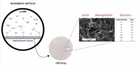
2.2. SrAp Biomineralization

The biomimetic method incorporated the apatite modified with Sr2+ in the membrane. The simulated body fluid (SBF) was produced following the methodology proposed by Bohner and Lemaitre [40], with the replacement of the calcium chloride solution (CaCl2) present in its composition by strontium chloride (SrCl2), preserving the same mass of the substituted ions (Sr2+ → Ca2+). OBC membranes were cut (1 cm2) and immersed in 10 mL of this modified SBF solution (m-SBF) in a Dubnoff Shaking Water Bath (Q226M, Quimis, Diadema, SP, Brazil) at 37.0 ± 0.5 °C with slight agitation, for periods of 7 and 14 days. After the pre-established periods, the membranes were obtained and washed with distilled water, frozen and subsequently dried by lyophilization for 48 h and stored for future tests.
Luz et al. [30] described the methodology used to observe the incorporated amount of strontium in the hybrid matrices. Aliquots of the m-SBF solutions were collected, and the residual concentrations of strontium were measured by Atomic Absorption Spectrometry (AAS) (AA240FS FAST Sequential Atomic Absorption Spectrometer, Varian, Palo Alto, CA, USA) at a wavelength of 460 nm, corresponding to strontium absorption. The calculation of the amount of Sr adsorbed by the membrane is provided in Equation (1):
Q a d s = ( C 0 C t ) V m
where Qads is the adsorbed amount of Sr per unit of mass of the membrane sample at time t (mg·g−1); C0 is the initial concentration of Sr in the m-SBF solution (mg·L−1); Ct is the concentration of Sr in the m-SBF solution at time t (mg·L−1); V is the volume of the m-SBF solution used in the experiment (L); and m is the dry mass of the membrane sample used (g).

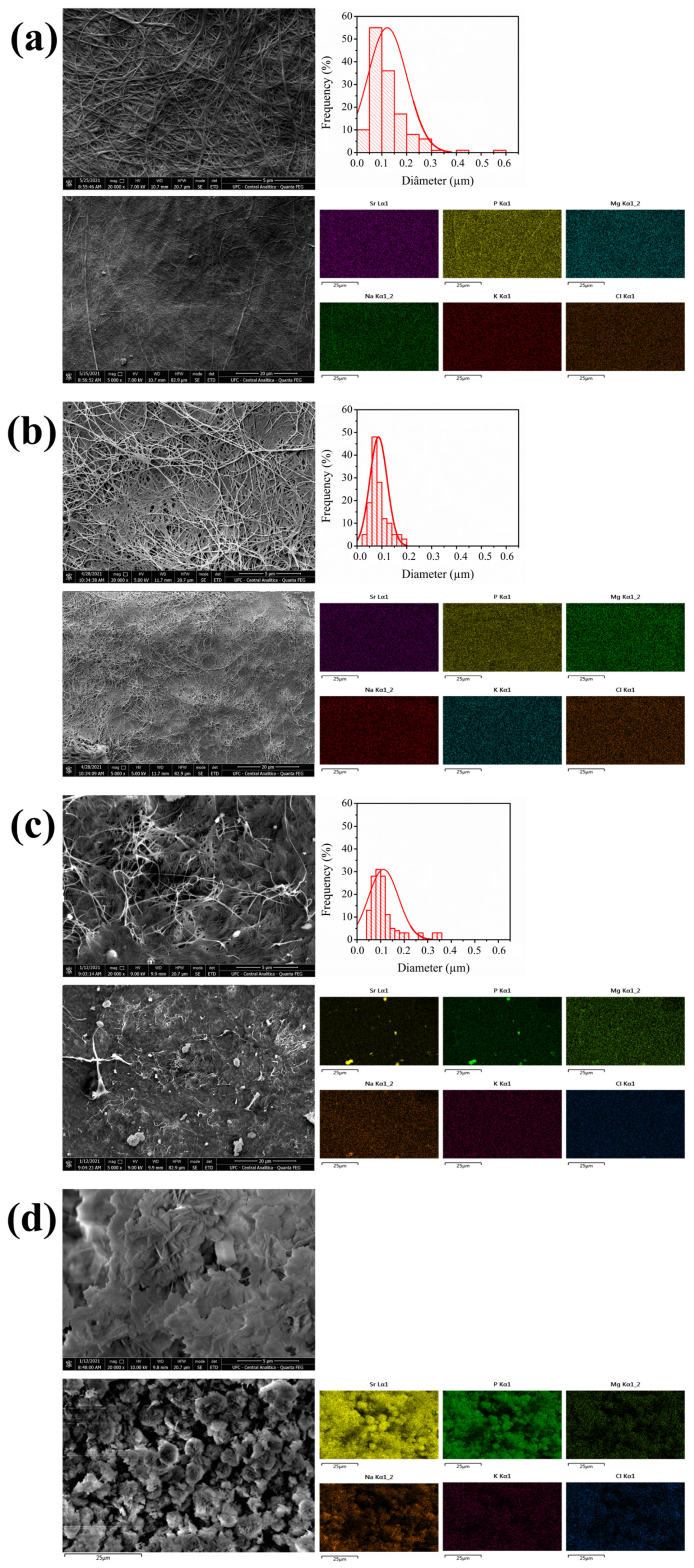
2.3. Scanning Electron Microscopy–Energy Dispersive Spectroscopy (SEM-EDS)

SEM micrographs accompanied by EDS data were obtained to investigate the morphology and chemical composition present on the surface of materials. The samples were mounted in stubs using carbon tape and covered with a thin layer of gold (20 nm) using a sputter applicator (K650, Emitech, Montigny-le-Bretonneux, Yvelines, France) and observed using a Quanta 450 FEG scanning electron microscope (FEI Company, Hillsboro, OR, USA). Diameters of membrane fibers were measured from the images obtained by SEM using ImageJ 1.54d software (public domain). One hundred and thirty-five points were measured for each of the images observed.

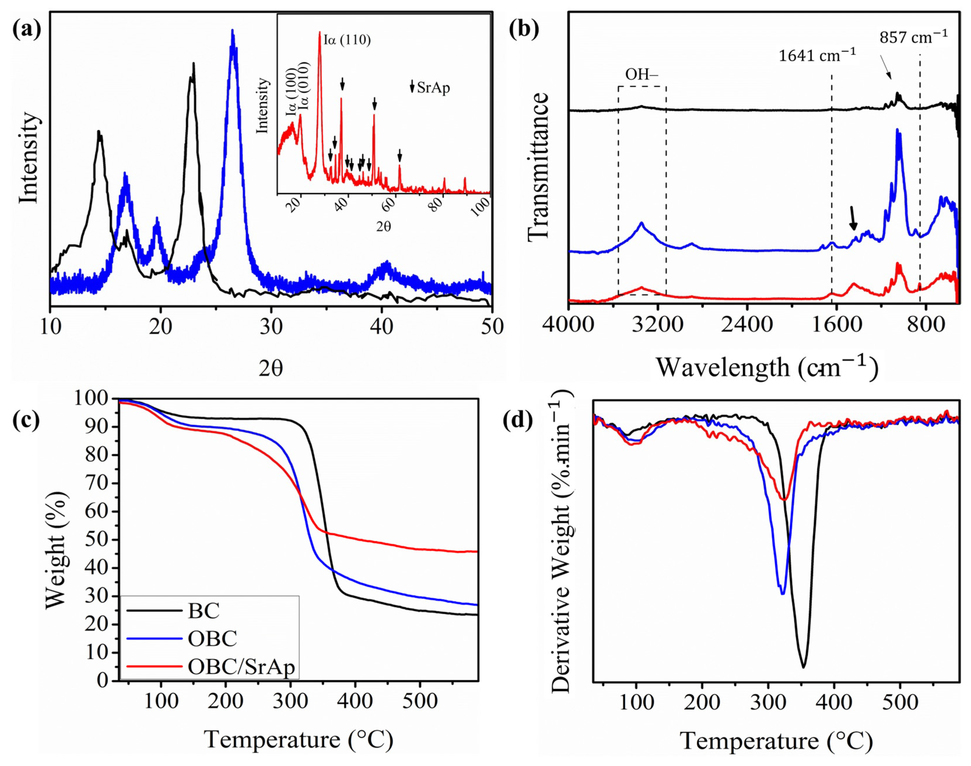
2.4. X-Ray Diffraction (XRD)

To analyze the crystallinity of the membranes studied, the XRD spectra were obtained using an X’Pert PRO MPD (40 kV, 40 mA, PANalytical BV, Almelo, EA, The Netherlands) with CoKα radiation (λ = 1.79 Å), with a sweep speed of 0.5°·min−1 and angle of 2θ, ranging from 10°–100°. Data resulting from this process were fitted using OriginPro 8 SR4 v8.0951 software (OriginLab Corp, Northampton, MA, USA), the baseline was subtracted, a 10-point smoothing was performed using the Savitxky–Golay methodology to reduce noise in the graphics, and the results were analyzed using the Segal method [41], adapting Equation (2).
C r I = I 200 I am I 200 100
where I200 and Iam are the maximum intensity of the main diffraction peak of the crystalline region at 2θ ~ 26° and the minimum intensity of the amorphous region located at 2θ ~ 21°, respectively.

2.5. Fourier Transform Infrared Spectroscopy (FTIR)

FTIR spectra were carried out to confirm the presence of functional groups related to the interaction between the membranes and SrAp. A Perkin Elmer® model spectrum two was used. The samples were ground and compacted with potassium bromide (KBr) (5% m·m−1). The resolution used in the equipment was 4 cm−1, and 32 scans were performed in the reading range of 4000–400 cm−1.

2.6. Thermal Analysis

TGA was performed to investigate the thermal stability of the materials produced, in addition to identifying the percentage of biomineralized material in the samples. For this thermal characterization, the Simultaneous Thermal Analyzer equipment (Netzsch STA 449 F3 Jupiter, Pomerode, SC, Brazil) was used. The membrane samples were weighed (approximately 3.5 mg per sample) and transferred to aluminum crucibles (Al), which were taken to the equipment for analysis. The equipment settings were a heating rate of 10 °C·min−1 at 30 to 600 °C under a synthetic air atmosphere (20% O2: 80% N2 at 40 mL·min−1). These results were treated in the Origin8 software (OriginLab Corp, Northampton, MA, USA). Differential thermogravimetry (DTG) curves were obtained by deriving the TGA curves.

2.7. In Vitro Degradability Test

For the in vitro degradation test, samples were cut (100 mm2), weighed and placed in a 50 mL centrifuge tube containing 10 mL of PBS for 1, 2, 3, 5, 10, 15, 30, 45 and 60 days. The process conditions were slow stirring at a temperature of 37 °C.
The supernatant was analyzed using a Thermo Finnigan Surveyor chromatography (HPLC System, Thermo Fisher Scientific, San Jose, CA, USA) with a refractive index detector and a Supelco 610 H column at 65 °C and 0.005 mol·L−1 H2SO4 in Milli-Q water as the mobile phase, with a flow rate of 0.6 mL·min−1.

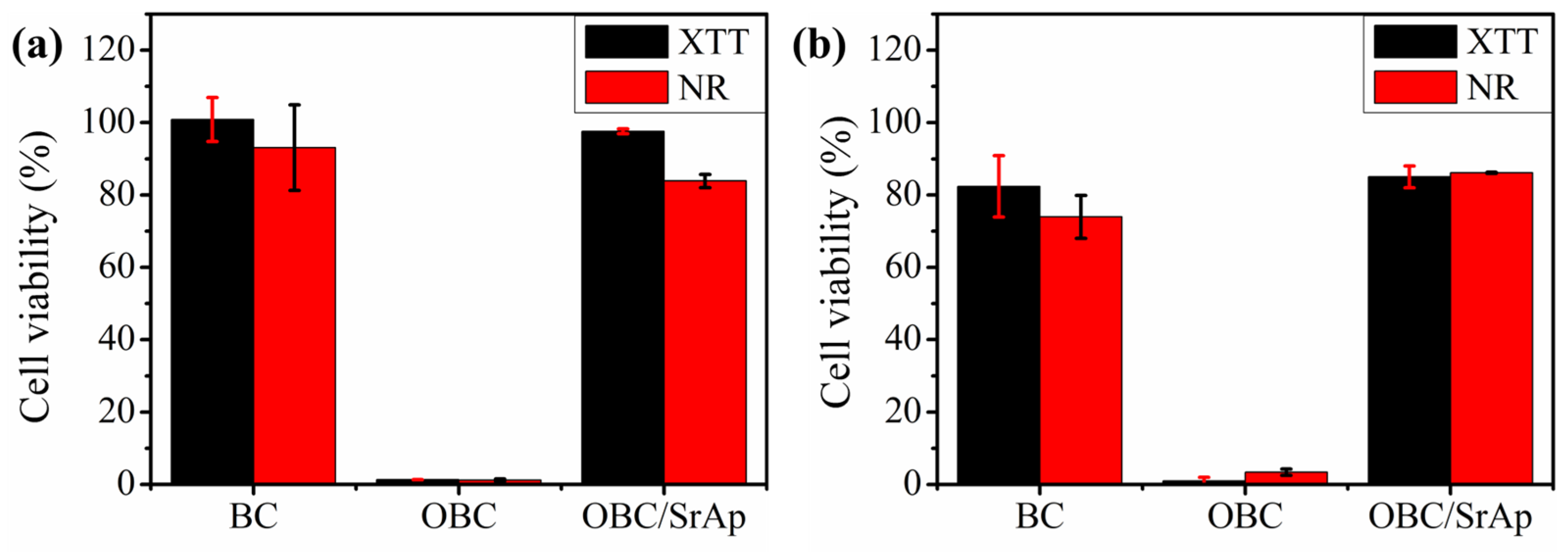
2.8. In Vitro Viability Assay

The cytotoxicity of the materials was evaluated by the indirect method, following the methodology established by the regulatory standard ISO 10993-12 [42], in which cell viability is measured after exposure of cells to sample extracts. For the preparation of extracts, firstly, the samples were cut in a square format with an approximate area of 100 mm2 and sterilized by autoclaving at a temperature of 121 °C for 20 min. Samples of each material were then placed in 24-well plates (each in a different well) with 1 mL of culture medium. A part of the samples was immersed in supplemented DMEM (5% FBS and 1% penicillin-streptomycin) for the assay with fibroblasts, and the other part in supplemented α-MEM (5% FBS and 1% penicillin-streptomycin) for the assay with osteoblasts. Afterward, samples were incubated at 37 °C for 24 h (5% CO2 and 95% humidity). After incubation, the extracts obtained were collected and stored in Falcon-type tubes. The cytotoxicity assay followed the methodology proposed in the In Cytotox LDHe-XTT-NR Kit (Xenometrix AG, Allschwil, Switzerland) (Xenometrix AG 2015) without performing the LDHe test.
Mouse fibroblast (L-929) and osteoblast (MC3T3-E1 Subclone 14) cells were seeded in supplemented DMEM medium (5% FBS and 1% penicillin-streptomycin) and supplemented α-MEM medium (5% FBS and 1% penicillin-streptomycin), respectively, in 96-well plates with a density of 20 × 103 cells·well−1 (0.2 mL), followed by incubation at 37 °C (5% CO2 and 95% humidity) for a period of 24 h. After this period, the culture medium was removed from the wells, and then 0.2 mL of extracts were added to the wells, and the plates were incubated again at 37 °C for 24 h (5% CO2 and 95% humidity).
After these periods, the extracts were removed from the wells and washed with a PBS buffer solution (pH 7.4). Subsequently, 0.2 mL of the new medium and 0.05 mL of the mixture of XTT solutions (solutions XTT II and XTT I in a ratio of 1:100) were added. Finally, the plate was incubated for 3 h, and the XTT reading was performed with a microplate reader (SpectraMax i3x, Molecular Devices, San Jose, CA, USA) with slow orbital agitation for 10 s and using an absorbance of 450 nm with a reference of 690 nm.
After reading, the supernatant was discarded, and the wells were washed with 0.3 mL of the NR I wash solution. An amount of 200 µL of the 1% NR II solution (prepared with the culture medium) was added, and the microplate was incubated for 3 h. Subsequently, 0.1 mL of NR III solution was added to each well and discarded after 1 min, and finally, 0.2 mL of NR IV solubilization solution was added, and plates were incubated for 15 min at room temperature. The NR reading was done with slow orbital agitation for 10 s and an absorbance of 540 nm with a reference of 690 nm.
Cell viability was calculated by Equation (3), as instructed by the protocol [43].
% V C = ( O D X O D 690 ) M S ( O D X O D 690 ) M B ( O D X O D 690 ) M G c
where OD is the optical density; x is the wavelength referring to the test (450 for XTT or 540 for NR); MS is the sample mean; MB is the mean of the blank; and MGc is the mean of the growth control.

2.9. Artemia Salina Lethality Assay

The Artemia salina lethality assay was performed using the methodology described by Meyer et al. [44] and the ISO/TS 20787 [45]. A sodium chloride solution (30 g·L−1) was prepared, and the pH was adjusted between 8.0 and 9.0 using a 0.1 mol·L−1 NaOH solution. This solution was used to hatch the Artemia salina eggs and to prepare the other dilutions. The eggs were placed to hatch in saline solution for 48 h, with constant aeration at 25 °C. Approximately 10 Artemia salina larvae were transferred using automatic pipettes to 24-well plates containing the saline solution (negative control) and samples to be tested at the following concentrations of the aqueous extract: 10, 100 and 1000 µg·mL−1 for each group. The assay was performed in sextuplicate. To obtain the results, the relative values were counted in percentages (%) through the absolute values of the immobile nauplii (dead) over the total nauplii (after fixation in 10% formalin) in periods of 24 h and 48 h for each replicate and each experimental condition. Animals were counted using a 3× magnifying glass (stereomicroscope). Abbott’s formula [46] was used to quantify the Artemia salina mortality:
% d e a t h s = t e s t n e g a t i v e c o n t r o l n e g a t i v e c o n t r o l

3. Results and Discussion

3.1. Obtaining Oxidized Bacterial Cellulose

OBC membranes showed a significant difference in size compared to BC membranes before the oxidation reaction. It was possible to observe the decrease in the membrane diameter, which lost approximately 31.3 ± 0.5% of its original diameter. The oxidation reaction conditions (temperature of 55 °C and reaction time of 6 h, oxidation degree of 50%) were studied by Vasconcelos et al. [21], who obtained a material that presented a more significant number of aldehydes in its structure without any significant impact on its structure. These authors also noted that, under stronger reaction conditions, the aldehyde groups are degraded to ketone and carboxylic groups, further reducing the cellulose mass. This effect was also evaluated by Fu et al. [47] using NaClO as an oxidizing agent. The authors reported that 10 mmol of NaClO per g of cellulose was the optimal ratio to obtain OBC with the highest carboxyl content, maintaining the membrane structure. However, for higher oxidizing agent concentrations, the carboxyl group content did not increase insignificantly, and cellulose degradation occurred, which was not useful for some applications.

3.2. SrAp Biomineralization

For preparing the m-SBF solution, it was possible to replace CaCl2 with SrCl2 due to the similarity of the Ca2+ and Sr2+ ions. These ions have a close atomic radius, equal valence, and common biochemical metabolic routes. The incorporation of SrAp onto the OBC membranes was due to the deposition of Sr2+ and phosphates (PO34−) on these surfaces, forming strontium phosphates with intramolecular interactions of the ionic type. In addition, cellulose has many free hydroxyl groups (OH) on its surface, providing an ionic bond between hydroxyl groups (negatively charged) and Sr2+ (positively charged). This deposition was investigated by evaluating the adsorbed amount (Qads) of Sr on OBC membranes after 7 and 14 days of immersion; adsorption was 3359 ± 727 mg·g−1 and 3773 ± 727 mg·g−1, respectively, with no significant difference in Sr adsorption with increasing time. These results are below the values obtained for the non-oxidized samples, which were 5981 ± 90 mg·g−1 for 7 days and 6233 ± 90 mg·g−1 for 14 days. This may have occurred due to the preferential deposition of other elements present in the m-SBF solution, such as magnesium, which can also be used in bone regeneration processes [48].

3.3. Scanning Electron Microscopy–Energy Dispersive Spectroscopy (SEM-EDS)

In Figure 1a,b, the presence of many fibers can be observed in both membranes; however, OBC presents a deformation, characterized by the appearance of pores between the fibers, while there is a large interconnection between them. This deformation is due to the oxidation of the material, consistent with the observations from a previous study that showed differences in membrane diameter, resulting from fiber degradation/compression due to oxidation reaction [6]. Nevertheless, the fiber diameter distributions of the membranes had similar normal distribution profiles, with a higher mean diameter for BC than OBC. The BC and OBC membranes had fibers with maximum diameters of 0.581 and 0.186 µm, minimum diameters of 0.028 and 0.028 µm, and mean diameters of 0.101 and 0.077 µm, respectively.
Figure 1c,d show OBC membranes after immersion in m-SBF solution for 7 (OBC7dSrAp) and 14 days (OBC14dSrAp) for biomineralization of SrAp on the surface. An increase in crystal deposition can be seen on the material, causing deformations in its structure since the deposition occurs on the entire surface of the scaffold. The fibers are almost invisible in the OBC14dSr membranes; according to the OBC7dSrAp fiber diameter distribution, the mean fiber diameter was 0.097 ± 0.064 µm, the minimum diameter was 0.048 µm and its maximum was 0.345 µm.
EDS showed that on the first days, SrAp was preferentially biomineralized. However, with the progression of the immersion time, other crystals were deposited on the membrane structure, such as Mg3(PO4)2, which, although it plays a fundamental role in bone metabolism, can inhibit the formation of apatite [36], negatively affecting the viability of bone cells [37]. This could explain the lower percentage of Sr obtained by EDS for OBC14dSrAp (Figure 1d). In contrast, SEM images showed that the membrane’s surface was entirely coated by the biomineralized layer. The inverse is verified by observing the data of OBC7dSrAp (Figure 1c), which demonstrate a higher superficial appearance of Sr and while retaining its fibrous structure.

3.4. X-Ray Diffraction (XRD)

This analysis was performed to investigate the effect of the presence of SrAp on the crystallinity of the materials. As seen previously, SrAp biomineralization for 14 days did not significantly affect the amount of Sr2+ adsorbed by the membrane; on the other hand, there was a great deformation in the membrane structure, possibly due to the coating of its fibers by other apatite crystals, which can inhibit the growth of the apatite of interest. Given this finding, a longer mineralization time is unnecessary, and other tests focused only on the study of OBC7dSrAp, which we call OBC/SrAp. The X-ray diffractograms of the samples of interest were then compiled in Figure 2a [19], with the addition of data referring to the BC before oxidation for a better discussion of these results.
The XRD spectra demonstrated no significant differences between the diffractograms formed for the BC and OBC membranes; however, they show a clear difference in the crystallinity of OBC/SrAp. The samples present characteristic peaks of type I cellulose; for BC, the peaks were located at approximately 14.5°, 16.9°, and 22.8°; for OBC, at approximately 16.8°, 19.7° and 26.5°. However, for OBC/SrAp, they present values of 16.2°, 19.8° and 27.9°. Correlating these data with the study proposed by Vasconcelos et al. [21], we can infer that they are the crystallographic planes represented by the Miller indices (100) (010) and (110), respectively. This difference may be due to the use of different radiation, where Vasconcelos used CuKα and the present work used CoKα; changes in the diffraction angle may occur. The XRD spectrum for the biomineralized membranes showed new peaks between 32° and 61°, which, according to the literature [49,50], may correspond to the presence of SrAp crystals in the membranes. These data are directly related to the thermal stability of the scaffolds (Figure 2c). It can be observed that the polymeric materials exhibit similar behavior, unlike the biomineralized material. While the latter displays additional crystallinity due to the inorganic portion, its organic component shows lower thermal stability than the pure membranes, resulting in slightly higher thermal instability and a greater percentage of residual mass.
The crystallinity index of these membranes was calculated using the Segal method [40], which can describe the polymer well but not the ceramic. For this reason, we cannot directly compare the two elements that make up the biomaterial. The crystallinity indices calculated by the Segal method for the samples OBC and OBC/SrAp demonstrated remarkable similarity, 91.97% and 91.78%, respectively, as these correlated only with the polymeric fraction. An increase in the crystallinity of the OBC (91.97%) in relation to the BC (86.2%) produced by Vasconcelos et al. [21] can be observed, unlike the expected results described in the literature by Fu et al. [47], Luz et al. [19] and Vasconcelos et al. [51]. In this study, there is an increase in the intensity and narrowing of the peaks, indicating a decrease in the partial amorphous phase of the structure. Luo et al. [22] obtained similar behavior for BC oxidized by TEMPO, attributing the increase in crystallinity to the fact that the aldehyde can be generated in disordered regions, combining with neighboring hydroxyl groups to form inter-acetal bonds that led to the partial loss of amorphous regions. This can also affect the rigidity of the membrane, facilitating the production of a scaffold that mimics the characteristics of natural bone, thus providing mechanical support for bone regeneration.

3.5. Fourier Transform Infrared Spectroscopy (FTIR)

Figure 2b represents the vibrational spectra of the membranes studied before and after the incorporation of SrAp for 7 days. The vibrational band between 3300 and 3500 cm−1 is present in all samples, as it refers to the stretching of the hydroxyl group (O–H) present in the cellulose chemical structure. BC presents the lowest intensity of this band, which may be caused by dehydration caused by the drying process of the sample, while OBC presents the highest intensity of this band, which may be related to the oxidation process, where glucose rings open, generating new reactive sites, resulting in the formation of more hydroxyl groups. When analyzing the mineralized material, a decrease in this band can be observed since, during biomineralization, the free hydroxyl groups can be replaced by strontium or phosphate ions that bind to the membrane surface [33].
The membranes also present common low frequency, vibrational bands among the polymers, which are close to the 2940 and 2900 cm−1 ranges, related to asymmetric and symmetrical CH2− stretches. The band that appears close to 1650 cm−1 represents the C=O (carbonyl) stretch, referring to the aldehyde group formed in the BC oxidation process. In addition to the bands expressed above, it was possible to observe the presence of a band close to 1430 cm−1 (indicated by the black arrow), representing the angular deformation of CH2 and the band 1557 cm−1 (indicated by the blue arrow) representing the C–O stretching [52].
Some bands indicate the changes caused by SrAp mineralization on the surface of these materials. For example, the appearance of a peak in the range of 850 cm−1, which refers to the elongation of the phosphate group (PO43−) [53], which may be directly related to the presence of Ap in the membrane, including SrAp [50], corroborating the results discussed previously.

3.6. Thermal Analysis

Figure 2c,d present the TGA and DTG graphs, respectively. These analyses provide information about the thermal stability of the material and the interactions of cellulose fibrils and the inorganic components present in the membranes studied.
TGA shows that the mass loss occurs in two thermal events for all samples. In the first event, between 30 and 150 °C, the evaporation of the most volatile compounds arises as the residual water. The materials had a similar mass loss; BC had a loss of 6.64%, OBC had a loss of 9.63%, and OBC7dSrAp presented a loss of 11.02%. In the second event, polymer degradation mainly occurs, related to the depolymerization and decomposition of glycosidic units [19,54]. The most significant mass loss occurred during the second thermal event, with a Tonset of 333 °C (~62%), 291 °C (~49%) and 261 °C (~36%) for the BC, OBC and OBC7dSrAp membranes, respectively. OBC7dSrAp had a lower Tonset value than OBC, indicating a decrease in thermal stability due to the reduction of surface crystallinity seen in the XRD data [21]. The residual mass of OCB7dSrAp was also found to be approximately 1.7 times greater than that of OBC, which can be explained by the residue of biomineralized material, confirming the inorganic matter present in the final membrane. It can also be seen from the curve derivatives (DTG) that the highest degradation peak occurs at 353, 321 and 323 °C for the OBC and OBC7dSrAp samples, respectively.

3.7. In Vitro Degradability Test

The OBC degradation, before and after immersion in m-SBF, was quantified by HPLC, measuring the amount of glucose (the fundamental unit of cellulose) in the PBS supernatant aliquots. Figure 3 compares these experimental results with data from the literature for BC degradation [31], demonstrating that the chemical modification by the addition of the aldehyde group affected the OBC degradation, increasing the glucose concentration dispersed in the medium. It was also compared with data obtained by Luz et al. [19] for biomineralized bacterial cellulose membranes with strontium apatite obtained by the immersion cycle method.
OBC and OBC7dSrAp released amounts of glucose of 4.16 ± 0.31 and 4.44 ± 0.35 g·L−1, respectively, in 60 days of degradation. Luz et al. [31] observed that, in the same period, pure bacterial cellulose released approximately 0.24 g·L–1 of glucose in its aliquots, while OBC/SrAP [19], approximately 2.13 g·L−1. The materials produced in this work released an amount of glucose approximately 18 times greater than pure BC and at least 2 times greater than the biomineralized material described in the literature.
It also identified low amounts of acetic acid as a cellulose degradation product. For the OBC membranes, 0.09 ± 0.01 g·L−1 and 0.42 ± 0.12 g·L−1 of acetic acid were identified on the first day and within 60 days, respectively. For the OBC7dSrAp membranes, acetic acid was not identified on the first day of testing, but 0.31 ± 0.1 g·L−1 acetic acid was identified after 60 days.
During the first days, the glucose concentration released by the composites was found to be slightly lower than that of the OBC samples. As discussed earlier, SrAp is chemically bonded to the OBC membrane because of negatively charged groups on its surface, strengthening interactions between fibrils. The degradation of this compound can be attributed to the detachment of oxidized fibrils during agitation [55].

3.8. In Vitro Viability Assay

Figure 4 shows the results for mitochondrial dehydrogenase activity (XTT assay) and membrane integrity (NR assay) of mouse fibroblast (L-929) and osteoblasts (MC3T3-E1 Subclone 14) cells after contact with the BC, OBC and OBC7dSrAp extract samples for 24 h.
The XTT assay is based on the ability of mitochondrial enzymes from metabolically active cells to reduce 2,3-bis(2-methoxy-4-nitro-5-sulfophenyl)-2H-tetrazolium-5-carboxanilide (XTT) to a soluble salt of formazan [55]. The NR assay evaluates the ability of viable cells to incorporate and bind to a weak cationic supravital dye that penetrates cell membranes by non-ionic diffusion and accumulates predominantly intracellularly in lysosomes [56,57].
The samples produced were tested after 24 h of incubation. The BC membranes showed high viability in both cells tested, as expected; as previously mentioned, this polymer has high biocompatibility. The OBC membrane showed an extremely cytotoxic result for both tests, resulting from the oxidation process. The possible degradation of this membrane can explain the presence of residues from the oxidation process or the presence of reactive groups on its surface. Although the degradability of the biomaterial is an exciting and desirable characteristic, this process can also be associated with increased toxicity, which, in the case of oxidized cellulose, could release acidic products such as butyric and acetic acid, subsequently decreasing the pH of the medium and consequently decreasing cell viability, as previously reported [19,32]. Using the biodegradability test, it was possible to observe, within just 24 h of the test, a slight degradation of the material, releasing, in addition to glucose, small amounts of acetic acid; this phenomenon did not occur with the composite.
The membranes functionalized with SrAp had high cell viability, with XTT values of 85.01 ± 3.0% and 97.58 ± 0.64% for osteoblasts and fibroblasts, respectively, and NR values of 86.15 ± 0.15% and 83.87 ± 1.82% for osteoblasts and fibroblasts, respectively. This fact can be explained by the precipitation of salts present in the m-SBF solution, which could neutralize possible reactive groups present on the surface of these materials since the degradation of the material is not greatly affected by the biomineralization of apatite.
The presence of strontium apatite in this material induces an improvement in its effects on cell viability and, therefore, has the potential to increase bone formation in vivo through the Wnt/β-catenin signaling pathway, which regulates several phenomena, including those related to cell differentiation, polarization, and migration [58].

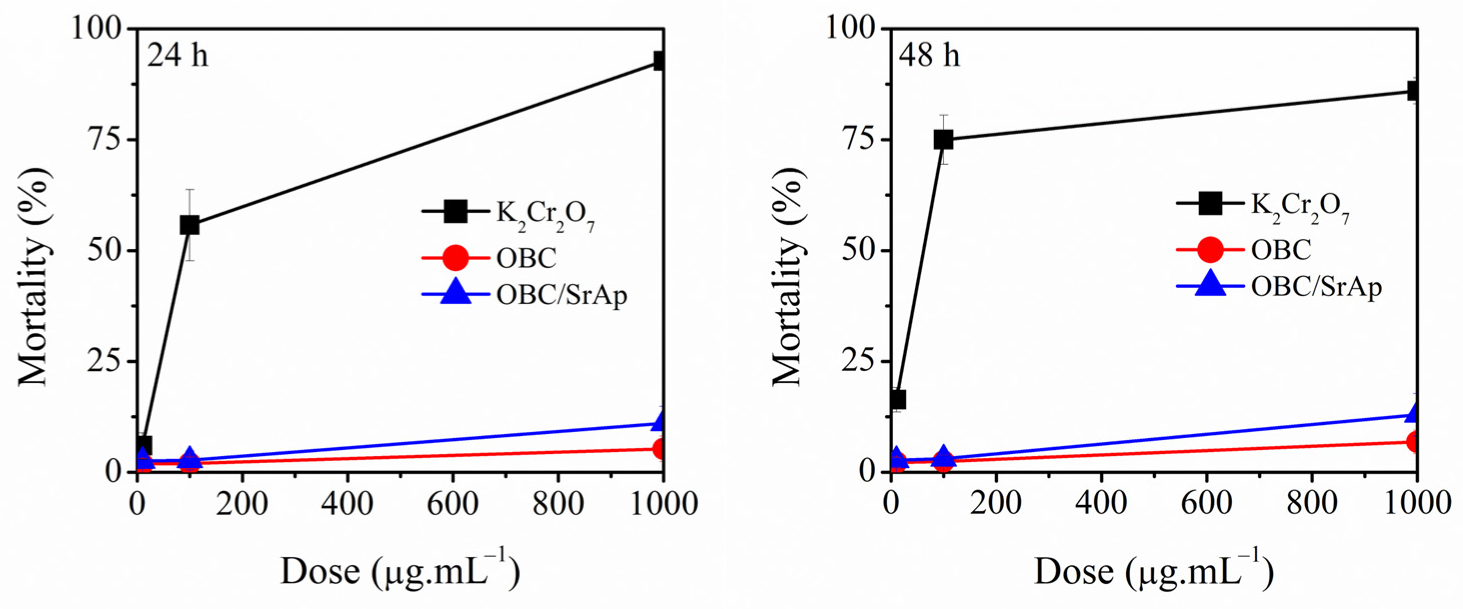
3.9. Artemia Salina Lethality Assay

The results of this test are shown in Figure 5 and showed that the two test groups (OBC before and after SrAp immobilization) present significantly lower toxicities to Artemia salina (p < 0.001) compared to the positive toxicity control (potassium dichromate). Furthermore, the cell mortality in the presence of these biomaterials was below 13%, even at the highest concentrations.
Despite being minimally toxic compared to the positive control, the two test groups also showed significant differences between them (p < 0.001), where, in general, the OBC7dSrAp group exhibited twice the mean toxicity observed with the OBC group at a dose of 1.000 µg·mL−1. Furthermore, OBC7dSrAp presented a mortality rate of 11.03 ± 3.79% in 24 h and 12.94 ± 4.77% in 48 h, whereas OBC presented a mortality rate of 5.27 ± 2.86% in 24 h and 6.8 3± 2.66% in 48 h. This result can be explained by the presence of biologically active compounds in the composite, increasing the lethality against Artemia salina. Although the results show minimal mortality, we can compare this behavior to the material studied by Kiani et al. [59], who produced Ag2O nanostructures with the addition of various concentrations of Sr, obtaining a proportionally increasing mortality rate, reaching up to 93.6%.
The times of 24 and 48 h showed significant toxicity variation only for the positive control (potassium dichromate) at doses of 10 and 100 µg·mL−1. There was no significant change for the tested biomaterials at the three doses tested (10, 100 and 1000 µg·mL−1) between the two experimental times (24 h and 48 h), suggesting that the acute biological response tends to remain unchanged over the first 48 h, according to the concentrations tested in this experimental model.

4. Conclusions

The method used for SrAp biomineralization proved effective, as evidenced by the identification of this metal and phosphate through chemical and morphological characterizations. Notably, the apatite layer obtained via the biomimetic method demonstrated greater stability than that produced by the immersion cycle method, as the biomimetic approach replicates conditions similar to those found in the human body. This study also revealed that SrAp mineralization over 7 days achieved maximum incorporation of Sr2+. Furthermore, SrAp biomineralization mitigated the cytotoxic effects observed with OBC and resulted in a low mortality rate for Artemia salina, highlighting the advantages of producing composites that reduce the toxicity of individual materials. Although in vivo tests are necessary for effective validation of this scaffold, this composite shows great promise for bone tissue engineering, demonstrating potential for creating a biomaterial with high cell viability and controlled degradation. The material is expected to degrade as SrAp fulfills its osteogenic role without significantly impacting the surrounding environment.

Author Contributions

All authors contributed to the study design. The material production and characterization, as well as the writing of the article, were carried out by A.L.d.B.S. I.I.C.-S. performed the lethality test for Artemia salina and helped with writing the manuscript. E.P.C.G.L. provided support in obtaining data and helped with corrections to the article. R.R.d.C.M., F.K.A. and R.S.V. commented on subsequent versions of the manuscript and coordinated research and funding. All authors have read and agreed to the published version of the manuscript.

Funding

The authors would like to thank the Coordination for improving Higher Education Personnel (CAPES) for funding this research, supported by the project PROCAD/CAPES (88881.068439/2014-01).

Institutional Review Board Statement

Not applicable.

Data Availability Statement

Data are contained within the article and available upon request.

Acknowledgments

The authors would like to thank Central Analytica-UFC for the SEM/EDS analysis, the UFC X-Ray Laboratory (LRX) for the DRX analysis, Embrapa Agroindústria Tropical for the FTIR analysis and Northeast Strategic Technologies Center (CETENE-Recife/PE) for the thermal analysis.

Conflicts of Interest

The authors declare no conflicts of interest.

References

  1. Black, C.R.M.; Goriainov, V.; Gibbs, D.; Kanczler, J.; Tare, R.S.; Oreffo, R.O.C. Bone Tissue Engineering. Curr. Mol. Biol. Rep. 2015, 1, 132–140. [Google Scholar] [CrossRef] [PubMed]
  2. Windhager, R.; Hobusch, G.M.; Matzner, M. Allogene Transplantate Für Biologische Rekonstruktionen von Knochendefekten. Orthopade 2017, 46, 656–664. [Google Scholar] [CrossRef]
  3. Yang, M.; Zhen, W.; Chen, H.; Shan, Z. Biomimetic Design of Oxidized Bacterial Cellulose-Gelatin-Hydroxyapatite Nanocomposites. J. Bionic Eng. 2016, 13, 631–640. [Google Scholar] [CrossRef]
  4. Fragal, E.H.; Cellet, T.S.P.; Fragal, V.H.; Witt, M.A.; Companhoni, M.V.P.; Ueda-Nakamura, T.; Silva, R.; Rubira, A.F. Biomimetic Nanocomposite Based on Hydroxyapatite Mineralization over Chemically Modified Cellulose Nanowhiskers: An Active Platform for Osteoblast Proliferation. Int. J. Biol. Macromol. 2019, 125, 133–142. [Google Scholar] [CrossRef] [PubMed]
  5. Oliveira Barud, H.G.; Barud, H.d.S.; Cavicchioli, M.; do Amaral, T.S.; Junior, O.B.d.O.; Santos, D.M.; Petersen, A.L.d.O.A.; Celes, F.; Borges, V.M.; de Oliveira, C.I.; et al. Preparation and Characterization of a Bacterial Cellulose/Silk Fibroin Sponge Scaffold for Tissue Regeneration. Carbohydr. Polym. 2015, 128, 41–51. [Google Scholar] [CrossRef]
  6. Andrade, F.K.; Silva, J.P.; Carvalho, M.; Castanheira, E.M.S.; Soares, R.; Gama, M. Studies on the Hemocompatibility of Bacterial Cellulose. J. Biomed. Mater. Res. A 2011, 98A, 554–566. [Google Scholar] [CrossRef]
  7. Treesuppharat, W.; Rojanapanthu, P.; Siangsanoh, C.; Manuspiya, H.; Ummartyotin, S. Synthesis and Characterization of Bacterial Cellulose and Gelatin-Based Hydrogel Composites for Drug-Delivery Systems. Biotechnol. Rep. 2017, 15, 84–91. [Google Scholar] [CrossRef]
  8. Weyell, P.; Beekmann, U.; Küpper, C.; Dederichs, M.; Thamm, J.; Fischer, D.; Kralisch, D. Tailor-Made Material Characteristics of Bacterial Cellulose for Drug Delivery Applications in Dentistry. Carbohydr. Polym. 2019, 207, 1–10. [Google Scholar] [CrossRef]
  9. Stumpf, T.R.; Yang, X.; Zhang, J.; Cao, X. In Situ and Ex Situ Modifications of Bacterial Cellulose for Applications in Tissue Engineering. Mater. Sci. Eng. C 2018, 82, 372–383. [Google Scholar] [CrossRef]
  10. Pang, M.; Huang, Y.; Meng, F.; Zhuang, Y.; Liu, H.; Du, M.; Ma, Q.; Wang, Q.; Chen, Z.; Chen, L.; et al. Application of Bacterial Cellulose in Skin and Bone Tissue Engineering. Eur. Polym. J. 2020, 122, 109365. [Google Scholar] [CrossRef]
  11. Medronho, B.; Romano, A.; Miguel, M.G.; Stigsson, L.; Lindman, B. Rationalizing Cellulose (in)Solubility: Reviewing Basic Physicochemical Aspects and Role of Hydrophobic Interactions. Cellulose 2012, 19, 581–587. [Google Scholar] [CrossRef]
  12. Rodríguez, K.; Gatenholm, P.; Renneckar, S. Electrospinning Cellulosic Nanofibers for Biomedical Applications: Structure and in Vitro Biocompatibility. Cellulose 2012, 19, 1583–1598. [Google Scholar] [CrossRef]
  13. Chavan, Y.V.; Panesar, P.S.; Kumar, V. Application of Bacterial Cellulose in the Food Industry. In Bacterial Cellulose; CRC Press: Boca Raton, FL, USA, 2023; pp. 123–140. [Google Scholar] [CrossRef]
  14. Faria, M.; Cunha, C.; Gomes, M.; Mendonça, I.; Kaufmann, M.; Ferreira, A.; Cordeiro, N. Bacterial Cellulose Biopolymers: The Sustainable Solution to Water-Polluting Microplastics. Water Res. 2022, 222, 118952. [Google Scholar] [CrossRef]
  15. Mathew, A.; Poulose, A.; Gopakumar, D.A.; Pasquini, D.; Grohens, Y.; George, J.J. Nanocellulose-Based Ultralight Porous Material for Various Environmental Applications. In Nanomaterials for Air- and Water Purification; Wiley: Hoboken, NJ, USA, 2024; pp. 373–397. [Google Scholar] [CrossRef]
  16. Bianchet, R.T.; Vieira Cubas, A.L.; Machado, M.M.; Siegel Moecke, E.H. Applicability of Bacterial Cellulose in Cosmetics—Bibliometric Review. Biotechnol. Rep. 2020, 27, e00502. [Google Scholar] [CrossRef]
  17. Lima, N.F.; Maciel, G.M.; Lima, N.P.; Fernandes, I.d.A.A.; Haminiuk, C.W.I. Bacterial Cellulose in Cosmetic Innovation: A Review. Int. J. Biol. Macromol. 2024, 275, 133396. [Google Scholar] [CrossRef] [PubMed]
  18. Cao, S.; Li, Q.; Zhang, S.; Liu, K.; Yang, Y.; Chen, J. Oxidized Bacterial Cellulose Reinforced Nanocomposite Scaffolds for Bone Repair. Colloids Surf. B Biointerfaces 2022, 211, 112316. [Google Scholar] [CrossRef]
  19. Luz, E.P.C.G.; das Chagas, B.S.; de Almeida, N.T.; de Fátima Borges, M.; Andrade, F.K.; Muniz, C.R.; Castro-Silva, I.I.; Teixeira, E.H.; Popat, K.; de Freitas Rosa, M.; et al. Resorbable Bacterial Cellulose Membranes with Strontium Release for Guided Bone Regeneration. Mater. Sci. Eng. C 2020, 116, 111175. [Google Scholar] [CrossRef]
  20. Jun, S.-H.; Park, S.-G.; Kang, N.-G. One-Pot Method of Synthesizing TEMPO-Oxidized Bacterial Cellulose Nanofibers Using Immobilized TEMPO for Skincare Applications. Polymers 2019, 11, 1044. [Google Scholar] [CrossRef]
  21. Vasconcelos, N.F.; Andrade, F.K.; Vieira, L.d.A.P.; Vieira, R.S.; Vaz, J.M.; Chevallier, P.; Mantovani, D.; Borges, M.d.F.; Rosa, M.d.F. Oxidized Bacterial Cellulose Membrane as Support for Enzyme Immobilization: Properties and Morphological Features. Cellulose 2020, 27, 3055–3083. [Google Scholar] [CrossRef]
  22. Luo, H.; Xiong, G.; Hu, D.; Ren, K.; Yao, F.; Zhu, Y.; Gao, C.; Wan, Y. Characterization of TEMPO-Oxidized Bacterial Cellulose Scaffolds for Tissue Engineering Applications. Mater. Chem. Phys. 2013, 143, 373–379. [Google Scholar] [CrossRef]
  23. Hori, S.; Hirano, M.; Ohta, R. Electrodeposition of Strontium Apatite Nanorod Arrays and Their Cell Compatibility. Ceram. Int. 2017, 43, 9047–9052. [Google Scholar] [CrossRef]
  24. Jiménez, M.; Abradelo, C.; San Román, J.; Rojo, L. Bibliographic Review on the State of the Art of Strontium and Zinc Based Regenerative Therapies. Recent Developments and Clinical Applications. J. Mater. Chem. B 2019, 7, 1974–1985. [Google Scholar] [CrossRef] [PubMed]
  25. Anastasiou, A.D.; Nerantzaki, M.; Gounari, E.; Duggal, M.S.; Giannoudis, P.V.; Jha, A.; Bikiaris, D. Antibacterial Properties and Regenerative Potential of Sr2+ and Ce3+ Doped Fluorapatites; a Potential Solution for Peri-Implantitis. Sci. Rep. 2019, 9, 14469. [Google Scholar] [CrossRef] [PubMed]
  26. Cochis, A.; Barberi, J.; Ferraris, S.; Miola, M.; Rimondini, L.; Vernè, E.; Yamaguchi, S.; Spriano, S. Competitive Surface Colonization of Antibacterial and Bioactive Materials Doped with Strontium and/or Silver Ions. Nanomaterials 2020, 10, 120. [Google Scholar] [CrossRef]
  27. Ullah, I.; Li, W.; Lei, S.; Zhang, Y.; Zhang, W.; Farooq, U.; Ullah, S.; Ullah, M.W.; Zhang, X. Simultaneous Co-Substitution of Sr2+/Fe3+ in Hydroxyapatite Nanoparticles for Potential Biomedical Applications. Ceram. Int. 2018, 44, 21338–21348. [Google Scholar] [CrossRef]
  28. Ullah, I.; Gloria, A.; Zhang, W.; Ullah, M.W.; Wu, B.; Li, W.; Domingos, M.; Zhang, X. Synthesis and Characterization of Sintered Sr/Fe-Modified Hydroxyapatite Bioceramics for Bone Tissue Engineering Applications. ACS Biomater. Sci. Eng. 2020, 6, 375–388. [Google Scholar] [CrossRef] [PubMed]
  29. Ullah, I.; Zhang, W.; Yang, L.; Ullah, M.W.; Atta, O.M.; Khan, S.; Wu, B.; Wu, T.; Zhang, X. Impact of Structural Features of Sr/Fe Co-Doped HAp on the Osteoblast Proliferation and Osteogenic Differentiation for Its Application as a Bone Substitute. Mater. Sci. Eng. C 2020, 110, 110633. [Google Scholar] [CrossRef]
  30. Luz, E.P.C.G.; Borges, M.d.F.; Andrade, F.K.; Rosa, M.d.F.; Infantes-Molina, A.; Rodríguez-Castellón, E.; Vieira, R.S. Strontium Delivery Systems Based on Bacterial Cellulose and Hydroxyapatite for Guided Bone Regeneration. Cellulose 2018, 25, 6661–6679. [Google Scholar] [CrossRef]
  31. Luz, E.P.C.G.; Chaves, P.H.S.; Vieira, L.d.A.P.; Ribeiro, S.F.; Borges, M.d.F.; Andrade, F.K.; Muniz, C.R.; Infantes-Molina, A.; Rodríguez-Castellón, E.; Rosa, M.d.F.; et al. In Vitro Degradability and Bioactivity of Oxidized Bacterial Cellulose-Hydroxyapatite Composites. Carbohydr. Polym. 2020, 237, 116174. [Google Scholar] [CrossRef]
  32. Luz, E.P.C.G.; Soares, A.L.d.B.; Souza, F.F.P.d.; Andrade, F.K.; Castro-Silva, I.I.; Rosa, M.d.F.; Vieira, R.S. Implantable Matrixes of Bacterial Cellulose and Strontium Apatite: Preclinical Analysis of Cytotoxicity and Osteoconductivity. Mater. Today Commun. 2022, 33, 104871. [Google Scholar] [CrossRef]
  33. Soares, A.L.d.B.; Luz, E.P.C.G.; Vieira, R.S. Adsorption Processes for Forming Biomaterials of Cellulose and Hydroxyapatite for Applications in Bone Tissue Regeneration. Adsorption 2024, 30, 595–607. [Google Scholar] [CrossRef]
  34. Maia, M.T.; De Brito Soares, A.L.; Caetano, M.A.; Andrade, F.K.; Rodríguez-Castellón, E.; Vieira, R.S. Biomimetic Strontium Substituted Calcium Phosphate Coating for Bone Regeneration. Coatings 2021, 11, 908. [Google Scholar] [CrossRef]
  35. Li, B.; Chen, Y.; He, J.; Shu, Y.; Yang, H.; Liu, J.; Zhang, C.; Xiao, W.; Liu, Z.; Liao, X. Silk Fibroin/Methacrylated Gelatine/Hydroxyapatite Biomimetic Nanofibrous Membranes for Guided Bone Regeneration. Int. J. Biol. Macromol. 2024, 263, 130380. [Google Scholar] [CrossRef] [PubMed]
  36. Gaulke, E.; Garcia, M.C.F.; Segat, B.; Apati, G.P.; Schneider, A.L.d.S.; Pezzin, A.P.T.; Cesca, K.; Porto, L.M. Evaluation of Potential Biomaterials for Application in Guide Bone Regeneration from Bacterial Nanocellulose/Hydroxyapatite. Polímeros 2023, 33, e20230034. [Google Scholar] [CrossRef]
  37. Kaya, Y.; Jodati, H.; Evis, Z. Effects of Biomimetic Synthesis Route and Sintering Temperature on Physicochemical, Microstructural, and Mechanical Properties of Hydroxyapatite. J. Aust. Ceram. Soc. 2021, 57, 1117–1129. [Google Scholar] [CrossRef]
  38. Baltatu, M.S.; Sandu, A.V.; Nabialek, M.; Vizureanu, P.; Ciobanu, G. Biomimetic Deposition of Hydroxyapatite Layer on Titanium Alloys. Micromachines 2021, 12, 1447. [Google Scholar] [CrossRef]
  39. Hestrin, S.; Schramm, M. Synthesis of Cellulose by Acetobacter Xylinum. 2. Preparation of Freeze-Dried Cells Capable of Polymerizing Glucose to Cellulose. Biochem. J. 1954, 58, 345–352. [Google Scholar] [CrossRef]
  40. Bohner, M.; Lemaitre, J. Can Bioactivity Be Tested in Vitro with SBF Solution? Biomaterials 2009, 30, 2175–2179. [Google Scholar] [CrossRef]
  41. Segal, L.; Creely, J.J.; Martin, A.E.; Conrad, C.M. An Empirical Method for Estimating the Degree of Crystallinity of Native Cellulose Using the X-Ray Diffractometer. Text. Res. J. 1959, 29, 786–794. [Google Scholar] [CrossRef]
  42. ISO 10993-12:2021; Biological Evaluation of Medical Devices—Part 12: Sample Preparation and Reference Materials. European Committee for Standardization, Europe: Brussels, Belgium, 2012.
  43. XENOMETRIX AG. Instructions for Use In Cytotox—LDH-XTT-NR 3—Parameter Cytotoxicity Kit; Roche: Basel, Switzerland, 2015. [Google Scholar]
  44. Meyer, B.; Ferrigni, N.; Putnam, J.; Jacobsen, L.; Nichols, D.; McLaughlin, J. Brine Shrimp: A Convenient General Bioassay for Active Plant Constituents. Planta Med. 1982, 45, 31–34. [Google Scholar] [CrossRef]
  45. ISO/TS 20787:2017; Nanotechnologies—Aquatic Toxicity Assessment of Manufactured Nanomaterials in Saltwater Lakes Using Artemia Sp. Nauplii. European Committee for Standardization, Europe: Brussels, Belgium, 2017.
  46. Abbott, W.S. A Method of Computing the Effectiveness of an Insecticide. J. Econ. Entomol. 1925, 18, 265–267. [Google Scholar] [CrossRef]
  47. Fu, R.; Ren, Y.; Fang, K.; Sun, Y.; Zhang, Z.; Luo, A. Preparation, Characterization and Biocompatibility of Chitosan/TEMPO-Oxidized Bacterial Cellulose Composite Film for Potential Wound Dressing Applications. Fibers Polym. 2021, 22, 1790–1799. [Google Scholar] [CrossRef]
  48. Zhou, H.; Liang, B.; Jiang, H.; Deng, Z.; Yu, K. Magnesium-Based Biomaterials as Emerging Agents for Bone Repair and Regeneration: From Mechanism to Application. J. Magnes. Alloys 2021, 9, 779–804. [Google Scholar] [CrossRef]
  49. Ozeki, K.; Hoshino, T.; Aoki, H.; Masuzawa, T. Phase Composition of Sputtered Film from a Mixture Target of Hydroxyapatite and Strontium-Apatite. J. Mater. Sci. Technol. 2013, 29, 1–6. [Google Scholar] [CrossRef]
  50. AL-Wafi, R.; Jafer, R.; Yahia, I.S.; Al-Ghamdi, A.A.; Al-ghamdi, M.A.; El-Naggar, A.M. Fast and Easy Synthesis of Novel Strontium Apatite Nanostructured Phase: Structure, Spectroscopy, and Dielectric Analysis. Ceram. Int. 2017, 43, 17153–17159. [Google Scholar] [CrossRef]
  51. Vasconcelos, N.F.; Feitosa, J.P.A.; Andrade, F.K.; Miranda, M.A.R.; Sasaki, J.M.; Morais, J.P.S.; e Silva, L.M.A.; Canuto, K.M.; Rosa, M.d.F. Chemically Modified Cellulose Nanocrystals as Polyanion for Preparation of Polyelectrolyte Complex. Cellulose 2019, 26, 1725–1746. [Google Scholar] [CrossRef]
  52. Tureck, B.C.; Hackbarth, H.G.; Neves, E.Z.; Garcia, M.C.F.; Apati, G.P.; Recouvreux, D.d.O.S.; Pezzin, A.P.T.; Schneider, A.L.d.S. Obtaining and Characterization of Bacterial Cellulose Synthesized by Komagataeibacter Hansenii from Alternative Sources of Nitrogen and Carbon. Matéria 2021, 26. [Google Scholar] [CrossRef]
  53. Motalebi Moghanjougi, Z.; Rezazadeh Bari, M.; Alizadeh Khaledabad, M.; Almasi, H.; Amiri, S. Bio-Preservation of White Brined Cheese (Feta) by Using Probiotic Bacteria Immobilized in Bacterial Cellulose: Optimization by Response Surface Method and Characterization. LWT 2020, 117, 108603. [Google Scholar] [CrossRef]
  54. Abdelghany, A.M.; El-Damrawi, G.; Oraby, A.H.; Madshal, M.A. Optical and FTIR Structural Studies on CoO-Doped Strontium Phosphate Glasses. J. Non Cryst. Solids 2018, 499, 153–158. [Google Scholar] [CrossRef]
  55. Solomevich, S.O.; Dmitruk, E.I.; Aharodnikau, U.E.; Salamevich, D.A.; Bychkovsky, P.M.; Golub, N.V.; Yurkshtovich, T.L. Characterization of H3PO4/HNO3–NANO2 Oxidized Bacterial Cellulose and Its Usage as a Carrier for the Controlled Release of Cephalexin. Cellulose 2021, 28, 9425–9439. [Google Scholar] [CrossRef]
  56. Hutchens, S.A.; Benson, R.S.; Evans, B.R.; Rawn, C.J.; O’Neill, H. A Resorbable Calcium-Deficient Hydroxyapatite Hydrogel Composite for Osseous Regeneration. Cellulose 2009, 16, 887–898. [Google Scholar] [CrossRef]
  57. Scelza, M.Z.; Linhares, A.B.; da Silva, L.E.; Granjeiro, J.M.; Alves, G.G. A Multiparametric Assay to Compare the Cytotoxicity of Endodontic Sealers with Primary Human Osteoblasts. Int. Endod. J. 2012, 45, 12–18. [Google Scholar] [CrossRef] [PubMed]
  58. Alexandrescu, L.; Syverud, K.; Gatti, A.; Chinga-Carrasco, G. Cytotoxicity Tests of Cellulose Nanofibril-Based Structures. Cellulose 2013, 20, 1765–1775. [Google Scholar] [CrossRef]
  59. Kiani, F.A.; Shamraiz, U.; Badshah, A.; Tabassum, S.; Ambreen, M.; Patujo, J.A. Optimization of Ag2O Nanostructures with Strontium for Biological and Therapeutic Potential. Artif. Cells Nanomed. Biotechnol. 2018, 46 (Suppl. 3), 1083–1091. [Google Scholar] [CrossRef]
Figure 1. Scanning electron microscopy (SEM) of BC (a), OBC (b), OBC7dSrAp (c) and OBC14dSrAp (d) surfaces, their respective energy dispersive spectroscopy (EDS), and the fiber diameter distribution with their respective normal distribution curves.
Polysaccharides 06 00023 g001
Figure 2. Characterizations performed before and after immersion in m-SBF for 7 days: X-ray diffraction patterns (a); FTIR spectra (b); TGA (c) and DTG curves (d). XRD data of natural BC were plotted by Vasconcelos et al. [21] with the aid of the PlotDigitizer application.
Polysaccharides 06 00023 g002
Figure 3. Glucose concentration in aliquots after the in vitro biodegradability test of BC [22], BC/SrAp [19] and OBC samples before and after immersion of the OBC in m-SBF.
Polysaccharides 06 00023 g003
Figure 4. XTT and NR from mouse (a) fibroblasts (L-929) and (b) osteoblasts (MC3T3-E1 Subclone 14) cells.
Polysaccharides 06 00023 g004
Figure 5. Mortality rate of Artemia salina in contact with OBC before and after SrAp immobilization.
Polysaccharides 06 00023 g005
Table 1. Properties incorporated into different scaffolds with the biomineralization of apatites by the biomimetic method.
ScaffoldIonsAggregate PropertiesRef.
Cellulose acetateSr2+Delayed long-term degradation, increased crystallinity, increased biocompatibility, and high osteoblast viability.[34]
Silk fibroin/methacrylated gelatineCa2+Increased mechanical strength and hydrophilicity, lower degradation rates. In vivo studies demonstrated almost complete repair of most bone defect areas studied after 12 weeks.[35]
Bacterial nanocelluloseMg2+High cell viability of mouse fibroblasts (line- L929).[36]
Cu2+Antimicrobial action against E. coli and S. aureus.
-Ca2+It achieves high purity, with mechanical properties and density comparable to natural human femur bone when sintered at 900 °C.[37]
Titanium alloysCa2+It presented biocompatibility, nanometric crystals and microindentation with Young’s modulus between 55.35 ± 0.3 GPa and 56.45 ± 0.3 GPa, showing similarity to human bone.[38]
Disclaimer/Publisher’s Note: The statements, opinions and data contained in all publications are solely those of the individual author(s) and contributor(s) and not of MDPI and/or the editor(s). MDPI and/or the editor(s) disclaim responsibility for any injury to people or property resulting from any ideas, methods, instructions or products referred to in the content.

