Article

Effects of Fucoidan and Fucoidan Oligosaccharides in Growth and Quorum Sensing Mediated Virulence Factor of Campylobacter Jejuni

by
Sharon Palafox Félix
1,
Giovanna Sandoval Larios
1,
Rosina Cabrera
2,
Alfonso García-Galaz
1,
José Ángel Huerta-Ocampo
1,
Ana María Guzmán-Partida
1,
Rosa Idalia Armenta Corral
1,
Jose Andrei Sarabia-Sainz
3 and
Gabriela Ramos Clamont Montfort
1,*
1
Coordinación de Ciencia de los Alimentos, Centro de Investigación en Alimentación y Desarrollo A.C., Carretera Gustavo Enrique Astiazarán # 46, la Victoria, Hermosillo 83304, Mexico
2
SECIHTI-Centro de Investigación en Alimentación y Desarrollo A.C. Subsede Hidalgo, Blvd. Santa Catarina, San Agustín Tlaxcala, Hidalgo 42163, Mexico
3
Departamento de Investigación en Física, Universidad de Sonora, Hermosillo 83190, Mexico
*
Author to whom correspondence should be addressed.
Polysaccharides 2025, 6(2), 24; https://doi.org/10.3390/polysaccharides6020024
Submission received: 9 December 2024 / Revised: 24 January 2025 / Accepted: 17 March 2025 / Published: 27 March 2025

Abstract

:
Fucoidan is a sulfated fucan marine polysaccharide with potential therapeutic applications, including antibacterial activity and the control of virulence factors associated with quorum sensing. This study investigates the bioactivity of fucoidan derived from the brown algae Ascophyllum nodosum, as well as their fucoidan oligosaccharides (OFuc; <3 kDa), on the growth, motility, biofilm formation, and adhesion of Campylobacter jejuni, the leading cause of bacterial gastroenteritis worldwide. The results showed that fucoidan decreased the growth rate of C. jejuni at concentrations greater than 25 µg/mL, while no effect was observed with different concentrations (5–100 µg/mL) of OFuc. Neither compound affected bacterial motility. Both fucoidan and OFuc inhibited abiotic biofilm formation and diminished pathogen adhesion in a concentration-dependent manner. The study also found that C. jejuni recognized the fucoidan molecule through an enzyme-like lectin assay (ELLA) showing a lectin-like adhesin-carbohydrate recognition. Overall, these results suggest the potential of fucoidan from A. nodosum for controlling abiotic biofilm formation in the food industry, and they open new avenues for research into the use of fucoidan as a molecule aimed at blocking infections caused by C. jejuni.

Graphical Abstract

1. Introduction

C. jejuni, a zoonotic Gram-negative enteropathogen, is the world’s leading bacterial etiological agent of human infectious gastroenteritis [1]. It affects all people, although children, the elderly, and immunocompromised people are more susceptible. C. jejuni causes diarrhea, vomiting, and fever, and in some cases it triggers important complications such as Guillain–Barré syndrome, Miller-Fisher syndrome, pancreatitis, reactive arthritis, myocarditis, and septicemia [2]. The high infection rates of C. jejuni, along with the duration of infection and possible sequelae, make campylobacteriosis critical from a socioeconomic perspective [1]. C. jejuni is widely widespread in nature. The main reservoir of these bacteria is the digestive tract of healthy, wild, and domestic animals, mainly chickens, beef, pork, and sheep. Poultry meat is the primary source of foodborne campylobacteriosis. Raw and contaminated milk, contaminated ice and water, and contact with animal reservoirs are also sources of infection [3].
The persistence of C. jejuni in the food chain and unfavorable environments is an enigma, considering that it is a microaerobic microorganism that cannot multiply in the natural aerobic environment [4]. Campylobacter bacteria communicate and interact through quorum sensing (QS), coordinating their growth and activating genes to express virulence factors such as biofilm formation, motility, and adherence [5]. Biofilm formation is increased in aerobic conditions, suggested as a critical preservation mechanism used by C. jejuni to survive in unfavorable conditions during its transmission from contaminated foods to humans [6]. The severity of campylobacteriosis depends on the strain-specific virulence factors expressed by C. jejuni, including chemotaxis, motility, adhesion to the intestinal wall, and colonization of the digestive tract [7].
QS can be blocked by quorum quenching (QQ), mitigating these virulence factors. Among the QQ candidates are fucose and other fucose-based substances, since they have been shown to act as chemo-attractants and putative adhesion receptors for C. jejuni [8,9]. On the other hand, there are reports that fucose can be used as a carbon source for some C. jejuni strains, promoting their growth [10].
While most naturally occurring monosaccharides in existing organisms are typically found in the D-configuration, fucose (C6H12O5 or 6-deoxy-L-galactose) is present in the L-configuration. L-fucose monosaccharide is often linked to glycoproteins and glycolipids, which assist critical functions such as adhesion, immunomodulation, and other molecular recognition processes [11]. In addition, structural polysaccharides that are rich in L-fucose, known as fucans, are synthesized in brown algae, diatoms, marine bacteria such as Enterobacter sakazakii, and fungi like Ganoderma lucidum and Auricularia auricula-judae [11,12].
Among the various sources of fucose that can be used to counteract the virulence factors of Campylobacter jejuni, fucoidan, a sulfated fucan, provides significant advantages compared to other natural fucans. This sulfated heteropolysaccharide is a component of the cell walls of brown algae from the class Phaeophyceae and is also present in some echinoderm tissues [13,14]. Fucoidan is extracted industrially from brown seaweeds like Ascophyllum nodosum. It is a safe, soluble, biocompatible, and environmentally friendly compound that has received approval from the Food and Drug Administration (FDA) as Generally Recognized as Safe (GRAS) [15].
Fucoidan consists mainly of fucose, some of which is sulfated (SO3) at C-2 or C-4 and rarely at C-3. The degree of sulfation of fucoidan is species-specific, imparting the molecule with a negative charge [16]. Fucoidans derived from Fucales, such as Ascophyllum nodosum, represent the simplest form of algal fucoidans. They consist of polymers made up of alternating α-(1→3)- and α-(1→4)-linked l-fucopyranoside repeating units, specifically: [→4)-α-l-Fucp-(1→3)-α-l-Fucp-(1→4)-α-l-Fucp-(1→3)-α-l-Fucp(1→] [17]. Other monosaccharides present in the fucoidan molecule may be galactoses linked to fucoses via β-bonds (1 → 6) and branched with mannoses linked via β-bonds (1 → 2) [13,16]. In addition, trace amounts of glucose, glucuronic acid, and xylose may be present [18].
Fucoidan possesses several biological activities, including anticoagulant, anti-inflammatory, immunomodulatory, and anti-cancer effects, and can prevent metastasis. Various factors, such as molecular weight, influence these bioactivities. Some studies indicate that low molecular weight oligosaccharides exhibit more significant bioactivity. Consequently, it is essential to develop methods for obtaining these fucoidan oligosaccharides to compare their bioactivity with high molecular weight fucoidan [16]. Current methodologies for producing these oligosaccharides include enzymatic processes and the Fenton reaction with hydrogen peroxide (H2O2) [19,20]. Additionally, mild hydrolysis with low hydrochloric acid (HCl) has been shown to preserve the bioactivity of the resulting oligosaccharides [20]. These oligosaccharides can then be separated using ultrafiltration, allowing efficient and scalable production [21].
This study examines the effect of fucoidan and its oligosaccharides on the growth and virulence factors of Campylobacter jejuni.

2. Materials and Methods

2.1. Materials

Campylobacter jejuni (ATCC 43442) isolated from human feces was obtained from the American Type Culture Collection (ATCC). Ascophyllum nodosum fucoidan was purchased from Byosynth, Carbosynth, UK. Brucella and Muller–Hinton (MH) broths for C. jejuni culture were obtained from Becton, Dickinson & Company (BD) (Franklin Lakes, NJ, USA). The gas generator system, CampyGen bags, anaerogen, and sealing clips for C. jejuni culture were purchased from Hardy Diagnostics (Santa Maria, CA, USA). Fucose and the remaining reagents used were reagent-grade reagents obtained from Sigma-Aldrich (St. Louis, MO, USA) unless otherwise specified.

2.2. Preparation of Fucoidan-Oligosaccharides

Fucoidan oligosaccharides (OFuc) were prepared using a modified method based on the procedure described by Sandoval Larios et al. (2023) [9]. The OFuc were obtained through mild acid hydrolysis of reactive-grade fucoidan from Ascophyllum nodosum. The fucoidan used exhibited the following characteristics: a purity of 85 ± 9.2%, a molecular weight cutoff of 300 kDa, a carbohydrate content of 61 ± 0.5%, a sulfate group content of 33.8 ± 2.1%, and a protein content of 2.2 ± 0.1%. Forty mg of fucoidan were dissolved in 2 mL of 0.03 N HCl and vortexed at 2-min intervals at moderate speed until complete dissolution. The mixture was incubated at 100 °C for 3 h in a water bath with continuous stirring. At the end of this period, the reaction was stopped by neutralization with 0.01 N NaOH and cooling for 10 min in an ice bath with salt. Then, the mixture was centrifuged in an Eppendorf™ Centrifuge 5430 R (Fisher Scientific, Hampton, NH, USA) at 5585× g for 25 min at 4 °C. Subsequently, the supernatant was fractionated with an Amicon (Merck Millipore MA, USA) stirred ultrafiltration cell using an ultrafiltration 3.0 kDa cut-off cellulose membrane (Millipore, Corp., Billerica, MA, USA) [21]. The <3 kDa fraction was desalted by Sephadex G-10, frozen at −40 °C for 24 h, freeze-dried at −54 °C, 0.1 m bar for 48 h (Virtis Benchtop, Model 2312, Gardiner, NY, USA) and stored at 4 °C until analysis.

2.3. Characterization of Fucoidan-Oligosaccharides

OFuc were characterized by Fourier transform infrared spectroscopy in attenuated total reflection mode (ATR-FTIR) and by dynamic light scattering, obtaining their particle size and zeta potential. The ATR-FTIR spectra were obtained with a Cary 630 FTIR spectroscope (Agilent, Santa Clara, CA, USA). A 4000 to 650 cm−1 spectral range with a resolution of 4 cm−1 was used. The resulting values were plotted with SigmaPlot software version 14.0 (Systat, software, Inc., San Jose, CA, USA).
The particle size and zeta potential of fucoidan and its OFuc were obtained by dynamic light scattering using a Zetasizer Nano ZS 90 (Malvern Instruments Ltd., Malvern UK). Concentrations of 1 mg/mL were prepared in deionized water. The measurements were made in triplicate at 25 °C.

2.4. Growth of C. jejuni in the Presence of Fucose-Based Compounds

Pre-tests were carried out to determine the best growth conditions for the C. jejuni strain studied. Growth was monitored in both Brucella and MH broth under microaerobic conditions (10% CO2, 5% O2, 85% N2). Different inoculum (1 × 10²–1 × 10⁸ CFU/mL) were tested. Under these conditions, the bacteria grew best in Brucella broth using an inoculum of 1 × 10⁸ CFU/mL. Therefore, to determine the effect of the presence of fucoidan and its oligosaccharides on the growth of C. jejuni, Brucella broth was chosen using an inoculum of 1 × 10⁸ CFU/mL. Different concentrations (5, 10, 25, 50, or 100 µg/mL) of each fucose-based compound were tested and compared with their controls (without fucose-based compounds). The exact concentration of fucose was tested as a second control. Growth kinetics were performed at 37 °C under microaerobic conditions (10% CO2, 5% O2, 85% N2) using 200 µL microtubes (CRMGlobe Int., Tlacoquemécatl, Mexico). The assay was carried out for 48 h. A specific microtube was taken every hour, and measurements were performed at 620 nm (Anthos Labtee Instruments, Madrid, Spain).

2.5. Effect of Fucose-Based Compounds on Abiotic Film Formation

For the abiotic film formation assay, individual cultures of C. jejuni were grown in MH or Brucella broth and then left to incubate for 72 h at 37 °C. The bacterial cells were collected by centrifugation at 2300× g for 10 min at 25 °C and then suspended in a phosphate-buffered saline solution (PBS 0.01 M; pH 7.4 ± 0.2) and adjusted to 0.5 OD at 600 nm in the same buffer. Following this, 300 µL of this bacterial suspension was added to 24-well polystyrene plates (Corning-Costar, Merck, USA), with each well containing 2700 µL of Brucella broth supplemented with varying concentrations of fucoidan or its OFuc (5, 10, 25, 50, or 100 µg/mL). The plates were then incubated at 37 °C under both microaerobic and aerobic conditions for 48 h while maintaining sterile conditions. A medium without fucose-based compounds was used as a control. The same concentration of fucose used for fucose-based compounds was tested as a second control.
The medium was removed along with unattached planktonic cells, and then the wells were washed twice with 3000 µL of sterile deionized water. After that, the plates were dried at 55 °C for 15 min. Next, 3000 µL of 0.1% (w/v) crystal violet was added and incubated for 5 min at 25 °C. Two washes with sterile deionized water removed the unattached crystal violet. The attached violet crystal was decolorized with an 80:20 ethanol-acetone solution (v/v), and the biofilms were recovered. To determine biofilm formation, 200 µL of the removed solution was placed in a 96-well plate, and the absorbance at 620 nm was measured with low agitation for 60 s [22].
The biofilms were characterized by ATR-FTIR according to the methodology described above and compared with the spectra of planktonic bacteria. The morphology of the biofilms was analyzed on a JEOL scanning electron microscope (JSM-7600, Tokyo, Japan). Sample measurements were made within an 80 s readout, and the average scanned areas were 1500 mm2. Before analysis, the samples were coated with a gold/palladium conductive plate.
The optimal conditions for biofilm formation were selected to investigate the impact of fucose-based compounds. Various concentrations (5, 10, 25, 50, or 100 µg/mL) of fucose, fucoidan, or fucoidan-oligosaccharides were examined, and the formation of biofilm in their presence was compared with biofilms without fucose-based compounds.

2.6. Bacterial Mobility Assays

The impact of fucose, fucoidan, and fucoidan-oligosaccharides on the motility of C. jejuni was assessed using Petri dishes containing Brucella media or semi-solid MH media supplemented with the same concentrations of fucose-based compounds (5, 10, 25, 50, or 100 µg/mL) as tested in previous studies. The plates were prepared according to the experimental design outlined in Table 1. Subsequently, a 1 µL aliquot of C. jejuni bacterial culture suspension was inoculated into the center of each plate. The Petri dishes were then incubated for 24 h and 48 h at 37 °C under microaerobic conditions [23]. The motility of the bacteria was determined by measuring colony displacement on the agar surface, compared to control plates (bacteria without fucose-based compounds), and by comparing positive controls obtained from other motile bacteria.

2.7. Microbial Adhesion to Hydrocarbons

The effect of fucose-based compounds on the surface hydrophobicity of C. jejuni was assessed using the MATH (Microbial Adhesion to Hydrocarbons) technique, as described by Shen et al. (2023) [24], with some adjustments. The C. jejuni culture was centrifuged at 5585× g for 10 min, washed three times with sterile PBS at pH 7.4, and then adjusted with sterile PBS to achieve an OD (optical density) of 0.5 (measured using Anthos Zenyth 340st, Anthos Labtee Instruments, Madrid, Spain). Subsequently, 3 mL of C. jejuni suspension was added to 9 mL PBS solutions of each fucose-based compound with varying final concentrations (5, 10, 25, 50, or 100 µg/mL). Each mixture was then incubated for 1 h at 37 °C. A layer of 2 mL of p-xylene was added on top of the bacterial solution, and tubes were vortexed for 120 s and then left undisturbed for 15 min at 25 °C to allow for phase separation. The percentage of cells adhering to p-xylene (indicating hydrophobicity) was calculated using the following formula:
%   of   a d h e s i o n   =   ( A 0 A ) A 0   ×   100 %
where A0 is the optical density at 620 nm of the bacterial suspension with the carbohydrate under study before mixing with the solvent, and A is the absorbance of the aqueous phase after mixing and phase separation. The experiment was performed three independent times with two observations per replica. PBS was used as a negative control.

2.8. Enzyme-Linked Lectin Assay

In addition to MATH analysis, a second adhesion assay was performed using both fucoidan and desulfated fucoidan to evaluate the protein–carbohydrate recognition of fucoidan’s fucose residues by C. jejuni ATCC 43442 adhesins through an enzyme-linked lectin assay (ELLA). Desulfation of fucoidan was achieved through solvolysis, according to Frenette and Weiss [25], with the following modifications: 100 mg of fucoidan was dissolved in 10 mL of deionized water and allowed to stand for 48 h. Using a vacuum pump, the fucoidan solution was filtered through a 0.45 µm pore size filter. Following filtration, the solution was mixed with 1 g of Dowex 50W X8 Ion Exchange Resin (Supelco 69011-20-7, Bellefonte, PA, USA). It was then neutralized (pH 7.2 ± 0.2) with pyridine (Sigma-Aldrich 360570) and lyophilized at −54 °C and 0.1 mbar. The resulting pyridinium salt was dissolved in 10 mL of a dimethyl sulfoxide/methanol/pyridine mixture (in a ratio of 87:10:3, v/v/v) and heated at 100 °C for 4 h. The desulfated fucoidan was then thoroughly dialyzed against PBS. Confirmation of desulfation was performed using FTIR-ATR analysis.
Enzyme-linked lectin assays are ELISA-modified procedures that use lectins instead of antibodies [19]. This method also determines lectin-like adhesion interactions between bacterial adhesins and their natural carbohydrate ligands or potential glycomimetic inhibitors that may interfere with bacterial adhesion to epithelial cells [9,26,27,28,29,30]. ELLA analyses were performed following the protocol described by Sandoval Larios et al. [9,27], using a biotin-avidin-peroxidase system. For carbohydrate biotinylation, 10 mg of the assayed polysaccharide (fucoidan or desulphated fucoidan) was dissolved in 200 μL deionized water. Following this, 50 μL of NaIO4 (100 mM, sodium acetate, pH 5.5) was added, and the mixture was incubated for 15 min at 25 °C. After incubation, any excess NaIO4 was removed using a PD-10 desalting column (Cytiva Life Sciences, Marlborough, MA, USA), pre-conditioned with 100 mM CH3COONa, pH 5.5. Next, the polysaccharide eluted from the column was mixed with biotin-hydrazide (Sigma-Aldrich, B7639) to achieve a final concentration of 5 mM. This mixture was incubated for 2 h at 25 °C in the dark. After incubation, any unconjugated biotin-hydrazide was removed using another PD-10 desalting column. The resulting biotinylated fucoidan was then frozen at −40 °C, lyophilized at −54 °C under 0.1 mbar pressure (using a Virtis Benchtop, Model 2312, Gardiner, NY, USA), and stored at 4 °C, then protected from light until needed.
To detect adhesin recognition, a bacterial suspension of C. jejuni in PBS was adjusted to 0.6 absorbance units at 600 nm (~10 CFU/mL). The bacteria were then immobilized in 96-well ELISA plates (Costar, 3590, Corning, NY, USA) using 1% glutaraldehyde and incubated for 1 h at 25 °C. After incubation, three washes were performed with PBS pH 7.4 with CaCl2 and MgCl2. Non-specific interactions were blocked with 1.5% bovine serum albumin for 24 h at 4 °C. At the end of this time, three washes were performed with PBS (CaCl2 and MgCl2, pH 7.4). Subsequently, the bacteria were incubated with 40 μg/mL biotinylated polysaccharides (fucoidan or desulfated fucoidan) for 2 h at 25 °C in the dark. After three washes with PBS (CaCl2 and MgCl2, pH 7.4), the plates were incubated with avidin-peroxidase (1:1000, v/v) for 1 h at 25 °C, washed again with PBS (CaCl2 and MgCl2, pH 7.4), and O-phenylenediamine (OPD sigmafast, P9187 Sigma Aldrich, USA) was used to detect bacterial adhesion, reading the absorbances at a wavelength of 450 nm, in an ELISA reader (Anthos Zenyth 340ST, Madrid, Spain).

2.9. Data Analysis

All experiments were performed in triplicate. Size and zeta potential analyses were presented as mean and standard errors. A one-way analysis of variance (ANOVA) was conducted to determine significant differences between the effects of fucose-based compounds on the growth of C. jejuni, abiotic film formation, C. jejuni surface hydrophobicity, and ELLA assay (NCSS 2020, software, LLC, Kaysville, UT, USA, 2020). The Tukey–Kramer multiple comparison test (α < 0.05) was carried out to identify significant differences between the treatment means (NCSS 2020, software, LLC, Kaysville, UT, USA, 2020). Prism 8 GraphPad software (GraphPad Software, Inc., San Diego, CA, USA) was used to determine the specific growth rate (µ) of C. jejuni.

3. Results and Discussion

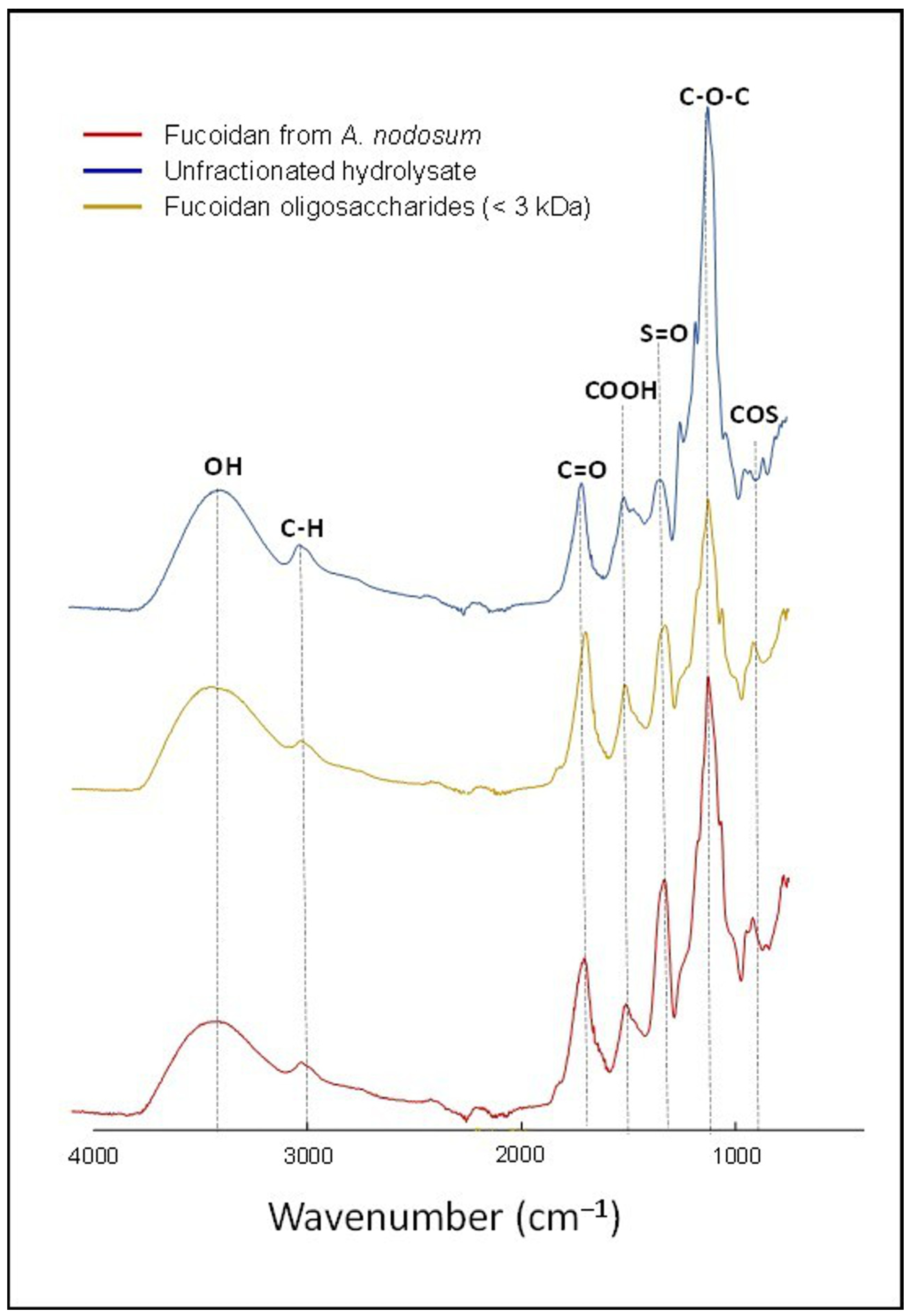
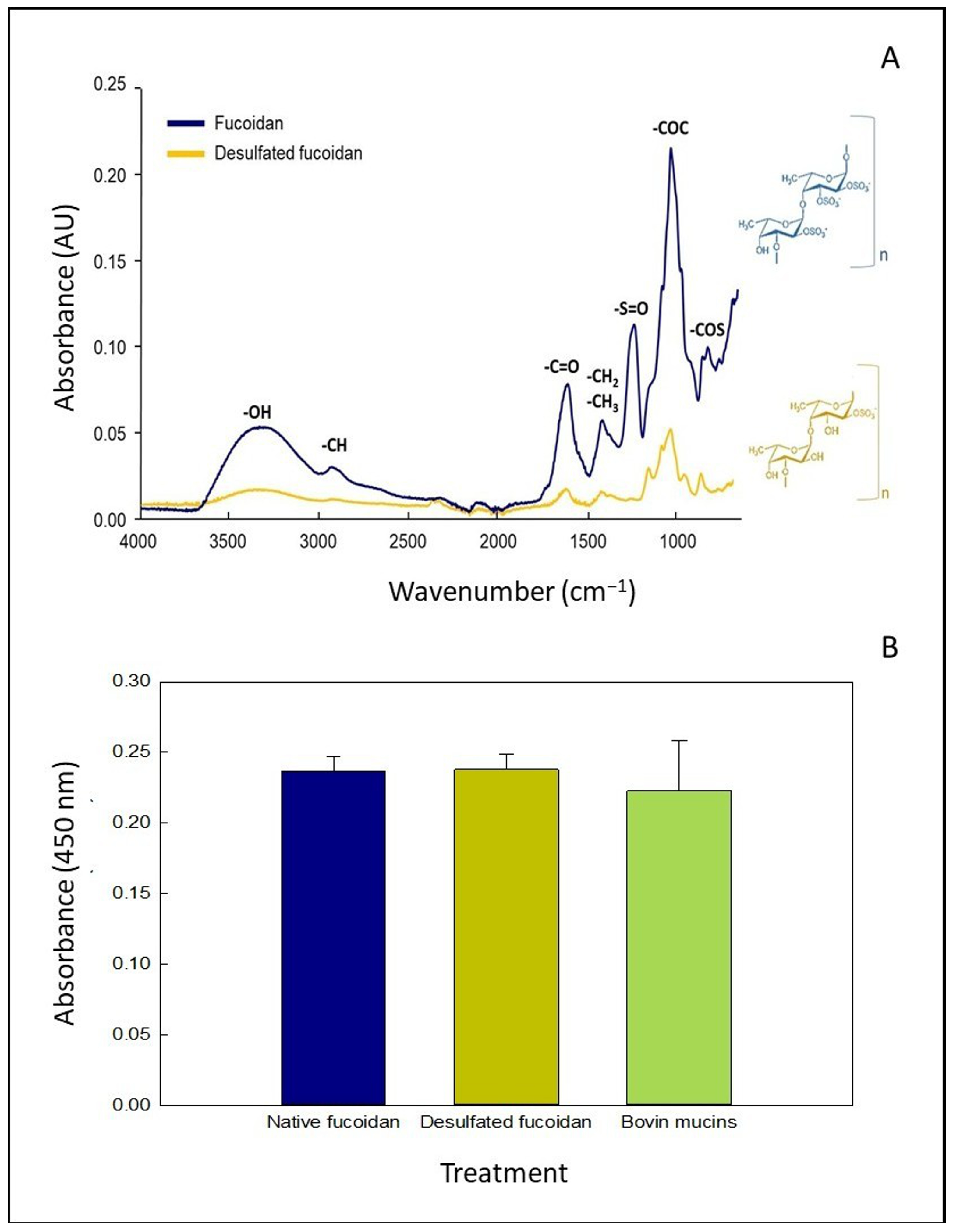
3.1. Characterization of OFuc

OFuc oligosaccharides were obtained by partial hydrolysis of fucoidan and further separation by ultrafiltration through a 3 kDa cut-off membrane. 65.2 ± 1.8% of the hydrolysate passed through the membrane, constituting the <3 kDa fraction. Due to its high yield and characteristics, this fraction was chosen to continue the experiments. Figure 1 shows the ATR-FTIR spectra of the <3 kDa fraction compared to the native fucoidan spectra. Both spectra were very similar; therefore, it can be assumed that the structure of the fucose was preserved. However, the intensity of the signals emitted by the glycosidic bonds (C-O-C) and the sulfate groups (S=O; COS) of the hydrolyzed fractions were lower than the intensity of these signals in the native fucoidan spectra. This effect was also observed by Sandoval Larios et al. (2023) [9] when hydrolyzing fucoidan from Macrocystis pyrifera. According to Pomin et al. (2005) [31], acid hydrolysis in sulfated polysaccharides starts with a desulfation of the molecule, followed by the cleavage of the glycosidic bond of the non-sulfated unit, resulting in homogeneous oligosaccharides.
The zeta potential and particle size of fucoidan and its OFuc are shown in Table 2. The OFuc showed lower zeta potential and smaller particle size than the untreated fucoidan. The changes in zeta potential are attributed to desulfation, while the changes in particle size are due to hydrolysis, being compatible with FTIR results [9]. In addition, an increase in OFuc polydispersity was observed due to the presence of populations with different sizes obtained by the fragmentation of the acid treatment.

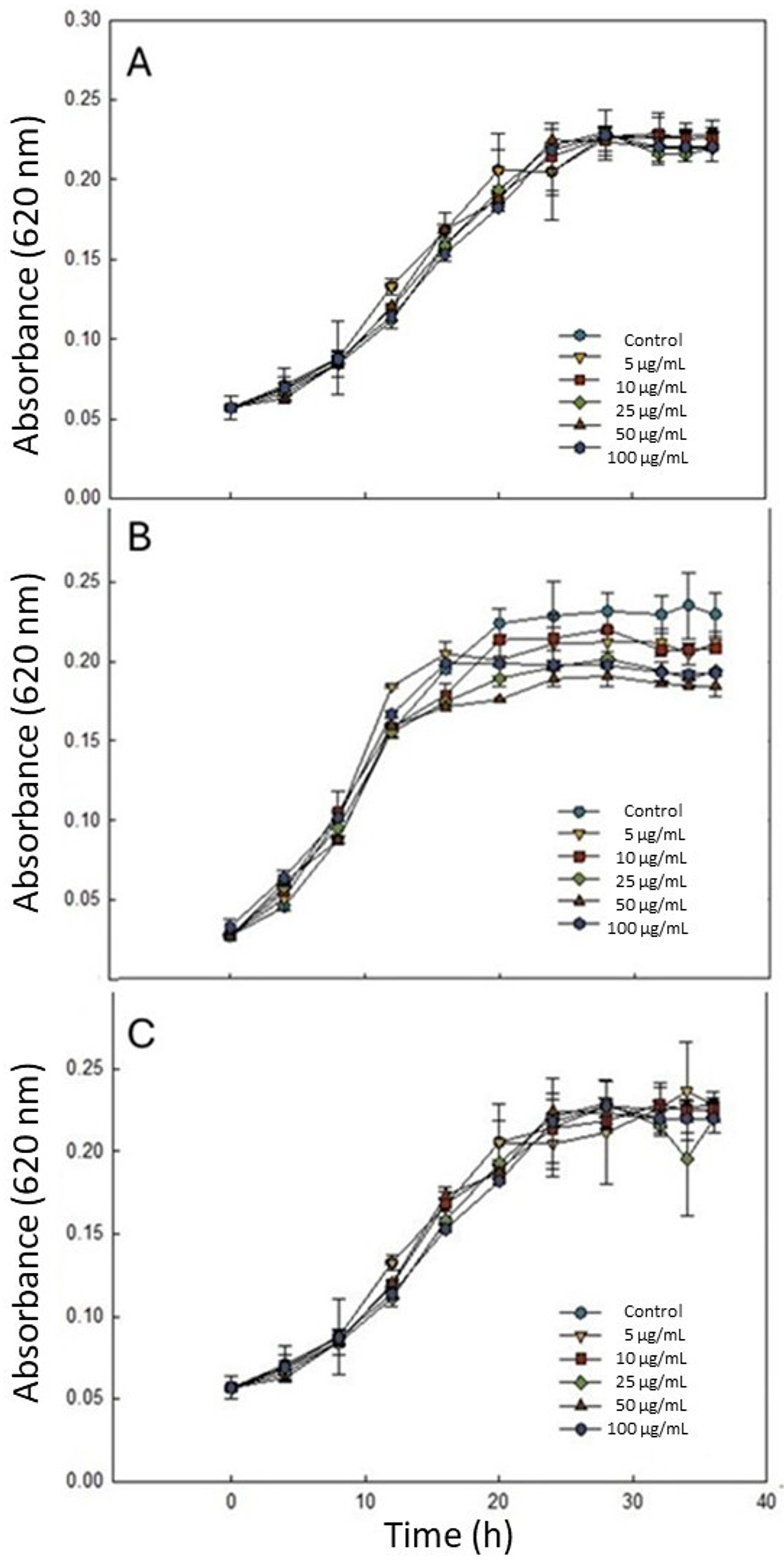
3.2. Effect of Fucose-Based Compounds on C. jejuni Growth

The presence of fucose (Figure 2) did not affect the specific growth rate of the pathogen. Campylobacter was previously considered unable to oxidize or ferment carbohydrates [32]. The primary energy sources for these bacteria are amino acids, citric acid cycle intermediates, and short-chain fatty acids [32]. However, various studies have concluded that some C. jejuni strains can utilize the carbohydrate L-fucose [33,34]. In addition, Muraoka et al. (2011) [35], Garber et al. (2020) [10], and Middendorf et al. (2024) [36] found that various Campylobacter strains carry the fucose utilization cluster Cj0480c–Cj0489 (fuc+). Moreover, the activation of the L-fucose utilization cluster allows specific C. jejuni strains to metabolize L-fucose, resulting in enhanced survival and in vitro higher invasion of C. jejuni to Caco-2 cells [36]. L-fucose is a common component of glycans present in intestinal mucins and enterocytes. Although these bacteria lack fucosidases to utilize fucose from fucose-based intestinal glycans, it has been shown that C. jejuni takes advantage of the fucose from the action of the fucosidases produced by spices of the intestinal microbiota [10]. Hence, future experiments are needed to determine if fucoidan and its oligosaccharides can be utilized as a carbon source by C. jejuni fuc+.
Two potential mechanisms have been proposed regarding how sulfated fucans affect the growth of pathogenic microorganisms [37]. Firstly, fucans may adhere to the cell membrane, which could ultimately lead to its destruction. This interaction might result in the leakage of cellular components, such as proteins [38] and nucleic acids [39], ultimately culminating in cell death. The second mechanism suggests that fucoidans exert an antibacterial effect by trapping nutrients, such as cationic minerals, in the nutrient medium. This occurs through negatively charged molecules of sulfated polysaccharides, which reduces the bioavailability of nutrients for microorganisms [37].
When examining the factors that influence the antibacterial activity of fucoidan, two of the most significant are molecular weight and sulfate content. Numerous studies have shown that fucoidans with low molecular weights (ranging from 5 kDa to 50 kDa) and high sulfate content (exceeding 20%) exhibit more significant bioactivity [37,39]. These results differ from our study, where no inhibitory effect was observed after 36 h of incubating C. jejuni at 37 °C in different concentrations (5, 10, 25, 50, or 100 µg/mL) of OFuc (Figure 2). One possible explanation for this behavior is that the concentration of OFuc used may not have been sufficient. Other studies have demonstrated an inhibitory effect of Gram-negative pathogens at concentrations of 6 mg/mL [39]. Therefore, it is essential to conduct further research using higher concentrations and to complete the characterization of the OFuc obtained in this study to better understand its behavior.
Incubation in the presence of fucoidan exhibited a bacteriostatic effect in a dose-dependent manner (≥25 µg/mL) (Figure 2). These findings were consistent with the specific growth rates obtained from the Prism-GraphPad software analysis (Table 3). Ayrapetyan et al. (2021) [37] reported a bacteriostatic effect of fucoidan on Escherichia coli, Staphylococcus epidermidis, S. aureus, and Bacillus licheniformis, which they attributed to the negative surface charge of the sulfate groups of fucoidan. These groups trap cationic minerals necessary for the bacteria or bind to functional groups of cell walls, causing their breakdown. Graikini et al. (2023) [40] observed complete inhibition of C. jejuni when incubated with 200 µg/mL of fucoidan extracted from Fucus vesiculosus. This finding underscores the importance of evaluating higher concentrations of fucoidan derived from A. nodosum to identify the specific concentration that effectively inhibits the growth of this pathogen.

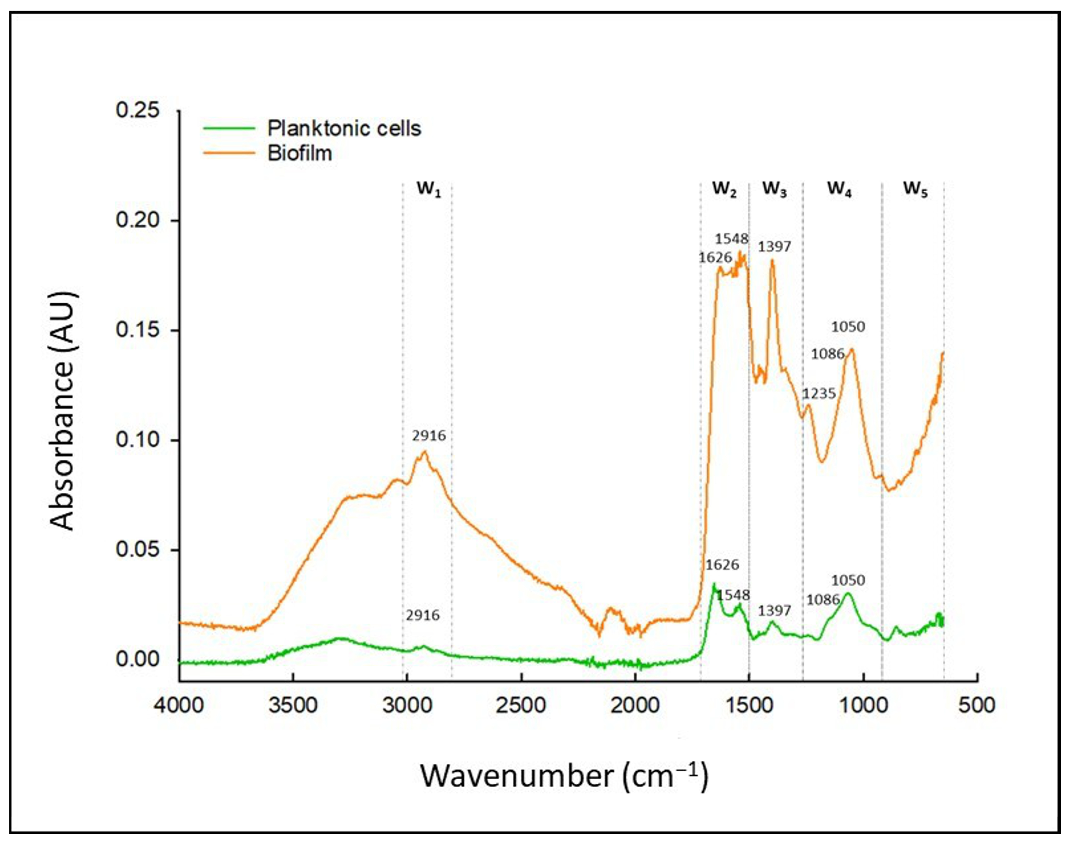
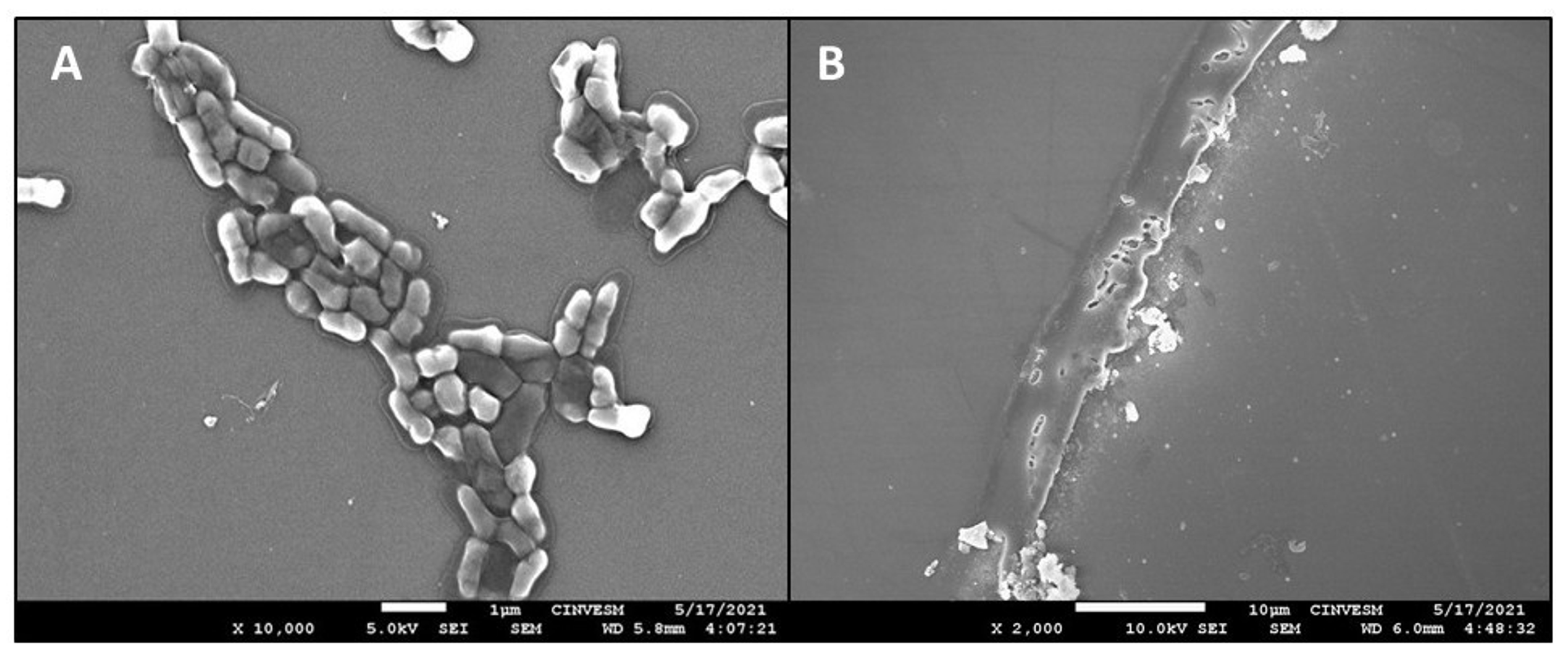
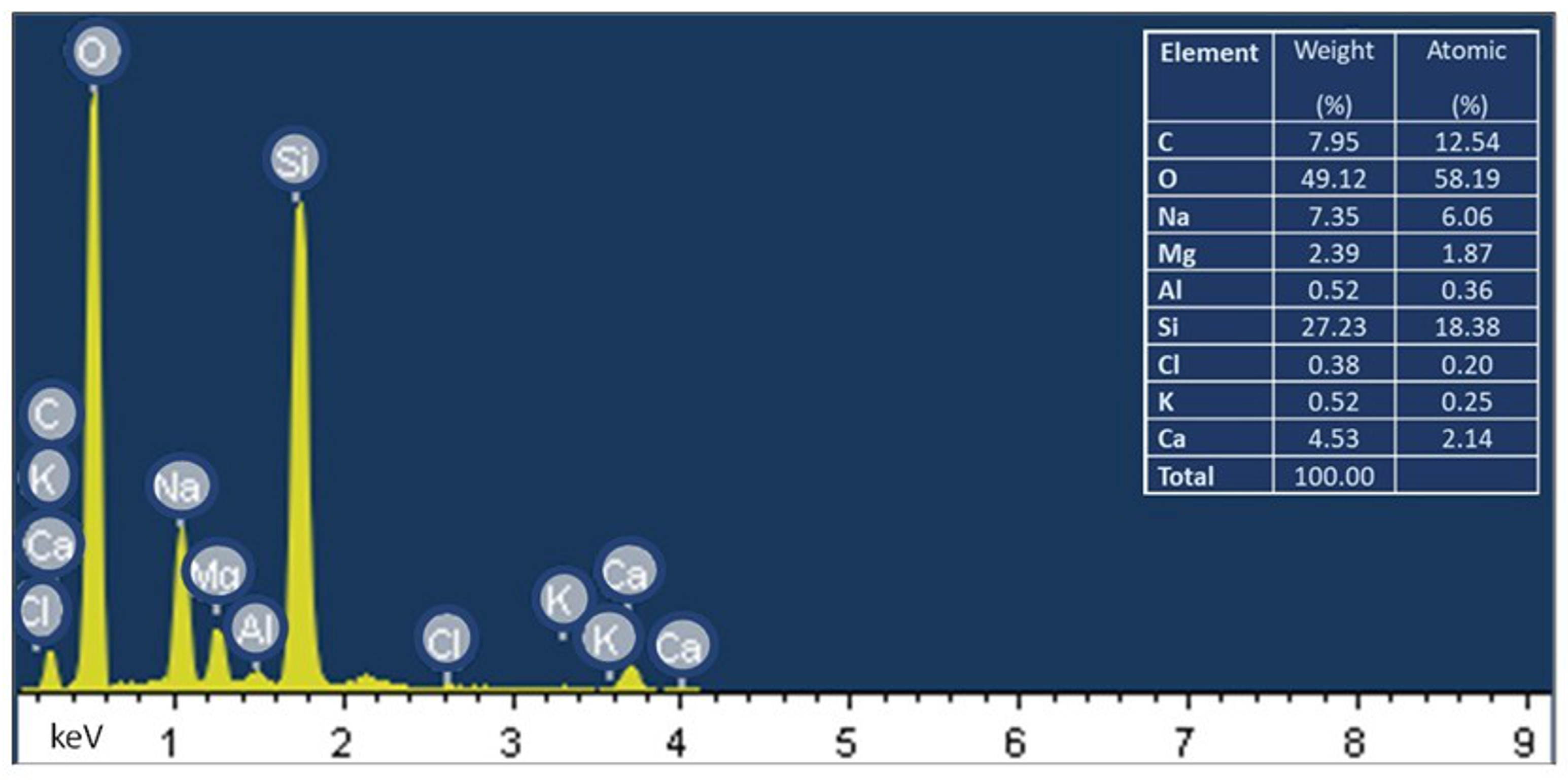
3.3. Characterization of Abiotic Biofilms of C. jejuni

Campylobacter jejuni can form abiotic biofilms on surfaces like glass, plastics, and stainless steel [6]. However, the degree of biofilm formation in C. jejuni is strain-specific; some strains cannot even form biofilms. Additionally, it is easier for Campylobacter to join the biofilms of other bacteria than to create their own [22,41].
In this study, the biofilm formation assays were initially conducted in MH media because it is reported that C. jejuni does not produce biofilms in a richer nutrient media like Brucella broth [15]. However, the growth of C. jejuni in MH was scarce, making it difficult to reach the necessary number of bacteria for each test. Because biofilm formation in C. jejuni is promoted by oxidative stress, it was decided to grow the bacteria in Brucella broth under aerobic conditions [42]. In this aerobic environment, biofilm formation was detected at 48 h of culture (Figure 3). Therefore, this time was established for the rest of the analyses.
The C. jejuni biofilms were characterized by ATR-FTIR, scanning electron microscopy (SEM), and energy dispersive spectroscopy (EDS) analysis. The typical spectrum of planktonic C. jejuni cells and their biofilms are presented in Figure 4. The classical studies of Helm et al. (1991) [43] for the characterization of planktonic cells by FTIR determine five spectral windows corresponding to the absorption expressed in wavenumbers (cm−1). Window 1 (3000–2800 cm−1) corresponds to the vibrations of functional groups usually present in fatty acids. Window 2 (1800–1500 cm−1) is assigned to proteins (here are the vibrations of the amide I and amide II bands). Window 3 (1500–1200 cm−1) is a mixed region with signals from functional groups present in proteins, fatty acids, nucleic acids, and phosphate-bearing compounds. Window 4 (1200–900 cm−1) is dominated by absorption bands of groups present in carbohydrates located in the cell wall. Window 5 (900–700 cm−1) is the fingerprint region. This region shows particular spectral patterns for each bacterium, which have not yet been assigned to cellular components or functional groups. Windows 3 and 4 are the most discriminatory for bacterial identification [43,44,45].
The biofilm spectra (Figure 4) showed an increase in the intensity of the signals assigned to proteins and peptides (Amide I, 1647–1626 cm−1; Amide II; 1548 cm−1), and in those assigned to carbohydrates signals, mainly at 1397 cm−1 (COOH groups) and 1086 and 1050 cm−1, which appear overlapping with the signal assigned to C=O groups and C-O-C glycosidic bonds, respectively [46]. These changes can be identified as important markers of biofilm formation in C. jejuni [47,48].
The chemical composition of C. jejuni abiotic biofilm depends on various factors such as the incubation temperature, strain type, presence or absence of O2, growth medium, and biofilm age [47,49]. Generally, the biofilm matrix comprises lipids, proteins, extracellular DNA, and carbohydrates [50]. An increase in amylogenic proteins is observed as C. jejuni biofilms mature [51,52]. This could explain the increase in the bands assigned to Amide I (1647–1626 cm−1) and Amide II (1548 cm−1) in the biofilm spectrum. Amylogenic proteins are now believed to be various bacteria’s prevailing structural components of the biofilm matrix. Their ability to self-assemble and polymerize confers high resistance to proteolytic cleavage and harsh denaturing conditions [52]. The C. jejuni biofilms spectrum also presented an increase in the signal assigned to phosphate groups (1235 cm−1) related to DNA presence [48]. Extracellular DNA plays an essential structural role in the biofilm matrix. It is also necessary for biofilm maturation [53].
Carbohydrates are also a critical constituent of C. jejuni biofilm. The increase in the signals corresponding to the carbohydrate region could be due to the production of exopolysaccharides linked by β1-3 and/or β1-4 bonds, which protect bacteria from harsh conditions [44]. In addition, Turonova et al. (2016) [43] identified the presence of N-acetylglucosamine, galactose, and N-acetylgalactosamine in the matrix of C. jejuni biofilms using fluorescence lectin-binding assays. Several genes involved in carbohydrate synthesis are expressed differentially during Campylobacter jejuni biofilm formation [52,54].
Figure 5 shows micrographs of the initial stages of biofilm formation. The most successful biofilm formation by Campylobacter jejuni was observed after 48 h at 37 °C under aerobic conditions in Brucella broth. At this time, C. jejuni entered a viable but not culturable state, losing its spiral shape (5A). Figure 5B shows the primary production of the biofilm matrix. Campylobacter biofilm communities display phenotypic differences compared to planktonic cultures, such as shrinking in shape, which can result in a coccus-like shape [55].
The energy dispersive spectroscopy spectra of the biofilms showed the presence of C, O, K, Na, Cl, and Mg (Figure 6). Cationic metals are abundant in the Campylobacter biofilm matrix. These cations can be toxic for Campylobacter planktonic cells at high concentrations. However, their absorption and accumulation stabilize and preserve biofilm matrix integrity and prevent erosion [56,57].

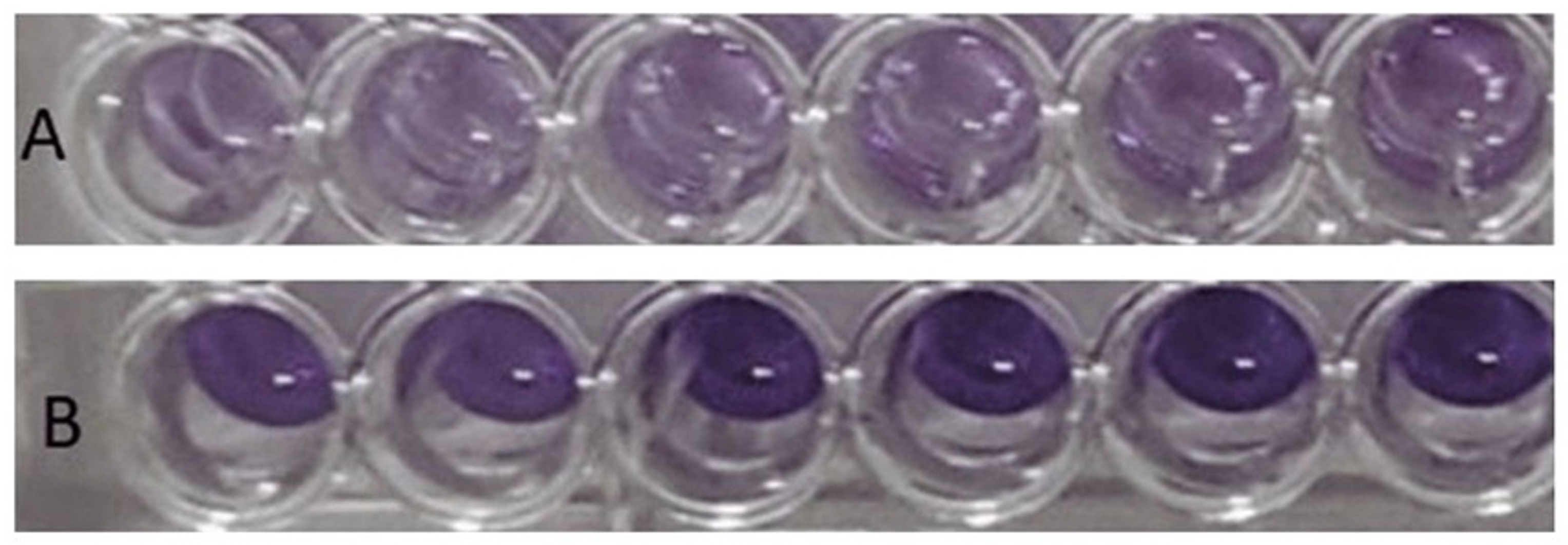
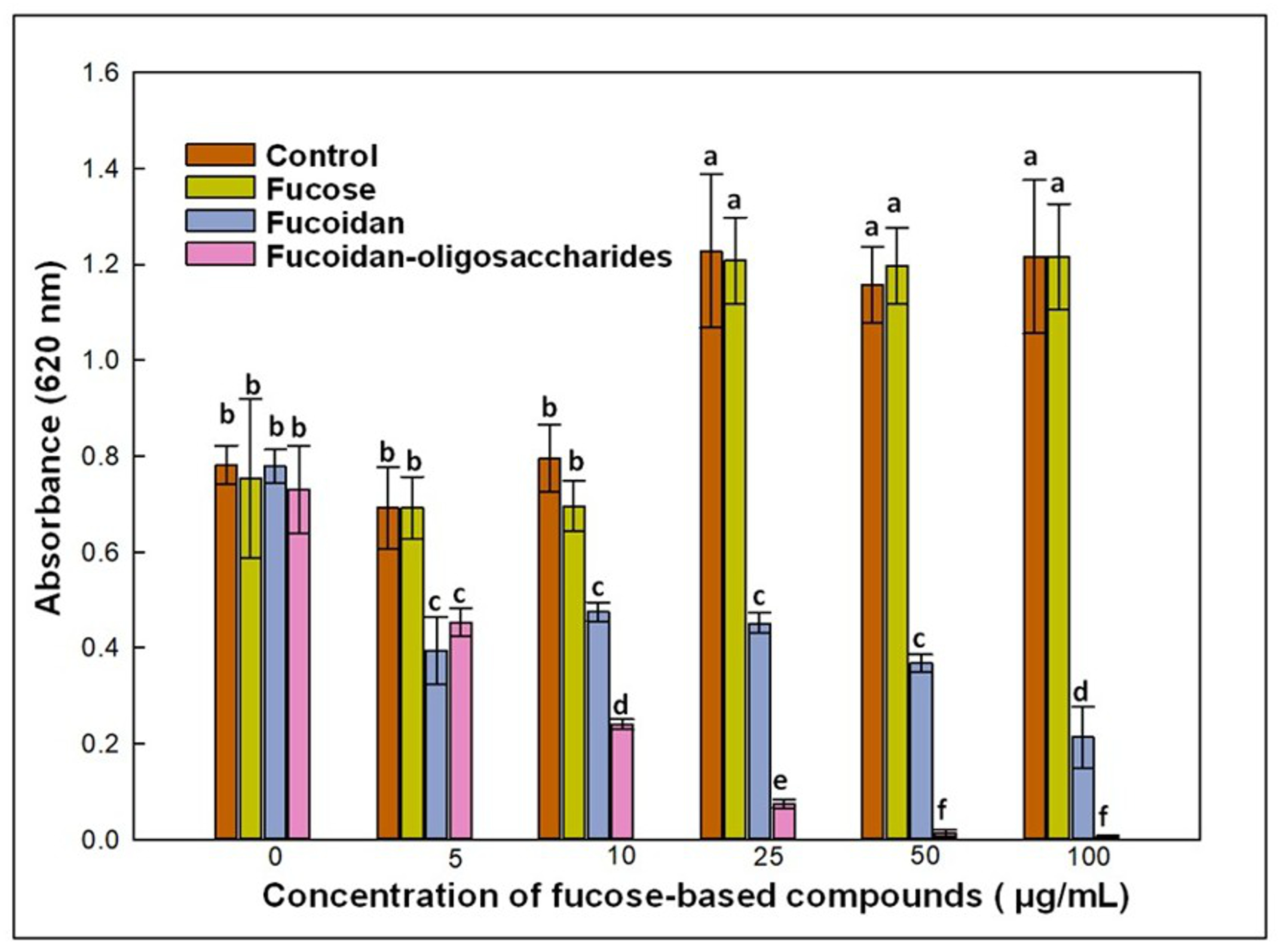
3.4. Effect of Fucose-Based Compounds on Biofilm Formation

The impact of fucose-based compounds on biofilm formation is shown in Figure 7. Fucose did not decrease biofilm formation at any of the concentrations tested. These results contrast those obtained by Dwivedi et al. (2016) [8], who observed a 50% reduction in C. jejuni NCTC11168 biofilm formation in 25 mM L-fucose. This strain possesses a fuc+ locus that allows it to utilize fucose as an energy source [36]. According to Dwivedi et al. (2016) [8], if the strain can detect and subsequently use the fucose present, most bacterial cells will remain in their planktonic form and not form biofilms. Further studies are required to detect the presence of the fuc locus in C. jejuni ATCC 43442 and to test the impact on biofilm formation using the concentrations used in Dwivedi’s study (approximately 4.10 g instead of 100 µg/mL).
Both the presence of fucoidan and fucoidan-oligosaccharides decreased the C. jejuni biofilm formation. Biofilm formation was severely reduced when bacteria grew with a fucoidan concentration of 100 µg. In contrast, the impact of fucoidan-oligosaccharides was more significant since they decreased the biofilm production at concentrations greater than 10 µg. Other studies have tested the anti-biofilm-forming effect of fucoidan. Jun et al. (2018) [58] proved that fucoidan from Fucus vesiculosus at concentrations above 250 µg/mL−1 completely suppressed Streptococcus mutans and S. sobrinus biofilm formation. Khan et al. (2019) [59] synthesized Au-fucoidan nanoparticles and found a minimum Pseudomonas aeruginosa biofilm inhibition concentration of 128 µg/mL−1.
The mechanism by which fucoidan inhibits biofilm production is unknown but may be linked to the disruption of quorum sensing (QS) signals that regulate biofilm formation [60]. This effect is attributed to fucoidan’s ability to interact with various molecules and receptors on bacterial cell surfaces [13,16,60]. The release of autoinducer molecules such as AI-2 regulates biofilm production. Fucoidan could employ similar strategies to those of QS inhibitors, like flavonoids and furanone, that target these signaling molecules’ receptors, either inactivating them or competing for binding [61]. This suggests that fucoidan may act by mimicking QS signals and disrupting the bacterial QS system [60,61]. Nonetheless, further research is needed to elucidate better the mechanisms of quorum sensing (QS) and quorum quenching (QQ) in Campylobacter jejuni.
The ability to form biofilms significantly enhances the survival of Campylobacter jejuni in harsh environments and plays a crucial role in the survival and transmission of this pathogen to humans. Furthermore, biofilm formation has been identified as a potential source of antimicrobial resistance [22]. Many C. jejuni strains increasingly resist the most commonly prescribed antibiotics. In this context, applying fucoidan alongside other compounds that inhibit C. jejuni biofilm formation—such as trans-cinnamaldehyde, eugenol, carvacrol, and citrus compounds—may offer a potential treatment for controlling the persistence of this bacteria on abiotic surfaces [46]. Additionally, fucoidan or OFuc treatments to food surfaces could control C. jejuni biofilm formation without affecting the taste of the food.

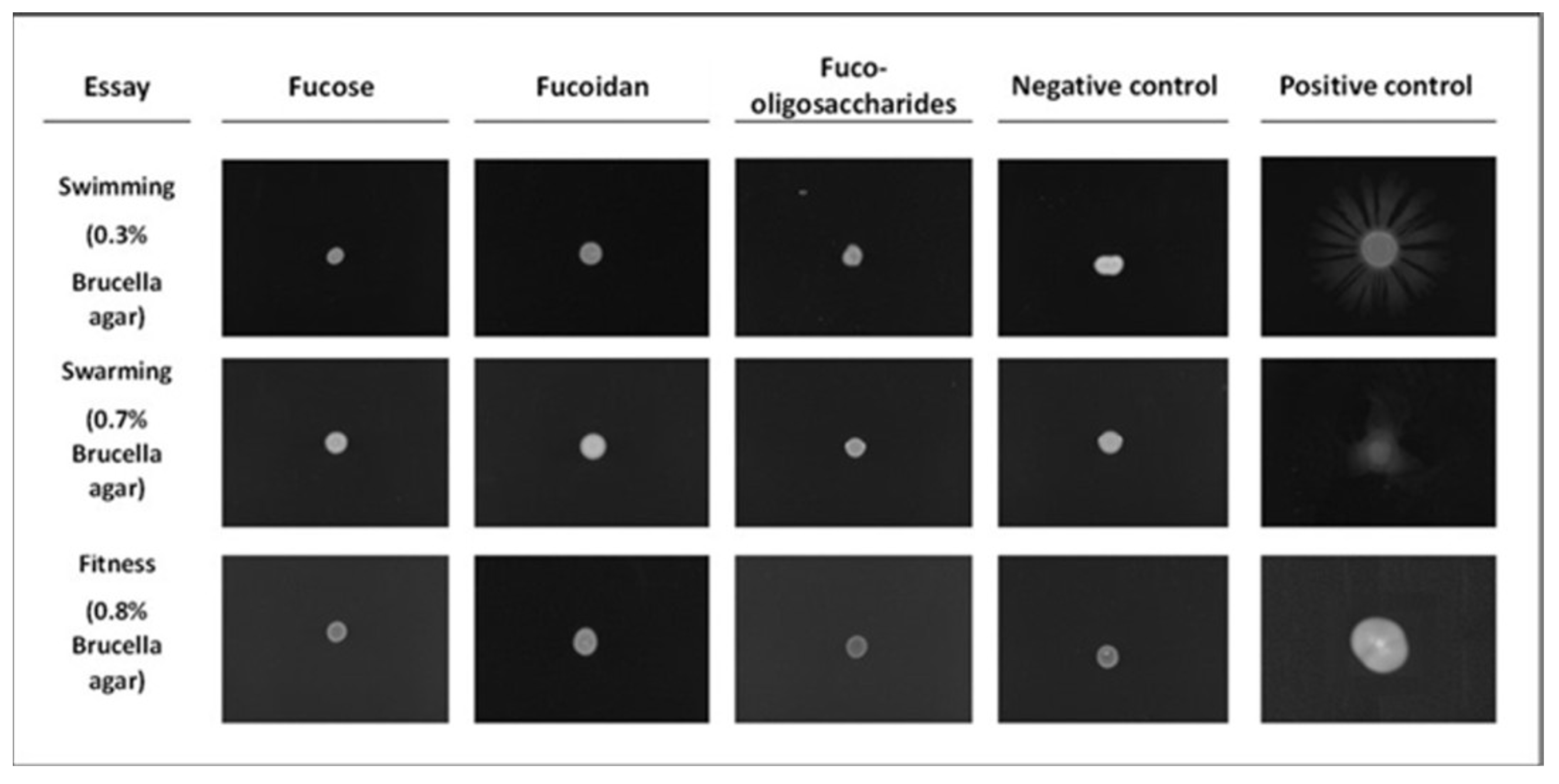
3.5. Motility Assays

Chemotaxis plays an essential role in the pathogenicity of C. jejuni, as it allows the pathogen to reach the site of infection through the motility imparted by its polar flagellum. Mutants lacking the chemotaxis signal transduction (che) pathway are less virulent and exhibit reduced colonization in animal models [62,63]. L-fucose is the only chemoattractant carbohydrate for C. jejuni [36], and its chemotaxis mutants, such as cheA, do not swim toward this sugar [64].
The results of the motility assays performed in this work showed the absence of swimming, swarming, and fitness in C. jejuni grown on Brucella agar and in MH media. Similar results were reported by Cohen et al. (2020) [65] when applying the same techniques. Fucose, fucoidan, or fucoidan-oligosaccharides did not affect movement at any probed concentrations. Figure 8 shows an example of the results obtained using the highest concentrations (100 µg) of fucose-based compounds.
An alternative to studying the mobility of C. jejuni is the use of more sensitive techniques. For example, using the video technique, Shigematsu et al. (1998) [66] studied the swimming patterns of C. jejuni in low and high-viscosity environments. In low-viscosity media, C. jejuni swam at an average speed of 39.3 µm/s with frequent direction changes. The speed of C. jejuni increased significantly in a medium with a viscosity of around 40 centipoises, exhibiting repeated back-and-forth (spiral) swimming patterns similar to the swimming pattern of spirochetes. This implies that the pathogen changes its motility pattern depending on the viscosity of the medium. The spiral swimming shape of C. jejuni could significantly influence its ability to swim in highly viscous media such as the mucous layer of the intestinal tract. Therefore, it is advisable to use this technique in further investigations to see how the bacteria behave in the presence of fucose and fucoidan and its oligosaccharides.

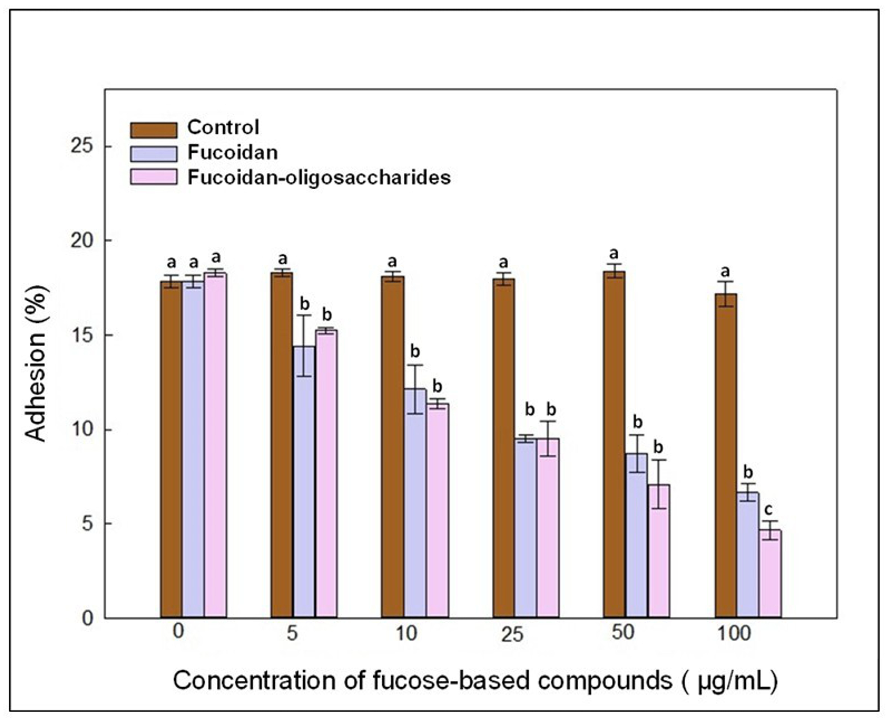
3.6. Adhesion Analysis

To investigate how fucose-based compounds affect the adhesion of Campylobacter jejuni, we used the Microbial Adhesion to Hydrocarbons (MATH) technique, which measures the percentage of bacteria adhering to p-xylene. Figure 9 shows the results obtained with different concentrations of fucoidan and fucoidan-oligosaccharides. The results related to fucose were inconsistent and not reproducible. Overall, a decrease in pathogen adhesion was observed with both fucoidan and fucoidan-oligosaccharides, and this effect was dose-dependent. Previous studies have demonstrated that C. jejuni can bind to fucose-based complex carbohydrates [67,68].
Moreover, human milk fucosylated oligosaccharides inhibit the binding of Campylobacter jejuni to human enterocytes, inferring the presence of fucose-specific lectin-like adhesins [69].
Lectin-like adhesins are important virulence factors that allow bacteria to attach to specific glycan receptors found on the plasma membranes of host cells. One strategy for preventing and treating infections is to block these binding sites using carbohydrate analogs that mimic the glycan receptors [70]. From a structural standpoint, binding to lectin-like adhesins involves carbohydrates forming non-covalent, reversible interactions that are specific regarding geometry and charge complementarity. Evidence indicates that adhesins on the surface of Campylobacter jejuni can recognize fucose [67,68,69]. In this context, a fucose-containing molecule, such as fucoidan, could function as an analog for the fucosylated receptors to which the bacteria attach. However, the presence of negatively charged sulfate groups within the fucoidan molecule may hinder the interaction between fucose and lectin. An ELLA assay was conducted to evaluate the recognition of protein–carbohydrate interactions between the fucose residues of fucoidan and the adhesins of C. jejuni ATCC 43442. The study used both fucoidan and desulfated fucoidan. Desulfation was performed to investigate the impact of sulfate groups on the interaction between bacteria and fucoidan. Figure 10A compares the FTIR-ATR spectrum of fucoidan and desulfated fucoidan, which reveals a notable reduction in the characteristic signals of sulfated groups in the desulfated molecule. Interestingly, both sulfated and desulfated fucoidans exhibited the same level of affinity when recognized by C. jejuni, as shown in Figure 10B. This suggests that the presence of sulfated groups does not hinder their interaction. However, complementary studies are necessary to confirm this observation. Other lectins that recognize fucoidan include Ulex europaeus agglutinin I, Anguilla anguilla agglutinin, L-Selectin, Ly-49 C-Type lectin, and p-Selectin [71,72].
These results support the hypothesis that fucoidan and its fucoidan-oligosaccharides can function as anchor molecules, disrupting the adhesion of C. jejuni to the intestine and thereby preventing infection [9]. However, further specific assays are required to verify this.

4. Conclusions

Fucoidan and its oligosaccharides exhibited a potent inhibitory effect on the abiotic biofilm formed by C. jejuni. This biofilm significantly enhances the pathogen’s ability to survive in extreme conditions, facilitating its transmission to humans. These biofilms also serve as a reservoir of Campylobacter jejuni strains resistant to antibiotics, especially those used to treat campylobacteriosis. Given the impact of campylobacteriosis and the emergence of antimicrobial resistance in Campylobacter, the application of fucoidan or its oligosaccharides, whether alone or in combination with other antibiofilm agents, could aid in controlling this pathogen in the food industry. In vitro studies show that C. jejuni binds to fucoidan through a lectin–carbohydrate recognition mechanism. This finding provides a new approach to combat Campylobacter infections by using fucoidan as a glycomimetic antagonist, potentially preventing the adhesion of this pathogen to fucosylated receptors that allow the bacterium union to intestinal cells and their subsequent infection.

Author Contributions

Conceptualization, G.R.C.M.; methodology, S.P.F., G.S.L., R.I.A.C., A.M.G.-P., A.G.-G. and R.C.; investigation, supervision, and validation, J.A.S.-S., J.Á.H.-O., R.C. and A.G.-G.; data curation, J.A.S.-S., J.Á.H.-O., R.C. and A.G.-G.; writing—original draft preparation, G.S.L. and G.R.C.M.; writing—review and editing, G.R.C.M. and J.Á.H.-O.; project administration, G.R.C.M.; funding acquisition, G.R.C.M. All authors have read and agreed to the published version of the manuscript.

Funding

This work was supported by the Secretariat of Science, Humanities, Technology, and Innovation of Mexico (SECIHTI) under project CBA1S-25043. S.P.F. and G.S.L. received postgraduate scholarship grants from SECIHTI, with grant numbers 1012066 and 935067, respectively.

Institutional Review Board Statement

Not applicable.

Data Availability Statement

Data are contained within the article.

Acknowledgments

The authors thank the Secretariat of Science, Humanities, Technology, and Innovation of Mexico (SECIHTI) and the Institutional Analytical Platform (PAI-10363) of the Centro de Investigación en Alimentación y Desarrollo A.C.

Conflicts of Interest

The authors declare that they have no known competing financial interests or personal relationships that could have appeared to influence the work reported in this paper.

References

  1. World Health Organization. Campylobacter. 2024. Available online: https://www.who.int/news-room/fact-sheets/detail/campylobacter (accessed on 9 March 2024).
  2. Igwaran, A.; Okoh, A.I. Human campylobacteriosis: A public health concern of global importance. Heliyon 2019, 5, e02814. [Google Scholar] [CrossRef] [PubMed]
  3. Davys, G.; Marshall, J.C.; Fayaz, A.; Weir, R.P.; Benschop, J. Campylobacteriosis associated with the consumption of unpasteurized milk: Findings from a sentinel surveillance site. Epidemiol. Infect. 2020, 148, e16. [Google Scholar] [CrossRef] [PubMed]
  4. Bronowski, C.; James, C.E.; Winstanley, C. Role of environmental survival in transmission of Campylobacter jejuni. FEMS Microbiol. Lett. 2014, 356, 8–19. [Google Scholar] [CrossRef]
  5. Šimunović, K.; Ramić, D.; Xu, C.; Smole Možina, S. Modulation of Campylobacter jejuni motility, adhesion to polystyrene surfaces, and invasion of INT407 cells by Quorum-Sensing inhibition. Microorganisms 2020, 8, 104. [Google Scholar] [CrossRef]
  6. Ma, L.; Feng, J.; Zhang, J.; Lu, X. Campylobacter biofilms. Microbiol. Res. 2022, 264, 127149. [Google Scholar] [CrossRef]
  7. Haddad, N.; Marce, C.; Magras, C.; Cappelier, J.M. An overview of methods used to clarify pathogenesis mechanisms of Campylobacter jejuni. J. Food Prot. 2010, 73, 786–802. [Google Scholar] [CrossRef] [PubMed]
  8. Dwivedi, R.; Nothaft, H.; Garber, J.; Xin Kin, L.; Stahl, M.; Flint, A.; van Vliet, A.H.; Stintzi, A.; Szymanski, C.M. L-fucose influences chemotaxis and biofilm formation in Campylobacter jejuni. Mol. Microbiol. 2016, 101, 575–589. [Google Scholar] [CrossRef]
  9. Sandoval Larios, G.; Huerta Ocampo, J.Á.; Sarabia Sainz, J.A.; García Galaz, A.; Guzmán-Partida, A.M.; Madera Santana, T.J.; Ramos-Clamont Montfort, G. Core-shell nanoparticles from fucoidan neoglycans: Synthesis, characterization with capability of glycomimetic ligands for Campylobacter jejuni. Carbohydr. Polym. Technol. Appl. 2023, 6, 100384. [Google Scholar]
  10. Garber, J.M.; Nothaft, H.; Pluvinage, B.; Stahl, M.; Bian, X.; Porfirio, S.; Enriquez, A.; Butcher, J.; Huang, H.; Glushka, J.; et al. The gastrointestinal pathogen Campylobacter jejuni metabolizes sugars with potential help from commensal Bacteroides vulgatus. Commun. Biol. 2020, 3, 2. [Google Scholar] [CrossRef]
  11. Meng, J.; Zhu, Y.; Wang, N.; Zhang, W.; Mu, W. Recent advances in a functional deoxy hexose L-fucose: Occurrence, physiological effects, and preparation. Trends Food Sci. 2023, 138, 16–26. [Google Scholar] [CrossRef]
  12. Xiao, M.; Ren, X.; Yu, Y.; Gao, W.; Zhu, C.; Sun, H.; Kong, Q.; Fu, X.; Mou, H. Fucose-containing bacterial exopolysaccharides: Sources, biological activities, and food applications. Food. Chem. X 2022, 13, 100233. [Google Scholar] [CrossRef] [PubMed]
  13. Jiao, G.; Yu, G.; Zhang, J.; Ewart, H.S. Chemical structures and bioactivities of sulfated polysaccharides from marine algae. Mar. Drugs 2011, 9, 196–223. [Google Scholar] [CrossRef] [PubMed]
  14. Fitton, H.J.; Stringer, D.S.; Park, A.Y.; Karpiniec, S.N. Therapies from Fucoidan: New Developments. Mar. Drugs. 2019, 17, 571. [Google Scholar] [CrossRef]
  15. Citkowska, A.; Szekalska, M.; Winnicka, K. Possibilities of fucoidan utilization in the development of pharmaceutical dosage forms. Mar. Drugs 2019, 17, 458. [Google Scholar] [CrossRef] [PubMed]
  16. Ale, M.T.; Mikkelsen, J.D.; Meyer, A.S. Important determinants for fucoidan bioactivity: A critical review of structure-function relations and extraction methods for fucose-containing sulfated polysaccharides from brown seaweeds. Mar. Drugs 2011, 9, 2106–2130. [Google Scholar] [CrossRef]
  17. Zayed, A.; El-Aasr, M.; Ibrahim, A.-R.S.; Ulber, R. Fucoidan characterization: Determination of purity and physicochemical and chemical properties. Mar. Drugs 2020, 18, 571. [Google Scholar] [CrossRef]
  18. van Weelden, G.; Bobiński, M.; Okła, K.; van Weelden, W.J.; Romano, A.; Pijnenborg, J.M.A. Fucoidan structure and activity in relation to anti-cancer mechanisms. Mar. Drugs 2019, 17, 32. [Google Scholar] [CrossRef]
  19. Silchenko, A.S.; Kusaykin, M.I.; Kurilenko, V.V.; Zakharenko, A.M.; Isakov, V.V.; Zaporozhets, T.S.; Isakov, V.V.; Zvyagintseva, T.N. Hydrolysis of fucoidan by fucoidanase isolated from the marine bacterium, Formosa algae. Mar. Drugs 2013, 11, 2413–2430. [Google Scholar] [CrossRef]
  20. Pielesz, A.; Biniaś, W.; Paluch, J. Mild acid hydrolysis of fucoidan: Characterization by electrophoresis and FT-Raman spectroscopy. Carbohydr. Res. 2011, 346, 1937–1944. [Google Scholar] [CrossRef]
  21. Mousavi, N.S.; Tabarsa, M.; You, S.; Ghandehari Yazdi, A.P.; Cao, R.; Gavlighi, H.A.; Babakhani, A. Hydrolysis and cross-flow ultrafiltration as an alternative process to isolate fucoidans from edible seaweed Nizamuddinia zarnardinii with enhanced immunostimulatory efficacy. Algal Res. 2024, 82, 103632. [Google Scholar] [CrossRef]
  22. Reeser, R.J.; Medler, R.T.; Billington, S.J.; Jost, B.H.; Joens, L.A. Characterization of Campylobacter jejuni biofilms under defined growth conditions. Appl. Environ. Microbiol. 2007, 73, 1908–1913. [Google Scholar] [CrossRef] [PubMed]
  23. Elvers, K.T.; Park, S.F. Quorum sensing in Campylobacter jejuni: Detection of a luxS encoded signaling molecule. Microbiology 2002, 148, 1475–1481. [Google Scholar] [CrossRef]
  24. Shen, G.; Yang, L.; Lv, X.; Zhang, Y.; Hou, X.; Li, M.; Zhou, M.; Pan, L.; Chen, A.; Zhang, Z. Antibiofilm activity and mechanism of linalool against food spoilage Bacillus amyloliquefaciens. Int. J. Mol. Sci. 2023, 24, 10980. [Google Scholar] [CrossRef] [PubMed]
  25. Frenette, P.S.; Weiss, L. Sulfated glycans induce rapid hematopoietic progenitor cell mobilization: Evidence for selectin-dependent and independent mechanisms. Blood 2000, 96, 2460–2468. [Google Scholar] [CrossRef] [PubMed]
  26. McCoy, J.P.; Varani, J.; Goldstein, I.J. Enzyme-linked lectin assay (ELLA). II. Detection of carbohydrate groups on the surface of unfixed cells. Exp. Cell Res. 1984, 151, 96–103. [Google Scholar] [CrossRef]
  27. Sandoval Larios, G.; Palafox Félix, S.; Armenta Corral, R.I.; García-Hernández, J.; Balandrán-Quintana, R.R.; Ramos Clamont Montfort, G. El fucoidan de Sargassum sinicola es reconocido in vitro por la bacteria Campylobacter jejuni. TIP Rev. Espec. Cienc. Químico-Biológicas 2024, 27, 1–13. [Google Scholar] [CrossRef]
  28. Wojtczak, K.; Byrne, J.P. Structural considerations for building synthetic glycoconjugates as inhibitors for Pseudomonas aeruginosa lectins. Chem. Med. Chem. 2022, 17, e202200081. [Google Scholar] [CrossRef]
  29. Hauck, D.; Joachim, I.; Frommeyer, B.; Varrot, A.; Philipp, B.; Möller, H.M.; Imberty, A.; Exner, T.E.; Titz, A. Discovery of two classes of potent glycomimetic inhibitors of Pseudomonas aeruginosa LecB with distinct binding modes. ACS Chem. Biol. 2013, 8, 1775–1784. [Google Scholar] [CrossRef]
  30. Richards, S.J.; Gibson, M.I. Toward glycomaterials with selectivity as well as affinity. JACS Au 2021, 1, 2089–2099. [Google Scholar] [CrossRef]
  31. Pomin, V.H.; Valente, A.P.; Pereira, M.S.; Mourão, P.A. Mild acid hydrolysis of sulfated fucans: A selective 2-desulfation reaction and an alternative approach for preparing tailored sulfated oligosaccharides. Glycobiology 2005, 15, 1376–1385. [Google Scholar] [CrossRef]
  32. Wagley, S.; Newcombe, J.; Laing, E.; Yusuf, E.; Sambles, C.M.; Studholme, D.J.; La Ragione, R.M.; Titball, R.W.; Champion, O.L. Differences in carbon source utilization distinguish Campylobacter jejuni from Campylobacter coli. BMC Microbiol. 2014, 14, 262. [Google Scholar] [CrossRef] [PubMed]
  33. Stahl, M.; Friis, L.M.; Nothaft, H.; Liu, X.; Li, J.; Szymanski, C.M.; Stintzi, A. L-fucose utilization provides Campylobacter jejuni with a competitive advantage. Proc. Natl. Acad. Sci. USA 2011, 108, 7194–7199. [Google Scholar] [CrossRef]
  34. Velayudhan, J.; Kelly, D.J. Analysis of gluconeogenic and anaplerotic enzymes in Campylobacter jejuni: An essential role for phosphoenolpyruvate carboxykinase. Microbiology 2002, 148, 685–694. [Google Scholar] [CrossRef]
  35. Muraoka, W.T.; Zhang, Q. Phenotypic and genotypic evidence for L-fucose utilization by Campylobacter jejuni. J. Bacteriol. 2011, 193, 1065–1075. [Google Scholar] [CrossRef]
  36. Middendorf, P.S.; Wijnands, L.M.; Boeren, S.; Zomer, A.L.; Jacobs-Reitsma, W.F.; den Besten, H.M.W.; Abee, T. Activation of the L-fucose utilization cluster in Campylobacter jejuni induces proteomic changes and enhances Caco-2 cell invasion and fibronectin binding. Heliyon 2024, 10, e34996. [Google Scholar] [CrossRef]
  37. Ayrapetyan, O.N.; Obluchinskaya, E.D.; Zhurishkina, E.V.; Skorik, Y.A.; Lebedev, D.V.; Kulminskaya, A.A.; Lapina, I.M. Antibacterial properties of fucoidans from the brown algae Fucus vesiculosus L. of the Barents Sea. Biology 2021, 10, 67. [Google Scholar] [CrossRef]
  38. Palanisamy, S.; Vinosha, M.; Rajasekar, P.; Anjali, R.; Sathiyaraj, G.; Marudhupandi, T.; Selvam, S.; Prabhu, N.M.; You, S. Antibacterial efficacy of a fucoidan fraction (Fu-F2) extracted from Sargassum polycystum. Int. J. Biol. Macromol. 2019, 125, 485–495. [Google Scholar] [CrossRef] [PubMed]
  39. Liu, M.; Liu, Y.; Cao, M.J.; Liu, G.M.; Chen, Q.; Sun, L.; Chen, H. Antibacterial activity and mechanisms of depolymerized fucoidans isolated from Laminaria japonica. Carbohydr. Polym. 2017, 172, 294–305. [Google Scholar] [CrossRef] [PubMed]
  40. Graikini, D.; Soro, A.B.; Sivagnanam, S.P.; Tiwari, B.K.; Sánchez, L. Bioactivity of fucoidan-rich extracts from Fucus vesiculosus against rotavirus and foodborne pathogens. Mar. Drugs 2023, 21, 478. [Google Scholar] [CrossRef]
  41. Heimesaat, M.M.; Backert, S.; Alter, T.; Bereswill, S. Molecular targets in Campylobacter infections. Biomolecules 2023, 13, 409. [Google Scholar] [CrossRef]
  42. Püning, C.; Su, Y.; Lu, X.; Gölz, G. Molecular mechanisms of Campylobacter biofilm formation and Quorum Sensing. Curr. Top. Microbiol. Immunol. 2021, 431, 293–319. [Google Scholar] [CrossRef] [PubMed]
  43. Helm, D.; Labischinski, H.; Schallehn, G.; Naumann, D. Classification and identification of bacteria by Fourier-transform infrared spectroscopy. J. Gen. Microbiol. 1991, 137, 69–79. [Google Scholar] [CrossRef] [PubMed]
  44. Novais, Â.; Freitas, A.R.; Rodrigues, C.; Peixe, L. Fourier transform infrared spectroscopy: Unlocking fundamentals and prospects for bacterial strain typing. Eur. J. Clin. Microbiol. Infect. Dis. 2019, 38, 427–448. [Google Scholar] [CrossRef]
  45. Sousa, C.; Novais, Â.; Magalhães, A.; Lopes, J.; Peixe, L. Diverse high-risk B2 and D Escherichia coli clones depicted by Fourier transform infrared spectroscopy. Sci. Rep. 2013, 3, 3278. [Google Scholar] [CrossRef]
  46. Wang, H.; Ding, S.; Wang, G.; Xu, X.; Zhou, G. In situ characterization and analysis of Salmonella biofilm formation under meat processing environments using a combined microscopic and spectroscopic approach. Int. J. Food Microbiol. 2013, 167, 293–302. [Google Scholar] [CrossRef]
  47. Melo, R.T.; Mendonça, E.P.; Monteiro, G.P.; Siqueira, M.C.; Pereira, C.B.; Peres, P.; Fernandez, H.; Rossi, D.A. Intrinsic and extrinsic aspects on Campylobacter jejuni biofilms. Front. Microbiol. 2017, 8, 1332. [Google Scholar] [CrossRef]
  48. Tram, G.; Klare, W.P.; Cain, J.A.; Mourad, B.; Cordwell, S.J.; Korolik, V.; Day, C.J. RNA sequencing data sets identifying differentially expressed transcripts during Campylobacter jejuni biofilm formation. Microbiol. Resour. Announce 2020, 9. [Google Scholar] [CrossRef]
  49. Turonova, H.; Briandet, R.; Rodrigues, R.; Hernould, M.; Hayek, N.; Stintzi, A.; Pazlarova, J.; Tresse, O. Biofilm spatial organization by the emerging pathogen Campylobacter jejuni: Comparison between NCTC 11168 and 81-176 strains under microaerobic and oxygen-enriched conditions. Front. Microbiol. 2015, 6, 709. [Google Scholar] [CrossRef]
  50. Turonova, H.; Neu, T.R.; Ulbrich, P.; Pazlarova, J.; Tresse, O. The biofilm matrix of Campylobacter jejuni determined by fluorescence lectin-binding analysis. Biofouling 2016, 32, 597–608. [Google Scholar] [CrossRef]
  51. Taglialegna, A.; Lasa, I.; Valle, J. Amyloid structures as biofilm matrix scaffolds. J. Bacteriol. 2016, 198, 2579–2588. [Google Scholar] [CrossRef]
  52. Matilla-Cuenca, L.; Toledo-Arana, A.; Valle, J. Anti-biofilm molecules targeting functional amyloids. Antibiotics 2021, 10, 795. [Google Scholar] [CrossRef] [PubMed]
  53. Svensson, S.L.; Pryjma, M.; Gaynor, E.C. Flagella-mediated adhesion and extracellular DNA release contribute to biofilm formation and stress tolerance of Campylobacter jejuni. PLoS ONE 2014, 9, e106063. [Google Scholar] [CrossRef]
  54. McLennan, M.K.; Ringoir, D.D.; Frirdich, E.; Svensson, S.L.; Wells, D.H.; Jarrell, H.; Szymanski, C.M.; Gaynor, E.C. Campylobacter jejuni biofilms up-regulated in the absence of the stringent response utilize a calcofluor white-reactive polysaccharide. J. Bacteriol. 2008, 190, 1097–1107. [Google Scholar] [CrossRef] [PubMed]
  55. Zhong, X.; Wu, Q.; Zhang, J.; Ma, Z.; Wang, J.; Nie, X.; Ding, Y.; Xue, L.; Chen, M.; Wu, S.; et al. Campylobacter jejuni biofilm formation under aerobic conditions and inhibition by ZnO nanoparticles. Front. Microbiol. 2020, 11, 207. [Google Scholar] [CrossRef]
  56. Oh, E.; Andrews, K.J.; Jeon, B. Enhanced biofilm formation by ferrous and ferric iron through oxidative stress in Campylobacter jejuni. Front. Microbiol. 2018, 9, 1204. [Google Scholar] [CrossRef]
  57. Grumbein, S.; Opitz, M.; Lieleg, O. Selected metal ions protect Bacillus subtilis biofilms from erosion†. Metallomics 2014, 6, 1441–1450. [Google Scholar] [CrossRef]
  58. Jun, J.Y.; Jung, M.J.; Jeong, I.H.; Yamazaki, K.; Kawai, Y.; Kim, B.M. Antimicrobial and antibiofilm activities of sulfated polysaccharides from marine algae against dental plaque bacteria. Mar. Drugs 2018, 16, 301. [Google Scholar] [CrossRef]
  59. Khan, F.; Manivasagan, P.; Lee, J.W.; Pham, D.T.N.; Oh, J.; Kim, Y.M. Fucoidan-stabilized gold nanoparticle-mediated biofilm inhibition, attenuation of virulence and motility properties in Pseudomonas aeruginosa PAO1. Mar. Drugs 2019, 17, 208. [Google Scholar] [CrossRef]
  60. Mani, G.; Rajendran, I.; Jayakumar, T.; Mani, A.; Govindaraju, R.; Dhayalan, S. Evaluation of antibiofilm and antiquorum sensing activities of fucoidan characterized from Padina boryana against nosocomial pathogens. Appl. Biochem. Biotech. 2024, 196, 4727–4744. [Google Scholar] [CrossRef]
  61. Vashistha, A.; Sharma, N.; Nanaji, Y.; Kumar, D.; Singh, G.; Barnwal, R.P.; Yadav, A.K. Quorum sensing inhibitors as therapeutics: Bacterial biofilm inhibition. Bioorganic Chem. 2023, 136, 106551. [Google Scholar] [CrossRef]
  62. Hendrixson, D.R.; DiRita, V.J. Identification of Campylobacter jejuni genes involved in commensal colonization of the chick gastrointestinal tract. Mol. Microbiol. 2004, 52, 471–484. [Google Scholar] [CrossRef]
  63. Chang, C.; Miller, J.F. Campylobacter jejuni colonization of mice with limited enteric flora. Infect. Immun. 2006, 74, 5261–5271. [Google Scholar] [CrossRef] [PubMed]
  64. Reuter, M.; van Vliet, A.H. Signal balancing by the CetABC and CetZ chemoreceptors controls energy taxis in Campylobacter jejuni. PLoS ONE 2013, 8, e54390. [Google Scholar] [CrossRef]
  65. Cohen, E.J.; Nakane, D.; Kabata, Y.; Hendrixson, D.R.; Nishizaka, T.; Beeby, M. Campylobacter jejuni motility integrates specialized cell shape, flagellar filament, and motor, to coordinate action of its opposed flagella. PLoS Pathog. 2020, 16, e1008620. [Google Scholar] [CrossRef]
  66. Shigematsu, M.; Umeda, A.; Fujimoto, S.; Amako, K. Spirochaete-like swimming mode of Campylobacter jejuni in a viscous environment. J. Med. Microbiol. 1998, 47, 521–526. [Google Scholar] [CrossRef] [PubMed]
  67. Ruiz-Palacios, G.M.; Cervantes, L.E.; Ramos, P.; Chavez-Munguia, B.; Newburg, D.S. Campylobacter jejuni binds intestinal H(O) antigen (Fuc alpha 1, 2Gal beta 1, 4GlcNAc), and fucosyloligosaccharides of human milk inhibit its binding and infection. J. Biol. Chem. 2003, 278, 14112–14120. [Google Scholar] [CrossRef]
  68. Day, C.J.; Tiralongo, J.; Hartnell, R.D.; Logue, C.A.; Wilson, J.C.; von Itzstein, M.; Korolik, V. Differential carbohydrate recognition by Campylobacter jejuni strain 11168: Influences of temperature and growth conditions. PLoS ONE 2009, 4, e4927. [Google Scholar] [CrossRef]
  69. Yu, Z.T.; Nanthakumar, N.N.; Newburg, D.S. The Human milk oligosaccharide 2′-fucosyllactose quenches Campylobacter jejuni-induced inflammation in human epithelial cells HEp-2 and HT-29 and in mouse intestinal mucosa. J. Nutr. 2016, 146, 1980–1990. [Google Scholar] [CrossRef]
  70. Sattin, S.; Bernardi, A. Glycoconjugates and glycomimetics as microbial anti-adhesives. Trends Biotechnol. 2016, 34, 483–495. [Google Scholar] [CrossRef]
  71. Gange, C.T.; Quinn, J.M.W.; Zhou, H.; Kartsogiannis, V.; Gillespie, M.T.; Ng, K.W. Characterization of sugar binding by osteoclast inhibitory lectin. JBC 2004, 279, 29043–29049. [Google Scholar] [CrossRef]
  72. Lin, Z.; Tan, X.; Zhang, Y.; Li, F.; Luo, P.; Liu, H. Molecular targets and related biologic activities of fucoidan: A review. Mar Drugs. 2020, 18, 376. [Google Scholar] [CrossRef] [PubMed]
Figure 1. Normalized ATR-FTIR spectra for fucoidan and its fucoidan-oligosaccharides.
Polysaccharides 06 00024 g001
Figure 2. Growth curves of Campylobacter jejuni in the presence of different fucose-based compound concentrations. (A) Fucose, (B) Fucoidan (C) Fucoidan-oligosaccharides.
Polysaccharides 06 00024 g002
Figure 3. Biofilm formation of C. jejuni in Brucella medium using violet crystal staining technique. (A). After 24 h of incubation in aerobiosis; (B). After 48 h of incubation in aerobiosis.
Polysaccharides 06 00024 g003
Figure 4. Fourier Transform Infrared Spectrophotometry Spectrum from Campylobacter jejuni planktonic cells and their abiotic biofilms.
Polysaccharides 06 00024 g004
Figure 5. Representative scanning electron microscopy (SEM) images of the initial stages of biofilm formation. (A) SEM image of C. jejuni biofilm cultured under microaerobic conditions (10,000×). Bar = 1 µm. (B) SEM image of C. jejuni biofilm cultured under aerobic conditions (2000×). Bar = 1 µm.
Polysaccharides 06 00024 g005
Figure 6. Energy dispersive spectroscopy spectra of the Campylobacter jejuni biofilm matrix.
Polysaccharides 06 00024 g006
Figure 7. Effect of fucose-based compounds on C. jejuni ATCC 43442 biofilm formation. Different letters mean statistically significant differences between treatments according to Tukey’s test (p < 0.05).
Polysaccharides 06 00024 g007
Figure 8. Motility assays for Campylobacter jejuni growing in Brucella broth containing 100 µg of fucose-based compounds.
Polysaccharides 06 00024 g008
Figure 9. Campylobacter jejuni adhesion to hydrocarbon (MATH assay) in the presence of fucoidan and fucoidan oligosaccharides. Different letters mean statistically significant differences between treatments according to Tukey’s test (p < 0.05).
Polysaccharides 06 00024 g009
Figure 10. Recognition of fucoidan (F) and desulfated fucoidan (DF) by Campylobacter jejuni. (A) Infrared spectrum of F and DF. (B) Enzyme-like lectin assay assessing carbohydrate recognition by the bacteria.
Polysaccharides 06 00024 g010
Table 1. Conditions for the assessment of motility phenotypes in C. jejuni.
TestAgar Concentration (%)Culture Media
Swimming0.3BrucellaMH
Swarming0.7BrucellaMH
Fitness0.8BrucellaMH
Standard1.5BrucellaMH
Table 2. Characterization of fucoidan-oligosaccharides by dynamic light scattering.
Zeta Potential (mV)Size (d.nm)Polydispersity Index
Fucoidan from
A. nodosum
−66.6 ± 0.5450.6 ± 42.30.48
Unfractionated hydrolysate−44.5 ± 10.4257.1 ± 67.90.92
Fraction < 3 kDa−41.9 ± 8.5121.3 ± 95.91.00
Table 3. Differences between treatments in the specific growth rate of C. jejuni kinetics.
Concentration
mg/mL
Fucose
μ (η−1)
Fucoidan μ (η−1)Fucoidan-Oligosaccharides
μ (η−1)
1000.060 ± 0.011 a0.127 ± 0.022 b0.0994± 0.085 a
500.064 ± 0.022 a0.129 ± 0.033 b0.0998± 0.060 a
250.067 ± 0.013 a0.127 ± 0.029 b0.0727± 0.056 a
100.064 ± 0.012 a0.144± 0.046 a0.0757± 0.043 a
50.066 ± 0.018 a0.154 ± 0.130 a0.0857± 0.035 a
Control0.065 ± 0.012 a0.154 ± 0.130 a0.0724 ± 0.039 a
Values indicate the mean ± the fiducial limit of two replicates. Horizontally different literals indicate statistical difference (p < 0.05). Control indicates growth in the absence of fucose-based compounds.
Disclaimer/Publisher’s Note: The statements, opinions and data contained in all publications are solely those of the individual author(s) and contributor(s) and not of MDPI and/or the editor(s). MDPI and/or the editor(s) disclaim responsibility for any injury to people or property resulting from any ideas, methods, instructions or products referred to in the content.

