
Review

Adipose Tissue–Brain Crosstalk: The Role of Adipokines in the Regulation of Cognitive Function

by
Mateusz Żołyniak
1,2,*,†,
Julia Soczyńska
1,2,†,
Klaudia Kania
2,
Jagoda Szwach
2,
Mateusz Kurowski
2,
Wiktor Gawełczyk
1,2 and
Sławomir Woźniak
3
1
Student Scientific Society Anatomia-Klinika Nauka, Division of Anatomy, Department of Human Morphology and Embryology, Wroclaw Medical University, 50-367 Wroclaw, Poland
2
Student Scientific Organisation, Institute of Heart Diseases, Wroclaw Medical University, 50-367 Wroclaw, Poland
3
Division of Anatomy, Department of Human Morphology and Embryology, Wroclaw Medical University, 50-367 Wroclaw, Poland
*
Author to whom correspondence should be addressed.
These authors contributed equally to this work.
Obesities 2026, 6(3), 32; https://doi.org/10.3390/obesities6030032
Submission received: 24 March 2026 / Revised: 18 May 2026 / Accepted: 23 May 2026 / Published: 28 May 2026

Abstract

Obesity is recognized as a significant risk factor for cognitive impairment. A growing number of studies point to the role of communication between adipose tissue and the brain, in which adipokines play a key role. Adipokines are biologically active molecules secreted by adipose tissue that participate in the regulation of metabolic processes and inflammation. This review presents the mechanisms of communication within the adipose tissue–brain axis, including transport across the blood–brain barrier, neuronal signaling, the involvement of extracellular vesicles, and the modulation of inflammatory processes. We describe the roles of selected adipokines, such as leptin, adiponectin, resistin, and irisin, in modulating synaptic plasticity, neurogenesis, and neuronal metabolism. We present the results of current clinical studies on factors modulating adipokine concentrations and on the potential significance of adipokines as biomarkers for early risk of cognitive impairment or Alzheimer’s disease. Despite promising research findings, we still do not fully understand all aspects of adipokine function in the central nervous system. Further research is crucial for a full understanding of their role in the pathogenesis of cognitive impairment and their potential application in clinical practice.

1. Introduction

In recent decades, there has been a rapid increase in the prevalence of obesity in the population among both women and men [1]. Data from the World Health Organization (WHO) indicate that nearly 2 billion adults are overweight, and obesity affects over 650 million adults globally [2]. As early as 1997, obesity was recognized by the WHO as a global epidemic [3]. According to WHO guidelines, a person is considered overweight if their body mass index (BMI) is between 25 and 29.9, and obese if their BMI is above 30 [4]. Projections predict an increase in the prevalence of overweight and obesity in the global population from 38% to 51% [5].
At the same time, the incidence of dementia, particularly Alzheimer’s disease (AD), is rising. It is estimated that approximately 50 million worldwide adults suffer from dementia, and this number is expected to triple by 2050 [6]. Progression of the disease leads to impairments in cognitive functions such as learning, memory, reasoning, and behavior [7]. Many studies point to the role of obesity in cognitive impairment. Particularly in adolescents and middle-aged adults, in whom obesity correlates with a decline in cognitive function [8]. The literature points to the roles of increased oxidative stress, inflammatory changes, and insulin resistance in the early development of AD in individuals with visceral obesity [9]. Given these associations, obesity is recognized as a modifiable risk factor for cognitive impairment [10].
Adipose tissue is a heterogeneous organ whose primary role is to store energy. It is now known that it also plays a significant role in regulating metabolic processes in the human body, influencing thermoregulation and insulin sensitivity [11,12]. The histological classification of adipose tissue includes white adipose tissue (WAT), brown adipose tissue (BAT), and beige adipose tissue (BeAT). WAT plays a role in storing energy and regulating metabolic processes, while the most important function of BAT is thermogenesis [13,14]. The literature indicates that BeAT is a hybrid combining the characteristics of WAT and BAT, which, in response to diet or cold exposure, performs functions similar to BAT, and when these stimuli subside, it begins to exhibit WAT functions [15].
In recent years, an increasing number of studies have shown that, in addition to its storage function, WAT also performs endocrine functions through the secretion of adipokines by fat cells [16,17]. Adipokines are proteins that involve in signaling the state of adipose tissue, regulate inflammation, appetite, and satiety, as well as the body’s overall homeostasis [18]. The literature highlights the roles of adipokines in metabolic inflammation leading to the development of insulin resistance, cardiovascular diseases, and liver dysfunction [19]. The role of adipokines in communication between adipose tissue and the liver, pancreas, and other organs of the body has been demonstrated [18]. For years, researchers have been striving to fully understand the communication between adipose tissue and the brain [16,18,20]. The literature suggests the potential future use of adipokines as early biomarkers for AD [21]. It has also been demonstrated that extracellular vesicles and microRNAs secreted by adipose tissue penetrate the brain and lead to impaired cognitive function and a reduction in the number of synapses. Correlations have also been observed between a reduction in the number of these vesicles and the alleviation of cognitive impairments, indicating the significant role of the adipose tissue–brain axis in this regard [22]. Further understanding of these signaling pathways and their role in the pathogenesis of AD may, in the future, lead to the development of new effective treatments for AD [20].
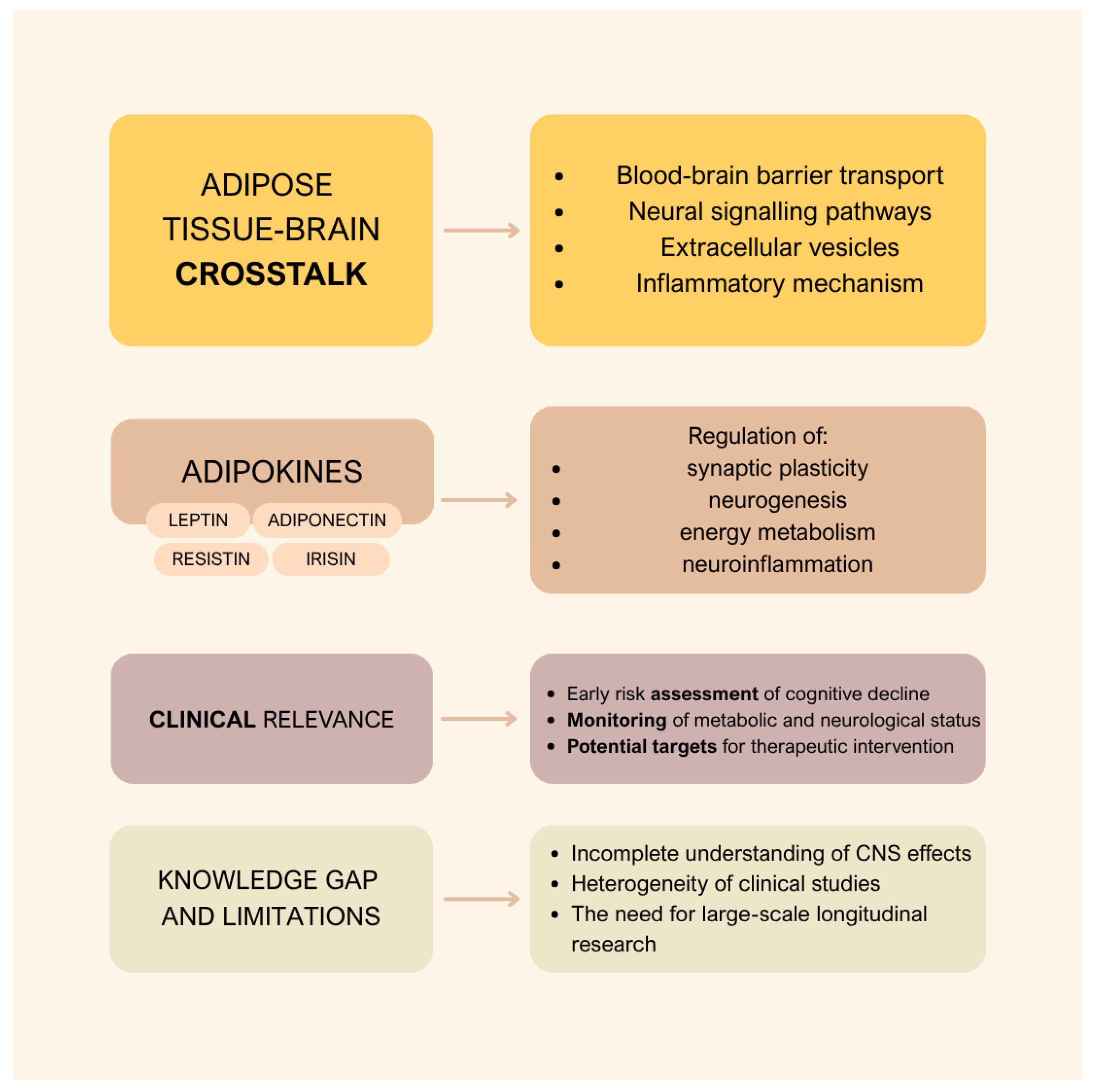
The aim of this review is to present the role of adipokines in the adipose tissue–brain axis and their significance for cognitive function. This review focuses on four selected adipose-derived molecules: leptin, adiponectin, resistin, and irisin. These four molecules were selected based on numerous studies demonstrating their importance in neuroinflammation, cognitive function, and the pathophysiology of AD, which we describe in this manuscript. We discuss the mechanisms of signal transduction from adipose tissue to the brain and characterize selected adipokines, taking into account their physiological effects. Finally, we present an overview of the clinical evidence regarding individual adipokines and the factors influencing their levels. Figure 1 presents the key topics we will discuss in this review.

2. Mechanisms of Communication Between Adipose Tissue and the Brain

2.1. Transport Across the Blood–Brain Barrier

One of the key mechanisms by which adipose tissue communicates with the central nervous system (CNS) is the transport of adipokines across the blood–brain barrier (BBB). The BBB is a highly selective structure located between the blood vessels and neural tissue that protects the nervous system from harmful factors such as toxins and pathogens. The transport of molecules across the BBB may occur through passive diffusion in the case of small lipophilic molecules or through membrane transporters and receptor-mediated mechanisms [23,24,25]. The transport of adipokines across the BBB is a highly selective and tightly regulated process. Among adipokines, leptin represents one of the best-characterized examples of molecules transported across the BBB. Adipokines cross the barrier through specific protein transporters located in the endothelial cells of cerebral blood vessels. For many years, the leptin receptor (ObR) receptor was considered the primary transporter responsible for leptin passage across the BBB [26]. However, more recent studies suggest that although this receptor participates in leptin binding and signal transduction within endothelial cells, it is not the sole transporter involved in this process [27,28,29].
An alternative mechanism for adipokine transport across the BBB involves specialized glial cells known as tanycytes. Tanycytes constitute a distinct cell population located at the floor of the third ventricle, particularly within the median eminence of the hypothalamus. These cells capture circulating adipokines such as leptin adipokines from blood vessels and transport them directly into the cerebrospinal fluid or deliver them to hypothalamic neurons. This process is tightly regulated by ERK kinase phosphorylation [30,31].

2.2. Neural Signaling Between Adipose Tissue and the Brain

An equally important signaling pathway involves communication between adipose tissue and the CNS through specialized neural pathways, including efferent pathways (from the brain to the tissue) and afferent pathways (from the tissue to the brain). Sympathetic fibers innervate adipocytes in both WAT and BAT [32]. The release of norepinephrine from sympathetic nerve terminals activates β3-adrenergic receptors on the surface of adipocytes, leading to the activation of two principal processes: lipolysis and thermogenesis. During lipolysis, triglycerides are broken down into free fatty acids, which can be utilized locally or released into the circulation as an energy source [33]. In BAT, sympathetic activation stimulates mitochondria to produce heat, forming the basis of the thermogenic process [34]. The existence of this pathway was confirmed in a study by Morii et al. [35]. Detailed anatomical examinations of postmortem specimens revealed that supraclavicular BAT is innervated by multiple nerve pathways, including branches of the third and fourth cervical nerves as well as the cervical sympathetic plexus [35]. A study by Williams et al. [36] investigated the regulation of thermogenesis by the CNS. The authors examined the role of the spliced form of X-box binding protein 1 (XBP1s), a transcription factor activated during the unfolded protein response and involved in the regulation of cellular energy and metabolic homeostasis. Increased hypothalamic XBP1s expression was shown to enhance BAT thermogenesis through modulation of efferent signals from the sympathetic nervous system. Experiments conducted in animal models demonstrated that elevated hypothalamic XBP1s expression protected against obesity induced by a high-fat diet. The XBP1s dependent signaling pathway therefore represents a key component of communication between the hypothalamus and adipose tissue, integrating metabolic signals and regulating adaptive mechanisms of energy expenditure [36,37].
Adipose tissue not only receives signals from the brain but also has the capacity to transmit signals to the CNS via afferent pathways. Leptin is one of the best characterized mediators of adipose tissue–brain communication. After entering the CNS, leptin acts through receptors expressed in hypothalamic and extra-hypothalamic regions, contributing to the regulation of appetite, satiety, energy expenditure, and systemic metabolic homeostasis [38,39]. In addition to leptin, pro-inflammatory mediators derived from adipose tissue, including TNF-α and IL-6, may contribute to CNS dysfunction by promoting neuroinflammatory signaling and altering autonomic regulation, particularly in the context of obesity-associated metabolic disturbances [40,41]. Besides adipokine-mediated signaling, neural afferent pathways also participate in adipose tissue–brain communication. A study by Xiong et al. [42] investigated the role of the adipose afferent reflex (AAR) in the development of obesity-induced hypertension. The results demonstrated that this reflex is significantly enhanced in obese rats compared with healthy controls. This finding indicates that, in obesity, sensory signals originating from adipose tissue excessively stimulate the brain, leading to increased activity of the sympathetic nervous system and, consequently, to persistently elevated blood pressure [42]. These studies provide evidence for bidirectional communication between the CNS and adipose tissue.

2.3. Extracellular Vesicles

In addition to hormonal and neural signaling, extracellular vesicles (EVs) are increasingly recognized as an additional mechanism of communication between adipose tissue and the brain. EVs are particles released by cells into the extracellular space. They represent a heterogeneous group of lipid bilayer–bound vesicles that lack the ability to replicate. The MISEV 2024 guidelines distinguish two main groups of EVs based on their mechanism of formation: exosomes (formation via the endosomal pathway) and ectosomes (produced by direct budding from the plasma membrane) [43]. EVs are present in many body fluids, including blood, cerebrospinal fluid, and breast milk. One of their key functions is the transport of biologically active molecules between cells and tissues. As a result, they can influence the regulation of gene expression as well as the function of target cells [44,45].
Studies have shown that adipose tissue is one of the sources of circulating EVs. These vesicles can transport biologically active molecules derived from adipose tissue, including adipokines, miRNAs and lipids capable of modulating processes occurring in the brain, including neuroinflammatory signaling pathways and the kinetics of amyloid β aggregation [46]. Studies have shown that EVs released by adipose tissue can carry adipokines such as ADPN and leptin as part of their molecular cargo and deliver them to distant target tissues, including the CNS, thereby providing an additional mechanism of communication between adipose tissue and the brain [47,48]. Although miRNAs and lipids are not traditionally classified as adipokines, they are recognized as important mediators of adipose tissue—brain communication [49,50]. Evidence also suggests that EVs may contribute to the pathogenesis of AD, as they can transport amyloid precursor protein (APP) as well as enzymes involved in its processing [51,52,53]. Amyloid β accumulation and impaired clearance are key pathological features of AD, contributing to neurodegeneration process [54]. Interestingly, at the same time, some studies indicate that EVs may also participate in the clearance of amyloid β [55].

2.4. Inflammatory Pathways

Adipokines and cytokines secreted by adipose tissue also function as modulators of inflammatory pathways. They can be broadly classified into pro-inflammatory adipokines and cytokines (e.g., TNF-α, IL-6, leptin, resistin) and anti-inflammatory adipokines and cytokines such as ADPN. Inflammatory signals originating from adipose tissue influence the CNS through several mechanisms, including transport across the BBB, activation of immune cells within the brain, and induction of neuroinflammation [56].
During this process, microglia and astrocytes become activated. Activation of the NF-κB signaling pathway leads to the production of inflammatory mediators, thereby amplifying the immune response. At the same time, cytokines secreted by adipose tissue can activate the JAK/STAT signaling pathway, which further enhances glial cell activity and the inflammatory response [57,58,59].
In summary, these mechanisms highlight the multilevel nature of communication between adipose tissue and the CNS. Metabolic disturbances may significantly contribute to cognitive decline and are likely associated with an increased risk of neurodegenerative diseases.

3. Adipokines

Adipokines, such as leptin, ADPN, resistin and irisin, are biologically active signalling molecules of the nature of protein hormones and cytokines that are involved in multi-organ communication. They are secreted mainly by adipose tissue, and in the case of irisin, also by muscles [60]. Although the classification of certain molecules within the adipokine family is not fully consistent, they are functionally grouped as adipokines due to their significant secretion from adipose tissue or their key role in the adipose tissue–brain metabolic axis [18,19,20]. In humans, resistin is predominantly secreted by monocytes and macrophages; however, its circulating levels are closely associated with adiposity and obesity-related metabolic dysfunction, supporting its inclusion in adipokine-related signalling networks [61,62]. Similarly, irisin, originally characterised as a myokine, has also been detected in adipose tissue and is increasingly considered an adipomyokine, reflecting its involvement in systemic energy homeostasis and metabolic regulation [63,64].
Adipokines demonstrate the ability to cross the BBB and modulate neurobiological processes, including synaptic plasticity, neuronal metabolism and the immune response [65]. In this section, we present an analysis of key adipokines, with particular emphasis on their mechanisms of action.

3.1. Leptin—Biological Role and CNS Effects

Leptin is synthesised primarily by WAT and acts as a key regulator of energy homeostasis [66,67]. It is released into the bloodstream in amounts proportional to adipose tissue stores or total stored energy, and its fasting plasma concentration in healthy individuals is generally higher in women than in men [68]. It is secreted in pulses and follows a circadian rhythm, resulting in dynamic changes in its blood levels throughout the day [69].
By acting on the hypothalamus, leptin regulates appetite and the body’s energy balance [70,71]. It can cross the BBB via a saturable transport mechanism and can bind to six different ObR isoforms, particularly the long ObRb isoform, which is mainly expressed in the hypothalamus [70,71]. Studies show that leptin activates the JAK/STAT, PI3K/Akt, and mitogen-activated protein kinase (MAPK) signaling pathways, thereby promoting neuronal survival and hippocampal plasticity [66,72]. The PI3K/Akt pathway mediates the anti-apoptotic and neuroprotective effects of leptin by regulating cell survival pathways and synaptic function, whilst activation of the MAPK/ERK cascade supports hippocampal plasticity by modulating dendritic spine formation and long-term potentiation (LTP), processes essential for learning and memory mechanisms [73,74]. Leptin has been shown to increase hippocampal neurogenesis and dendritic spine formation, as well as to enhance LTP via the modulation of NMDA receptors [66,67].
In obesity, chronic overproduction of leptin leads to leptin resistance, characterised by reduced transport across the (BBB) and impaired intracellular signaling [71,75]. This resistance is further exacerbated by the induction of SOCS3, which inhibits the neuroprotective JAK2/STAT3 pathway [76,77]. In the context of AD, leptin resistance facilitates neurodegeneration by increasing β-secretase activity and tau hyperphosphorylation, thereby accelerating amyloid plaque and tau pathology [74,78].
At the behavioural level, it has been shown that leptin administration improves memory in animal models. In rodents, intrahippocampal leptin administration following training enhances performance in maze avoidance tasks [79]. Conversely, leptin deficiency or resistance, as observed in db/db or Zucker rodents with a ObR mutation, causes impairment of hippocampal structure. These models exhibit lower dendritic spine density, which is associated with the development of spatial memory deficits [79,80].
In summary, leptin signalling promotes synaptic plasticity and the energy balance of neurons. Its disruption, due to factors such as low concentrations or receptor resistance, contributes to a decline in cognitive function.

3.2. Adiponectin—Biological Role and CNS Effects

ADPN, which is synthesized in WAT, belongs to the complement 1q family of proteins and occurs in the form of three multimeric complexes: a low molecular weight (LMW) form, a medium molecular weight (MMW) form, and a high molecular weight (HMW) form. Among these isoforms, HMW ADPN is considered the most biologically active and is primarily associated with insulin sensitisation, glucose uptake, fatty acid oxidation, and anti-inflammatory effects. MMW ADPN appears to exert intermediate metabolic and immunomodulatory functions, whereas LMW adiponectin is thought to participate mainly in energy homeostasis and vascular protection [81,82,83]. Each of these performs different functions in various organs of the body [84,85]. Its serum concentration is inversely proportional to total body fat mass and is reduced in many metabolic and neurodegenerative diseases, such as type 2 diabetes, cardiovascular diseases, AD, strokes and cancers [86].
Although ADPN itself is not synthesised in the brain, studies have shown that it can cross the BBB into the CNS. There, it signals via AdipoR1, AdipoR2 and T-cadherin receptors in regions such as the hippocampus, hypothalamus and cerebral cortex [85,86]. In the hypothalamus, ADPN via AdipoR1 activates the AMP-activated protein kinase (AMPK) signalling pathway, thereby regulating food intake and energy expenditure [87]. Furthermore, the AdipoR1 receptor is also abundantly present in cardiomyocytes and skeletal muscle, where ADPN plays a significant role in regulating lipid oxidation, glucose transport, tissue insulin sensitivity and in modulating the inflammatory response [84].
Of particular note is the role of ADPN in reducing insulin resistance and increasing glucose uptake by tissues via the ADPN/APPL1/PI3K/Akt or ADPN/AMPK/PPARα pathways [86]. The AdipoR2 receptor is predominantly expressed in the liver and activates the peroxisome proliferator-activated receptor alpha (PPARα) pathway [88]. Its role in the brain remains largely unknown; however, studies in a rodent model of post-traumatic stress disorder (PTSD) suggest it plays a part in the regulation of emotions. Mice lacking the AdipoR2 gene exhibit stronger consolidation of fear memories and impaired extinction of the fear response. This suggests a possible role for this receptor in the regulation of fear memory [89]. Binding of ADPN to receptors in the hippocampus leads to increased synaptic plasticity [90]. It promotes neurogenesis by activating p38 MAPK, whilst its deficiency results in reduced formation of new neurons [91].
Another function of ADPN is its potent anti-inflammatory effect. Studies have shown that it inhibits pro-inflammatory cytokines, such as TNF-α, IL-6 and reactive oxygen species, whilst simultaneously inducing the anti-inflammatory cytokine IL-10 [92]. Other studies on the role of ADPN in AD have shown that ADPN, via the AdipoR1/NF-κB signalling, reduces soluble Aβ oligomers and inhibits the release of IL-1β and TNF-α by microglial cells [93]. It should be emphasised that ADPN may also have pro-inflammatory effects in certain conditions—in rheumatoid arthritis (RA) or chronic kidney disease (CKD), it causes an increase in CRP, TNF-α or IL-6 [94].
Another interesting aspect is the effect of ADPN on cognitive function. In animal models of AD, it has been demonstrated that ADPN improves neurocognitive function, restores impaired glucose uptake in the brain, reduces pathogenic levels of Aβ42 and leads to significant improvements in learning and memory [95]. Consequently, clinically, higher levels of ADPN are associated with better cognitive function, whilst its deficiency may contribute to a decline in mental performance.
In summary, ADPN is an adipokine with anti-inflammatory properties that enhances tissue sensitivity to insulin. It regulates energy metabolism, protects the cardiovascular system and supports neurogenesis. It counteracts changes typical of AD, supports cognitive function and limits the accumulation of harmful amyloid plaques.

3.3. Resistin—Biological Role and CNS Effects

Resistin is a pro-inflammatory adipokine belonging to the FIZZ (Found In Inflammatory Zone) family of proteins [96]. Unlike in animal models, in humans it is secreted mainly by immune system cells—particularly monocytes and macrophages—in response to pro-inflammatory mediators, i.e., TNF-α, IL-6 and IL-1β [61]. The mechanism of action of resistin involves binding to its receptors: Toll-like receptor 4 (TLR4) and adenylate cyclase-associated protein 1 (CAP1), which are expressed both in peripheral tissues and throughout the CNS [61]. When the BBB is compromised, resistin is able to cross into the CNS, where it acts directly on neurons and glial cells to induce a local immune response and cause neuroinflammation [97]. As a natural insulin antagonist, resistin plays a key role in the development of insulin both systemic and neuronal insulin resistance. By activating TLR4 and CAP1 on brain cells receptors and subsequently stimulating the NF-κB and ERK1/2 pathways, resistin impairs the phosphorylation of the IRS-1 protein. This leads to the blocking of the Akt signalling cascade within the neurons, preventing cells from responding properly to insulin and resulting in reduced glucose uptake [97,98]. Abnormal expression of this adipokine is considered an important factor contributing to the development of metabolic diseases associated with modern lifestyles, particularly type 2 diabetes and obesity [62]. In experimental models using rat hippocampal slices, it has been observed that resistin negatively affects glucose metabolism specifically in nerve cells [99]. This effect is due, amongst other things, to a reduction in hexokinase activity. As this enzyme plays a key role in glycolysis, its inhibition by resistin significantly disrupts the energy metabolism of neurons [100].
By stimulating inflammatory processes and oxidative stress, resistin exerts a negative effect on brain tissue and leads to direct neuronal damage [96,98]. A particularly vulnerable area is the hippocampus, where resistin disrupts carbohydrate metabolism, impairing memory and learning processes. This phenomenon is characteristic of mild cognitive impairment (MCI) [98]. Due to the close correlation between high resistin levels and insulin resistance, this adipokine is considered a key factor increasing the risk of developing AD in patients with type 2 diabetes [100].
In summary, resistin acts as a pro-inflammatory mediator. By promoting central insulin resistance and disrupting glucose metabolism in the brain, it contributes to the development of neurological disorders.

3.4. Irisin—Biological Role and CNS Effects

Irisin is a myokine produced by the proteolytic cleavage of its precursor protein, fibronectin type III domain-containing protein 5 (FNDC5) [101,102]. It is released into the bloodstream mainly in response to endurance training [103]. Its biological functions are mediated via receptors known as integrins [102]. Because integrins are distributed throughout virtually the entire body, irisin can actively influence not only muscle and adipose tissue, but also the liver and the CNS [103]. Irisin promotes the conversion of WAT into BAT, a process known in scientific terms as “browning”. This process occurs through the induction of thermogenin (UCP1) expression, which is achieved by activating the MAPK signalling pathways, specifically the p38 and ERK pathways. UCP1 converts stored energy into heat, thereby supporting the metabolism of the entire body [101,103].
Irisin plays an equally important role in terms of its anti-inflammatory properties, which are crucial for maintaining cognitive function. It has the ability to cross the BBB and modulate the immune response within the brain [104]. It can effectively reduce levels of pro-inflammatory markers such as TNF-α, IL-6 and IL-1β, and, via PPAR-γ- and Nrf2-dependent pathways, promotes the polarisation of microglia from the destructive M1 phenotype to the anti-inflammatory M2 phenotype [104,105]. In addition to inhibiting neuroinflammation, irisin stimulates repair processes in the brain by increasing levels of brain-derived neurotrophic factor (BDNF) and cAMP response element-binding protein (CREB). The increase in these neuroprotective factors means that irisin supports synaptic plasticity, which translates into improved memory and learning ability [106].
In summary, irisin acts as a link between the muscles and the brain. It has a multifaceted effect—it both modulates energy metabolism by inducing the ‘browning’ of adipose tissue and influences the CNS. On the one hand, it reduces inflammation, and on the other, it stimulates repair processes in neurons. It is therefore recognised as one of the key mediators of the beneficial effects of physical activity on brain function.

3.5. Other Adipokines—Biological Role and CNS Effects

In addition to the adipokines described above, the literature also mentions other factors that transmit metabolic information from adipose tissue to the brain such as chemerin, visfatin, apelin, lipocalin-2 (LCN2) and omentin [99]. These substances, which often have opposing effects, influence inflammatory processes in the body, metabolic homeostasis and insulin signalling [107]. Some of them interact with immune system cells or modulate the permeability of the BBB [108]. Their direct impact on cognitive function remains the subject of intensive research; however, current evidence suggests that they may exert bidirectional effects, depending on the metabolic context [99]. An example of a pro-inflammatory adipokine is chemerin, which can attract immune cells expressing the CMKLR1 receptor [109]. It stimulates the migration of macrophages and NK cells [110] and may exacerbate neuroinflammation [111]. Similar negative effects are also attributed to LCN2, which can disrupt the BBB and promote glial reactivity, thereby facilitating neurodegenerative processes [108]. On the other hand, adipokines such as omentin exhibit anti-inflammatory and anti-apoptotic effects, limiting damage to nerve cells [112]. Another example is apelin, which may support neuronal survival by influencing the vascular system and modulating insulin signalling pathways [113]. These adipokines differ from one another, but their ultimate impact on neuronal function likely depends on metabolic status and stressors.
A summary of the key properties of selected adipokines and their effects on selected physiological and pathological processes is presented in Table 1.

4. Clinical Significance of the Adipose Tissue–Brain Axis

Increasingly, the scientific literature contains references to metabolic dysfunctions as factors playing a role in disturbances of CNS functioning [97]. Adipokines mediating these mechanisms, in addition to acting as disease promoters, may also perform modulatory or protective functions in specific disease entities [99]. Adipokines are described as biomarkers in the assessment of disease risk at an early stage and in therapy monitoring, as well as potential therapeutic targets [97]. Different levels and functions of adipokines are associated with various factors. These include, among others, sex, insulin resistance, inflammation, lifestyle, the use of antidiabetic medications, and natural compounds [116,117,118,119,120].
Below we describe the potential clinical role of adipokines. Particular attention is given to factors modulating their levels and the consequences thereof, as well as their position among biomarkers. The studies included in this review were selected based on their scientific relevance, recency, and impact within the field.

4.1. Leptin

4.1.1. Factors Influencing Leptin Levels

A study by De Frutos González et al. [121] conducted in mice aged 3, 6, and 12 months demonstrated that the early stages of leptin signaling pathways weaken with age. In this context, the authors describe the possible influence of neuroinflammation in the brainstem on leptin resistance [121]. Others indicate that leptin resistance resulting from age-related changes, as well as from a high-fat diet, is associated with impaired synaptic plasticity and hippocampal function, contributing to cognitive deficits [66].
With regard to sex differences, the analysis by Varma et al. [122] of 1811 adults showed that higher levels of free testosterone and lower levels of sex hormone-binding globulin were associated with higher leptin levels in women when only demographic variables were considered; however, the authors note a weakening of this relationship after adjustment for other variables. In men, higher leptin levels were observed in situations of higher free testosterone and lower sex hormone-binding globulin (full adjustment including all variables [122]. Stepanov et al. [123] report an influence of insulin on leptin levels, explaining that prolonged hyperinsulinemia leads to an increase in leptin concentration in plasma. Moreover, in their analysis concerning patients with gastroesophageal reflux disease, leptin concentrations increased along with the increase in adipose tissue mass and BMI. The authors emphasize enhanced leptin production in subcutaneous adipocytes compared with visceral adipose tissue [123]. The results of the meta-analysis by Li and He are noteworthy, indicating higher serum and plasma leptin concentrations in patients with obstructive sleep apnea and a positive correlation with the apnea–hypopnea index [124]. It is also worth mentioning the associations of leptin with depressive symptoms in patients with metabolic syndrome or type 2 diabetes mellitus. To the best of our knowledge, at present, in accordance with the observation by Chirinos et al. [125], studies examining associations between leptin levels and depressive symptoms in individuals with obstructive sleep apnea remain lacking [125]. Importantly, several of the determinants of circulating leptin discussed above—namely metabolic syndrome, insulin resistance, obstructive sleep apnea, and depressive symptoms—have been considered risk factors for cognitive decline and AD [126,127,128]. This supports the hypothesis that impaired leptin signaling may represent a shared pathophysiological pathway linking these conditions with neurodegeneration.

4.1.2. Review of the Latest Clinical Studies—Leptin in the Context of the Nervous System

Most recent clinical studies related to the fields of neurology or psychiatry involving leptin present its use primarily as a biomarker. In a study including 2262 healthy participants, concentrations of leptin, soluble ObR, and their ratio were measured using enzyme-linked immunosorbent assays. Brain magnetic resonance imaging data were available for 2028 participants. The results indicate higher leptin bioavailability in cases with better preservation of white matter microstructure. The authors conclude that higher leptin availability in midlife may represent potential protection against dementia and note the possibility of enhanced benefits in individuals with visceral obesity [129]. Another study including 171 participants assigned to groups with and without obesity demonstrated a negative association between cognitive function and both leptin levels and the leptin–ADPN ratio. These correlations were clearly present in individuals with obesity, whereas they were not observed in those without obesity. The authors concluded that obesity may influence the manner in which adipokines affect the brain and cognitive functions. They also noted that in older patients experiencing metabolic stress, the leptin–ADPN ratio might represent a more advantageous risk indicator in AD [130].
Analyses by Thacker et al. [131] of patients with cognitive impairment and those with preserved cognitive function demonstrated that among participants with normal BMI, lower leptin levels were associated with an increased risk of cognitive impairment. In contrast, in the case of obesity, a higher risk of these impairments was associated with higher ADPN levels. In this case, the details are discussed in Section 4.2. The conclusions suggest a complex role of adipokines and BMI in cognitive health [131]. The results of a study of 118 individuals aged 18–35 years indicate a negative correlation between cognitive status and both BMI and leptin levels in individuals with obesity without neurodegenerative disorders. In this project, a positive correlation between BMI and leptin, as well as inflammatory markers, was also demonstrated [132]. Interesting findings may emerge from a study involving patients in whom obesity was diagnosed at least 5 years earlier with a current BMI of 30–40 kg/m2, the aim of which is to determine the effect of synbiotic supplementation on anxiety and depressive symptoms as well as the glycemic profile and insulin resistance. The objective is to include 60 individuals with anxiety or depressive disorders and 60 without these disorders. According to the study protocol, among other parameters, leptin and ADPN will be assessed. The planned completion of the project is scheduled for 2027 [133].
Another study, the results of which may be expected soon, concerns idiopathic intracranial hypertension and will examine patients with this condition who are newly diagnosed, untreated, and present with transverse sinus stenosis. It is mentioned here due to reports of elevated leptin concentrations in patients suffering from this disorder. The study assumes measurement of leptin as a marker alongside numerous other parameters [134].

4.2. ADPN

4.2.1. Factors Influencing ADPN Levels

Differences in ADPN levels between women and men are well documented: women usually exhibit higher concentrations. A role of estrogen is suspected, as it stimulates the production of high-molecular-weight ADPN, which is considered the most biologically active isoform. Androgens, particularly testosterone, have been shown to inhibit ADPN production [135]. A study by Guan et al. [136] concerning women suggests that ADPN is associated with adipose tissue distribution and the concentration of high-density lipoprotein cholesterol, independently of physical activity levels and insulin sensitivity [136]. A study by Ohman-Hanson et al. [137] demonstrated a decreasing trend in ADPN during late puberty. At the same time, a more pronounced decline is observed in adult men [137]. The influence of insulin resistance on ADPN levels is also considered. The literature indicates a positive correlation between ADPN levels and insulin sensitivity, as well as a negative correlation between ADPN levels and fasting proinsulin concentrations and the proinsulin-to-insulin ratio [138].
Interestingly, studies indicate that ADPN production may be stimulated by selected intestinal bacteria; however, the relationship between the microbiota and adipokines is complex and dependent on numerous factors [60].

4.2.2. Review of the Latest Clinical Studies—ADPN

Sun et al. [139] conducted a two-sample analysis using Mendelian randomization with verification through proteomic analysis. Data from genome-wide association studies were used for ADPN, leptin, resistin, and monocyte chemoattractant protein-1. The results were as follows: genetically predicted higher ADPN concentrations were associated with a lower risk of AD (OR = 0.79, 95% CI = 0.65–0.96, p = 0.019). The proteomic analysis confirmed these findings. No similar associations were observed for leptin, resistin, or monocyte chemoattractant protein-1. The authors indicate ADPN as a potential biomarker and therapeutic target in AD [139]. Evidence for a causal effect of ADPN on the risk of AD is also provided by the results of an analysis using Mendelian randomization with genetic proxies from genome-wide association studies. A causal effect of ADPN on the risk of AD was demonstrated (OR: 0.850; 95% CI: 0.731–0.990; p = 0.037), without a similar association between AD and resistin (OR: 0.936; 95% CI: 0.851–1.029; p = 0.171) [140]. Wennberg et al. [141] examined the relationship between plasma ADPN levels and the results of neuroimaging and cognitive assessments in 535 individuals aged 70 years and older without dementia. Higher ADPN levels were observed in women than in men. In women, higher ADPN levels were also associated with smaller hippocampal volume (B = −0.595; 95% CI −1.19, −0.005), poorer performance in the language domain (B = −0.676; 95% CI −1.23, −0.121), and poorer global cognitive function (B = −0.459; 95% CI −0.915, −0.002). A higher probability of MCI diagnosis was identified (OR = 6.23; 95% CI 1.20, 32.43) [141]. Another study involving 283 cognitively normal individuals aged 55–90 years demonstrated a positive association between serum ADPN concentration and global deposition of beta-amyloid protein. The authors suggest that ADPN may potentially represent a preventive and therapeutic target in AD [142]. In order to determine the relationship between cognitive test results and ADPN levels prior to the development of type 2 diabetes mellitus, 938 individuals without diabetes in middle age (45.7 ± 4.9 years) were examined. The results suggest that ADPN may indicate disturbances in cognitive function at an early stage, representing a potential therapeutic target for protection against dementia [143]. The observational CALERIE Legacy Study concerning caloric restriction in humans without obesity will also investigate, among other outcomes, cognitive functions alongside total ADPN and HMW ADPN. Completion of the project is planned for 2027 [144].

4.3. Resistin

4.3.1. Factors Influencing Resistin Levels

When considering modifying factors, we refer to the study by Chen et al. [145] concerning resistin concentrations in the development of type 2 diabetes mellitus, which included 359 apparently healthy postmenopausal women and 170 apparently healthy men, after which matched controls were selected. The results indicated higher baseline resistin levels in women and in patients with diabetes compared with the control group [145]. Resistin levels are described as being associated with insulin resistance and type 2 diabetes mellitus. The literature reports elevated resistin concentrations in patients with diabetes as well as positive correlations between resistin and insulin resistance, particularly in the context of type 2 diabetes mellitus and obesity, although not all results are consistent in this regard [146]. The analysis by Zhao et al. [119], which included 49 results, indicates that administration of metformin significantly contributes to the reduction in resistin levels compared with control groups [119].

4.3.2. Review of the Latest Clinical Studies—Resistin

In a study including 26 patients suffering from AD, 21 patients with frontotemporal dementia, and 23 patients with other non-neurodegenerative neurological disorders, lower resistin concentrations were observed in patients with dementia compared with the control group, particularly in AD (p < 0.001). A direct correlation was observed between resistin and the concentration of Aβ1-42 in cerebrospinal fluid (p < 0.001, r = 0.50). Furthermore, the risk of AD diagnosis was described as inversely associated with resistin after consideration of confounding factors [147]. The study of 945 participants without dementia and 177 individuals in whom dementia was diagnosed during follow-up, described by Mooldijk et al. [114], strengthens the hypothesis regarding the role of adipokines in dementia. According to the results, higher levels of leptin and resistin were associated with a lower risk of this disease: for leptin HR 0.85; 95% CI 0.72–1.00; p = 0.056 per standard deviation increase in Model 3, and for resistin HR 0.82; 95% CI 0.71–0.95 in Model 3. Similar results were observed with regard to the risk of AD [114].

4.4. Irisin and Others (Visfatin, Omentin)

4.4.1. Factors Influencing Irisin Levels

The analysis by Torabi et al. [148] demonstrated an increase in serum irisin concentrations in overweight and obese individuals who performed exercise compared with control participants. High-intensity interval training had the greatest impact [148]. Another study including obese and non-obese individuals with low and high caloric intake, respectively, showed no association between physical activity and irisin levels. The authors indicated a positive correlation between irisin and fat-free mass in the non-obese group and associations between irisin and concentrations of thyroid-stimulating hormone and glucose in individuals with obesity [149]. In individuals with obesity and diabetes mellitus, lower irisin concentrations are observed. The authors also describe a negative association between irisin and interleukin-6, while emphasizing the need for verification of these findings in larger samples [150].

4.4.2. Review of the Latest Clinical Studies—Irisin, Visfatin, Omentin

One study aimed to determine whether plasma biomarkers could distinguish individuals with normal cognitive function from those with MCI. The study included 124 patients with MCI and 126 individuals with normal cognitive function. The results indicated an association between higher irisin concentration and an increased probability of MCI (OR: 1.06, p = 0.004). Additionally, concentrations of irisin and BDNF increased with age, whereas the BDNF –to–irisin ratio remained stable [151]. The results of the meta-analysis by Han et al. [152] indicate a positive correlation between irisin levels and global cognitive function (r = 0.26, 95% CI: 0.10–0.41). The authors did not identify correlations between irisin levels and specific cognitive domains (including immediate memory, short-term memory, language, and others). The analysis also mentioned a correlation between BDNF levels and global cognitive function (r = 0.27, 95% CI: 0.12–0.42) [152]. The number of studies concerning visfatin is more limited compared with the adipokines discussed previously. One notable study from 2014 involving 61 Caucasian patients with primary obesity (BMI > 30) examined the hypothesis of a neuroprotective effect of visfatin in individuals with obesity. The results suggested a protective effect of visfatin on neural tissue in the prefrontal cortex in individuals with obesity; however, the authors emphasize that visfatin may participate in both harmful and beneficial biological processes in this population [153]. In another study including 106 patients (including 62 with cognitive impairment) aged 75 years and older, the group with cognitive impairment was characterized by higher serum concentrations of tumor necrosis factor-α, interleukin-6, and visfatin. The authors identify handgrip strength, negatively correlated with tumor necrosis factor-α and interleukin-6, as an independent predictor of cognitive impairment [154]. With regard to omentin, studies concerning cognitive functions and the brain are based mainly on preclinical research, and studies conducted in humans are still needed. Table 2 summarizes studies on the relationship between adipokines and cognitive function and the risk of dementia.

5. Discussion

The literature increasingly includes references to the role of adipose tissue as a neurological modulator. When focusing on cognitive functions and other aspects related to brain health, the neuro–metabo–inflammatory axis is considered. Much attention is devoted to factors secreted by adipose tissue as an endocrine organ, the description of which was presented in the previous section—adipokines. Authors highlight their regulatory functions in the context of metabolism and inflammatory processes, as well as their potential to influence brain functions. Among those mentioned are leptin, ADPN, resistin, and others discussed in detail earlier. Some of them are described as potential biomarkers for early risk assessment or monitoring of therapeutic outcomes, as well as possible therapeutic targets [59,97]. For example, there are reports of disturbances in leptin and ADPN signaling pathways in the brains of individuals suffering from AD. In this context, enhancement of these signaling pathways could represent a potential therapeutic strategy [67].
Although scientific understanding of adipokine function in metabolic processes has advanced considerably, their influence in the field of neurology still requires further clarification. Many issues remain unresolved [67]. Despite the growing number of studies, many existing investigations are limited with regard to the number of participants or the duration of observation. There is a need for studies that distinguish effects dependent on sex, age, and other factors influencing adipokine levels described earlier, in order to precisely determine the potential of adipokines as biomarkers or therapeutic targets. For some adipokines, such as omentin, research is still largely limited to preclinical studies, and similar investigations in humans are required. We conclude that there is a need to investigate interactions between adipokines and other factors of the neuro–metabo–inflammatory axis. An interesting aspect remains the microbiota. Its relationship with adipokines is complex and remains insufficiently investigated.

6. Conclusions

In this review, we discussed the role of the adipose tissue–brain axis in cognitive dysfunction. Adipokines play a key role in this communication by influencing synaptic plasticity, neuronal metabolism, and the immune response. Current evidence points to the role of obesity in the deterioration of cognitive function and the development of neurodegenerative diseases such as AD. Despite the rapid advancement of research on adipokines, their role as biomarkers and therapeutic targets remains limited and requires further investigation. A thorough examination of adipokine interactions within the adipose tissue–brain axis will enable the development of new therapeutic strategies in the future to preserve cognitive function and prevent neurodegeneration.

Author Contributions

Conceptualization, M.Ż. and J.S. (Julia Soczyńska).; validation, M.Ż., J.S. (Julia Soczyńska) and S.W.; formal analysis, M.Ż., J.S. (Jagoda Szwach) and K.K.; investigation, M.Ż., W.G., M.K. and K.K.; data curation, M.Ż., K.K., J.S. (Jagoda Szwach) and M.K.; writing—original draft preparation, M.Ż., J.S. (Julia Soczyńska), K.K., J.S. (Jagoda Szwach), M.K., W.G. and S.W.; writing—review and editing, M.Ż., J.S. (Julia Soczyńska), K.K., J.S. (Jagoda Szwach), M.K., W.G. and S.W.; visualization, M.Ż., J.S. (Julia Soczyńska) and S.W.; supervision, S.W. All authors have read and agreed to the published version of the manuscript.

Funding

This research received no external funding.

Institutional Review Board Statement

Not applicable.

Informed Consent Statement

Not applicable.

Data Availability Statement

The contributions presented in this study are included in the article. Further inquiries can be directed to the corresponding authors.

Conflicts of Interest

The authors declare no conflicts of interest.

Abbreviations

The following abbreviations are used in this manuscript:
WHOWorld Health Organization
ADAlzheimer’s Disease
BBBBlood–brain barrier
BMIBody mass index
WATWhite adipose tissue
BATBrown adipose tissue
ObRLeptin receptor
ADPNAdiponectin
CNSCentral nervous system
AMPKAMP-activated protein kinase
MCIMild cognitive impairment
MAPKMitogen-activated protein kinase
BDNFBrain-derived neurotrophic factor
LCN2Lipocalin-2
PTSDPost-traumatic stress disorder
RARheumatoid arthritis
CKDChronic kidney disease
SBP1sX-box binding protein 1
EVsExtracellular vesicles
APPAmyloid precursor protein
PPARaPeroxisome proliferator-activated receptor alpha
FIZZFound In Inflammatory Zone
TLR4Toll-like receptor 4
CAP1Adenylate cyclase-associated protein 1
FNDC5Fibronectin type III domain-containing protein 5
cAMPcAMP response element-binding protein
LMWLow molecular weight
MMWMedium molecular weight
HMWHigh molecular weight
LTPLong-term potentiation
AARAdipose afferent reflex

References

  1. Lin, X.; Li, H. Obesity: Epidemiology, Pathophysiology, and Therapeutics. Front. Endocrinol. 2021, 12, 706978. [Google Scholar] [CrossRef] [Scilit]
  2. Haththotuwa, R.N.; Wijeyaratne, C.N.; Senarath, U. Worldwide Epidemic of Obesity. In Obesity and Obstetrics; Elsevier: Amsterdam, The Netherlands, 2020; pp. 3–8. [Google Scholar] [CrossRef] [Scilit]
  3. Sormani, L.E.; Palacio, T.L.N.; Corrêa, C.R. From Evolutionary Advantage to Global Epidemic: The Multisystemic Impact of Obesity on Metabolic Health and the Potential of Bergamot (Citrus bergamia). Nutrire 2025, 50, 83. [Google Scholar] [CrossRef] [Scilit]
  4. Mohajan, D.; Mohajan, H.K. Body Mass Index (BMI) Is a Popular Anthropometric Tool to Measure Obesity Among Adults. J. Innov. Med. Res. 2023, 2, 25–33. [Google Scholar] [CrossRef] [Scilit]
  5. Koliaki, C.; Dalamaga, M.; Liatis, S. Update on the Obesity Epidemic: After the Sudden Rise, Is the Upward Trajectory Beginning to Flatten? Curr. Obes. Rep. 2023, 12, 514–527. [Google Scholar] [CrossRef] [Scilit] [PubMed]
  6. Scheltens, P.; De Strooper, B.; Kivipelto, M.; Holstege, H.; Chételat, G.; Teunissen, C.E.; Cummings, J.; van der Flier, W.M. Alzheimer’s Disease. Lancet 2021, 397, 1577–1590. [Google Scholar] [CrossRef] [Scilit]
  7. Safiri, S.; Ghaffari Jolfayi, A.; Fazlollahi, A.; Morsali, S.; Sarkesh, A.; Daei Sorkhabi, A.; Golabi, B.; Aletaha, R.; Motlagh Asghari, K.; Hamidi, S.; et al. Alzheimer’s Disease: A Comprehensive Review of Epidemiology, Risk Factors, Symptoms Diagnosis, Management, Caregiving, Advanced Treatments and Associated Challenges. Front. Med. 2024, 11, 1474043. [Google Scholar] [CrossRef] [Scilit]
  8. Cecot, J.; Zarzecki, K.; Mandryk, M.; Jerczyński, P.; Dzik, D.; Piasta, M. Obesity as a Risk Factor for Dementia and Cognitive Impairment—A Systematic Review. J. Educ. Health Sport 2024, 67, 55063. [Google Scholar] [CrossRef] [Scilit]
  9. Al-Kuraishy, H.M.; Al-Gareeb, A.I.; Alsayegh, A.A.; Hakami, Z.H.; Khamjan, N.A.; Saad, H.M.; Batiha, G.E.S.; De Waard, M. A Potential Link Between Visceral Obesity and Risk of Alzheimer’s Disease. Neurochem. Res. 2022, 48, 745–766. [Google Scholar] [CrossRef] [Scilit] [PubMed]
  10. Costache, A.D.; Ignat, B.E.; Grosu, C.; Mastaleru, A.; Abdulan, I.; Oancea, A.; Roca, M.; Leon, M.M.; Badescu, M.C.; Luca, S.; et al. Inflammatory Pathways in Overweight and Obese Persons as a Potential Mechanism for Cognitive Impairment and Earlier Onset Alzeihmer’s Dementia in the General Population: A Narrative Review. Biomedicines 2023, 11, 3233. [Google Scholar] [CrossRef] [Scilit]
  11. Sakers, A.; De Siqueira, M.K.; Seale, P.; Villanueva, C.J. Adipose-Tissue Plasticity in Health and Disease. Cell 2022, 185, 419–446. [Google Scholar] [CrossRef] [Scilit]
  12. Lopez-Yus, M.; Hörndler, C.; Borlan, S.; Bernal-Monterde, V.; Arbones-Mainar, J.M. Unraveling Adipose Tissue Dysfunction: Molecular Mechanisms, Novel Biomarkers, and Therapeutic Targets for Liver Fat Deposition. Cells 2024, 13, 380. [Google Scholar] [CrossRef] [Scilit]
  13. Pilkington, A.C.; Paz, H.A.; Wankhade, U.D. Beige Adipose Tissue Identification and Marker Specificity—Overview. Front. Endocrinol. 2021, 12, 599134. [Google Scholar] [CrossRef] [Scilit]
  14. Reyes-Farias, M.; Fos-Domenech, J.; Serra, D.; Herrero, L.; Sánchez-Infantes, D. White Adipose Tissue Dysfunction in Obesity and Aging. Biochem. Pharmacol. 2021, 192, 114723. [Google Scholar] [CrossRef] [Scilit] [PubMed]
  15. Ghesmati, Z.; Rashid, M.; Fayezi, S.; Gieseler, F.; Alizadeh, E.; Darabi, M. An Update on the Secretory Functions of Brown, White, and Beige Adipose Tissue: Towards Therapeutic Applications. Rev. Endocr. Metab. Disord. 2023, 25, 279–308. [Google Scholar] [CrossRef] [Scilit] [PubMed]
  16. Bettinetti-Luque, M.; Trujillo-Estrada, L.; Garcia-Fuentes, E.; Andreo-Lopez, J.; Sanchez-Varo, R.; Garrido-Sánchez, L.; Gómez-Mediavilla, Á.; López, M.G.; Garcia-Caballero, M.; Gutierrez, A.; et al. Adipose Tissue as a Therapeutic Target for Vascular Damage in Alzheimer’s Disease. Br. J. Pharmacol. 2024, 181, 840–878. [Google Scholar] [CrossRef] [Scilit]
  17. Martins, F.F.; Amarante, M.d.S.M.; Oliveira, D.S.; Vasques-Monteiro, I.M.L.; Souza-Mello, V.; Daleprane, J.B.; Camillo, C.d.S. Obesity, White Adipose Tissue, and Adipokines Signaling in Male Reproduction. Mol. Nutr. Food Res. 2025, 69, e70054. [Google Scholar] [CrossRef] [Scilit] [PubMed]
  18. Fasshauer, M.; Blüher, M. Adipokines in Health and Disease. Trends Pharmacol. Sci. 2015, 36, 461–470. [Google Scholar] [CrossRef] [Scilit]
  19. Tilg, H.; Ianiro, G.; Gasbarrini, A.; Adolph, T.E. Adipokines: Masterminds of Metabolic Inflammation. Nat. Rev. Immunol. 2025, 25, 250–265. [Google Scholar] [CrossRef] [Scilit]
  20. Huang, X.; Wang, Y.J.; Xiang, Y. Bidirectional Communication between Brain and Visceral White Adipose Tissue: Its Potential Impact on Alzheimer’s Disease. eBioMedicine 2022, 84, 104263. [Google Scholar] [CrossRef] [Scilit]
  21. García-García, I.; Fernández-Andújar, M.; Narváez, M.; García-Casares, N. Assessing Adipokines as Potential Biomarkers of Dementia, Alzheimer’s Disease, and Mild Cognitive Impairment: A Systematic Review and Meta-Analysis. Obes. Rev. 2023, 24, e13573. [Google Scholar] [CrossRef] [Scilit]
  22. Chen, B.; Schneeberger, M. Neuro-Adipokine Crosstalk in Alzheimer’s Disease. Int. J. Mol. Sci. 2024, 25, 5932. [Google Scholar] [CrossRef] [Scilit]
  23. Banks, W. The Blood-Brain Barrier as a Cause of Obesity. Curr. Pharm. Des. 2008, 14, 1606–1614. [Google Scholar] [CrossRef] [Scilit] [PubMed]
  24. Pan, W.; Kastin, A.J. Adipokines and the Blood-Brain Barrier. Peptides 2007, 28, 1317–1330. [Google Scholar] [CrossRef] [Scilit] [PubMed]
  25. Whelan, R.; Hargaden, G.C.; Knox, A.J.S. Modulating the Blood–Brain Barrier: A Comprehensive Review. Pharmaceutics 2021, 13, 1980. [Google Scholar] [CrossRef] [Scilit] [PubMed]
  26. Pan, W.; Hsuchou, H.; He, Y.; Sakharkar, A.; Cain, C.; Yu, C.; Kastin, A.J. Astrocyte Leptin Receptor (ObR) and Leptin Transport in Adult-Onset Obese Mice. Endocrinology 2008, 149, 2798–2806. [Google Scholar] [CrossRef] [Scilit]
  27. Gonzalez-Carter, D.; Goode, A.E.; Fiammengo, R.; Dunlop, I.E.; Dexter, D.T.; Porter, A.E. Inhibition of Leptin–ObR Interaction Does Not Prevent Leptin Translocation Across a Human Blood–Brain Barrier Model. J. Neuroendocrinol. 2016, 28, 6. [Google Scholar] [CrossRef] [Scilit]
  28. Wauman, J.; Zabeau, L.; Tavernier, J. The Leptin Receptor Complex: Heavier than Expected? Front. Endocrinol. 2017, 8, 30. [Google Scholar] [CrossRef] [Scilit]
  29. Banks, W.A. The Blood-Brain Barrier as an Endocrine Tissue. Nat. Rev. Endocrinol. 2019, 15, 444–455. [Google Scholar] [CrossRef] [Scilit]
  30. Balland, E.; Dam, J.; Langlet, F.; Caron, E.; Steculorum, S.; Messina, A.; Rasika, S.; Falluel-Morel, A.; Anouar, Y.; Dehouck, B.; et al. Hypothalamic Tanycytes Are an ERK-Gated Conduit for Leptin into the Brain. Cell Metab. 2014, 19, 293–301. [Google Scholar] [CrossRef] [Scilit]
  31. Di Spiezio, A.; Sandin, E.S.; Dore, R.; Müller-Fielitz, H.; Storck, S.E.; Bernau, M.; Mier, W.; Oster, H.; Jöhren, O.; Pietrzik, C.U.; et al. The LepR-Mediated Leptin Transport across Brain Barriers Controls Food Reward. Mol. Metab. 2018, 8, 13–22. [Google Scholar] [CrossRef] [Scilit]
  32. Bartness, T.J.; Liu, Y.; Shrestha, Y.B.; Ryu, V. Neural Innervation of White Adipose Tissue and the Control of Lipolysis. Front. Neuroendocrinol. 2014, 35, 473–493. [Google Scholar] [PubMed]
  33. Braun, K.; Oeckl, J.; Westermeier, J.; Li, Y.; Klingenspor, M. Non-Adrenergic Control of Lipolysis and Thermogenesis in Adipose Tissues. J. Exp. Biol. 2018, 121, jeb165381. [Google Scholar] [CrossRef] [Scilit] [PubMed]
  34. Kajimura, S.; Spiegelman, B.M.; Seale, P. Brown and Beige Fat: Physiological Roles beyond Heat Generation. Cell Metab. 2015, 22, 546–559. [Google Scholar] [CrossRef] [Scilit]
  35. Mori, S.; Beyer, R.S.; de Souza, B.B.; Sorg, J.M.; Hoover, D.B.; Sacks, H.S.; Fishbein, M.C.; Chang, G.; Peacock, W.J.; St John, M.A.; et al. Sympathetic Innervation of the Supraclavicular Brown Adipose Tissue: A Detailed Anatomical Study. PLoS ONE 2023, 18, e0290455. [Google Scholar] [CrossRef] [Scilit]
  36. Williams, K.W.; Liu, T.; Kong, X.; Fukuda, M.; Deng, Y.; Berglund, E.D.; Deng, Z.; Gao, Y.; Liu, T.; Sohn, J.W.; et al. Xbp1s in Pomc Neurons Connects ER Stress with Energy Balance and Glucose Homeostasis. Cell Metab. 2014, 20, 471–482. [Google Scholar] [CrossRef] [Scilit]
  37. Piperi, C.; Adamopoulos, C.; Papavassiliou, A.G. XBP1: A Pivotal Transcriptional Regulator of Glucose and Lipid Metabolism. Trends Endocrinol. Metab. 2016, 27, 119–122. [Google Scholar] [CrossRef] [Scilit] [PubMed]
  38. Flak, J.N.; Myers, M.G. Minireview: CNS Mechanisms of Leptin Action. Mol. Endocrinol. 2016, 30, 3–12. [Google Scholar] [CrossRef] [Scilit]
  39. Park, H.K.; Ahima, R.S. Physiology of Leptin: Energy Homeostasis, Neuroendocrine Function and Metabolism. Metabolism 2015, 64, 24–34. [Google Scholar] [CrossRef] [Scilit]
  40. Coppack, S.W. Pro-Inflammatory Cytokines and Adipose Tissue. Proc. Nutr. Soc. 2001, 60, 349–356. [Google Scholar] [CrossRef] [Scilit]
  41. Thaler, J.P.; Yi, C.X.; Schur, E.A.; Guyenet, S.J.; Hwang, B.H.; Dietrich, M.O.; Zhao, X.; Sarruf, D.A.; Izgur, V.; Maravilla, K.R.; et al. Obesity Is Associated with Hypothalamic Injury in Rodents and Humans. J. Clin. Investig. 2012, 122, 153–162. [Google Scholar] [CrossRef] [Scilit]
  42. Xiong, X.-Q.; Chen, W.-W.; Han, Y.; Zhou, Y.-B.; Zhang, F.; Gao, X.-Y.; Zhu, G.-Q. Enhanced Adipose Afferent Reflex Contributes to Sympathetic Activation in Diet-Induced Obesity Hypertension. Hypertension 2012, 60, 1280–1286. [Google Scholar] [CrossRef] [Scilit]
  43. Welsh, J.A.; Goberdhan, D.C.I.; O’Driscoll, L.; Buzas, E.I.; Blenkiron, C.; Bussolati, B.; Cai, H.; Di Vizio, D.; Driedonks, T.A.P.; Erdbrügger, U.; et al. Minimal Information for Studies of Extracellular Vesicles (MISEV2023): From Basic to Advanced Approaches. J. Extracell. Vesicles 2024, 13, e12404. [Google Scholar] [CrossRef] [Scilit]
  44. Visnovitz, T. Extracellular Vesicles: Biology and Therapeutic Applications. Int. J. Mol. Sci. 2024, 25, 13034. [Google Scholar] [CrossRef] [Scilit]
  45. Zhu, J.; Wang, S.; Yang, D.; Xu, W.; Qian, H. Extracellular Vesicles: Emerging Roles, Biomarkers and Therapeutic Strategies in Fibrotic Diseases. J. Nanobiotechnol. 2023, 21, 164. [Google Scholar] [CrossRef] [Scilit]
  46. Paneru, B.D.; Hill, D.A. The Role of Extracellular Vesicle-Derived MiRNAs in Adipose Tissue Function and Metabolic Health. Immunometabolism 2023, 5, e00027. [Google Scholar] [CrossRef] [Scilit]
  47. Rome, S.; Blandin, A.; Le Lay, S. Adipocyte-Derived Extracellular Vesicles: State of the Art. Int. J. Mol. Sci. 2021, 22, 1788. [Google Scholar] [CrossRef] [Scilit]
  48. Funcke, J.B.; Scherer, P.E. Beyond Adiponectin and Leptin: Adipose Tissue-Derived Mediators of Inter-Organ Communication. J. Lipid Res. 2019, 60, 1648–1697. [Google Scholar] [CrossRef] [Scilit] [PubMed]
  49. Diez-Roda, P.; Perez-Navarro, E.; Garcia-Martin, R. Adipose Tissue as a Major Launch Spot for Circulating Extracellular Vesicle-Carried MicroRNAs Coordinating Tissue and Systemic Metabolism. Int. J. Mol. Sci. 2024, 25, 13488. [Google Scholar] [CrossRef] [Scilit]
  50. Camino, T.; Lago-Baameiro, N.; Pardo, M. Extracellular Vesicles as Carriers of Adipokines and Their Role in Obesity. Biomedicines 2023, 11, 422. [Google Scholar] [CrossRef] [Scilit] [PubMed]
  51. Arif, S.; Qazi, T.J.; Quan, Z.; Ni, J.; Li, Z.; Qiu, Y.; Qing, H. Extracellular Vesicle-Packed MicroRNAs Profiling in Alzheimer’s Disease: The Molecular Intermediary between Pathology and Diagnosis. Ageing Res. Rev. 2025, 104, 102614. [Google Scholar] [CrossRef] [Scilit] [PubMed]
  52. Li, W.; Zheng, Y. MicroRNAs in Extracellular Vesicles of Alzheimer’s Disease. Cells 2023, 12, 1378. [Google Scholar] [CrossRef] [Scilit]
  53. Sun, M.; Chen, Z. Unveiling the Complex Role of Exosomes in Alzheimer’s Disease. J. Inflamm. Res. 2024, 17, 3921–3948. [Google Scholar] [CrossRef] [Scilit] [PubMed]
  54. Hampel, H.; Hardy, J.; Blennow, K.; Chen, C.; Perry, G.; Kim, S.H.; Villemagne, V.L.; Aisen, P.; Vendruscolo, M.; Iwatsubo, T.; et al. The Amyloid-β Pathway in Alzheimer’s Disease. Mol. Psychiatry 2021, 26, 5481–5503. [Google Scholar] [CrossRef] [Scilit]
  55. Yuyama, K.; Sun, H.; Mitsutake, S.; Igarashi, Y. Sphingolipid-Modulated Exosome Secretion Promotes Clearance of Amyloid-β by Microglia. J. Biol. Chem. 2012, 287, 10977–10989. [Google Scholar] [CrossRef] [Scilit]
  56. Ouchi, N.; Parker, J.L.; Lugus, J.J.; Walsh, K. Adipokines in Inflammation and Metabolic Disease. Nat. Rev. Immunol. 2011, 11, 85–97. [Google Scholar] [CrossRef] [Scilit]
  57. Ageeva, T.; Rizvanov, A.; Mukhamedshina, Y. NF-ΚB and JAK/STAT Signaling Pathways as Crucial Regulators of Neuroinflammation and Astrocyte Modulation in Spinal Cord Injury. Cells 2024, 13, 581. [Google Scholar] [CrossRef] [Scilit] [PubMed]
  58. Kirichenko, T.V.; Markina, Y.V.; Bogatyreva, A.I.; Tolstik, T.V.; Varaeva, Y.R.; Starodubova, A.V. The Role of Adipokines in Inflammatory Mechanisms of Obesity. Int. J. Mol. Sci. 2022, 23, 14982. [Google Scholar] [CrossRef] [Scilit]
  59. Li, J.; Li, Z.; Chen, K.; Wu, Y.; Yang, X.; He, Z.; Xiang, H. Adipose Tissue and Central Nervous System Crosstalk: Roles in Pain and Cognitive Dysfunction. Biomedicines 2025, 14, 54. [Google Scholar] [CrossRef] [Scilit]
  60. Clemente-Suárez, V.J.; Redondo-Flórez, L.; Beltrán-Velasco, A.I.; Martín-Rodríguez, A.; Martínez-Guardado, I.; Navarro-Jiménez, E.; Laborde-Cárdenas, C.C.; Tornero-Aguilera, J.F. The Role of Adipokines in Health and Disease. Biomedicines 2023, 11, 1290. [Google Scholar] [CrossRef] [Scilit]
  61. Acquarone, E.; Monacelli, F.; Borghi, R.; Nencioni, A.; Odetti, P. Resistin: A Reappraisal. Mech. Ageing Dev. 2019, 178, 46–63. [Google Scholar] [CrossRef] [Scilit] [PubMed]
  62. Su, K.Z.; Li, Y.R.; Zhang, D.; Yuan, J.H.; Zhang, C.S.; Liu, Y.; Song, L.M.; Lin, Q.; Li, M.W.; Dong, J. Relation of Circulating Resistin to Insulin Resistance in Type 2 Diabetes and Obesity: A Systematic Review and Meta-Analysis. Front. Physiol. 2019, 10, 1399. [Google Scholar] [CrossRef] [Scilit] [PubMed]
  63. Roca-Rivada, A.; Castelao, C.; Senin, L.L.; Landrove, M.O.; Baltar, J.; Crujeiras, A.B.; Seoane, L.M.; Casanueva, F.F.; Pardo, M. FNDC5/Irisin Is Not Only a Myokine but Also an Adipokine. PLoS ONE 2013, 8, e60563. [Google Scholar] [CrossRef] [Scilit] [PubMed]
  64. Moreno-Navarrete, J.M.; Ortega, F.; Serrano, M.; Guerra, E.; Pardo, G.; Tinahones, F.; Ricart, W.; Fernández-Real, J.M. Irisin Is Expressed and Produced by Human Muscle and Adipose Tissue in Association with Obesity and Insulin Resistance. J. Clin. Endocrinol. Metab. 2013, 98, E769–E778. [Google Scholar] [CrossRef] [Scilit]
  65. Prodaniuc, A.; Amalinei, C.; Caruntu, I.D.; Riscanu, A.L.; Grigoraș, A. Parallel Pathways, Divergent Outcomes: Adipose Tissue–Neural Crosstalk in Depression and Obesity. J. Clin. Med. 2025, 14, 8307. [Google Scholar] [CrossRef] [Scilit]
  66. Valladolid-Acebes, I. Hippocampal Leptin Resistance and Cognitive Decline: Mechanisms, Therapeutic Strategies and Clinical Implications. Biomedicines 2024, 12, 2422. [Google Scholar] [CrossRef] [Scilit]
  67. Lee, T.H.Y.; Cheng, K.K.Y.; Hoo, R.L.C.; Siu, P.M.F.; Yau, S.Y. The Novel Perspectives of Adipokines on Brain Health. Int. J. Mol. Sci. 2019, 20, 5638. [Google Scholar] [CrossRef] [Scilit]
  68. Korczynska, J.; Czumaj, A.; Chmielewski, M.; Swierczynski, J.; Sledzinski, T. The Causes and Potential Injurious Effects of Elevated Serum Leptin Levels in Chronic Kidney Disease Patients. Int. J. Mol. Sci. 2021, 22, 4685. [Google Scholar] [CrossRef] [Scilit]
  69. McGuire, M.J.; Ishii, M. Leptin Dysfunction and Alzheimer’s Disease: Evidence from Cellular, Animal, and Human Studies. Cell. Mol. Neurobiol. 2016, 36, 203. [Google Scholar] [CrossRef] [Scilit]
  70. Gruzdeva, O.; Borodkina, D.; Uchasova, E.; Dyleva, Y.; Barbarash, O. Leptin Resistance: Underlying Mechanisms and Diagnosis. Diabetes Metab. Syndr. Obes. 2019, 12, 191. [Google Scholar] [CrossRef] [Scilit]
  71. Abbasi Kasbi, N.; Stopschinski, B.E.; Polyak, A.G.; Malladi, A.R.; Manouchehri, N.; Scherer, P.E.; Stuve, O. Leptin as a Potential Modifier of Neuroinflammation: Contrasting Roles in Alzheimer’s Disease and Multiple Sclerosis. Int. J. Mol. Sci. 2025, 27, 168. [Google Scholar] [CrossRef] [Scilit] [PubMed]
  72. Tan, R.; Hu, X.; Wang, X.; Sun, M.; Cai, Z.; Zhang, Z.; Fu, Y.; Chen, X.; An, J.; Lu, H. Leptin Promotes the Proliferation and Neuronal Differentiation of Neural Stem Cells through the Cooperative Action of MAPK/ERK1/2, JAK2/STAT3 and PI3K/AKT Signaling Pathways. Int. J. Mol. Sci. 2023, 24, 15151. [Google Scholar] [CrossRef] [Scilit]
  73. McGregor, G.; Harvey, J. Food for Thought: Leptin Regulation of Hippocampal Function and Its Role in Alzheimer’s Disease. Neuropharmacology 2018, 136, 298–306. [Google Scholar] [CrossRef] [Scilit]
  74. Forny-Germano, L.; De Felice, F.G.; Do Nascimento Vieira, M.N. The Role of Leptin and Adiponectin in Obesity-Associated Cognitive Decline and Alzheimer’s Disease. Front. Neurosci. 2019, 12, 1027. [Google Scholar] [CrossRef] [Scilit] [PubMed]
  75. Cui, H.; López, M.; Rahmouni, K. The Cellular and Molecular Bases of Leptin and Ghrelin Resistance in Obesity. Nat. Rev. Endocrinol. 2017, 13, 338–351. [Google Scholar] [CrossRef] [Scilit]
  76. Liu, H.; Du, T.; Li, C.; Yang, G. STAT3 Phosphorylation in Central Leptin Resistance. Nutr. Metab. 2021, 18, 39. [Google Scholar] [CrossRef] [Scilit]
  77. Cao, L.; Wang, Z.; Wan, W. Suppressor of Cytokine Signaling 3: Emerging Role Linking Central Insulin Resistance and Alzheimer’s Disease. Front. Neurosci. 2018, 12, 417. [Google Scholar] [CrossRef] [Scilit]
  78. de Candia, P.; Matarese, G. Leptin and Ghrelin: Sewing Metabolism onto Neurodegeneration. Neuropharmacology 2018, 136, 307–316. [Google Scholar] [CrossRef] [Scilit]
  79. Harvey, J. Therapeutic Potential of Leptin in Neurodegenerative Disease. Biomedicines 2025, 13, 2969. [Google Scholar] [CrossRef] [Scilit]
  80. Manuel Sánchez, D.M.; Limón, D.; Silva Gómez, A.B. Obese Male Zucker Rats Exhibit Dendritic Remodeling in Neurons of the Hippocampal Trisynaptic Circuit as Well as Spatial Memory Deficits. Hippocampus 2022, 32, 828–838. [Google Scholar] [CrossRef] [Scilit] [PubMed]
  81. van Andel, M.; Heijboer, A.C.; Drent, M.L. Adiponectin and Its Isoforms in Pathophysiology. Adv. Clin. Chem. 2018, 85, 115–147. [Google Scholar] [CrossRef] [Scilit] [PubMed]
  82. Roy, B.; Palaniyandi, S.S. Tissue-Specific Role and Associated Downstream Signaling Pathways of Adiponectin. Cell Biosci. 2021, 11, 77. [Google Scholar] [CrossRef] [Scilit]
  83. Khoramipour, K.; Chamari, K.; Hekmatikar, A.A.; Ziyaiyan, A.; Taherkhani, S.; Elguindy, N.M.; Bragazzi, N.L. Adiponectin: Structure, Physiological Functions, Role in Diseases, and Effects of Nutrition. Nutrients 2021, 13, 1180. [Google Scholar] [CrossRef] [Scilit]
  84. Achari, A.E.; Jain, S.K. Adiponectin, a Therapeutic Target for Obesity, Diabetes, and Endothelial Dysfunction. Int. J. Mol. Sci. 2017, 18, 1321. [Google Scholar] [CrossRef] [Scilit] [PubMed]
  85. Rizzo, M.R.; Fasano, R.; Paolisso, G. Adiponectin and Cognitive Decline. Int. J. Mol. Sci. 2020, 21, 2010. [Google Scholar] [CrossRef] [Scilit]
  86. Guo, H.H.; Ou, H.N.; Yu, J.S.; Rosa, J.M.E.; Formolo, D.A.; Cheng, T.; Yau, S.Y.; Tsang, H.W.H. Adiponectin as a Potential Mediator of the Pro-Cognitive Effects of Physical Exercise on Alzheimer’s Disease. Neural Regen. Res. 2025, 21, 96. [Google Scholar] [CrossRef] [Scilit] [PubMed]
  87. Baldelli, S.; Aiello, G.; Mansilla Di Martino, E.; Campaci, D.; Muthanna, F.M.S.; Lombardo, M. The Role of Adipose Tissue and Nutrition in the Regulation of Adiponectin. Nutrients 2024, 16, 2436. [Google Scholar] [CrossRef] [Scilit]
  88. Bais, S.; Patel, N.J. Role of Adiponectin and Its Target Receptors to Control Deposition of Fat in Obesity Related Disorders. Obes. Med. 2019, 16, 100148. [Google Scholar] [CrossRef] [Scilit]
  89. Zhang, D.; Wang, X.; Wang, B.; Garza, J.C.; Fang, X.; Wang, J.; Scherer, P.E.; Brenner, R.; Zhang, W.; Lu, X.Y. Adiponectin Regulates Contextual Fear Extinction and Intrinsic Excitability of Dentate Gyrus Granule Neurons through AdipoR2 Receptors. Mol. Psychiatry 2016, 22, 1044. [Google Scholar] [CrossRef] [Scilit]
  90. Pousti, F.; Ahmadi, R.; Mirahmadi, F.; Hosseinmardi, N.; Rohampour, K. Adiponectin Modulates Synaptic Plasticity in Hippocampal Dentate Gyrus. Neurosci. Lett. 2018, 662, 227–232. [Google Scholar] [CrossRef] [Scilit]
  91. Genini, P.; D’Aprile, I.; Petrillo, G.; Di Benedetto, M.G.; Begni, V.; Cattane, N.; Cattaneo, A. From Fat to Brain: Adiponectin as a Mediator of Neuroplasticity in Depression. Biomolecules 2025, 15, 1642. [Google Scholar] [CrossRef] [Scilit]
  92. Bloemer, J.; Pinky, P.D.; Govindarajulu, M.; Hong, H.; Judd, R.; Amin, R.H.; Moore, T.; Dhanasekaran, M.; Reed, M.N.; Suppiramaniam, V. Role of Adiponectin in Central Nervous System Disorders. Neural Plast. 2018, 2018, 4593530. [Google Scholar] [CrossRef] [Scilit]
  93. Jian, M.; Kwan, J.S.-C.; Bunting, M.; Ng, R.C.-L.; Chan, K.H. Adiponectin Suppresses Amyloid-β Oligomer (AβO)-Induced Inflammatory Response of Microglia via AdipoR1-AMPK-NF-ΚB Signaling Pathway. J. Neuroinflamm. 2019, 16, 110. [Google Scholar] [CrossRef] [Scilit]
  94. Choi, H.M.; Doss, H.M.; Kim, K.S. Multifaceted Physiological Roles of Adiponectin in Inflammation and Diseases. Int. J. Mol. Sci. 2020, 21, 1219. [Google Scholar] [CrossRef] [Scilit]
  95. Cisternas, P.; Gherardelli, C.; Gutierrez, J.; Salazar, P.; Mendez-Orellana, C.; Wong, G.W.; Inestrosa, N.C. Adiponectin and Resistin Modulate the Progression of Alzheimer’s Disease in a Metabolic Syndrome Model. Front. Endocrinol. 2023, 14, 1237796. [Google Scholar] [CrossRef] [Scilit]
  96. Reynoso-Roa, A.S.; Gutiérrez-Rubio, S.A.; Magallón-Gastélum, E.; García-Iglesias, T.; Suárez-Rico, D.O.; García-Cobián, T.A. The Role of Resistin in Macrovascular and Microvascular Complications of Type 2 Diabetes. Life 2025, 15, 585. [Google Scholar] [CrossRef] [Scilit]
  97. Ormazabal, P.; Bastías-Pérez, M.; Inestrosa, N.C.; Cisternas, P. Adipokines at the Metabolic–Brain Interface: Therapeutic Modulation by Antidiabetic Agents and Natural Compounds in Alzheimer’s Disease. Pharmaceuticals 2025, 18, 1527. [Google Scholar] [CrossRef] [Scilit] [PubMed]
  98. Wang, C.; Huang, X.; Tian, S.; Huang, R.; Guo, D.; Lin, H.; Wang, J.; Wang, S. High Plasma Resistin Levels Portend the Insulin Resistance-Associated Susceptibility to Early Cognitive Decline in Patients with Type 2 Diabetes Mellitus. J. Alzheimer’s Dis. 2020, 75, 807–815. [Google Scholar] [CrossRef] [Scilit] [PubMed]
  99. Huber, K.; Szerenos, E.; Lewandowski, D.; Toczylowski, K.; Sulik, A. The Role of Adipokines in the Pathologies of the Central Nervous System. Int. J. Mol. Sci. 2023, 24, 14684. [Google Scholar] [CrossRef] [Scilit]
  100. Cisternas, P.; Martinez, M.; Ahima, R.S.; William Wong, G.; Inestrosa, N.C. Modulation of Glucose Metabolism in Hippocampal Neurons by Adiponectin and Resistin. Mol. Neurobiol. 2018, 56, 3024. [Google Scholar] [CrossRef] [Scilit]
  101. Rabiee, F.; Lachinani, L.; Ghaedi, S.; Nasr-Esfahani, M.H.; Megraw, T.L.; Ghaedi, K. New Insights into the Cellular Activities of Fndc5/Irisin and Its Signaling Pathways. Cell Biosci. 2020, 10, 51. [Google Scholar] [CrossRef] [Scilit] [PubMed]
  102. Ma, M.; Jing, G.; Tian, Y.; Yin, R.; Zhang, M. Irisin: Emerging Therapeutic Targets for Cognitive Impairment-Related Diseases. Expert Rev. Mol. Med. 2025, 27, e23. [Google Scholar] [CrossRef] [Scilit]
  103. Liu, S.; Cui, F.; Ning, K.; Wang, Z.; Fu, P.; Wang, D.; Xu, H. Role of Irisin in Physiology and Pathology. Front. Endocrinol. 2022, 13, 962968. [Google Scholar] [CrossRef] [Scilit]
  104. Minuti, A.; Raffaele, I.; Scuruchi, M.; Lui, M.; Muscarà, C.; Calabrò, M. Role and Functions of Irisin: A Perspective on Recent Developments and Neurodegenerative Diseases. Antioxidants 2025, 14, 554. [Google Scholar] [CrossRef] [Scilit]
  105. Chistiakov, D.A.; Myasoedova, V.A.; Revin, V.V.; Orekhov, A.N.; Bobryshev, Y.V. The Impact of Interferon-Regulatory Factors to Macrophage Differentiation and Polarization into M1 and M2. Immunobiology 2018, 223, 101–111. [Google Scholar] [CrossRef] [Scilit]
  106. Chen, K.; Wang, K.; Wang, T. Protective Effect of Irisin against Alzheimer’s Disease. Front. Psychiatry 2022, 13, 967683. [Google Scholar] [CrossRef] [Scilit]
  107. Reilly, S.M.; Saltiel, A.R. Adapting to Obesity with Adipose Tissue Inflammation. Nat. Rev. Endocrinol. 2017, 13, 633–643. [Google Scholar] [CrossRef] [Scilit]
  108. Mondal, A.; Bose, D.; Saha, P.; Sarkar, S.; Seth, R.; Kimono, D.; Albadrani, M.; Nagarkatti, M.; Nagarkatti, P.; Chatterjee, S. Lipocalin 2 Induces Neuroinflammation and Blood-Brain Barrier Dysfunction through Liver-Brain Axis in Murine Model of Nonalcoholic Steatohepatitis. J. Neuroinflamm. 2020, 17, 201. [Google Scholar] [CrossRef] [Scilit]
  109. De Henau, O.; Degroot, G.N.; Imbault, V.; Robert, V.; De Poorter, C.; McHeik, S.; Galés, C.; Parmentier, M.; Springael, J.Y. Signaling Properties of Chemerin Receptors CMKLR1, GPR1 and CCRL2. PLoS ONE 2016, 11, e0164179. [Google Scholar] [CrossRef] [Scilit]
  110. Helfer, G.; Wu, Q.F. Chemerin: A Multifaceted Adipokine Involved in Metabolic Disorders. J. Endocrinol. 2018, 238, R79. [Google Scholar] [CrossRef] [Scilit]
  111. Chen, Y.; Liu, Z.; Gong, P.; Zhang, H.; Chen, Y.; Yao, S.; Li, W.; Zhang, Y.; Yu, Y. The Chemerin/CMKLR1 Axis Is Involved in the Recruitment of Microglia to Aβ Deposition through P38 MAPK Pathway. Int. J. Mol. Sci. 2022, 23, 9041. [Google Scholar] [CrossRef] [Scilit]
  112. Shi, W.Z.; Li, W.; Cheng, Y.; Zhang, M.; Niu, X.C.; Gao, Q.W.; Lu, Y.; Tian, T.; Du, S.; Mi, Y.; et al. The Cytoprotective Role of Omentin against Oxidative Stress-Induced PC12 Apoptosis. Artif. Cells Nanomed. Biotechnol. 2021, 49, 483–492. [Google Scholar] [CrossRef] [Scilit]
  113. Luo, H.; Xiang, Y.; Qu, X.; Liu, H.; Liu, C.; Li, G.; Han, L.; Qin, X. Apelin-13 Suppresses Neuroinflammation Against Cognitive Deficit in a Streptozotocin-Induced Rat Model of Alzheimer’s Disease Through Activation of BDNF-TrkB Signaling Pathway. Front. Pharmacol. 2019, 10, 395. [Google Scholar] [CrossRef] [Scilit]
  114. Mooldijk, S.S.; Ikram, M.K.; Ikram, M.A. Adiponectin, Leptin, and Resistin and the Risk of Dementia. J. Gerontol. A Biol. Sci. Med. Sci. 2021, 77, 1245. [Google Scholar] [CrossRef] [Scilit]
  115. Obata, Y.; Kita, S.; Koyama, Y.; Fukuda, S.; Takeda, H.; Takahashi, M.; Fujishima, Y.; Nagao, H.; Masuda, S.; Tanaka, Y.; et al. Adiponectin/T-Cadherin System Enhances Exosome Biogenesis and Decreases Cellular Ceramides by Exosomal Release. JCI Insight 2018, 3, e99680. [Google Scholar] [CrossRef] [Scilit]
  116. Becerril, S.; Rodríguez, A.; Catalán, V.; Ramírez, B.; Mentxaka, A.; Neira, G.; Gómez-Ambrosi, J.; Frühbeck, G. Sex- and Age-Dependent Changes in the Adiponectin/Leptin Ratio in Experimental Diet-Induced Obesity in Mice. Nutrients 2023, 15, 73. [Google Scholar] [CrossRef] [Scilit]
  117. Gupta, A.; Gupta, P.; Singh, A.K.; Gupta, V. Association of Adipokines with Insulin Resistance and Metabolic Syndrome Including Obesity and Diabetes. GHM Open 2023, 3, 7. [Google Scholar] [CrossRef] [Scilit]
  118. Xin, X.; Guo, Y.; Liu, L.; Liu, Q.; Xie, J. Effects of Exercise Alone or Combined with Dietary Restriction on Leptin and Adiponectin in Overweight or Obese Individuals: A Network Meta-Analysis. BMC Sports Sci. Med. Rehabil. 2025, 17, 295. [Google Scholar] [CrossRef] [Scilit]
  119. Zhao, D.; Sohouli, M.H.; Rohani, P.; Fotros, D.; Velu, P.; Ziamanesh, F.; Fatahi, S.; Shojaie, S.; Li, Y. The Effect of Metformin on Adipokines Levels: A Systematic Review and Meta-Analysis of Randomized-Controlled Trials. Diabetes Res. Clin. Pract. 2024, 207, 111076. [Google Scholar] [CrossRef] [Scilit]
  120. Pacifici, F.; Malatesta, G.; Mammi, C.; Pastore, D.; Marzolla, V.; Ricordi, C.; Chiereghin, F.; Infante, M.; Donadel, G.; Curcio, F.; et al. A Novel Mix of Polyphenols and Micronutrients Reduces Adipogenesis and Promotes White Adipose Tissue Browning via UCP1 Expression and AMPK Activation. Cells 2023, 12, 714. [Google Scholar] [CrossRef] [Scilit]
  121. De Frutos González, E.; Lauzurica, N.; Ochoa Navarro, J.J.; García San Frutos, M.; Aguado, F.; Fernández-Agulló, T. Development of Insulin and Leptin Resistance in the Mouse Brainstem with Age. Mol. Neurobiol. 2026, 63, 367. [Google Scholar] [CrossRef] [Scilit]
  122. Varma, B.; Ogunmoroti, O.; Ndumele, C.E.; Kazzi, B.; Rodriquez, C.P.; Osibogun, O.; Allison, M.A.; Bertoni, A.G.; Michos, E.D. Associations between Endogenous Sex Hormone Levels and Adipokine Levels in the Multi-Ethnic Study of Atherosclerosis. Front. Cardiovasc. Med. 2023, 9, 1062460. [Google Scholar] [CrossRef] [Scilit] [PubMed]
  123. Stepanov, Y.M.; Mosiychuk, L.M.; Tatarchuk, O.M.; Shevtsova, O.M.; Petishko, O.P. Effects of Visceral Obesity on Cytokine and Hormonal Regulation in Patients with Gastroesophageal Reflux Disease. Gastroenterology 2023, 57, 135–140. [Google Scholar] [CrossRef] [Scilit]
  124. Li, X.; He, J. The Association Between Serum/Plasma Leptin Levels and Obstructive Sleep Apnea Syndrome: A Meta-Analysis and Meta-Regression. Front. Endocrinol. 2021, 12, 696418. [Google Scholar] [CrossRef] [Scilit] [PubMed]
  125. Chirinos, D.A.; Gurubhagavatula, I.; Broderick, P.; Chirinos, J.A.; Teff, K.; Wadden, T.; Maislin, G.; Saif, H.; Chittams, J.; Cassidy, C.; et al. Depressive Symptoms in Patients with Obstructive Sleep Apnea: Biological Mechanistic Pathways. J. Behav. Med. 2017, 40, 955. [Google Scholar] [CrossRef] [Scilit]
  126. Ezkurdia, A.; Ramírez, M.J.; Solas, M. Metabolic Syndrome as a Risk Factor for Alzheimer’s Disease: A Focus on Insulin Resistance. Int. J. Mol. Sci. 2023, 24, 4354. [Google Scholar] [CrossRef] [Scilit]
  127. Kuo, C.Y.; Hsiao, H.T.; Lo, I.H.; Nikolai, T. Association Between Obstructive Sleep Apnea, Its Treatment, and Alzheimer’s Disease: Systematic Mini-Review. Front. Aging Neurosci. 2021, 12, 591737. [Google Scholar] [CrossRef] [Scilit]
  128. Fernández Fernández, R.; Martín, J.I.; Antón, M.A.M. Depression as a Risk Factor for Dementia: A Meta-Analysis. J. Neuropsychiatry Clin. Neurosci. 2024, 36, 101–109. [Google Scholar] [CrossRef] [Scilit]
  129. Charisis, S.; Short, M.I.; Bernal, R.; Kautz, T.F.; Treviño, H.A.; Mathews, J.; Dediós, A.G.V.; Muhammad, J.A.S.; Luckey, A.M.; Aslam, A.; et al. Leptin Bioavailability and Markers of Brain Atrophy and Vascular Injury in the Middle Age. Alzheimer’s Dement. 2024, 20, 5849. [Google Scholar] [CrossRef] [Scilit] [PubMed]
  130. Ha, J.; Kwak, S.; Kim, K.Y.; Kim, H.; Cho, S.Y.; Kim, M.; Lee, J.Y.; Kim, E. Relationship Between Adipokines, Cognition, and Brain Structures in Old Age Depending on Obesity. J. Gerontol. Ser. A 2023, 78, 120–128. [Google Scholar] [CrossRef] [Scilit]
  131. Thacker, E.L.; Karki, R.; Gabor, R.; Blades, N.J.; Sawyer, R.P.; Kouambo Beckodro, C.N.; Preece, Q.; Prince, S.; Smith, M.M.; Gillett, S.R.; et al. Leptin, Adiponectin, Body Mass Index, and Incident Cognitive Impairment. J. Alzheimer’s Dis. 2025, 105, 90–106. [Google Scholar] [CrossRef] [Scilit]
  132. S D, D.; Andrew Rajaratnam, S.; Vijayan, V. Exploring the Correlationbetween Serum Leptin Levels and Obesity Associated Cognitive Dysfunctionin the Background of Systemic Inflammation—An Observational Study. South East. Eur. J. Public Health 2024, XXV, 1773–1779. [Google Scholar]
  133. Study Details|NCT06901739|Therapeutic Potential of a Synbiotic to Improve Mental Health in Subjects with Obesity.|ClinicalTrials.Gov. Available online: https://clinicaltrials.gov/study/NCT06901739#publications (accessed on 10 March 2026).
  134. Study Details|NCT06059703|Biomarkers in the Etiology of Idiopathic Intracranial Hypertension|ClinicalTrials.Gov. Available online: https://clinicaltrials.gov/study/NCT06059703#study-overview (accessed on 10 March 2026).
  135. Kazemi, T.; Mao, Y.; Zhang, T. The Cardioprotective Effects of Adiponectin in Diabetes. Curr. Diab. Rep. 2025, 25, 53. [Google Scholar] [CrossRef] [Scilit] [PubMed]
  136. Guan, Y.; Zuo, F.; Zhao, J.; Nian, X.; Shi, L.; Xu, Y.; Huang, J.; Kazumi, T.; Wu, B. Relationships of Adiponectin to Regional Adiposity, Insulin Sensitivity, Serum Lipids, and Inflammatory Markers in Sedentary and Endurance-Trained Japanese Young Women. Front. Endocrinol. 2023, 14, 1097034. [Google Scholar] [CrossRef] [Scilit] [PubMed]
  137. Ohman-Hanson, R.A.; Cree-Green, M.; Kelsey, M.M.; Bessesen, D.H.; Sharp, T.A.; Pyle, L.; Pereira, R.I.; Nadeau, K.J. Ethnic and Sex Differences in Adiponectin: From Childhood to Adulthood. J. Clin. Endocrinol. Metab. 2016, 101, 4808–4815. [Google Scholar] [CrossRef] [Scilit]
  138. Begum, M.; Choubey, M.; Tirumalasetty, M.B.; Arbee, S.; Mohib, M.M.; Wahiduzzaman, M.; Mamun, M.A.; Uddin, M.B.; Mohiuddin, M.S. Adiponectin: A Promising Target for the Treatment of Diabetes and Its Complications. Life 2023, 13, 2213. [Google Scholar] [CrossRef] [Scilit]
  139. Sun, W.; Luan, H.; Li, S.; Wang, P.; Gong, J.; Xu, C.; Han, X.; Wen, B.; Lv, S.; Wei, C. Circulating Adipokines Level and the Risk of Neurodegenerative Diseases: A Two-sample Mendelian Randomization Study and Proteomic Analysis. BMC Neurol. 2026, 26, 90. [Google Scholar] [CrossRef] [Scilit] [PubMed]
  140. Cai, X.; Wang, Y.; Li, Y.; Du, Z.; Wang, Z. The Causal Associations between Adipokines and Alzheimer’s Disease: A Two-Sample Mendelian Randomization Study. J. Alzheimer’s Dis. Rep. 2024, 8, 75–83. [Google Scholar] [CrossRef] [Scilit]
  141. Wennberg, A.M.V.; Gustafson, D.; Hagen, C.E.; Roberts, R.O.; Knopman, D.; Jack, C.; Petersen, R.C.; Mielke, M.M. Serum Adiponectin Levels, Neuroimaging, and Cognition in the Mayo Clinic Study of Aging. J. Alzheimer’s Dis. 2016, 53, 573. [Google Scholar] [CrossRef] [Scilit]
  142. Kim, J.W.; Byun, M.S.; Yi, D.; Kim, M.J.; Jung, J.H.; Kong, N.; Jung, G.; Ahn, H.; Lee, J.Y.; Kang, K.M.; et al. Serum Adiponectin and In Vivo Brain Amyloid Deposition in Cognitively Normal Older Adults: A Cohort Study. Aging Dis. 2023, 14, 904. [Google Scholar] [CrossRef] [Scilit]
  143. Cezaretto, A.; Kimie Suemoto, C.; Bensenor, I.; Lotufo, P.A.; De Almeida-Pititto, B.; Ferreira, S.R.G. Association of Adiponectin with Cognitive Function Precedes Overt Diabetes in the Brazilian Longitudinal Study of Adult Health: ELSA. Diabetol. Metab. Syndr. 2018, 10, 54. [Google Scholar] [CrossRef] [Scilit]
  144. Study Details|NCT05651620|The CALERIETM Legacy Study|ClinicalTrials.Gov. Available online: https://clinicaltrials.gov/study/NCT05651620 (accessed on 10 March 2026).
  145. Chen, B.H.; Song, Y.; Ding, E.L.; Roberts, C.K.; Manson, J.E.; Rifai, N.; Buring, J.E.; Gaziano, J.M.; Liu, S. Circulating Levels of Resistin and Risk of Type 2 Diabetes in Men and Women: Results From Two Prospective Cohorts. Diabetes Care 2009, 32, 329–334. [Google Scholar] [CrossRef] [Scilit]
  146. Ewell, D.; Fluitt, M.; Odonkor, W.; Gambir Kanwal, K. Revisiting Resistin: A Novel Signaling Molecule in the Development of Inflammation, Insulin Resistance, and Type 2 Diabetes Mellitus. J. Endocrinol. Disord. 2023, 7, 150. [Google Scholar] [CrossRef] [Scilit]
  147. Marcinnò, A.; Gallo, E.; Roveta, F.; Boschi, S.; Grassini, A.; Rainero, I.; Rubino, E. Decreased Resistin Plasmatic Concentrations in Patients with Alzheimer’s Disease: A Case-Control Study. Heliyon 2022, 8, e11738. [Google Scholar] [CrossRef] [Scilit] [PubMed]
  148. Torabi, A.; Reisi, J.; Kargarfard, M.; Mansourian, M. Differences in the Impact of Various Types of Exercise on Irisin Levels: A Systematic Review and Meta-Analysis. Int. J. Prev. Med. 2024, 15, 11. [Google Scholar] [CrossRef] [Scilit] [PubMed]
  149. Hejazi, J.; Ghobadian, B.; Ghasemi, N.; Sadeh, H.; Abedimanesh, N.; Rahimlou, M. Relationship of Serum Irisin Levels, Physical Activity, and Metabolic Syndrome Biomarkers in Obese Individuals with Low-Calorie Intake and Non-Obese Individuals with High-Calorie Intake. J. Health Popul. Nutr. 2025, 44, 2. [Google Scholar] [CrossRef] [Scilit] [PubMed]
  150. Khajebishak, Y.; Faghfouri, A.H.; Soleimani, A.; Ilaei, S.; Peyrovi, S.; Madani, S.; Payahoo, L. The Potential Relationship Between Serum Irisin Concentration With Inflammatory Cytokines, Oxidative Stress Biomarkers, Glycemic Indices and Lipid Profiles in Obese Patients With Type 2 Diabetes Mellitus: A Pilot Study. J. ASEAN Fed. Endocr. Soc. 2023, 38, 45–51. [Google Scholar] [CrossRef] [Scilit]
  151. Wan, Q.; Huang, X.; Zhang, S. Plasma BDNF/IRISIN Ratio Associates with Cognitive Function in Older Adults. Innov. Aging 2023, 7, 689. [Google Scholar] [CrossRef] [Scilit]
  152. Han, C.; Zhou, Z.; Kong, L.; Lu, J.; Li, X. Association Between Irisin Level and Cognitive Function: A Systematic Review and Meta-Analysis. Brain Behav. 2025, 15, e70662. [Google Scholar] [CrossRef] [Scilit]
  153. Bieliński, M.; Pulkowska-Ulfig, J.; Tretyn, A.; Junik, R.; Borkowska, A.; Sikora, M.; Kopczyńska, E.; Kamińska, A.; Tomaszewska, M. Putative Neuroprotective Role of Visfatin against Cognitive Dysfunction in Obese Patients Physical Education. Indian J. Res. (Phys. Educ.) 2014, 3, 149–152. [Google Scholar]
  154. Huang, H.H.; Chang, J.C.Y.; Liu, H.C.; Yang, Z.Y.; Yang, Y.J.; Chen, L.K.; Yen, D.H.T. Handgrip Strength, Tumor Necrosis Factor-α, Interlukin-6, and Visfatin Levels in Oldest Elderly Patients with Cognitive Impairment. Exp. Gerontol. 2020, 142, 111138. [Google Scholar] [CrossRef] [Scilit]
Figure 1. Topics discussed in the review.
Obesities 06 00032 g001
Table 1. Characteristics of selected adipokines and their molecular mechanisms of action in the context of central nervous system function and neurodegeneration.
AdipokineExperimental ModelPrimary Site of SecretionMolecular Mechanisms of ActionEffect on the CNS and Cognitive FunctionsReferences
LeptinHuman and rodent modelsWATActivation of the JAK/STAT, PI3K/Akt and MAPK pathways.Promotes neurogenesis and hippocampal plasticity; improves memory[66,67,68,69,70,71,72,73,74,75,76,77,78,79,80,114]
AdiponectinHuman and mouse modelsAdipose tissueActivation of AMPK and the p38 MAPK pathway.Has anti-inflammatory effects; supports neurogenesis and reduces amyloid deposits[81,82,83,84,85,86,87,88,89,90,91,92,93,94,95,115]
ResistinHuman studies and ex vivo rat hippocampal slice modelsMonocytes, macrophages (in humans)Induction of NF-κB; impairment of IRS-1 phosphorylation and the Akt cascade.Induces neuroinflammation; disrupts glucose metabolism in the hippocampus and impairs memory[61,62,96,97,98,99,100]
IrisinHuman and rodent modelsMuscles (training), adipose tissueInduction of BDNF and CREB; polarisation of microglia to the M2 phenotype.Supports synaptic plasticity; mediator of the beneficial effects of exercise on the brain[101,102,103,104,105,106]
ChemerinHuman and preclinical studiesAdipose tissueRecruitment of NK cells and macrophages.May exacerbate inflammation within the nervous system[99,109,110,111]
Lipocalin-2Animal and in vitro modelsAdipose tissuePromotes glial reactivity.Disrupts BBB integrity; promotes neurodegeneration[99,108]
OmentinHuman and preclinical studiesAdipose tissueAnti-apoptotic effect.Reduces neuronal damage; has anti-inflammatory effects.[99,112]
ApelinHuman and preclinical studiesAdipose tissueModulation of insulin signalling.Supports neuronal survival and influences the vascular system.[99,113]
Table 2. Summary of studies concerning the relationship between adipokines and cognitive functions as well as the risk of dementia.
AdipokineMain OutcomesReferencesEvidence Type
Leptin
-
Higher leptin bioavailability in cases of better preservation of white matter microstructure.
-
Higher leptin availability in midlife as a potential protection against dementia.
[129]human
-
A negative association between cognitive function and leptin levels as well as the leptin—ADPN ratio, with correlations clearly observed in individuals with obesity.
-
The possibility that obesity influences the manner in which adipokines affect the brain and cognitive functions.
[130]human
In participants with normal body mass, lower leptin levels were associated with an increased risk of cognitive impairment.[131]human
A negative correlation between cognitive status and both BMI and leptin levels in individuals with obesity without neurodegenerative disorders.[132]human
Higher leptin levels were associated with a lower risk of dementia and AD.[114]human
AdiponectinIn the case of obesity, higher ADPN levels were associated with a higher risk of cognitive impairment.[131]human
-
Genetically predicted higher ADPN concentrations were associated with a lower risk of AD.
-
ADPN was identified as a potential biomarker and therapeutic target in AD.
[139]human—GWAS data
Mendelian randomization analysis indicated a causal effect of ADPN on the risk of AD.[140]human—GWAS data
In women, higher ADPN levels were associated with smaller hippocampal volume, poorer performance in the language domain and global cognitive function, and a higher probability of MCI diagnosis.[141]human
-
A positive association between serum ADPN concentration and global beta-amyloid deposition.
-
ADPN may potentially serve as a preventive and therapeutic target in AD.
[142]human
ADPN may indicate early-stage cognitive dysfunction, representing a potential therapeutic target for protection against dementia.[143]human
Resistin
-
Lower resistin concentrations in patients with dementia compared with the control group, particularly in AD.
-
A direct correlation was observed between resistin and Aβ1-42 concentrations in cerebrospinal fluid
[147]human
Individuals with higher resistin levels had a lower risk of dementia and AD.[114]human
No causal relationship was observed between resistin and AD.[140]human—GWAS data
IrisinAn association between higher irisin levels and an increased probability of MCI.[151]human
The results of the meta-analysis indicate a positive correlation between irisin levels and global cognitive function.[152]meta-analysis
VisfatinThe results suggested a protective effect of visfatin on neural tissue in the prefrontal cortex in individuals with obesity; however, the authors note that visfatin may participate in both harmful and beneficial biological processes in this population.[153]human
The group with cognitive impairment was characterized by higher serum concentrations of TNF-α, IL-6, and visfatin.[154]human
Disclaimer/Publisher’s Note: The statements, opinions and data contained in all publications are solely those of the individual author(s) and contributor(s) and not of MDPI and/or the editor(s). MDPI and/or the editor(s) disclaim responsibility for any injury to people or property resulting from any ideas, methods, instructions or products referred to in the content.