Article

Distinct Bacterial Communities Among Halimeda Thalli, Seawater, Sediment, and Sea Cucumber Feces in a Halimeda-Dominated Habitat


by
Jatdilok Titioatchasai
1,
Komwit Surachat
2 and
Jaruwan Mayakun
3,*
1
Faculty of Innovative Agriculture, Fisheries and Food, Prince of Songkla University Surat Thani Campus, Surat Thani 84000, Thailand
2
Department of Biomedical Science & Biomedical Engineering, Faculty of Medicine, Prince of Songkla University, Songkhla 90110, Thailand
3
Division of Biological Science, Faculty of Science, Prince of Songkla University, Songkhla 90110, Thailand
*
Author to whom correspondence should be addressed.
Ecologies 2026, 7(3), 69; https://doi.org/10.3390/ecologies7030069
Submission received: 30 April 2026 / Revised: 12 July 2026 / Accepted: 13 July 2026 / Published: 15 July 2026

Abstract

Halimeda-dominated habitats are ecologically significant tropical benthic ecosystems whose structural complexity supports diverse marine organisms, particularly associated microbes. However, the characteristics and ecological roles of these bacterial communities and their connectivity within Halimeda-dominated habitats remain poorly understood. This study examined microbial diversity and composition across four microenvironments of a Halimeda meadow, namely, ambient seawater, sediment, Halimeda thalli, and Holothuria atra feces, using the V3–V4 region of the 16S rRNA gene. A total of 44 phyla, 733 genera, and 827 species were identified, with Proteobacteria, Cyanobacteria, Bacteroidota, Desulfobacterota, and Actinobacteriota dominating across all samples. Microbial diversity and composition differed significantly among microenvironments, with sediment showing the highest species diversity and seawater showing the lowest. Halimeda thalli were dominated by Alphaproteobacteria, particularly Phycisphaerae and Parcubacteria, associated with nutrient cycling on macroalgal surfaces. Sediment was enriched with Desulfobacteria, Actinobacteria, and Chloroflexi. Seawater was characterized by Synechococcus CC9902, a primary producer in tropical waters. H. atra feces were dominated by Bacteroidia and Bdellovibrionota, representing a distinct bacterial community shaped by both gut-associated taxa and microorganisms derived from ingested sediments. Only 6.95% of genera were shared across all microenvironments, and feces and sediment shared the highest overlap (47.28%), suggesting that H. atra acts as a biogeochemical bridge that repackages benthic organic matter without fundamentally altering its microbial identity. The low overlap between Halimeda and seawater, as well as between seawater and both sediment and sea cucumber feces, suggests strong habitat-specific bacterial community assembly, with algal-associated bacterial communities being distinct from pelagic communities and seawater communities differing from benthic and fecal communities.

1. Introduction

Microbial communities are major drivers of nutrient cycling and biogeochemical processes in marine ecosystems, mediating the transformation of carbon, nitrogen, phosphorus, and other essential elements involved in the cycling of organic and inorganic matter [1,2]. In marine benthic ecosystems, microbial abundance and distribution are often heterogeneous among habitats due to variation in physicochemical conditions, resource availability, and substrate characteristics [3,4,5,6]. Consequently, distinct microbial communities have been reported among sediments, seawater, and host-associated microhabitats, reflecting their differing environmental conditions and ecological functions [7,8]. In particular, host-associated bacterial communities can influence host growth, productivity, nutrient acquisition, stress tolerance, and health [9,10,11,12,13]. Despite growing interest in marine host-associated microbiomes, research has primarily focused on ecologically important coastal ecosystems, such as coral reefs, seagrass meadows, and mangrove forests, resulting in substantial advances in our understanding of microbial diversity, ecological functions, and environmental drivers within these systems [14,15,16], bacterial communities associated with Halimeda-dominated habitats remain comparatively understudied.
In addition to primary producers, benthic detritivores are important components of marine ecosystems and contribute substantially to sediment biogeochemical processes. Holothuria atra is a deposit-feeding sea cucumber commonly found in tropical coastal habitats, including seagrass meadows, coral reefs, and Halimeda-dominated environments [17,18,19]. Dense Halimeda patches trap sediments, accumulate organic matter, and provide habitat complexity that supports a variety of benthic organisms, including sea cucumbers [20,21]. Consequently, H. atra is frequently observed within Halimeda habitats, where it feeds on sediment-associated organic matter and detritus [19]. Through sediment ingestion and excretion, H. atra plays important roles in bioturbation, detritus processing, organic matter recycling, and nutrient redistribution [21,22,23]. These processes can alter sediment physicochemical properties and influence microbial diversity and community composition [22,23]. Furthermore, the unique physicochemical conditions within the digestive tract of sea cucumbers may selectively enrich, suppress, or transform specific microbial taxa [24]. As a result, bacterial communities in sea cucumber feces may differ substantially from those in surrounding sediments and may contribute to microbial redistribution and nutrient cycling within Halimeda-dominated benthic ecosystems.
Halimeda (Bryopsidales, Chlorophyta) is a genus of calcified green macroalgae widely distributed throughout tropical and subtropical marine ecosystems, including coral reefs and coastal intertidal and subtidal habitats [25]. As an important primary producer, Halimeda contributes substantially to ecosystem productivity, carbonate sediment formation, and carbon sequestration [20,26]. Halimeda has been identified as a major contributor to carbonate production on tropical reefs, with reported production rates of up to 4 kg CaCO3 m−2 yr−1, and it is increasingly recognized as an important component of blue carbon ecosystems [20]. In addition, the structural complexity of Halimeda patches provides habitat for a diverse assemblage of marine organisms, including fishes, sea slugs, sea cucumbers, and other invertebrates [21,27,28]. Beyond providing physical habitat, Halimeda thalli offer an extensive surface area for microbial colonization and create unique microenvironments that may support distinct bacterial communities [29,30]. Similar to other marine macrophytes, bacterial communities associated with Halimeda may contribute to nutrient cycling, organic matter degradation, host health maintenance, and other ecosystem processes [30,31,32,33]. Understanding the composition and distribution of bacterial communities associated with Halimeda is therefore important for elucidating host–microbe interactions and assessing the ecological role of microorganisms within Halimeda-dominated ecosystems.
Despite the ecological importance of Halimeda-dominated habitats and their associated benthic fauna, little is known about bacterial communities across habitat microenvironments that are ecologically linked through nutrient cycling, organic matter transfer, microbial dispersal, and trophic interactions. In particular, variation in bacterial assemblages among Halimeda thalli, surrounding seawater, sediment, and sea cucumber feces within dense Halimeda patches has received limited attention. Previous studies of Halimeda-associated bacterial communities have primarily focused on comparisons between algal surfaces and surrounding seawater. For example, Kuba et al. [30] reported distinct bacterial diversity and community composition between Halimeda thalli and adjacent seawater. However, studies simultaneously investigating bacterial communities across multiple interconnected microenvironments, including Halimeda thalli, seawater, sediment, and sea cucumber feces within Halimeda-dominated habitats, remain scarce.
Therefore, this study aimed to characterize and compare bacterial community structures across four interconnected microenvironments—Halimeda thalli, seawater, sediment, and sea cucumber feces—using 16S rRNA gene metabarcoding. It was hypothesized that bacterial communities associated with these microenvironments would differ significantly in both diversity and community composition because each habitat provides distinct physicochemical conditions, resource availability, and ecological niches that selectively favor different bacterial assemblages. By examining bacterial communities across multiple habitat compartments, this study provides new insights into microbial niche partitioning and potential ecological linkages within tropical benthic ecosystems. The findings improve our understanding of host–microbe and environment–microbe interactions and contribute to a more comprehensive understanding of microbial processes occurring within Halimeda-dominated coastal habitats.

2. Materials and Methods

2.1. Study Site

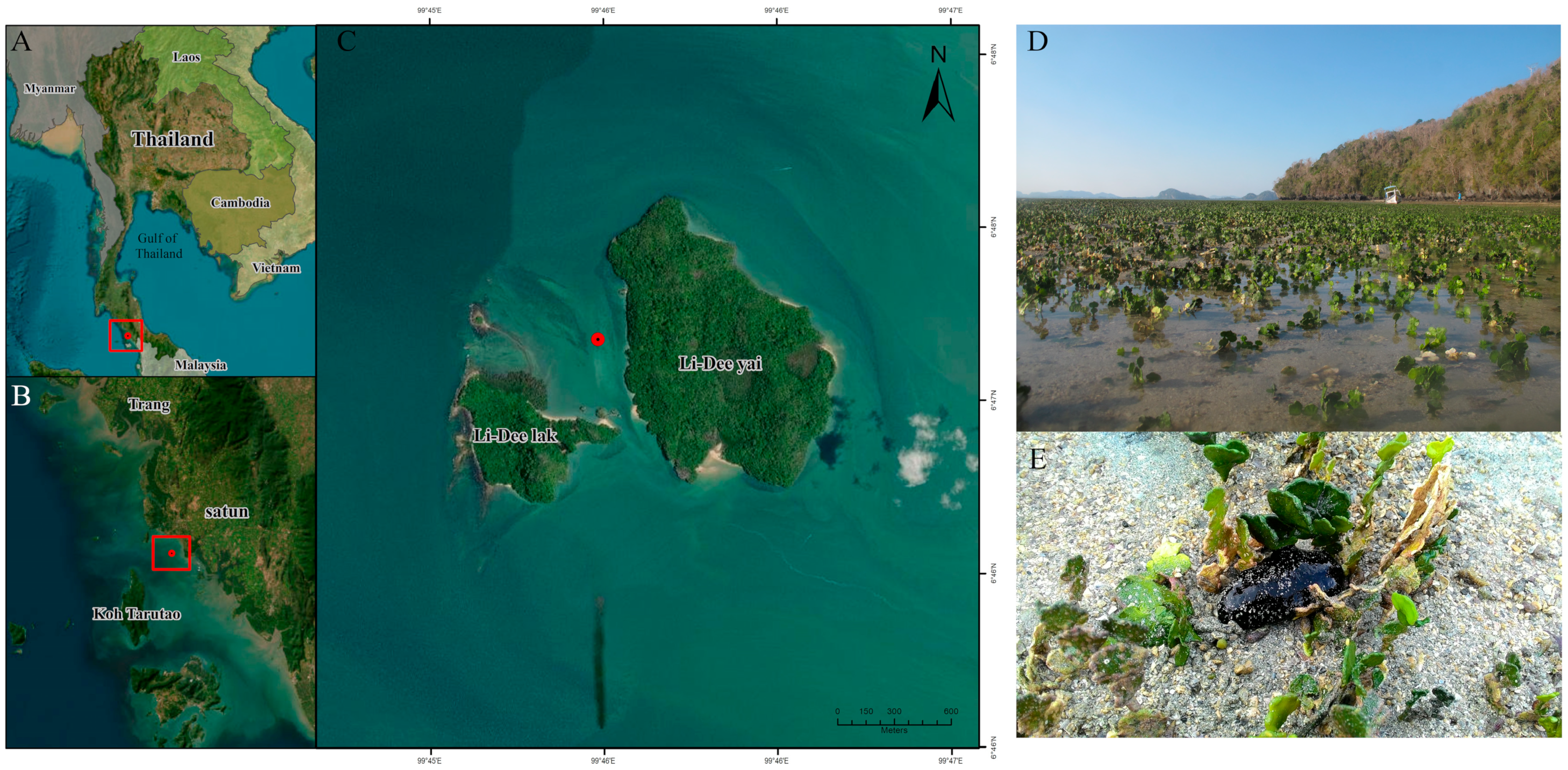
This study was conducted in the intertidal zone of Lidee Island (6°47′18″ N 99°46′17″ E), Satun Province, Andaman Sea, Thailand, in December 2020 (Figure 1). The study site was dominated by Halimeda macroloba Decaisne, which provides habitat for a variety of marine organisms, particularly the sea cucumber Holothuria atra. In addition, three seagrass species were present at this study site: Halophila ovalis (R. Brown) J. D. Hooker, Thalassia hemprichii (Ehrenberg) Ascherson, and Cymodocea rotundata Ascherson & Schweinfurth. Seawater temperature and salinity were approximately 30 °C and 30 ppt, respectively. Nitrate and phosphate concentrations were 7.37 ± 0.02 mg/L and 2.14 ± 0.39 mg/L, respectively. All samples, including seawater, sediment, H. macroloba thalli, and sea cucumber feces, were collected randomly within Halimeda patches at a depth of 1.5–2.0 m via snorkeling, with three independent replicates collected for each sample type. The distance between sampling points was approximately 5 m. This spacing was selected to obtain independent biological replicates while maintaining similar environmental conditions within the study area and minimizing potential confounding effects arising from small-scale habitat heterogeneity. Seawater samples were collected approximately 0.5 m above the sediment surface using sterilized 2 L bottles. Sediment samples were collected from a depth of approximately 5 cm using a corer with a diameter of 5 cm, and then they were transferred into sterile 50 mL centrifuge tubes. For H. macroloba and sea cucumber feces, each replicate was collected directly into sterile 50 mL centrifuge tubes. Immediately after collection, all samples were transferred to sterile sampling containers and maintained in an insulated cooler with ice packs during transport to the laboratory. The samples were transported to the Prince of Songkla University laboratory and subsequently stored at −20 °C until DNA extraction. The seawater samples were thawed and filtered through a sterile 0.2 μm membrane filter. The membrane filters containing the retained microbial biomass were subsequently used for DNA extraction. To ensure that subsequent analyses targeted the tightly associated bacterial community, the Halimeda samples were washed with sterile 3.5% artificial seawater prepared using commercial sea salt (Marinium, Bangkok, Thailand) to remove loosely attached epibionts and residual sediment [30].

2.2. DNA Extraction and 16s rRNA Sequencing

Microbial DNA was extracted from a total of 12 samples representing four sample types, with three biological replicates per sample type, using a DNeasy® PowerSoil Kit (QIAGEN, Hilden, Germany) according to the manufacturer’s instructions. DNA quantity and purity were assessed with a DS-11 Series spectrophotometer (DeNovix Inc., Wilmington, DE, USA). The purified DNA was then sent to GENEWIZ Biological Technology (Suzhou, China) for library preparation and sequencing. The V3–V4 hypervariable region of the 16S rRNA gene was selected because it is widely used for Illumina-based microbial community profiling and provides a balance between broad taxonomic coverage and sufficient resolution for community-level comparisons. This region was amplified using degenerate primers recommended by GENEWIZ/Azenta: forward 5′-CCTACGGRRBGCASCAGKVRVGAAT-3′ and reverse 5′-GGACTACNVGGGTWTCTAATCC-3′. These primers target the V3–V4 region of the prokaryotic 16S rRNA gene and are designed to amplify diverse bacterial taxa from complex environmental samples. Related Pro341F/Pro805R-type V3–V4 primers have been reported to provide a broad coverage of both Bacteria and Archaea [34]; however, as with all amplicon-based approaches, primer-associated bias may remain. Therefore, because the present study focused mainly on the bacterial community composition based on the downstream taxonomic classification, the results are interpreted primarily as bacterial community profiles. PCR amplification was performed as a single amplicon PCR with two annealing temperature phases rather than as a two-stage Illumina indexing PCR. Briefly, amplification began with an initial denaturation at 95 °C for 5 min, followed by 10 cycles of denaturation at 94 °C for 30 s, annealing at 57 °C for 45 s, and extension at 72 °C for 1 min. This was followed by 15 additional cycles using the same denaturation and extension conditions but with the annealing temperature reduced to 47 °C. A final extension was performed at 72 °C for 10 min. Each 25 µL reaction contained 2.5 µL TransStart Buffer (TransGen, Beijing, China), 2 µL dNTPs, 1 µL of each primer, and 20–30 ng template DNA. The primer sequences reported above represent the locus-specific V3–V4 primer regions. Illumina adaptor and index sequences were incorporated during library preparation by GENEWIZ/Azenta according to the provider’s standard amplicon sequencing workflow. The indexed amplicon libraries were quantified, purified using AMPure XP beads (Beckman Coulter, Indianapolis, IN, USA), and sequenced on an Illumina MiSeq platform (Illumina, San Diego, CA, USA) using a 2 × 300 bp paired-end configuration. Dedicated PCR-negative controls, extraction blanks, mock community controls, or positive control samples were not included in this study. Therefore, potential low-level background contamination from reagents or laboratory handling could not be directly assessed using control samples.

2.3. Bioinformatic and Statistical Analyses

All sequence data were processed using QIIME 2 [35]. Demultiplexed reads were initially inspected using the QIIME 2 demux summary to evaluate read quality profiles. Amplicon sequence variants (ASVs) were inferred using the DADA2 plugin [36]. Because the final ASV table was generated from single-end reads, denoising was performed using the QIIME 2 dada2 denoise-single command. Reads were trimmed by removing the first 10 bases from the 5′ end and truncated at 430 bp. Reads with ambiguous bases were discarded, and reads with expected errors greater than 2.0 were removed. Quality truncation was performed at the first base with a quality score of 2 or lower. ASVs were inferred using the independent pooling method, and chimeric sequences were removed using the consensus method. The minimum fold-parent-over-abundance parameter for chimera detection was set to 1.0, and 1,000,000 reads were used for error model learning. Taxonomic assignment was performed using the QIIME 2 feature-classifier classify-sklearn method with a naïve Bayes classifier trained on the SILVA 138 SSURef NR99 reference database [37]. The classifier was generated using RESCRIPt and q2-feature-classifier, and taxonomy was assigned with a confidence threshold of 0.7. After taxonomic classification, the ASVs were screened for non-target assignments, including Archaea, chloroplasts, and mitochondria. No ASVs were assigned to the domain Archaea, and no mitochondrial ASVs were detected. Chloroplast-assigned ASVs were detected and removed before downstream analyses. Non-bacterial and unassigned ASVs were also excluded. The final filtered bacterial ASV table was used for taxonomic composition, alpha-diversity, beta-diversity, Venn diagram, and LEfSe analyses. To normalize the sequencing depth prior to diversity analysis, the filtered bacterial ASV table was rarefied to an even depth of 24,000 sequences per sample. This rarefaction depth was selected based on the distribution of the final bacterial read counts after chloroplast removal, which ranged from 24,589 to 35,680 reads per sample, allowing all samples to be retained while minimizing the loss of sequences. The rarefied ASV table was used for alpha- and beta-diversity analyses, whereas taxonomic composition was summarized as relative abundance using the filtered bacterial ASV table. The bacterial community composition was expressed as relative abundance for each replicate across treatments. Alpha diversity was assessed using Simpson’s diversity index, observed features (observed ASV richness), Faith’s phylogenetic diversity, the Shannon diversity index, and Pielou’s evenness. Differences in alpha diversity among treatments were tested with the non-parametric Kruskal–Wallis test. The unweighted pair group method with arithmetic mean (UPGMA) based on Bray–Curtis dissimilarity was used to cluster the bacterial community at the phylum level. Beta diversity was evaluated using principal coordinate analysis (PCoA) based on unweighted UniFrac distances. Differences in community composition among treatments were determined using permutational multivariate analysis of variance (PERMANOVA) with 9999 permutations, after confirming the homogeneity of dispersions with PERMDISP. Differentially abundant taxa were identified using linear discriminant analysis effect size (LEfSe) [38], applying an LDA threshold of 4.5 and a significance level of 0.05. The overlap of core microbiota between treatments was visualized with a Venn diagram generated using the online tool jvenn (https://www.bioinformatics.com.cn/static/others/jvenn/example.html, accessed 20 July 2024). Differences in the relative abundance of microbial taxa at the phylum level among samples were tested using one-way analysis of variance (ANOVA), followed by Tukey’s HSD test for pairwise comparisons.

3. Results

A total of 382,400 high-quality bacterial reads were retained after demultiplexing, quality filtering, denoising, chimera removal, and removal of chloroplast-assigned ASVs. The sequencing depth ranged from 24,589 to 35,680 reads per sample (mean: 31,867; median: 32,871 reads per sample). Taxonomic classification in QIIME 2 assigned all recovered ASVs to the domain Bacteria, and no ASVs were assigned to Archaea. No mitochondrial ASVs were detected. Chloroplast-assigned ASVs accounted for 18,271 reads, representing 4.56% of the post-denoising dataset, and were removed before downstream bacterial community analyses. After filtering, the final bacterial dataset comprised 44 phyla, 104 classes, 222 orders, 363 families, 733 genera, and 827 species-level annotations. Sample-wise sequencing and DADA2 processing statistics are provided in Supplementary Table S1.
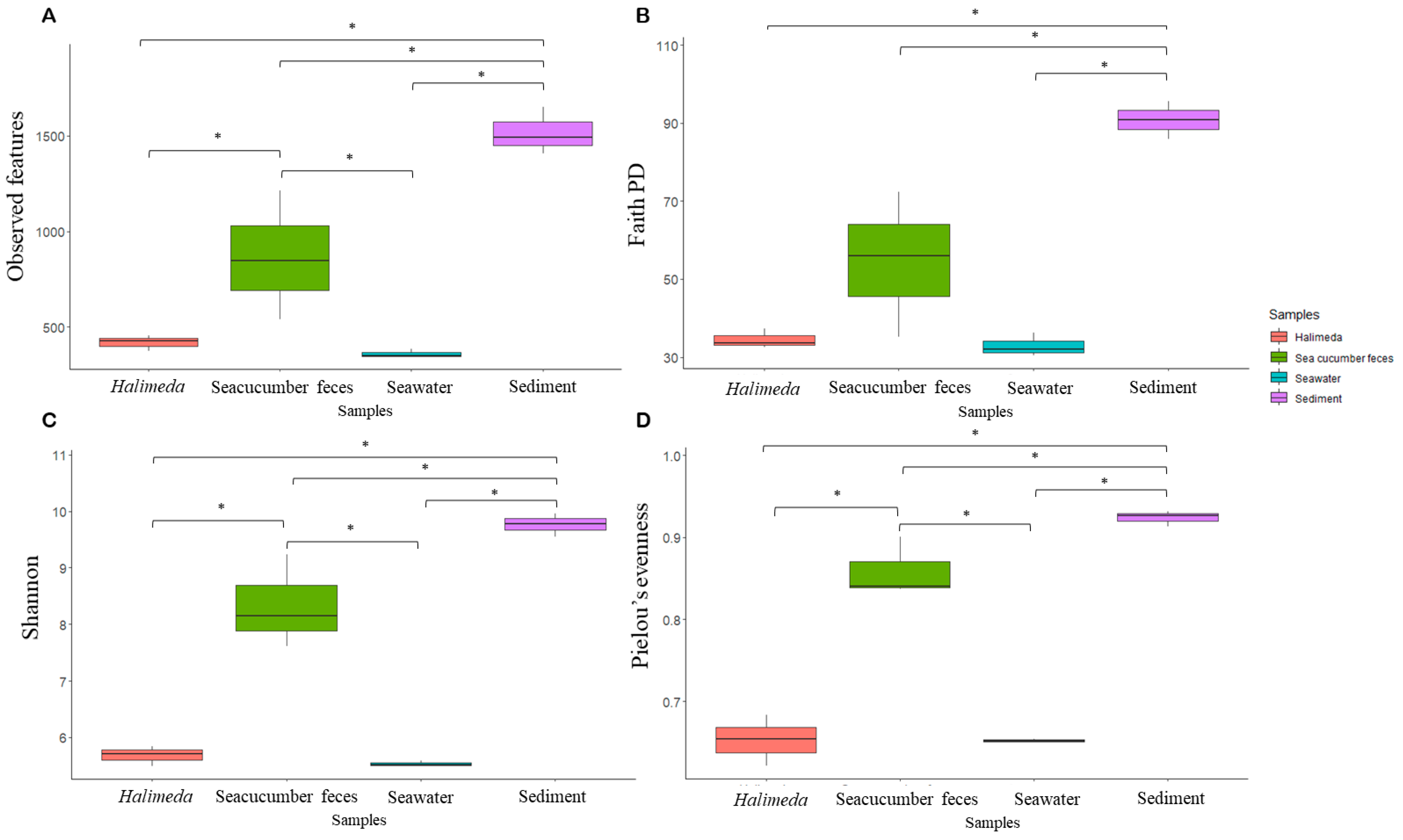
For bacterial alpha diversity, the sediment samples exhibited the highest richness and diversity, with an average of 1528.52 ± 73.62 observed features and a Shannon index of 9.77 ± 0.12, followed by the sea cucumber feces (870.26 ± 195.38 observed features; Shannon index = 8.33 ± 0.47) and Halimeda samples (426.87 ± 25.48 observed features; Shannon index = 5.69 ± 0.10). Ambient seawater showed the lowest alpha diversity, with an average of 363.67 ± 14.50 observed features and a Shannon index of 5.54 ± 0.02 (Figure 2A and C). Species evenness showed a similar pattern, with sediment (Pielou’s evenness = 0.924 ± 0.005), sea cucumber feces (0.859 ± 0.021), and Halimeda (0.653 ± 0.018) exhibiting higher evenness than ambient seawater, which showed the lowest value (0.652 ± 0.001) (Figure 2D). Overall, microbial richness, diversity, and evenness differed significantly among sample types (p < 0.05, Kruskal–Wallis). However, no significant differences were detected between the Halimeda and seawater samples for either diversity or evenness (p > 0.05, Dunn’s Test) (Figure 2).
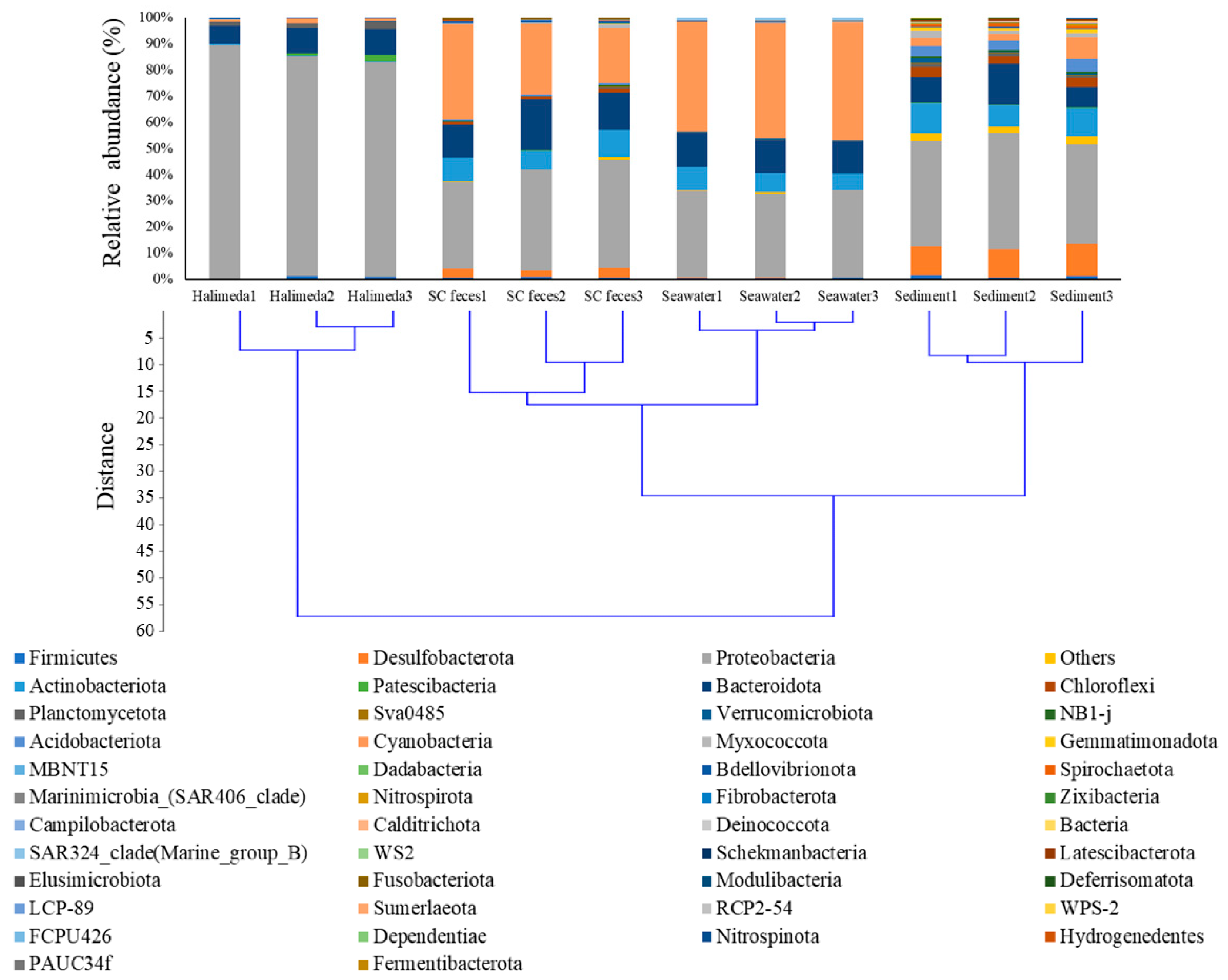
The bacterial community composition differed markedly among seawater, sediment, Halimeda thalli, and sea cucumber feces, indicating strong compartment-specific structuring of the bacterial communities (p < 0.05, PERMANOVA, Supplementary Table S2). At the phylum level, Proteobacteria, Cyanobacteria, Bacteroidota, Desulfobacterota, and Actinobacteriota were dominant across all sample types, comprising 32.32–89.39%, 0.96–45.07%, 6.98–19.65%, 0.02–12.50%, and 0.11–11.41% of the total community, respectively (Figure 3). At the class level, Alphaproteobacteria, Gammaproteobacteria, Cyanobacteriia, Bacteroidia, and Acidimicrobiia were the most abundant groups, accounting for 7.71–74.80%, 11.40–36.80%, 0.95–45.07%, 6.95–19.65%, and 0.09–10.54%, respectively (Figure 4). At lower taxonomic levels, several taxa showed marked variation among sample types. For example, the Synechococcus CC9902 lineage, Agarilytica rhodophyticola, and Lutibacter sp. reads were among the most abundant assignments, with relative abundances ranging from 0.05 to 28.64%, 0.00 to 10.42%, and 0.00 to 9.04%, respectively.
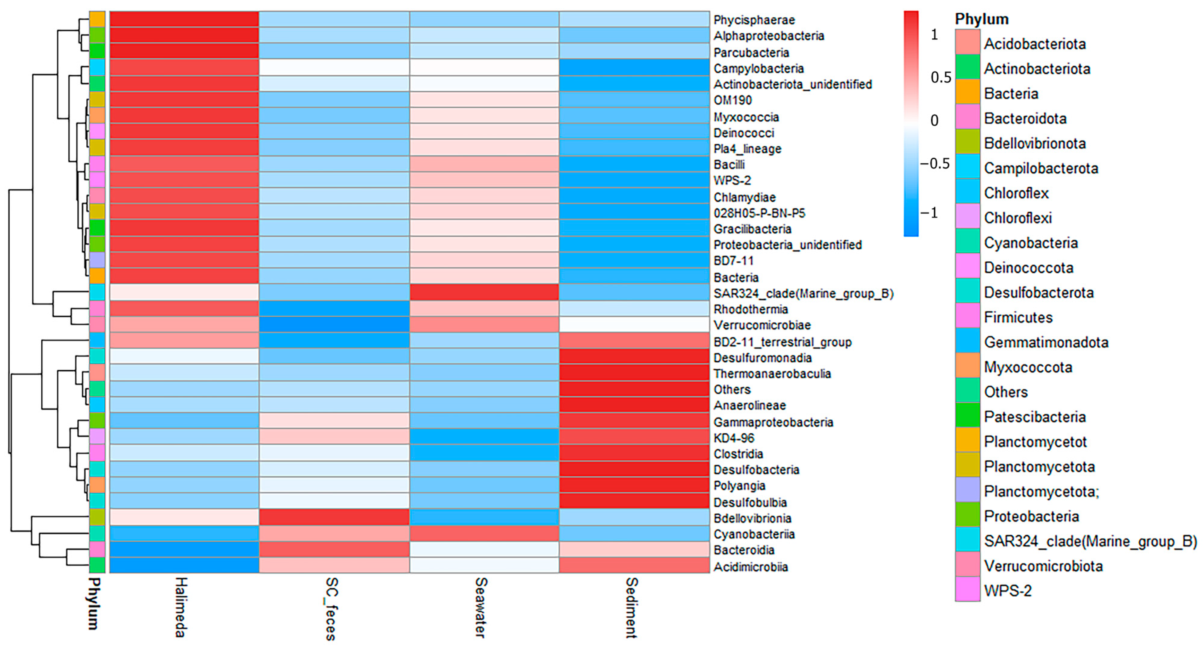
Taxonomic composition and bacterial community patterns differed among the four sample types, as shown in Figure 3. Hierarchical clustering using the unweighted pair group method with arithmetic mean (UPGMA) at the phylum level separated the samples into two major clusters. The first cluster comprised sediment, seawater, and sea cucumber feces, whereas the second cluster consisted exclusively of the Halimeda samples (Figure 3). Halimeda thalli were characterized by a high relative abundance of Proteobacteria and Planctomycetota, accounting for 85.17 ± 2.18% and 2.08 ± 0.47% of the community, respectively. The relative abundance of Proteobacteria in Halimeda was higher than that in ambient seawater, sediment, and sea cucumber feces (p < 0.05). Likewise, Planctomycetota were significantly more abundant in Halimeda than in seawater and sea cucumber feces (p < 0.05, Kruskal–Wallis). The seawater samples were characterized by high relative abundances of Cyanobacteria, the SAR324 clade (Marine Group B), and Marinimicrobia (SAR406 clade), accounting for 43.63 ± 0.97%, 0.93 ± 0.04%, and 0.42 ± 0.04%, respectively. Cyanobacteria was significantly more abundant in seawater than in Halimeda, sea cucumber feces, and sediment (p < 0.05). Additionally, the SAR324 clade (Marine Group B) and Marinimicrobia (SAR406 clade) were significantly more abundant in seawater than in Halimeda and sea cucumber feces (p < 0.05). The sediment samples showed elevated abundances of Desulfobacteria, Actinobacteria, Chloroflexi, Myxococcota, Acidobacteriota, and Firmicutes, with relative abundances of 11.38 ± 0.57%, 10.11 ± 0.95%, 3.46 ± 0.34%, 1.92 ± 0.41%, 4.06 ± 0.40%, and 1.13 ± 0.20%, respectively. Actinobacteria and Chloroflexi were significantly more abundant in sediment than in seawater and Halimeda (p < 0.05). Additionally, Acidobacteriota, Myxococcota, and Desulfobacterota were significantly more abundant in sediment than in Halimeda and seawater (p > 0.05). Firmicutes were also relatively abundant in sediment, although no significant differences were detected among sample types (p > 0.05). Sea cucumber feces were enriched in Bacteroidota, Fusobacteriota, and Bdellovibrionota, with relative abundances of 15.43 ± 2.17%, 0.87 ± 0.04%, and 0.71 ± 0.05%, respectively. Fusobacteriota were significantly more abundant in sea cucumber feces than in Halimeda and seawater (p < 0.05). Similarly, the abundance of Bdellovibrionota was higher in feces than in seawater (p < 0.05). In contrast, the abundance of Bacteroidota did not differ significantly among the sample types (Figure 3). The distribution of the 30 most abundant bacterial classes is shown in a heatmap in Figure 4, where the color gradient from dark blue to dark red indicates increasing relative abundance across samples. The Halimeda samples were enriched in Alphaproteobacteria, Phycisphaerae, Parcubacteria, Phycisphaerae, Bacilli, and Gracilibacteria, with relative abundances of 69.06 ± 3.39%, 1.96 ± 0.45%,1.05 ± 0.73%, 0.74 ± 0.38%, and 0.16 ± 0.09%, respectively. In sea cucumber feces, Bacteroidia and Bdellovibrionia were more abundant, accounting for 15.37 ± 2.19% and 0.86 ± 0.04, respectively. The seawater samples were dominated by Cyanobacteriia and the SAR324 clade (Marine group B), with significantly higher relative abundances (46.63 ± 0.97% and 0.93 ± 0.04%, respectively) than in the other sample types. In sediment, 19 bacterial classes were relatively abundant; among these, Gammaproteobacteria, Acidimicrobiia, Desulfobacteria, Anaerolineae, and Polyangia were the most dominant, with relative abundances of 30.88 ± 2.96%, 9.40 ± 0.82%, 4.23 ± 0.43%, 2.42 ± 0.29%, and 1.77 ± 0.38%, respectively (Figure 4). At the genus level, the three most dominant genera in seawater were Synechococcus CC9902, Cyanobium PCC-6307, and Candidatus Actinomarina, with relative abundances of 30.90 ± 0.35%, 7.56 ± 0.72%, and 5.69 ± 0.93%, respectively. In the sediment samples, Woeseia and Ilumatobacter were the dominant genera, accounting for 5.99 ± 0.25% and 1.80 ± 0.04%, respectively. Sea cucumber feces were dominated by Pleurocapsa PCC-7319, Lutibacter, and Vibrio, with relative abundances of 7.11 ± 1.89%, 4.69 ± 2.28%, and 3.15 ± 0.45%, respectively. In contrast, Halimeda thalli were primarily associated with Parvularcula, Kordiimonas, Agarilytica, Aestuariibacter, and Nisaea, accounting for 5.70 ± 1.09%, 5.45 ± 0.44%, 5.00 ± 2.09%, 4.68 ± 0.78%, and 3.24 ± 0.47% of the community, respectively (Supplementary Table S3).
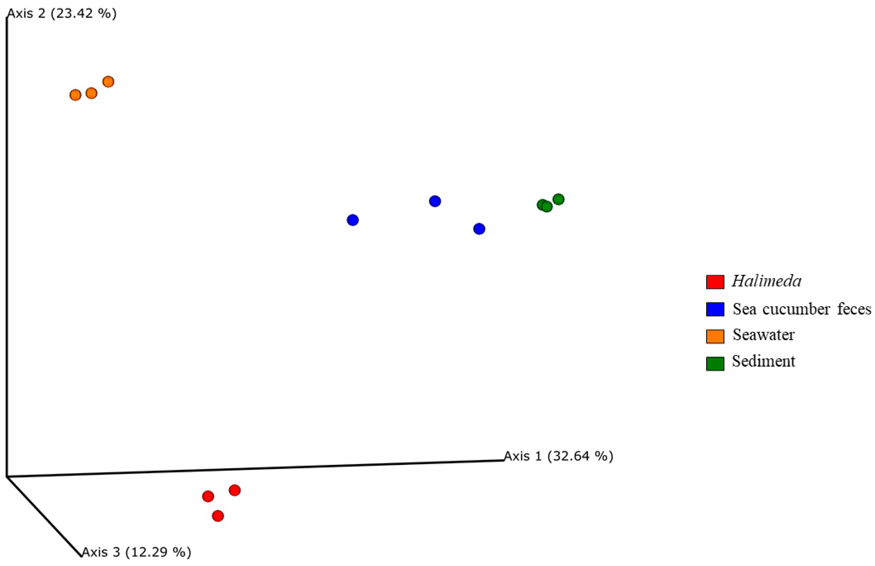
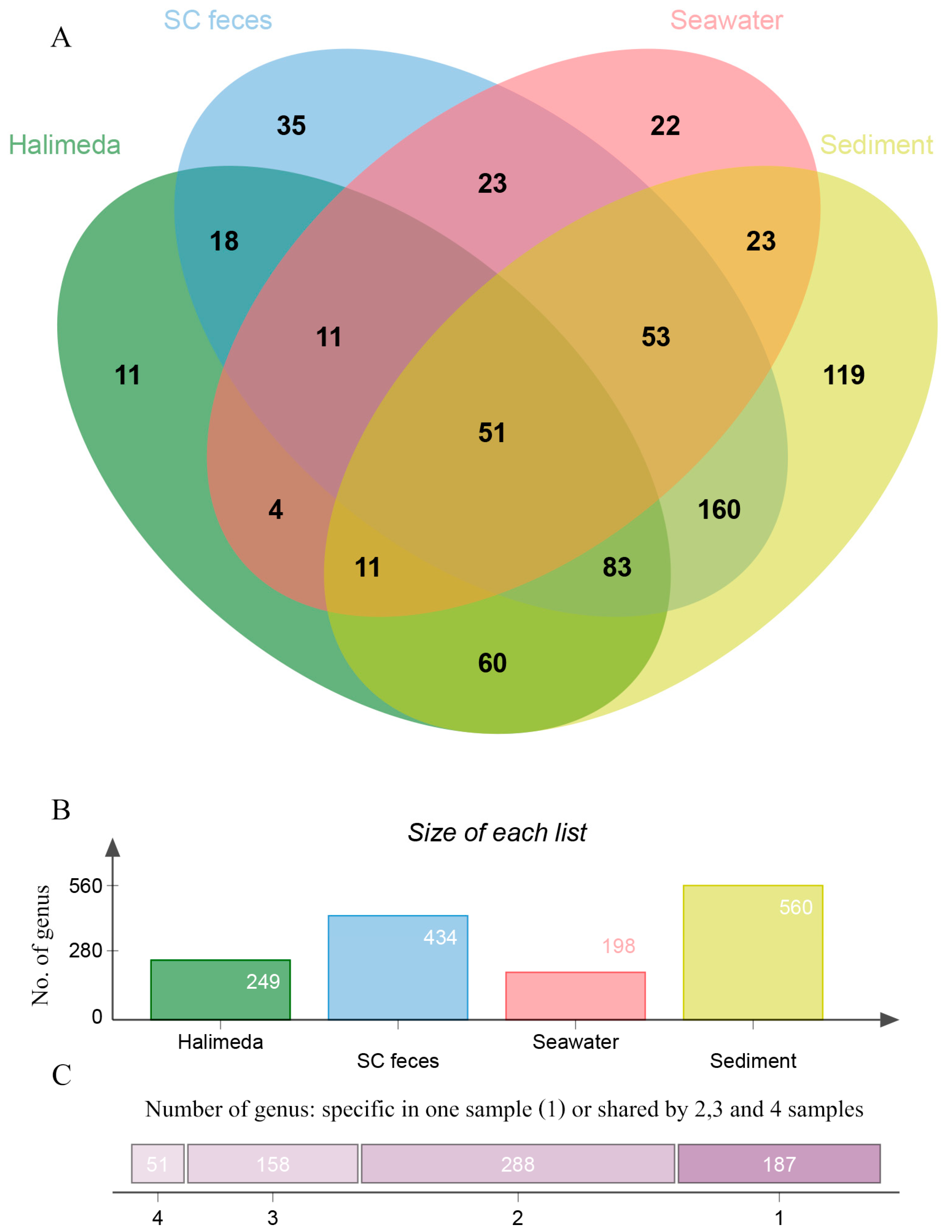
For beta diversity, principal coordinate analysis (PCoA) based on unweighted UniFrac distances showed clear separation of bacterial communities among the four sample types (Figure 5). In addition, PERMANOVA confirmed that the bacterial community composition differed significantly among sample types (p < 0.05, Supplementary Table S1). The Venn diagram revealed that 51 bacterial genera (6.95%) were shared across all sample types. Each microenvironment also contained unique bacterial taxa, including 119 genera in sediment (16.26%), 11 genera in Halimeda (1.49%), 35 genera in sea cucumber feces (4.77%), and 22 genera in ambient seawater (2.99%). Pairwise overlap in microbial communities also varied among sample types. Ambient seawater and sediment shared 138 genera (18.80%), whereas Halimeda and sediment shared 205 genera (27.92%). Halimeda and ambient seawater shared 77 genera (10.49%), while Halimeda and sea cucumber feces shared 163 genera (22.20%). Sea cucumber feces and ambient seawater shared 138 genera (18.80%). The greatest overlap was observed between sea cucumber feces and sediment, which shared 347 genera (47.28%) (Figure 6).
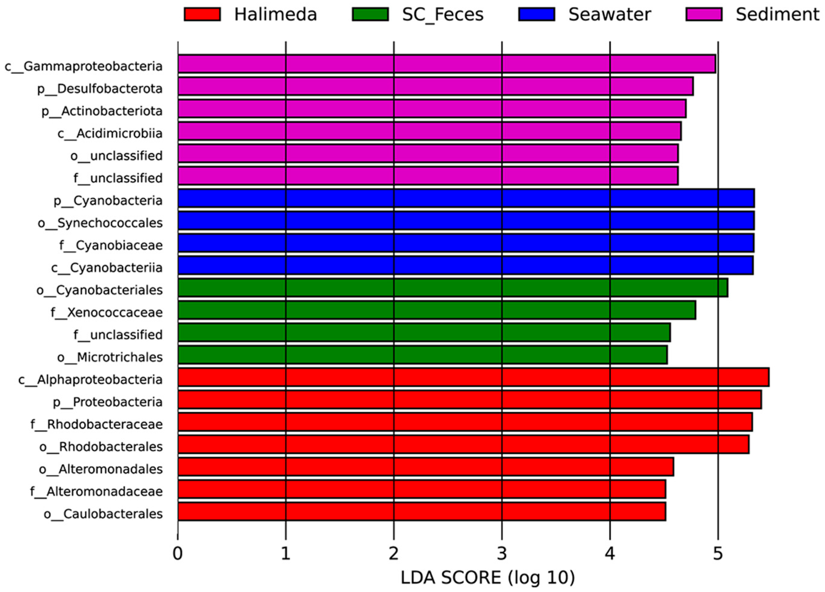
Linear discriminant analysis effect size (LEfSe) was used to identify bacterial taxa that were significantly enriched in each sample type. In sediment, the class Acidimicrobiia (phylum Actinobacteriota), together with the class Gammaproteobacteria (phylum Proteobacteria) and the phylum Desulfobacteriota, was significantly enriched. The seawater samples were characterized by the enrichment of the family Cyanobiaceae (phylum Cyanobacteria). In the Halimeda samples, several taxa were significantly enriched, including the order Caulobacterales and the families Rhodobacteraceae (within the class Alphaproteobacteria of the phylum Proteobacteria) and Alteromonadaceae (within the class Gammaproteobacteria of the phylum Proteobacteria). In contrast, sea cucumber feces were characterized by the enrichment of Xenococcaceae (family within the order Cyanobacteriales) and the order Microtrichales (class Actinobacteriota) (Figure 7).

4. Discussion

This study demonstrated that bacterial communities differed markedly among Halimeda macroloba thalli, seawater, sediment, and Holothuria atra feces, indicating strong habitat-specific microbial assembly within a tropical Halimeda-dominated ecosystem. The pronounced differentiation among these benthic microenvironments suggests that the bacterial community structure is shaped by a combination of environmental filtering, substrate characteristics, resource availability, and host-associated biological processes. Similar patterns of microbial differentiation among microenvironments have been reported in other tropical benthic ecosystems, including macroalgal habitats, seagrass meadows, coral reefs, and mangrove forests, where distinct physicochemical conditions and host-specific traits promote the establishment of specialized microbial assemblages [30,39,40,41,42,43,44]. Our findings extend this concept to Halimeda habitats and suggest that interactions among macroalgal surfaces, sediments, seawater, and benthic detritivores contribute to the development of ecologically distinct yet interconnected bacterial communities. These results highlight the potential importance of microbial niche partitioning in maintaining ecosystem functioning within tropical macroalgal habitats.
The bacterial community associated with Halimeda thalli was markedly different from that associated with the other microenvironments, accounting for more than 85% of the total bacterial community. Proteobacteria was the dominant bacterial phylum, with Alphaproteobacteria showing a significantly higher relative abundance on Halimeda thalli than in the other microenvironments, a result similar to that of Kuba et al. [30,41]. This may be because the surface of Halimeda can serve as a specialized niche, selecting for specific taxa. This pattern is consistent with that in previous studies reporting that this group is a common and often dominant component of the bacterial community of photosynthetic marine organisms, particularly marine macroalgae [45,46,47], especially Halimeda, which show a high abundance of Alphaproteobacteria [30,41,48]. LEfSe analysis revealed that the families Caulobacterales and Rhodobacteraceae were significantly more abundant in the Halimeda phycosphere than in the surrounding microenvironment. These two groups have been reported as core macroalgal surface-associated bacteria [48,49,50]. They perform a range of beneficial functions for their host, including cellulose decomposition, stabilization of the surface bacterial community, carbon and sulfur cycling, and synthesis of vitamins, all of which may enhance macroalgal growth, photosynthesis, health, pathogen resistance, and environmental adaptation [51,52,53]. The occurrence of Alteromonadaceae on Halimeda thalli may likewise reflect a close association between this bacterial family and marine macrophytes, as previously reported [54]. This bacterial group was highly abundant on the surface of Halimeda, likely because carbon compounds released by the macroalga can stimulate chemotaxis and support the growth of bacteria possessing genes involved in carbon utilization [54]. Furthermore, this bacterial group can form biofilms that may protect macroalgae from the settlement of algal spores, pathogenic bacteria, fungi, and invertebrate fouling organisms [55,56]. At the genus level, Parvularcula, Kordiimonas, Agarilytica, Nisaea, and Aestuariibacter were dominant on Halimeda. Parvularcula and Kordiimonas have been described as aerobic bacteria commonly associated with marine environments [57,58,59,60], whereas Aestuariibacter and Nisaea have previously been reported in seawater and sediment and may contribute to nutrient cycling, including nitrogen [61,62] and carbon cycling [63], and other ecological processes in coastal habitats [64,65,66,67,68]. Agarilytica has been reported in association with macrophytes [60]. One possible explanation for this is that the macroalgal surface is rich in polysaccharides, such as cellulose and xylans, which can serve as substrates for bacteria harboring genes involved in algal polysaccharide degradation [69]. The phylum Planctomycetota also showed the highest relative abundance on Halimeda thalli, consistent with previous studies reporting members of this phylum as common constituents of macroalgal surface biofilms [70,71]. Several macroalgae-associated Planctomycetota genera, including Algisphaera and Rhodopirellula, have been reported to possess sulfatases and other carbohydrate-active enzymes involved in the degradation of sulfated polysaccharides derived from algal tissues [72,73,74]. Furthermore, some Planctomycetota lineages have been implicated in carbon and nitrogen cycling in marine ecosystems [31,33]. These findings suggest that the enrichment of Planctomycetota on Halimeda thalli may be linked to the utilization and turnover of algal-derived organic matter. However, such functional roles should be considered tentative because they cannot be directly inferred from the 16S rRNA gene sequencing data generated in this study. This interpretation is supported by the UPGMA clustering results, which clearly separated the Halimeda samples from the other habitat microenvironment samples. In addition, alpha diversity was relatively low in the Halimeda samples, suggesting that Halimeda may structure its bacterial community in a manner similar to other host-associated systems. This pattern may reflect the influence of chemical compounds on the algal surface, which create a selective niche for particular microbial groups [75,76].
Seawater harbored a distinct bacterial community compared with the other sample types. At the genus level, Synechococcus CC9902, Cyanobium PCC-6307, and Candidatus Actinomarina were among the dominant taxa. The findings of the high relative abundance of Synechococcus CC9902 and Cyanobium PCC-6307, members of the class Cyanobacteriia, are consistent with those of previous reports showing that this taxon is widely distributed throughout seawater columns worldwide because of its ability to adapt to a broad range of temperature, salinity, and nutrient conditions [77,78,79]. Synechococcus plays a crucial role as a primary producer and contributes substantially to marine biogeochemical cycles [32,80]. Candidatus Actinomarina is also a typical pelagic bacterium. Candidatus Actinomarina is commonly distributed in the photic zone and oligotrophic seawater [81,82]. Together, these patterns may suggest that the seawater column within the Halimeda meadow is well lit and relatively low in nutrients. The SAR324 clade (Marine Group B) and Marinimicrobia (SAR406 clade) were significantly more abundant in seawater than in the other sample types. Both groups have frequently been reported in oxygen minimum zone environments [83,84]. Their presence in the present study may indicate intense microbial respiration within Halimeda patches, including in the sediment and on macroalgal surfaces. LEfSe analysis further identified Cyanobiaceae (a member of the order Synechococcales, class Cyanobacteriia) as a taxon enriched in seawater. Cyanobiaceae has been reported to be widely distributed across natural ecosystems [80,85]. This bacterial group is also considered a key contributor to nitrogen fixation in marine ecosystems [86]. Species diversity in seawater was the lowest among all sample types. This result is consistent with that of Mancuso et al. [87], who reported lower microbial diversity in seawater than in algal-associated habitats, which tend to be more stable over time. One possible explanation for this is that the shallow water column is frequently mixed, resulting in relatively homogeneous physical and chemical conditions. Such homogeneity may favor bacterial groups with broad environmental tolerance and a strong adaptive capacity [88,89]. The low overlap in bacterial genera between Halimeda and seawater further suggests that the algal surface does not allow colonization by all bacteria present in the water column but instead selectively recruits particular taxa through surface-associated chemical properties [75,90].
Sediment harbored the highest microbial diversity among the four microenvironments. The heterogeneity of the sediment structure and chemical properties likely creates a wide range of microhabitats [4,91]. In particular, the vertical gradient within the upper few millimeters of sediment, from the surface to deeper layers, generates oxic, suboxic, and anoxic conditions that shape distinct bacterial communities associated with each microenvironment [4,92]. The bacterial community in the sediment samples reflected this geochemical complexity, with Desulfobacterota, Actinobacteriota, Chloroflexi, and Acidobacteriota being the dominant phyla and Gammaproteobacteria, Acidimicrobiia, and Desulfobacteria being the dominant classes. Gammaproteobacteria (a member of the phylum Proteobacteria) and Acidimicrobiia (a member of the phylum Actinobacteriota) have been frequently reported as dominant bacterial classes in marine sediments [43,93,94], and both groups play crucial roles in sediment biogeochemistry, including carbon, nitrogen, and sulfur cycling [94,95,96]. Desulfobacteria (a member of the phylum Desulfobacterota) have been reported at high abundance in organic-rich sediments, where sulfate in pore water serves as an essential electron acceptor for anaerobic respiration during organic matter degradation [97]. Sediments beneath Halimeda meadows may contain elevated levels of organic matter due to the high productivity and rapid turnover of Halimeda [20]. Acidobacteriota and Chloroflexi were also detected at relatively high abundance in the sediment samples. These groups have frequently been reported in organic-rich and anoxic sediments [98], suggesting that Halimeda patches may contribute substantial organic inputs to the underlying sediment. At the genus level, Woeseia, a member of Gammaproteobacteria, was among the most abundant taxa in sediment. This genus is commonly found in seafloor sediments [99,100] and exhibits considerable metabolic plasticity, enabling it to inhabit sediments across a broad redox gradient [101]. Ilumatobacter (a member of Acidimicrobiia) is a bacterial group that is present in high abundance in marine sediments and plays important roles in organic matter turnover, biogeochemical cycling, and sulfide detoxification [94,102]. LEfSe analysis confirmed that Acidimicrobiia (a member of the phylum Actinobacteriota), Gammaproteobacteria, and Desulfobacterota were the principal biomarkers for the sediment samples from Halimeda patches. These findings indicate that sediments beneath Halimeda patches harbor diverse bacterial communities with potential roles in organic matter remineralization and biogeochemical cycling, suggesting their importance in sediment ecosystem functioning.
The bacterial community of Holothuria atra feces exhibited an intermediate level of microbial diversity, being higher than that of seawater and Halimeda thalli but lower than that of sediment. A high overlap in bacterial genera between H. atra feces and sediment was expected, as sea cucumbers feed directly on sediment and therefore ingest sediment-associated microorganisms into their digestive tract. However, once sediment passes through the gut, the bacterial community appears to be modified by gut physicochemical conditions, resulting in slightly lower diversity than that of the original sediment [103]. The enrichment of Bacteroidota, Fusobacteriota, and Bdellovibrionota in feces relative to that in seawater and Halimeda is particularly noteworthy. Bacteroidota and Fusobacteriota are commonly associated with the anaerobic fermentation of organic particles and are frequently found in animal gut environments, especially in the anterior intestine of sea cucumbers [104]. The hypoxic conditions within the sea cucumber gut may therefore provide a favorable environment for members of this phylum [104]. Bdellovibrionota, which are predatory bacteria that consume Gram-negative bacteria, were also relatively abundant in fecal samples. Their enrichment may indicate a high abundance of potential prey bacteria in the sea cucumber gut and active bacterial predation during gut transit [5,105]. At the class level, Bacteroidia was significantly enriched in feces. This group is commonly reported at high abundance in sea cucumber guts and is known to play an important role in the degradation of complex carbohydrates [106,107]. Because the sea cucumbers in this study inhabited dense Halimeda patches, where detrital material is likely rich in polysaccharides, the elevated abundance of Bacteroidia may reflect the digestion of polysaccharide-rich organic matter during sediment passage through the gut. Pleurocapsa PCC-7319 (family Xenococcaceae), Lutibacter (order Flavobacteriales), and Vibrio were identified as the dominant taxa in the sea cucumber fecal samples. The high relative abundances of Pleurocapsa PCC-7319 and Lutibacter in feces may reflect the capacity of these sediment-associated bacteria to survive and persist through gut transit conditions [108,109]. These findings are consistent with those of Yamazaki et al. [110], who reported that sea cucumber feces are enriched in Flavobacteriales—the order to which Lutibacter belongs—and suggested that fecal deposition contributes to the surrounding sediment bacterial community, potentially stimulating the mineralization of sedimentary organic matter [22,110]. The elevated abundance of Vibrio in excreted feces likely originates from the sea cucumber digestive tract, where this genus is commonly detected as a resident gut inhabitant [104,111]. Vibrio has been reported to play a probiotic role in sea cucumbers by enhancing the degradation of proteins and lipids [112,113]. Nevertheless, certain Vibrio species are recognized as opportunistic pathogens of sea cucumbers and other marine invertebrates, and their dispersal via fecal deposition may pose ecological risks to the surrounding benthic environment [114,115]. LEfSe analysis further identified Microtrichales (a member of the phylum Actinobacteriota) and Xenococcaceae (a member of the phylum Cyanobacteria) as differentially abundant taxa. Microtrichales has been reported in association with marine sediments [116], whereas Xenococcaceae has been found attached to macroalgal surfaces [117,118]. These two bacterial groups may be ingested by H. atra through feeding activity on sediment and macroalgae, subsequently passing through the gut where they may contribute to organic matter mineralization and carbon cycling in benthic ecosystems [116,118]. Collectively, these findings suggest that H. atra may play an important role in restructuring sediment-associated microbial communities [22,119,120]. This process may benefit Halimeda ecosystems by enhancing nutrient cycling, supporting more complete organic matter mineralization, and promoting nutrient regeneration.
According to the analysis of shared genera among the four sample types, only 60 genera (10.36%) were common across all compartments. This limited overlap suggests strong compartment-specific structuring of the bacterial communities. Such patterns may be influenced by differences in habitat characteristics among sediment, seawater, Halimeda thalli, and sea cucumber feces, including factors such as redox conditions, substrate properties, nutrient availability, and host-associated processes. However, these potential drivers were not measured in this study and should be investigated in future research. A comparison of genus-level overlap among sample types revealed the greatest overlap between feces and sediment (47.28%), supporting a strong connection between these two microenvironments through the non-selective deposit-feeding strategy of holothurians via sediment ingestion, whereby material is processed and redeposited [110]. This situation may indicate that sea cucumber plays a key role as an active mediator of benthic microbial recycling, linking sediment and consumer compartments within the biogeochemical cycling of the Halimeda-dominated habitat [110]. In contrast, Halimeda and sediment shared 27.92% of genera, which may reflect the deposition of senescent Halimeda fragments into the sediment, as well as the attachment of suspended sediment particles to the surface of living thalli. Feces and Halimeda shared 22.20% of genera, suggesting that sea cucumber foraging on Halimeda-derived detritus may create microbial connectivity between the algal surface and the benthic fecal niche. The lowest overlap was observed between seawater and Halimeda (10.46%), supporting the idea of host-specific microbial selection on the Halimeda surface [121]. Taken together, these patterns suggest that microbial communities in Halimeda patches are not isolated but rather connected through the movement of organic matter and biological activity within the benthic habitat. By ingesting sediment and depositing feces beneath Halimeda patches, sea cucumbers may function as biological vectors linking otherwise distinct microbial niches. In this way, Holothuria atra may facilitate the dispersal of bacteria among microenvironments, particularly within the benthic community. Bioturbation resulting from the deposit-feeding behavior of sea cucumbers can disturb and restructure sediment, potentially influencing the distribution of the resident bacterial community [110]. As sea cucumbers feed, move, and excrete at different locations, sediment particles and their associated microbiota are relocated and redistributed, potentially establishing a mobile link between the sediment and Halimeda-associated microbial pools within the benthic community. Moreover, sea cucumber feces serve as a nutrient-enriched substrate that promotes bacterial growth and colonization at the deposition site [107]. This process may, in turn, drive bacterial community assembly and influence nutrient cycling within the habitat [22,110,122].
Synthesizing bacterial community patterns across the four microenvironments revealed strong habitat-specific community structuring within the Halimeda-dominated ecosystem. Seawater communities were characterized by relatively high abundances of picocyanobacteria and oligotrophic heterotrophs, whereas sediment communities were dominated by taxa commonly associated with organic matter degradation and anaerobic metabolism. In contrast, Halimeda thalli harbored distinct host-associated bacterial assemblages, while sea cucumber feces supported bacterial communities that differed from those in the surrounding sediment and seawater. These patterns suggest that each microenvironment provides unique ecological niches that select for different bacterial taxa. Although the functional roles of these communities were not directly assessed, the observed taxonomic differentiation indicates the potential for distinct microbial contributions to ecological processes within Halimeda patches. Further studies incorporating environmental measurements and functional analyses are needed to clarify the ecological roles and interactions of microbial communities in Halimeda-dominated ecosystems.
The results of this study demonstrate that each microenvironment harbors unique functional bacteria that play important roles within the Halimeda-dominated habitat, implying that the loss of any single component could influence bacterial community structure and associated biogeochemical functions. In particular, sea cucumbers play a key role in shaping sediment-associated bacterial communities through feeding activity and feces deposition. Given this key ecological role, the conservation of sea cucumbers should be prioritized. These findings support the use of microbiome information as an indicator for monitoring and management frameworks in tropical Halimeda-dominated coastal ecosystems and potentially in other marine ecosystems. Such information could also inform marine protected area (MPA) zoning and restoration efforts aimed at preserving microbial connectivity among microenvironments, habitats, and overall ecosystem resilience.
However, several limitations of this study should be acknowledged. First, 16S rRNA amplicon sequencing targeting the V3–V4 hypervariable region was used to characterize the bacterial community composition based on the SILVA reference database. Although this approach provides valuable taxonomic information, it does not directly reflect the functional potential or metabolic activity of microbial communities in each sample type. Future studies should therefore incorporate shotgun metagenomics and metatranscriptomics to better resolve functional gene profiles and actively expressed metabolic pathways, thereby providing deeper insight into the mechanisms underlying bacterial community structure and function. Second, only three biological replicates were collected for each microenvironment, which may not fully capture the spatial heterogeneity of the bacterial communities within Halimeda-dominated habitats. Although clear differences among sample types were detected and replicate samples showed generally consistent community patterns, the limited replication reduces statistical power and may underestimate within-habitat variability. Therefore, the results should be interpreted as an initial characterization of bacterial communities associated with Halimeda surfaces, seawater, sediment, and sea cucumber feces. Third, this study represents only a single spatial and temporal snapshot. Microbial communities have been reported to vary markedly across seasons and geographic locations [123,124]. Consequently, the observed community patterns may not fully represent temporal or spatial variability within Halimeda-dominated ecosystems. Future studies incorporating larger sample sizes, multiple locations, environmental measurements, and seasonal sampling would provide a more comprehensive understanding of microbial dynamics and ecological processes in these habitats. Finally, dedicated PCR-negative controls, extraction blanks, and positive mock community controls were not included in this study. As a result, potential low-level reagent or laboratory contamination could not be directly identified or removed using control samples. However, the clear separation of microbial communities among sample types and the dominance of ecologically relevant taxa suggest that the major community patterns observed in this study were primarily associated with habitat-specific differences rather than contamination.

5. Conclusions

This study demonstrated clear differences in the bacterial community structure among ambient seawater, sediment, Halimeda thalli, and Holothuria atra feces within a tropical Halimeda-dominated benthic habitat. Sediment showed the highest microbial diversity, whereas seawater showed the lowest. Distinct bacterial assemblages characterized each microenvironment, with macroalgal-associated taxa dominating Halimeda thalli, remineralization-related taxa dominating sediment, Synechococcus CC9902 dominating seawater, and fecal samples enriched in taxa associated with gut processing and microbial restructuring. Limited overlap among microenvironments, together with the strong similarity between feces and sediment communities, indicates a close microbial relationship between these habitats and suggests that H. atra may influence bacterial community composition within the benthic environment. These findings improve our understanding of habitat-specific microbial assemblages and ecosystem functioning within Halimeda patches. The high proportion of bacterial overlap between H. atra feces and sediments suggests that sediment processing by sea cucumbers may influence benthic bacterial community dynamics. Such insights contribute to our understanding of microbial ecology in tropical coastal ecosystems and may support coastal management and blue carbon habitat conservation. Future studies using shotgun metagenomics and metatranscriptomics are needed to clarify the functional roles and activities of these microbial communities under natural conditions. Additionally, carbon connectivity across these four microenvironments should be investigated using stable isotope probing (SIP) to observe the flow through the loop of Halimeda–sediment–sea cucumber–feces.

Supplementary Materials

The following supporting information can be downloaded at: https://www.mdpi.com/article/10.3390/ecologies7030069/s1, Table S1: Sample-wise sequencing and DADA2 processing statistics; Table S2: Results of permutational multivariate analysis of variance (PERMANOVA) testing for differences in the bacterial community composition among the four microenvironments; Table S3: Relative abundance (%) of the top 30 bacterial genera detected across the four microenvironments.

Author Contributions

Conceptualization, J.M.; methodology, K.S., J.T. and J.M.; software, K.S. and J.T.; validation, J.M. and J.T.; formal analysis, K.S. and J.T.; investigation, J.T. and J.M.; resources, J.M. and K.S.; data curation, J.T. and J.M.; writing—original draft preparation, J.T.; writing—review and editing, J.T., K.S. and J.M.; visualization, K.S. and J.T.; supervision, J.M.; project administration, J.M.; funding acquisition, J.M. All authors have read and agreed to the published version of the manuscript.

Funding

This work was funded by the National Science and Research and Innovation Fund (NSRF), Prince of Songkla University (SCI6701053S). This research was also supported by the Faculty of Science Research Fund, Faculty of Science, Prince of Songkla University (2026).

Data Availability Statement

The QIIME 2 artifacts and visualization files generated during this study have been deposited in Zenodo and are publicly available at https://doi.org/10.5281/zenodo.19904043.

Acknowledgments

The authors would like to thank all members of the Seaweed and Seagrass Research Unit, Division of Biological Science, Faculty of Science, Prince of Songkla University, for their help during the field trip. The authors would also like to thank Thomas Duncan Coyne for assisting with the English text. During the preparation of this manuscript, the authors used Gemini and ChatGPT-5.6 for grammar checking. The authors have reviewed and edited the output and take full responsibility for the content of this publication.

Conflicts of Interest

The authors declare no conflicts of interest.

References

  1. Bourne, D.G.; Webster, N.S. Coral Reef Bacterial Communities. In The Prokaryotes; Rosenberg, E., DeLong, E.F., Lory, S., Stackebrandt, E., Thompson, F., Eds.; Springer: Berlin/Heidelberg, Germany, 2013; pp. 163–187. [Google Scholar]
  2. Lin, G.; Gao, D.; Yang, P.; Liu, S.; Sun, D.; Lin, X. Editorial: Linking Microbial-Driven Key Processes with Carbon and Nitrogen Cycling in Estuarine, Coastal, and the Nearshore Areas. Front. Microbiol. 2024, 15, 1382148. [Google Scholar] [CrossRef] [Scilit] [PubMed]
  3. Chen, J.; Hanke, A.; Tegetmeyer, H.E.; Kattelmann, I.; Sharma, R.; Hamann, E.; Hargesheimer, T.; Kraft, B.; Lenk, S.; Geelhoed, J.S.; et al. Impacts of Chemical Gradients on Microbial Community Structure. ISME J. 2017, 11, 920–931. [Google Scholar] [CrossRef] [Scilit] [PubMed]
  4. Hoshino, T.; Doi, H.; Uramoto, G.-I.; Wörmer, L.; Adhikari, R.R.; Xiao, N.; Morono, Y.; D’Hondt, S.; Hinrichs, K.-U.; Inagaki, F. Global Diversity of Microbial Communities in Marine Sediment. Proc. Natl. Acad. Sci. USA 2020, 117, 27587–27597. [Google Scholar] [CrossRef] [Scilit] [PubMed]
  5. Davis, C.L.; Venturelli, R.A.; Michaud, A.B.; Hawkings, J.R.; Achberger, A.M.; Vick-Majors, T.J.; Rosenheim, B.E.; Dore, J.E.; Steigmeyer, A.; Skidmore, M.L.; et al. Biogeochemical and Historical Drivers of Microbial Community Composition and Structure in Sediments from Mercer Subglacial Lake, West Antarctica. ISME Commun. 2023, 3, 8. [Google Scholar] [CrossRef] [Scilit] [PubMed]
  6. Deng, W.; Chen, S.; Chen, S.; Xing, B.; Chan, Z.; Zhang, Y.; Chen, B.; Chen, G. Impacts of Eutrophication on Microbial Community Structure in Sediment, Seawater, and Phyllosphere of Seagrass Ecosystems. Front. Microbiol. 2024, 15, 1449545. [Google Scholar] [CrossRef] [Scilit] [PubMed]
  7. Diner, R.E.; Zimmer-Faust, A.; Cooksey, E.; Allard, S.; Kodera, S.M.; Kunselman, E.; Garodia, Y.; Verhougstraete, M.P.; Allen, A.E.; Griffith, J.; et al. Host and Water Microbiota Are Differentially Linked to Potential Human Pathogen Accumulation in Oysters. Appl. Environ. Microbiol. 2023, 89, e0031823. [Google Scholar] [CrossRef] [Scilit] [PubMed]
  8. Von Jackowski, A.; Walter, M.; Spiegel, T.; Buttigieg, P.L.; Molari, M. Drivers of Pelagic and Benthic Microbial Communities on Central Arctic Seamounts. Front. Mar. Sci. 2023, 10, 1216442. [Google Scholar] [CrossRef] [Scilit]
  9. Khandavalli, L.V.N.S.; Lodha, T.; Abdullah, M.; Guruprasad, L.; Chintalapati, S.; Chintalapati, V.R. Insights into the Carbonic Anhydrases and Autotrophic Carbon Dioxide Fixation Pathways of High CO2 Tolerant Rhodovulum Viride JA756. Microbiol. Res. 2018, 215, 130–140. [Google Scholar] [CrossRef] [Scilit] [PubMed]
  10. Foong, C.P.; Higuchi-Takeuchi, M.; Numata, K. Optimal Iron Concentrations for Growth-Associated Polyhydroxyalkanoate Biosynthesis in the Marine Photosynthetic Purple Bacterium Rhodovulum sulfidophilum under Photoheterotrophic Condition. PLoS ONE 2019, 14, e0212654. [Google Scholar] [CrossRef] [Scilit] [PubMed]
  11. Mei, X.; Wu, C.; Zhao, J.; Yan, T.; Jiang, P. Community Structure of Bacteria Associated with Drifting Sargassum horneri, the Causative Species of Golden Tide in the Yellow Sea. Front. Microbiol. 2019, 10, 1192. [Google Scholar] [CrossRef] [Scilit] [PubMed]
  12. Sonnenschein, E.C.; Jimenez, G.; Castex, M.; Gram, L. The Roseobacter-Group Bacterium Phaeobacter as a Safe Probiotic Solution for Aquaculture. Appl. Environ. Microbiol. 2021, 87, e02581-20. [Google Scholar] [CrossRef] [Scilit] [PubMed]
  13. Bunbury, F.; Deery, E.; Sayer, A.P.; Bhardwaj, V.; Harrison, E.L.; Warren, M.J.; Smith, A.G. Exploring the Onset of B12-Based Mutualisms Using a Recently Evolved Chlamydomonas Auxotroph and B12-Producing Bacteria. Environ. Microbiol. 2022, 24, 3134–3147. [Google Scholar] [CrossRef] [Scilit] [PubMed]
  14. Bourne, D.G.; Morrow, K.M.; Webster, N.S. Insights into the Coral Microbiome: Underpinning the Health and Resilience of Reef Ecosystems. Annu. Rev. Microbiol. 2016, 70, 317–340. [Google Scholar] [CrossRef] [Scilit] [PubMed]
  15. Ohimain, E.I.; Turner, R.E.; Middleton, B.A. Mangrove Microbiomes as Drivers of Ecosystem Recovery and Restoration Success. Microorganisms 2026, 14, 1235. [Google Scholar] [CrossRef] [Scilit] [PubMed]
  16. Vadillo Gonzalez, S.; Jongen, R.; Thomas, T.; Marzinelli, E.M.; Gribben, P.E. Seagrass–Microbe Interactions: A Systematic Review of Current Research Trends and Mapping of the Core Microbiome. Biol. Rev. 2026, 101, 1334–1357. [Google Scholar] [CrossRef] [Scilit] [PubMed]
  17. Bellchambers, L.; Meeuwig, J.; Evans, S.; Legendre, P. Modelling Habitat Associations of 14 Species of Holothurians from an Unfished Coral Atoll: Implications for Fisheries Management. Aquat. Biol. 2011, 14, 57–66. [Google Scholar] [CrossRef] [Scilit]
  18. Dissanayake, D.C.T.; Stefansson, G. Habitat Preference of Sea Cucumbers: Holothuria atra and Holothuria edulis in the Coastal Waters of Sri Lanka. J. Mar. Biol. Assoc. 2012, 92, 581–590. [Google Scholar] [CrossRef] [Scilit]
  19. Kongsap, V.; Rattanachot, E.; Prathep, A.; Buaphol, W.; Mayakun, J. A High Abundance of Holothuria (Halodeima) Atra (Holothuroidea aspidochirotida) in a Halimeda Dominated Habitat. J. Mar. Sci. Eng. 2023, 11, 451. [Google Scholar] [CrossRef] [Scilit]
  20. Castro-Sanguino, C.; Bozec, Y.; Mumby, P. Dynamics of Carbonate Sediment Production by Halimeda: Implications for Reef Carbonate Budgets. Mar. Ecol. Prog. Ser. 2020, 639, 91–106. [Google Scholar] [CrossRef] [Scilit]
  21. McNeil, M.; Firn, J.; Nothdurft, L.D.; Pearse, A.R.; Webster, J.M.; Roland Pitcher, C. Inter-Reef Halimeda Algal Habitats within the Great Barrier Reef Support a Distinct Biotic Community and High Biodiversity. Nat. Ecol. Evol. 2021, 5, 647–655. [Google Scholar] [CrossRef] [Scilit] [PubMed]
  22. MacTavish, T.; Stenton-Dozey, J.; Vopel, K.; Savage, C. Deposit-Feeding Sea Cucumbers Enhance Mineralization and Nutrient Cycling in Organically-Enriched Coastal Sediments. PLoS ONE 2012, 7, e50031. [Google Scholar] [CrossRef] [Scilit] [PubMed]
  23. Ennas, C.; Pasquini, V.; Abyaba, H.; Addis, P.; Sarà, G.; Pusceddu, A. Sea Cucumbers Bioturbation Potential Outcomes on Marine Benthic Trophic Status under Different Temperature Regimes. Sci. Rep. 2023, 13, 11558. [Google Scholar] [CrossRef] [Scilit] [PubMed]
  24. Yang, X.; Gao, S.; Kong, X.; Gao, F.; Xu, Q. Host Filtering Overrides Environmental Heterogeneity in Shaping Sea Cucumber Gut Microbiomes. Environ. Microbiol. 2026, 28, e70294. [Google Scholar] [CrossRef] [Scilit] [PubMed]
  25. Hillis-Colinvaux, L. Ecology and Taxonomy of Halimeda: Primary Producer of Coral Reefs. In Advances in Marine Biology; Elsevier: Amsterdam, The Netherlands, 1980; Volume 17, pp. 1–327. [Google Scholar]
  26. Payri, C.E. Halimeda Contribution to Organic and Inorganic Production in a Tahitian Reef System. Coral Reefs 1988, 6, 251–262. [Google Scholar] [CrossRef] [Scilit]
  27. Roff, G.; Wabnitz, C.C.C.; Harborne, A.R.; Mumby, P.J. Macroalgal Associations of Motile Epifaunal Invertebrate Communities on Coral Reefs. Mar. Ecol. 2013, 34, 409–419. [Google Scholar] [CrossRef] [Scilit]
  28. Mateo-Ramírez, Á.; Máñez-Crespo, J.; Royo, L.; Tuya, F.; Castejón-Silvo, I.; Hernan, G.; Pereda-Briones, L.; Terrados, J.; Tomas, F. A Tropical Macroalga (Halimeda Incrassata) Enhances Diversity and Abundance of Epifaunal Assemblages in Mediterranean Seagrass Meadows. Front. Mar. Sci. 2022, 9, 886009. [Google Scholar] [CrossRef] [Scilit]
  29. Cleary, D.F.R.; Huang, Y.M. A Comparison of the Prokaryotic Communities Associated with Seven Seaweed Species, Sediment, and Sea. Mar. Biol. Res. 2020, 16, 744–761. [Google Scholar] [CrossRef] [Scilit]
  30. Kuba, G.M.; Spalding, H.L.; Hill-Spanik, K.M.; Fullerton, H. Microbiota-Macroalgal Relationships at a Hawaiian Intertidal Bench Are Influenced by Macroalgal Phyla and Associated Thallus Complexity. mSphere 2021, 6, e00665-21. [Google Scholar] [CrossRef] [Scilit] [PubMed]
  31. Van Niftrik, L.; Jetten, M.S.M. Anaerobic Ammonium-Oxidizing Bacteria: Unique Microorganisms with Exceptional Properties. Microbiol. Mol. Biol. Rev. 2012, 76, 585–596. [Google Scholar] [CrossRef] [Scilit] [PubMed]
  32. Flombaum, P.; Gallegos, J.L.; Gordillo, R.A.; Rincón, J.; Zabala, L.L.; Jiao, N.; Karl, D.M.; Li, W.K.W.; Lomas, M.W.; Veneziano, D.; et al. Present and Future Global Distributions of the Marine Cyanobacteria Prochlorococcus and Synechococcus. Proc. Natl. Acad. Sci. USA 2013, 110, 9824–9829. [Google Scholar] [CrossRef] [Scilit] [PubMed]
  33. Sun, Z.-Z.; Ji, B.-W.; Zheng, N.; Wang, M.; Cao, Y.; Wan, L.; Li, Y.-S.; Rong, J.-C.; He, H.-L.; Chen, X.-L.; et al. Phylogenetic Distribution of Polysaccharide-Degrading Enzymes in Marine Bacteria. Front. Microbiol. 2021, 12, 658620. [Google Scholar] [CrossRef] [Scilit] [PubMed]
  34. Takahashi, S.; Tomita, J.; Nishioka, K.; Hisada, T.; Nishijima, M. Development of a Prokaryotic Universal Primer for Simultaneous Analysis of Bacteria and Archaea Using Next-Generation Sequencing. PLoS ONE 2014, 9, e105592. [Google Scholar] [CrossRef] [Scilit] [PubMed]
  35. Bolyen, E.; Rideout, J.R.; Dillon, M.R.; Bokulich, N.A.; Abnet, C.C.; Al-Ghalith, G.A.; Alexander, H.; Alm, E.J.; Arumugam, M.; Asnicar, F.; et al. Reproducible, Interactive, Scalable and Extensible Microbiome Data Science Using QIIME 2. Nat. Biotechnol. 2019, 37, 852–857. [Google Scholar] [CrossRef] [Scilit] [PubMed]
  36. Callahan, B.J.; McMurdie, P.J.; Rosen, M.J.; Han, A.W.; Johnson, A.J.A.; Holmes, S.P. DADA2: High-Resolution Sample Inference from Illumina Amplicon Data. Nat. Methods 2016, 13, 581–583. [Google Scholar] [CrossRef] [Scilit] [PubMed]
  37. Quast, C.; Pruesse, E.; Yilmaz, P.; Gerken, J.; Schweer, T.; Yarza, P.; Peplies, J.; Glöckner, F.O. The SILVA Ribosomal RNA Gene Database Project: Improved Data Processing and Web-Based Tools. Nucleic Acids Res. 2012, 41, D590–D596. [Google Scholar] [CrossRef] [Scilit] [PubMed]
  38. Segata, N.; Izard, J.; Waldron, L.; Gevers, D.; Miropolsky, L.; Garrett, W.S.; Huttenhower, C. Metagenomic Biomarker Discovery and Explanation. Genome Biol. 2011, 12, R60. [Google Scholar] [CrossRef] [Scilit] [PubMed]
  39. Glasl, B.; Bourne, D.G.; Frade, P.R.; Thomas, T.; Schaffelke, B.; Webster, N.S. Microbial Indicators of Environmental Perturbations in Coral Reef Ecosystems. Microbiome 2019, 7, 94. [Google Scholar] [CrossRef] [Scilit] [PubMed]
  40. Yao, H.; Sun, X.; He, C.; Li, X.-C.; Guo, L.-D. Host Identity Is More Important in Structuring Bacterial Epiphytes than Endophytes in a Tropical Mangrove Forest. FEMS Microbiol. Ecol. 2020, 96, fiaa038. [Google Scholar] [CrossRef] [Scilit] [PubMed]
  41. Kuba, G.M.; Spalding, H.L.; Hill-Spanik, K.M.; Williams, T.M.; Paiano, M.O.; Sherwood, A.R.; Hauk, B.B.; Kosaki, R.K.; Fullerton, H. Characterization of Macroalgal-Associated Microbial Communities from Shallow to Mesophotic Depths at Manawai, Papahānaumokuākea Marine National Monument, Hawai‘i. PeerJ 2023, 11, e16114. [Google Scholar] [CrossRef] [Scilit] [PubMed]
  42. Mo, S.; Yan, B.; Gao, T.; Li, J.; Kashif, M.; Song, J.; Bai, L.; Yu, D.; Liao, J.; Jiang, C. Sulfur Metabolism in Subtropical Marine Mangrove Sediments Fundamentally Differs from Other Habitats as Revealed by SMDB. Sci. Rep. 2023, 13, 8126. [Google Scholar] [CrossRef] [Scilit] [PubMed]
  43. Titioatchasai, J.; Surachat, K.; Rattanachot, E.; Tuntiprapas, P.; Mayakun, J. Assessment of Diversity of Marine Organisms among Natural and Transplanted Seagrass Meadows. J. Mar. Sci. Eng. 2023, 11, 1928. [Google Scholar] [CrossRef] [Scilit]
  44. Rastelli, E.; Tangherlini, M.; Corinaldesi, C.; Dell’Anno, A.; Lo Martire, M.; Giorgetti, A.; De Luca, P.; Bakran-Petricioli, T.; Kipson, S.; Pajusalu, L.; et al. Seagrasses Host Unique and Vulnerable Microbiomes, Structured by Inter-Domain Microbial Interactions. iScience 2026, 29, 115757. [Google Scholar] [CrossRef] [Scilit] [PubMed]
  45. Serebryakova, A.; Aires, T.; Viard, F.; Serrão, E.A.; Engelen, A.H. Summer Shifts of Bacterial Communities Associated with the Invasive Brown Seaweed Sargassum Muticum Are Location and Tissue Dependent. PLoS ONE 2018, 13, e0206734. [Google Scholar] [CrossRef] [Scilit] [PubMed]
  46. Pang, M.; Huang, Z.; Lv, L.; Li, X.; Jin, G. Seasonal Succession of Bacterial Communities in Cultured Caulerpa lentillifera Detected by High-Throughput Sequencing. Open Life Sci. 2022, 17, 10–21. [Google Scholar] [CrossRef] [Scilit] [PubMed]
  47. Titioatchasai, J.; Darakrai, A.; Phetcharat, S.; Mayakun, J. Effects of Elevated Temperatures and Nutrient Enrichment on Microbial Communities Associated with Turf Algae Under Laboratory Culture. Oceans 2025, 6, 68. [Google Scholar] [CrossRef] [Scilit]
  48. Florez, J.Z.; Camus, C.; Hengst, M.B.; Buschmann, A.H. A Functional Perspective Analysis of Macroalgae and Epiphytic Bacterial Community Interaction. Front. Microbiol. 2017, 8, 2561. [Google Scholar] [CrossRef] [Scilit] [PubMed]
  49. Dogs, M.; Wemheuer, B.; Wolter, L.; Bergen, N.; Daniel, R.; Simon, M.; Brinkhoff, T. Rhodobacteraceae on the Marine Brown Alga Fucus Spiralis Are Abundant and Show Physiological Adaptation to an Epiphytic Lifestyle. Syst. Appl. Microbiol. 2017, 40, 370–382. [Google Scholar] [CrossRef] [Scilit] [PubMed]
  50. Brunet, M.; Le Duff, N.; Barbeyron, T.; Thomas, F. Year-Round Quantification, Structure and Dynamics of Epibacterial Communities from Diverse Macroalgae Reveal a Persistent Core Microbiota and Strong Host Specificities. Environ. Microbiol. Rep. 2025, 17, e70077. [Google Scholar] [CrossRef] [Scilit] [PubMed]
  51. Verastegui, Y.; Cheng, J.; Engel, K.; Kolczynski, D.; Mortimer, S.; Lavigne, J.; Montalibet, J.; Romantsov, T.; Hall, M.; McConkey, B.J.; et al. Multisubstrate Isotope Labeling and Metagenomic Analysis of Active Soil Bacterial Communities. mBio 2014, 5, e01157-14. [Google Scholar] [CrossRef] [Scilit] [PubMed]
  52. Simon, M.; Scheuner, C.; Meier-Kolthoff, J.P.; Brinkhoff, T.; Wagner-Döbler, I.; Ulbrich, M.; Klenk, H.-P.; Schomburg, D.; Petersen, J.; Göker, M. Phylogenomics of Rhodobacteraceae Reveals Evolutionary Adaptation to Marine and Non-Marine Habitats. ISME J. 2017, 11, 1483–1499. [Google Scholar] [CrossRef] [Scilit] [PubMed]
  53. Berrios, L. Plant-Growth-Promoting Caulobacter Strains Isolated from Distinct Plant Hosts Share Conserved Genetic Factors Involved in Beneficial Plant–Bacteria Interactions. Arch. Microbiol. 2022, 204, 43. [Google Scholar] [CrossRef] [Scilit] [PubMed]
  54. Selvarajan, R.; Sibanda, T.; Venkatachalam, S.; Ogola, H.J.O.; Christopher Obieze, C.; Msagati, T.A. Distribution, Interaction and Functional Profiles of Epiphytic Bacterial Communities from the Rocky Intertidal Seaweeds, South Africa. Sci. Rep. 2019, 9, 19835. [Google Scholar] [CrossRef] [Scilit] [PubMed]
  55. Holmström, C.; Egan, S.; Franks, A.; McCloy, S.; Kjelleberg, S. Antifouling Activities Expressed by Marine Surface Associated Pseudoalteromonas Species. FEMS Microbiol. Ecol. 2002, 41, 47–58. [Google Scholar] [CrossRef] [Scilit] [PubMed]
  56. Juhmani, A.-S.; Vezzi, A.; Wedyan, M.; Buosi, A.; Wahsha, M.; Pascale, F.D.; Al-Shara, B.; Schiavon, R.; Sfriso, A.; Sfriso, A.A. Response of Seaweed Associated Microbiome to Environmental Disturbances from the Gulf of Aqaba, Jordan. Egypt. J. Aquat. Res. 2025, 51, 172–180. [Google Scholar] [CrossRef] [Scilit]
  57. Cho, J.-C.; Giovannoni, S.J. Parvularcula bermudensis Gen. Nov., Sp. Nov., a Marine Bacterium That Forms a Deep Branch in the α-Proteobacteria. Int. J. Syst. Evol. Microbiol. 2003, 53, 1031–1036. [Google Scholar] [CrossRef] [Scilit] [PubMed]
  58. Yang, S.-H.; Kim, M.-R.; Seo, H.-S.; Lee, S.H.; Lee, J.-H.; Kim, S.-J.; Kwon, K.K. Description of Kordiimonas aquimaris Sp. Nov., Isolated from Seawater, and Emended Descriptions of the Genus Kordiimonas Kwon et al. 2005 Emend. Xu et al. 2011 and of Its Existing Species. Int. J. Syst. Evol. Microbiol. 2013, 63, 298–302. [Google Scholar] [CrossRef] [Scilit] [PubMed]
  59. Cavalcanti, G.; Thompson, F. The Family Parvularculaceae. In The Prokaryotes; Rosenberg, E., DeLong, E.F., Lory, S., Stackebrandt, E., Thompson, F., Eds.; Springer: Berlin/Heidelberg, Germany, 2014; pp. 349–354. [Google Scholar]
  60. Ling, S.-K.; Xia, J.; Liu, Y.; Chen, G.-J.; Du, Z.-J. Agarilytica rhodophyticola Gen. Nov., Sp. Nov., Isolated from Gracilaria blodgettii. Int. J. Syst. Evol. Microbiol. 2017, 67, 3778–3783. [Google Scholar] [CrossRef] [Scilit] [PubMed]
  61. Urios, L.; Michotey, V.; Intertaglia, L.; Lesongeur, F.; Lebaron, P. Nisaea denitrificans Gen. Nov., Sp. Nov. and Nisaea nitritireducens Sp. Nov., Two Novel Members of the Class Alphaproteobacteria from the Mediterranean Sea. Int. J. Syst. Evol. Microbiol. 2008, 58, 2336–2341. [Google Scholar] [CrossRef] [Scilit] [PubMed]
  62. Decleyre, H.; Heylen, K.; Van Colen, C.; Willems, A. Dissimilatory Nitrogen Reduction in Intertidal Sediments of a Temperate Estuary: Small Scale Heterogeneity and Novel Nitrate-to-Ammonium Reducers. Front. Microbiol. 2015, 6, 1124. [Google Scholar] [CrossRef] [Scilit] [PubMed]
  63. Wietz, M.; Wemheuer, B.; Simon, H.; Giebel, H.; Seibt, M.A.; Daniel, R.; Brinkhoff, T.; Simon, M. Bacterial Community Dynamics during Polysaccharide Degradation at Contrasting Sites in the Southern and Atlantic Oceans. Environ. Microbiol. 2015, 17, 3822–3831. [Google Scholar] [CrossRef] [Scilit] [PubMed]
  64. Tanaka, N.; Romanenko, L.A.; Frolova, G.M.; Mikhailov, V.V. Aestuariibacter litoralis Sp. Nov., Isolated from a Sandy Sediment of the Sea of Japan. Int. J. Syst. Evol. Microbiol. 2010, 60, 317–320. [Google Scholar] [CrossRef] [Scilit] [PubMed]
  65. Wang, Y.; Wang, H.; Liu, J.; Lai, Q.; Shao, Z.; Austin, B.; Zhang, X.-H. Aestuariibacter aggregatus Sp. Nov., a Moderately Halophilic Bacterium Isolated from Seawater of the Yellow Sea: Aestuariibacter aggregatus Sp. Nov. FEMS Microbiol. Lett. 2010, 309, 48–54. [Google Scholar] [CrossRef] [Scilit] [PubMed]
  66. Bacosa, H.P.; Kamalanathan, M.; Chiu, M.-H.; Tsai, S.-M.; Sun, L.; Labonté, J.M.; Schwehr, K.A.; Hala, D.; Santschi, P.H.; Chin, W.-C.; et al. Extracellular Polymeric Substances (EPS) Producing and Oil Degrading Bacteria Isolated from the Northern Gulf of Mexico. PLoS ONE 2018, 13, e0208406. [Google Scholar] [CrossRef] [Scilit] [PubMed]
  67. Ma, B.; Wang, Y.; Ye, S.; Liu, S.; Stirling, E.; Gilbert, J.A.; Faust, K.; Knight, R.; Jansson, J.K.; Cardona, C.; et al. Earth Microbial Co-Occurrence Network Reveals Interconnection Pattern across Microbiomes. Microbiome 2020, 8, 82. [Google Scholar] [CrossRef] [Scilit] [PubMed]
  68. Zhu, S.; Cheng, Y.; Guo, C.; Xie, F.; Jung, D.; Zhang, W.; He, S. Nisaea sediminum Sp. Nov., a Heavy Metal Resistant Bacterium Isolated from Marine Sediment in the East China Sea. Antonie Leeuwenhoek 2021, 114, 2113–2121. [Google Scholar] [CrossRef] [Scilit] [PubMed]
  69. Lu, D.-C.; Wang, F.-Q.; Amann, R.I.; Teeling, H.; Du, Z.-J. Epiphytic Common Core Bacteria in the Microbiomes of Co-Located Green (Ulva), Brown (Saccharina) and Red (Grateloupia, Gelidium) Macroalgae. Microbiome 2023, 11, 126. [Google Scholar] [CrossRef] [Scilit] [PubMed]
  70. Bengtsson, M.M.; Øvreås, L. Planctomycetes Dominate Biofilms on Surfaces of the Kelp Laminaria hyperborea. BMC Microbiol. 2010, 10, 261. [Google Scholar] [CrossRef] [Scilit] [PubMed]
  71. Bondoso, J.; Godoy-Vitorino, F.; Balagué, V.; Gasol, J.M.; Harder, J.; Lage, O.M. Epiphytic Planctomycetes Communities Associated with Three Main Groups of Macroalgae. FEMS Microbiol. Ecol. 2017, 93, fiw255. [Google Scholar] [CrossRef] [Scilit] [PubMed]
  72. Jeske, O.; Jogler, M.; Petersen, J.; Sikorski, J.; Jogler, C. From Genome Mining to Phenotypic Microarrays: Planctomycetes as Source for Novel Bioactive Molecules. Antonie Leeuwenhoek 2013, 104, 551–567. [Google Scholar] [CrossRef] [Scilit] [PubMed]
  73. Yoon, J.; Jang, J.-H.; Kasai, H. Algisphaera agarilytica Gen. Nov., Sp. Nov., a Novel Representative of the Class Phycisphaerae within the Phylum Planctomycetes Isolated from a Marine Alga. Antonie Leeuwenhoek 2014, 105, 317–324. [Google Scholar] [CrossRef] [Scilit] [PubMed]
  74. Faria, M.; Bordin, N.; Kizina, J.; Harder, J.; Devos, D.; Lage, O.M. Planctomycetes Attached to Algal Surfaces: Insight into Their Genomes. Genomics 2018, 110, 231–238. [Google Scholar] [CrossRef] [Scilit] [PubMed]
  75. Nylund, G.M.; Persson, F.; Lindegarth, M.; Cervin, G.; Hermansson, M.; Pavia, H. The Red Alga Bonnemaisonia asparagoides Regulates Epiphytic Bacterial Abundance and Community Composition by Chemical Defence: Chemical Defence against Bacterial Colonization. FEMS Microbiol. Ecol. 2010, 71, 84–93. [Google Scholar] [CrossRef] [Scilit] [PubMed]
  76. Egan, S.; Harder, T.; Burke, C.; Steinberg, P.; Kjelleberg, S.; Thomas, T. The Seaweed Holobiont: Understanding Seaweed–Bacteria Interactions. FEMS Microbiol. Rev. 2013, 37, 462–476. [Google Scholar] [CrossRef] [Scilit] [PubMed]
  77. Doré, H.; Leconte, J.; Guyet, U.; Breton, S.; Farrant, G.K.; Demory, D.; Ratin, M.; Hoebeke, M.; Corre, E.; Pitt, F.D.; et al. Global Phylogeography of Marine Synechococcus in Coastal Areas Reveals Strong Community Shifts. mSystems 2022, 7, e00656-22. [Google Scholar] [CrossRef] [Scilit] [PubMed]
  78. Zhang, X.; Cui, L.; Liu, S.; Li, J.; Wu, Y.; Ren, Y.; Huang, X. Seasonal Dynamics of Bacterial Community and Co-Occurrence with Eukaryotic Phytoplankton in the Pearl River Estuary. Mar. Environ. Res. 2023, 192, 106193. [Google Scholar] [CrossRef] [Scilit] [PubMed]
  79. Li, Y.; Wang, Q.; Chen, D.; Liu, X.; Chen, F. Salinity-Influenced Changes in the Community and Functional Composition of Zooplankton-Associated Bacteria in the Lakes of Inner Mongolia. Front. Microbiol. 2025, 16, 1529512. [Google Scholar] [CrossRef] [Scilit] [PubMed]
  80. Hernández-Zulueta, J.; Díaz-Pérez, L.; Echeverría-Vega, A.; Nava-Martínez, G.G.; García-Salgado, M.Á.; Rodríguez-Zaragoza, F.A. An Update of Knowledge of the Bacterial Assemblages Associated with the Mexican Caribbean Corals Acropora palmata, Orbicella faveolata, and Porites porites. Diversity 2023, 15, 964. [Google Scholar] [CrossRef] [Scilit]
  81. Ghai, R.; Mizuno, C.M.; Picazo, A.; Camacho, A.; Rodriguez-Valera, F. Metagenomics Uncovers a New Group of Low GC and Ultra-Small Marine Actinobacteria. Sci. Rep. 2013, 3, 2471. [Google Scholar] [CrossRef] [Scilit] [PubMed]
  82. López-Pérez, M.; Haro-Moreno, J.M.; Iranzo, J.; Rodriguez-Valera, F. Genomes of the “Candidatus Actinomarinales” Order: Highly Streamlined Marine Epipelagic Actinobacteria. mSystems 2020, 5, e01041-20. [Google Scholar] [CrossRef] [Scilit] [PubMed]
  83. Thrash, J.C.; Seitz, K.W.; Baker, B.J.; Temperton, B.; Gillies, L.E.; Rabalais, N.N.; Henrissat, B.; Mason, O.U. Metabolic Roles of Uncultivated Bacterioplankton Lineages in the Northern Gulf of Mexico “Dead Zone”. mBio 2017, 8, e01017-17. [Google Scholar] [CrossRef] [Scilit] [PubMed]
  84. Malfertheiner, L.; Martínez-Pérez, C.; Zhao, Z.; Herndl, G.J.; Baltar, F. Phylogeny and Metabolic Potential of the Candidate Phylum SAR324. Biology 2022, 11, 599. [Google Scholar] [CrossRef] [Scilit] [PubMed]
  85. Belykh, O.I.; Sorokovikova, E.G.; Tomberg, I.V.; Fedorova, G.A.; Kuzmin, A.V.; Krasnopeev, A.Y.; Suslova, M.Y.; Potapov, S.A.; Belykh, T.I.; Norovsuren, J.; et al. Water Quality, Toxicity and Diversity of Planktonic and Benthic Cyanobacteria in Pristine Ancient Lake Khubsugul (Hövsgöl), Mongolia. Toxins 2023, 15, 213. [Google Scholar] [CrossRef] [Scilit] [PubMed]
  86. Latysheva, N.; Junker, V.L.; Palmer, W.J.; Codd, G.A.; Barker, D. The Evolution of Nitrogen Fixation in Cyanobacteria. Bioinformatics 2012, 28, 603–606. [Google Scholar] [CrossRef] [Scilit] [PubMed]
  87. Mancuso, F.P.; D’Hondt, S.; Willems, A.; Airoldi, L.; De Clerck, O. Diversity and Temporal Dynamics of the Epiphytic Bacterial Communities Associated with the Canopy-Forming Seaweed Cystoseira compressa (Esper) Gerloff and Nizamuddin. Front. Microbiol. 2016, 7, 476. [Google Scholar] [CrossRef] [Scilit] [PubMed]
  88. Brown, M.V.; Ostrowski, M.; Grzymski, J.J.; Lauro, F.M. A Trait Based Perspective on the Biogeography of Common and Abundant Marine Bacterioplankton Clades. Mar. Genom. 2014, 15, 17–28. [Google Scholar] [CrossRef] [Scilit] [PubMed]
  89. Tucker, S.J.; Freel, K.C.; Monaghan, E.A.; Sullivan, C.E.S.; Ramfelt, O.; Rii, Y.M.; Rappé, M.S. Spatial and Temporal Dynamics of SAR11 Marine Bacteria across a Nearshore to Offshore Transect in the Tropical Pacific Ocean. PeerJ 2021, 9, e12274. [Google Scholar] [CrossRef] [Scilit] [PubMed]
  90. Zang, Y.; Liu, X.; Xue, S.; Yin, L.; Fan, S.; Miao, X.; Fu, M.; Xiao, J.; Wang, Z. A Comparative Analysis of Bacterial Community Characterization and Host–Bacteria Interactions between Bi-Macroalgal Blooms Caused by Ulva prolifera and Sargassum horneri. Front. Microbiol. 2025, 16, 1728378. [Google Scholar] [CrossRef] [Scilit] [PubMed]
  91. Qiao, Y.; Liu, J.; Zhao, M.; Zhang, X.-H. Sediment Depth-Dependent Spatial Variations of Bacterial Communities in Mud Deposits of the Eastern China Marginal Seas. Front. Microbiol. 2018, 9, 1128. [Google Scholar] [CrossRef] [Scilit] [PubMed]
  92. Dar, S.A.; Kleerebezem, R.; Stams, A.J.M.; Kuenen, J.G.; Muyzer, G. Competition and Coexistence of Sulfate-Reducing Bacteria, Acetogens and Methanogens in a Lab-Scale Anaerobic Bioreactor as Affected by Changing Substrate to Sulfate Ratio. Appl. Microbiol. Biotechnol. 2008, 78, 1045–1055. [Google Scholar] [CrossRef] [Scilit] [PubMed]
  93. Devereux, R.; Mosher, J.J.; Vishnivetskaya, T.A.; Brown, S.D.; Beddick, D.L.; Yates, D.F.; Palumbo, A.V. Changes in Northern Gulf of Mexico Sediment Bacterial and Archaeal Communities Exposed to Hypoxia. Geobiology 2015, 13, 478–493. [Google Scholar] [CrossRef] [Scilit] [PubMed]
  94. Silva-Solar, S.; Viver, T.; Wang, Y.; Orellana, L.H.; Knittel, K.; Amann, R. Acidimicrobiia, the Actinomycetota of Coastal Marine Sediments: Abundance, Taxonomy and Genomic Potential. Syst. Appl. Microbiol. 2024, 47, 126555. [Google Scholar] [CrossRef] [Scilit] [PubMed]
  95. Lenk, S.; Arnds, J.; Zerjatke, K.; Musat, N.; Amann, R.; Mußmann, M. Novel Groups of Gammaproteobacteria Catalyse Sulfur Oxidation and Carbon Fixation in a Coastal, Intertidal Sediment. Environ. Microbiol. 2011, 13, 758–774. [Google Scholar] [CrossRef] [Scilit] [PubMed]
  96. Stauffert, M.; Cravo-Laureau, C.; Duran, R. Structure of Hydrocarbonoclastic Nitrate-Reducing Bacterial Communities in Bioturbated Coastal Marine Sediments. FEMS Microbiol. Ecol. 2014, 89, 580–593. [Google Scholar] [CrossRef] [Scilit] [PubMed]
  97. Wöhlbrand, L.; Dörries, M.; Siani, R.; Medrano-Soto, A.; Schnaars, V.; Schumacher, J.; Hilbers, C.; Thies, D.; Kube, M.; Reinhardt, R.; et al. Key Role of Desulfobacteraceae in C/S Cycles of Marine Sediments Is Based on Congeneric Catabolic-Regulatory Networks. Sci. Adv. 2025, 11, eads5631. [Google Scholar] [CrossRef] [Scilit] [PubMed]
  98. Vuillemin, A.; Kerrigan, Z.; D’Hondt, S.; Orsi, W.D. Exploring the Abundance, Metabolic Potential and Gene Expression of Subseafloor Chloroflexi in Million-Year-Old Oxic and Anoxic Abyssal Clay. FEMS Microbiol. Ecol. 2020, 96, fiaa223. [Google Scholar] [CrossRef] [Scilit] [PubMed]
  99. Mußmann, M.; Pjevac, P.; Krüger, K.; Dyksma, S. Genomic Repertoire of the Woeseiaceae/JTB255, Cosmopolitan and Abundant Core Members of Microbial Communities in Marine Sediments. ISME J. 2017, 11, 1276–1281. [Google Scholar] [CrossRef] [Scilit] [PubMed]
  100. Banchi, E.; Del Negro, P.; Celussi, M.; Malfatti, F. Sediment Features and Human Activities Structure the Surface Microbial Communities of the Venice Lagoon. Front. Mar. Sci. 2021, 8, 762292. [Google Scholar] [CrossRef] [Scilit]
  101. Buongiorno, J.; Sipes, K.; Wasmund, K.; Loy, A.; Lloyd, K.G. Woeseiales Transcriptional Response to Shallow Burial in Arctic Fjord Surface Sediment. PLoS ONE 2020, 15, e0234839. [Google Scholar] [CrossRef] [Scilit] [PubMed]
  102. Bulleri, F.; Marzinelli, E.M.; Oliva, M.; Gribben, P.E. Variation in Sediment and Rhizosphere Microbial Communities of Posidonia Oceanica across Sites Exposed to Different Human Influences. Mar. Environ. Res. 2026, 213, 107674. [Google Scholar] [CrossRef] [Scilit] [PubMed]
  103. Maritan, A.J.; Clements, C.S.; Pratte, Z.A.; Hay, M.E.; Stewart, F.J. Sea Cucumber Grazing Linked to Enrichment of Anaerobic Microbial Metabolisms in Coral Reef Sediments. ISME J. 2025, 19, wraf088. [Google Scholar] [CrossRef] [Scilit] [PubMed]
  104. Pagán-Jiménez, M.; Ruiz-Calderón, J.F.; Dominguez-Bello, M.G.; García-Arrarás, J.E. Characterization of the Intestinal Microbiota of the Sea Cucumber Holothuria glaberrima. PLoS ONE 2019, 14, e0208011. [Google Scholar] [CrossRef] [Scilit] [PubMed]
  105. Romero-Rivera, M.; Fernández-de-Bobadilla, M.D.; Beltrán, M.; Del Campo, R.; Avendaño-Ortiz, J.; Herencias, C. Genome Assembly and Functional Predation Analysis of Novel Bdellovibrio Isolates from Human Gut Microbiota. Front. Microbiol. 2026, 17, 1752098. [Google Scholar] [CrossRef] [Scilit] [PubMed]
  106. Zhang, H.; Wang, Q.; Liu, S.; Huo, D.; Zhao, J.; Zhang, L.; Zhao, Y.; Sun, L.; Yang, H. Genomic and Metagenomic Insights Into the Microbial Community in the Regenerating Intestine of the Sea Cucumber Apostichopus japonicus. Front. Microbiol. 2019, 10, 1165. [Google Scholar] [CrossRef] [Scilit] [PubMed]
  107. Pan, W.; Wang, X.; Ren, C.; Jiang, X.; Gong, S.; Xie, Z.; Wong, N.-K.; Li, X.; Huang, J.; Fan, D.; et al. Sea Cucumbers and Their Symbiotic Microbiome Have Evolved to Feed on Seabed Sediments. Nat. Commun. 2024, 15, 8825. [Google Scholar] [CrossRef] [Scilit] [PubMed]
  108. Ludwig, M.; Bryant, D.A. Synechococcus Sp. Strain PCC 7002 Transcriptome: Acclimation to Temperature, Salinity, Oxidative Stress, and Mixotrophic Growth Conditions. Front. Microbiol. 2012, 3, 354. [Google Scholar] [CrossRef] [Scilit] [PubMed]
  109. Otsu, T.; Eki, T.; Hirose, Y. A Hybrid Type of Chromatic Acclimation Regulated by the Dual Green/Red Photosensory Systems in Cyanobacteria. Plant Physiol. 2022, 190, 779–793. [Google Scholar] [CrossRef] [Scilit] [PubMed]
  110. Yamazaki, Y.; Sakai, Y.; Mino, S.; Suda, W.; Hattori, M.; Meirelles, P.M.; Thompson, F.; Sawabe, T. Repeated Selective Enrichment Process of Sediment Microbiota Occurred in Sea Cucumber Guts. Environ. Microbiol. Rep. 2019, 11, 797–807. [Google Scholar] [CrossRef] [Scilit] [PubMed]
  111. Wang, Y.; Zhang, Y.; Jia, C.; Xu, Q.; Rong, Y.; Xu, Z.; Wang, Y.; Gao, F. Comparative Analysis of Gut Microbial Community Structure of Three Tropical Sea Cucumber Species. Diversity 2023, 15, 855. [Google Scholar] [CrossRef] [Scilit]
  112. De Schrijver, R.; Ollevier, F. Protein Digestion in Juvenile Turbot (Scophthalmus maximus) and Effects of Dietary Administration of Vibrio Proteolyticus. Aquaculture 2000, 186, 107–116. [Google Scholar] [CrossRef] [Scilit]
  113. Zhafirah, N.A.; Hiraoka, M.; Satoh, M.; Mukrimin, M.; Millang, S.; Ohnishi, K. Complete Genome Sequences of Vibrio Species, Degraders of Sulfated Polysaccharide Ulvan Extracted from a Green Algae Ulva ohnoi. Microbiol. Resour. Announc. 2026, 15, e0011326. [Google Scholar] [CrossRef] [Scilit] [PubMed]
  114. Liang, W.; Zhang, W.; Li, C. Vibrio splendidus Virulence to Apostichopus japonicus Is Mediated by hppD through Glutamate Metabolism and Flagellum Assembly. Virulence 2022, 13, 458–470. [Google Scholar] [CrossRef] [Scilit] [PubMed]
  115. Sampaio, A.; Silva, V.; Poeta, P.; Aonofriesei, F. Vibrio spp.: Life Strategies, Ecology, and Risks in a Changing Environment. Diversity 2022, 14, 97. [Google Scholar] [CrossRef] [Scilit]
  116. Miksch, S.; Meiners, M.; Meyerdierks, A.; Probandt, D.; Wegener, G.; Titschack, J.; Jensen, M.A.; Ellrott, A.; Amann, R.; Knittel, K. Bacterial Communities in Temperate and Polar Coastal Sands Are Seasonally Stable. ISME Commun. 2021, 1, 29. [Google Scholar] [CrossRef] [Scilit] [PubMed]
  117. Kučera, P.; Uher, B.; Komárek, O. Epiphytic Cyanophytes Xenococcus kerneri and Chamaesiphon minutus on the Freshwater Red Alga Paralemanea catenata (Rhodophyta). Biologia 2006, 61, 11–13. [Google Scholar] [CrossRef] [Scilit]
  118. Valdespino-Castillo, P.M.; Bautista-García, A.; Favoretto, F.; Merino-Ibarra, M.; Alcántara-Hernández, R.J.; Pi-Puig, T.; Castillo, F.S.; Espinosa-Matías, S.; Holman, H.-Y.; Blanco-Jarvio, A. Interplay of Microbial Communities with Mineral Environments in Coralline algae. Sci. Total Environ. 2021, 757, 143877. [Google Scholar] [CrossRef] [Scilit] [PubMed]
  119. Ward-Rainey, N.; Rainey, F.A.; Stackebrandt, E. A Study of the Bacterial Flora Associated with Holothuria atra. J. Exp. Mar. Biol. Ecol. 1996, 203, 11–26. [Google Scholar] [CrossRef] [Scilit]
  120. Plotieau, T.; Lavitra, T.; Gillan, D.C.; Eeckhaut, I. Bacterial Diversity of the Sediments Transiting through the Gut of Holothuria Scabra (Holothuroidea; Echinodermata). Mar. Biol. 2013, 160, 3087–3101. [Google Scholar] [CrossRef] [Scilit]
  121. Marzinelli, E.M.; Campbell, A.H.; Zozaya Valdes, E.; Vergés, A.; Nielsen, S.; Wernberg, T.; De Bettignies, T.; Bennett, S.; Caporaso, J.G.; Thomas, T.; et al. Continental-scale Variation in Seaweed Host-associated Bacterial Communities Is a Function of Host Condition, Not Geography. Environ. Microbiol. 2015, 17, 4078–4088. [Google Scholar] [CrossRef] [Scilit] [PubMed]
  122. Li, X.; Mou, A.; Dong, Y.; Mo, J.; Liao, X.; Wang, X.; Wang, Z.; Zhang, X.; Xu, Q. Bacterial Community Characterization and Relationship in the Gut of Deep-Sea Holothurians and Sediment from the Northern Indian Ocean. Deep. Sea Res. Part I Oceanogr. Res. Pap. 2025, 223, 104546. [Google Scholar] [CrossRef] [Scilit]
  123. Frade, P.R.; Glasl, B.; Matthews, S.A.; Mellin, C.; Serrão, E.A.; Wolfe, K.; Mumby, P.J.; Webster, N.S.; Bourne, D.G. Spatial Patterns of Microbial Communities across Surface Waters of the Great Barrier Reef. Commun. Biol. 2020, 3, 442. [Google Scholar] [CrossRef] [Scilit] [PubMed]
  124. Ho, C.T.; Tatsuya, U.; Nguyen, S.G.; Nguyen, T.-H.; Dinh, S.T.; Le, S.T.; Pham, T.-M.-H. Seasonal Change of Sediment Microbial Communities and Methane Emission in Young and Old Mangrove Forests in Xuan Thuy National Park. J. Microbiol. Biotechnol. 2024, 34, 580–588. [Google Scholar] [CrossRef] [Scilit] [PubMed]
Figure 1. Maps of Thailand (A), Satun Province (B), Lidee Island (C), a Halimeda patch in the intertidal zone of Lidee Island (D), and Holothuria atra inhabiting Halimeda patches in the natural environment (E). The red square with red dot in panel (A,B) and the red dot in panel (C) indicate the sample collection site. The sampling site was located at 6°47′03″ N, 99°46′01″ E.
Ecologies 07 00069 g001
Figure 2. Boxplots showing bacterial alpha diversity across the four microenvironments (Halimeda thalli, seawater, sediment, and sea cucumber feces), based on (A) observed features (richness), (B) Faith’s phylogenetic diversity (Faith’s PD), (C) Shannon diversity index, and (D) Pielou’s evenness index. Asterisks (*) indicate statistically significant differences between groups (p < 0.05).
Ecologies 07 00069 g002
Figure 3. UPGMA dendrogram based on the relative abundance of bacterial phyla across the four microenvironments (Halimeda thalli, seawater, sediment, and sea cucumber feces). The dendrogram illustrates the similarity in bacterial community composition among samples based on phylum-level relative abundance. Each colored bar represents a bacterial phylum, and the length of each colored segment corresponds to its relative abundance within each sample.
Ecologies 07 00069 g003
Figure 4. A hierarchically clustered heatmap of the microbial community composition. The heatmap illustrates the relative abundance of various bacterial taxa across all samples, namely, Halimeda, sea cucumber feces, seawater, and sediment. The rows represent individual microbial taxa, and the columns correspond to each of the samples. Data were row-scaled into Z-scores for visualization; for each specific taxon across all conditions, red indicates a higher-than-average relative abundance, while blue indicates a lower-than-average abundance. The dendrogram on the left displays the hierarchical clustering of taxa based on the Euclidean distance, grouping microbes that exhibit similar abundance profiles. The adjacent vertical color bar annotates the broader phylum classification for each taxon.
Ecologies 07 00069 g004
Figure 5. PCoA of bacterial community of all samples was based on an unweighted UniFrac distance matrix. Red, Halimeda; blue, sea cucumber feces; orange, seawater; and green, sediment. The bacterial community composition differed significantly among the four microenvironments according to PERMANOVA analysis (p < 0.05; Supplementary Table S2).
Ecologies 07 00069 g005
Figure 6. (A) A Venn diagram illustrating the distributions of shared and unique microbial genera across four distinct marine microenvironments: the macroalga Halimeda, sea cucumber feces, seawater, and sediment. (B) A bar chart displaying the total number of genera within each individual microenvironment. (C) A summary of the degree to which genera are specific to a single environment or shared across multiple microenvironments. The visualization was generated using the jvenn interactive tool (https://www.bioinformatics.com.cn/static/others/jvenn/example.html; accessed on 10 June 2026).
Ecologies 07 00069 g006
Figure 7. LEfSe analysis showing bacterial taxa that differed significantly among the four sample types. The histogram presents the linear discriminant analysis (LDA) scores of differentially abundant bacterial taxa. Taxa with an LDA score > 4.5 and p < 0.05 were considered significantly discriminative.
Ecologies 07 00069 g007
Disclaimer/Publisher’s Note: The statements, opinions and data contained in all publications are solely those of the individual author(s) and contributor(s) and not of MDPI and/or the editor(s). MDPI and/or the editor(s) disclaim responsibility for any injury to people or property resulting from any ideas, methods, instructions or products referred to in the content.