
Review

Autophagy–Proteasome Crosstalk in Neurodegenerative Diseases: Cellular Proteostasis, Neural Interactions, and Therapeutic Implications

1
Department of Pharmaceutical Chemistry, RIMT University, Mandi Gobindgarh 147301, India
2
Department of Pharmaceutical Analysis, Dasmesh College of Pharmacy, Faridkot 151203, India
3
Department of Biotechnology, Graphic Era Deemed to be University, Dehradun 248002, India
4
Centre of Research Impact and Outcome, Chitkara College of Pharmacy, Chitkara University, Rajpura 140401, India
5
Department of Pharmaceutical Sciences, Chauras Campus, HNB Garhwal University, Srinagar 246174, India
*
Author to whom correspondence should be addressed.
Biophysica 2026, 6(1), 12; https://doi.org/10.3390/biophysica6010012
Submission received: 4 December 2025 / Revised: 16 January 2026 / Accepted: 11 February 2026 / Published: 13 February 2026
(This article belongs to the Collection Feature Papers in Biophysics)

Abstract

Neurodegenerative conditions such as Alzheimer’s disease, Parkinson’s disease, Huntington’s disease, and amyotrophic lateral sclerosis (ALS) are devastating disorders with the gradual loss of neurons and cognitive or motor disability. This is a review article that develops the crucial functions of autophagy and proteostasis within the scope of the neurodegenerative disease. Autophagy is a very well-conserved cell process that poses a quality control checkpoint that allows for the degradation and recycling of damaged organelles and misfolded proteins. Chaperones, the ubiquitin–proteasome complexes, and endoplasmic reticulum-associated degradation (ERAD) are also referred to as proteostasis, which plays a key role in ensuring the correct protein folding properties and the prevention of toxic protein accumulation. This article offers a detailed discussion of the relationship between autophagy and proteostasis, illustrating the mechanisms of mutual cooperation of these processes, ensuring cellular homeostasis and inhibiting the formation of pathogenic protein aggregates. In addition, this review includes experimental data during preclinical studies and clinical trials and expounds on the therapeutic opportunities that show the potential of targeting autophagy and proteostasis to counter neurodegenerative disorders. Although research progress creates potential for new indicators, the issues and difficulties relating to the effects of regulating such complex cellular pathways are also discussed in the article. Finally, the review can add to the research of neurodegenerative disease mechanisms of autophagy and proteostasis as well as provide insights about the future of treatment and its implementation.

1. Introduction

Neurodegenerative disorders constitute a group of pathological conditions characterized by progressive neurodegeneration (loss of nerve cells) in the central nervous system (CNS). Such disorders cause a gradual loss/compromise of the cognitive, motor, and/or behavioral functioning abilities [1]. Complex mechanisms of pathogenesis of some neurodegenerative disorders exist, including Alzheimer’s disease (AD), Parkinson’s disease (PD), Huntington’s disease (HD), and amyotrophic lateral sclerosis (ALS). The majority of neurodegenerative disorders have not been defined as to their exact cause yet, and they often have multi-faceted etiological causes, including genetic, environmental, and lifestyle causes [2]
Neurodegenerative diseases represent a diverse group of progressive disorders characterized by selective neuronal loss and the accumulation of misfolded or aggregated proteins in the central nervous system. Neurons are particularly vulnerable to disturbances in protein homeostasis due to their post-mitotic nature, extended lifespan, complex morphology, and high metabolic demand. Unlike dividing cells, neurons cannot dilute damaged or misfolded proteins through cell division, making efficient protein quality control mechanisms essential for their long-term survival.
Autophagy is an extremely important cellular maintenance process involving the expression of the recycling and destruction of damaged organelles, misfolded proteins, and other cellular components by maintaining a cell in a state of homeostasis. It takes the role of quality control, ensuring that damaged pieces are removed and recycling the molecules required. Autophagy is a system that leads to the formation of the so-called autophagosomes, which accomplish the uptake of the intracellular debris and convey it into lysosomes to be dismantled and recycled [3]. Proteostasis is the process of ensuring that the cells have correct protein assembly, degradation, and protein folding. It entails a cellular system of processes that guarantees the synthesis of proteins in the right way, their folding process into functioning shapes, and their elimination when damaged or incorrectly folded. Proteostasis is very important in cellular physiology, as unfolded proteins can be aggregated and cause dysfunction and toxicity in cells [4].
Proteostasis and autophagy are two phenomena that go hand in hand with each other in the effort to preserve cellular well-being. The proteins that cannot be readily cleared by the proteasome, which is the cellular machinery responsible for removing short-lived proteins, ought to be cleared through autophagy [5]. By delivering these proteins to lysosomes, autophagy ensures their degradation and recycling, preventing their accumulation and potential toxic effects. Various studies show that dysfunction in autophagy can contribute to the pathogenesis of neurodegenerative diseases. Sometimes, aging or genetic factors might hinder autophagy, which causes harmful protein aggregates to build up inside neurons [6]. These aggregates may impair cellular function and contribute to neuronal death, a common feature of many neurodegenerative disorders. Similarly, disturbances in proteostasis mechanisms can also contribute to the development of neurodegenerative disorders. When the proteasome and other protein quality control systems are overwhelmed or compromised, misfolded proteins can accumulate, leading to protein aggregation and cellular dysfunction [7,8,9].
The research into the role of autophagy and proteostasis in neurodegenerative diseases has recently grown more comprehensive with new advances in molecular biology, imaging, and genetics. A clear molecular mechanism regulating autophagy processes and proteostasis in neurodegenerative diseases has been improved. Researchers are revealing the mechanisms that control such processes and their applicability in disease pathogenesis. Preclinical and clinical studies are underway on drug candidates and gene therapies that act on these pathways. Although great advances have been made, there are still some issues in the field. Neurodegenerative disorders are multifaceted diseases and hence need a multifaceted approach [10].
To sum up, the last several decades have experienced great progress in the knowledge about the mechanisms of autophagy and the role of proteostasis in neurodegenerative diseases. The situation nowadays signifies the increasing interest in the field of therapeutic interventions and precision medicine; however, the development of adequate medicine in line with these complicated conditions is still a problem. Studies have made progress to increase knowledge and approach closer to the possible discoveries of many aspects and methods for the management of neurodegeneration in the case of numerous targets and treatment options [10,11].
Here are some of the drugs and compounds that have been investigated for their potential effects on autophagy and proteostasis in the context of neurodegenerative diseases: [12].
  • Rapamycin (Sirolimus): Rapamycin is an immunosuppressant drug that acts via activation of autophagy through inhibition of the mTOR pathway. This drug has been widely explored for its neuroprotective potential against various neurodegenerative dysfunctions [13].
  • Trehalose: Trehalose is a natural disaccharide known to enhance autophagy. It has been explored for its ability to prevent protein aggregate accumulation via enhancing its clearance rate in neurodegenerative diseases [14].
  • Lithium: Lithium, used as a drug for bipolar disorder, is reported to induce autophagy and has been explored to determine its neuroprotective potential in AD [15].
  • Rolipram: It has been demonstrated that the phosphodiesterase-4 (PDE4) inhibitor rolipram improves autophagy and decreases protein aggregation in animal models of neurodegenerative disorders [16].
  • Rilmenidine: Rilmenidine is an antihypertensive drug that has been reported to enhance autophagy and reduce the aggregation of proteins in in vivo models of neurodegenerative diseases [17].
  • Nilotinib: Nilotinib is a tyrosine kinase inhibitor that is being explored for its potential to enhance autophagy and reduce toxic protein accumulation in neurodegenerative disorders like Parkinson’s disease [18].
  • Bexarotene: This is a retinoid X receptor (RXR) agonist that has been studied for its effects on proteostasis and autophagy in AD models [19].
  • Carnosine: Carnosine is a dipeptide that has anti-aggregation properties and may enhance autophagy to promote protein clearance in neurodegenerative diseases [20].
This article covers the crucial roles that proteostasis and autophagy play in neurodegenerative diseases. The natural history, disease processes, and possible treatment of diseases such as AD, PD, HD, ALS, and others will be discussed. Moreover, the process of autophagy and proteostasis will be reasonably discussed. It explores the molecular mechanisms, regulatory agents, and cellular molecules in these processes to be thorough in the knowledge. The management of these neurodegenerative diseases as prospective therapeutic measures using autophagy and proteostasis is within the scope of discussion of this review article. It has to be recognized that the issue is rather complicated, and the article discusses the possible complications concerning the existing research as well as the weaknesses of the contemporary coverage. It also discusses the possible future prospects and directions that have to be investigated to evolve in the field and create more competent treatments. In the review, a brief description of the cellular mechanisms of autophagy and proteostasis, and how they carry out their functions in cellular homeostasis and protein homeostasis, is given. It will explain the existing variants of autophagy and the main aspects of proteostasis machinery as well as their significance in normal cellular functioning. The article also critically reviews the current body of evidence that defines the links between the mentioned three components and neurodegenerative diseases [21,22,23].
Autophagy is a conserved lysosomal degradation pathway that removes damaged organelles and misfolded proteins via autophagosome lysosome fusion. The ubiquitin-proteasome system (UPS) selectively degrades short-lived or misfolded proteins that are tagged with polyubiquitin chains. Proteostasis refers to the integrated network (chaperones, UPS, autophagy, ER-associated degradation) that maintains the cellular proteome. Critically, autophagy and UPS are interdependent when UPS is overwhelmed or inhibited, autophagy is upregulated to compensate, and vice versa. This autophagy proteasome crosstalk is essential for neuronal survival, and its failure underlies proteinopathy in neurodegeneration.

2. Overview of Neurodegenerative Diseases

Neurodegenerative disease (a disorder of the nervous system) may be understood as long-term, progressive damage in the pathological process of long-term impaired functioning that is identified by the progressive loss of nerve cells throughout and within the CNS and peripheral organs with modified physicochemical properties. With the help of extra-pyramidal and pyramidal movement, which is the most common, one may roughly describe the clinical manifestation of neurodegenerative diseases by dividing them into cognitive or behavioral disorders. This notwithstanding, numerous process basics are related to neuron dysfunction as well as death, which can occur as a result of oxidative stress, programmed regulated cell death, neuroinflammation, and proteotoxic stress, and anomalies in the ubiquitin-proteasomal and autophagosome lysosomal systems [24].
According to the review article by Dugger et al. 2017, no current specific biomarker techniques are available to diagnose neurodegenerative diseases, with the exception of a few rare circumstances where a causal genetic mutation can be proven to be the disorder’s etiology [25]. Protein deposits are a feature of the majority of neurodegenerative illnesses, although not for all of them. For example, current methods do not enable the detection of specific protein inclusions in hereditary spastic paraplegia. Alterations in the genes that produce these proteins are the root cause of familial diseases [26].
Although neurodegenerative diseases such as Alzheimer’s disease, Parkinson’s disease, Huntington’s disease, and amyotrophic lateral sclerosis differ in their clinical manifestations and affected neuronal populations, they share common pathological hallmarks, including protein misfolding, aggregation, and progressive neuronal loss. These shared features arise from the disruption of cellular proteostasis mechanisms that normally maintain protein quality control. Importantly, while proteostasis failure is a unifying theme, the specific pathways involved and their pathological consequences vary significantly between diseases, underscoring the need for disease-specific mechanistic analysis [22].

2.1. Classification of Neurodegenerative Diseases

NDDs are divided into different categories on the basis of different parameters such as clinical appearance, the affected body parts, or type of cells, the proteins whose structure is altered and implicated in the pathogenetic process, and aetiology (i.e., genetic defects or acquired pathways).
  • Clinical signs based on the anatomical area exhibiting dysfunction of the neurons;
  • Proteins that build up in neurons or glial cells (intracellularly), as well as in extracellular regions, and show a variety of biochemical alterations [27].

2.1.1. Clinical Classification

When focusing on the early symptoms, clinical classification is useful. The clinical manifestation combinations are usually seen as an early sign of the disease and frequently as it progresses [28]. The following groups are distinguished below:
(a)
Neurodegenerative disorders are classified based on syndromic presentation and pathology. Major categories include: dementia-predominant (e.g., AD, frontotemporal lobar degeneration), movement disorder-predominant (e.g., PD, HD, multiple system atrophy), motor neuron diseases (e.g., ALS, spinal muscular atrophy), and atypical or mixed forms.
(b)
Movement disorders, which can also include hyperkinesia, hypokinesia, cerebellar, or motor neuronal dysfunction of the upper or lower region, involve lower motor neurons, including the spinal cord, thalamus, the basal ganglia, cerebral cortex, brainstem nuclei, and motor cortical areas [29].

2.1.2. Molecular Pathological Classification:

The accumulation of proteins occurring in this classification is the accumulation of synaptic, intracellular, and extracellular proteins. The sites in the subcellular space, where the intracellular deposits may lie, also have to be considered, i.e., in the nuclear, cytoplasmic, or cell process [30]. Since the diagnostic guidelines do not match the rate of new antibodies being discovered as quickly, all protein aggregates are not given the appropriate consideration during the classification of such disorders.
Although morphological parameters play a major role in disease subtyping, biochemical and gene studies are often needed along with immunohistochemistry. Notably, hereditary forms of all neurodegenerative proteinopathies have been identified; therefore, genetic investigations are needed, as well as an extensive family history, to complete the diagnosis of the disease [31].
The majority of sporadic and genetic adult-onset NDDs are linked to the proteins listed in Table 1 [28,29]:

2.2. Prevalence and Impact of Neurodegenerative Disease

Neurodegenerative disorders greatly affect individuals, families, and society as a whole and are becoming of great concern to the aging population. Now, it is time to examine the distribution of the cases and burden brought about by these diseases. Neurodegenerative diseases are killing millions of people across the globe. Although precise prevalence rates depend upon the geographical areas and involved disease, all of them combined form quite a large share of the disease burden on the planet. It is predicted that with an increasing life expectancy, the number of neurodegenerative diseases is set to grow, and therefore a subsequent need to provide more healthcare solutions and support to those affected [32]. People with a particular disease and at a particular stage may have various physical, cognitive, and emotional symptoms. The effects usually encountered are loss of memory, impaired movements, speech and language problems, alterations in personality and behavior, and the inability to perform daily activities, amongst many others. As they evolve, the diseases can make people ever more reliant on caregivers to undertake day-to-day activities and assist them [33]. Caregivers are usually relatives who offer aid and help to their infected family members. Their physically and emotionally demanding caregiving responsibilities can result in career stress, career burnout, and impaired quality of life. Sandwich caregivers may face the dilemma of work/life/caregiving balance, which results in economic disadvantage and even loneliness or isolation [34].
Problems of neurodegenerative diseases spread to health systems and economies. These disorders are complicated and chronic conditions that require long-term care, medical treatment, and support services that are expensive in most cases. Due to the growing rates of neurodegenerative diseases, healthcare resources might become overloaded. It is also known that there are less explicit costs, like lost productivity, when people and caregivers are under the effects of the disease [35]. The increasing rate of such conditions offers a challenge to healthcare systems, social services, and long-term care facilities. The policies and measures of public health must consult the needs of people affected and their caretakers. Fundraising and research work are deemed important to increase our knowledge about the diseases and create effective drugs and possible cures [36]. The incidence of neurodegenerative disorders is likely to spread even further; thus, we are poised to feel their effects more and more, as the worldwide population ages. The estimated increase highlights the necessity to invest in research, as well as medical facilities to support the still-expanding needs of affected people and their families [33].

Common Neurodegenerative Diseases

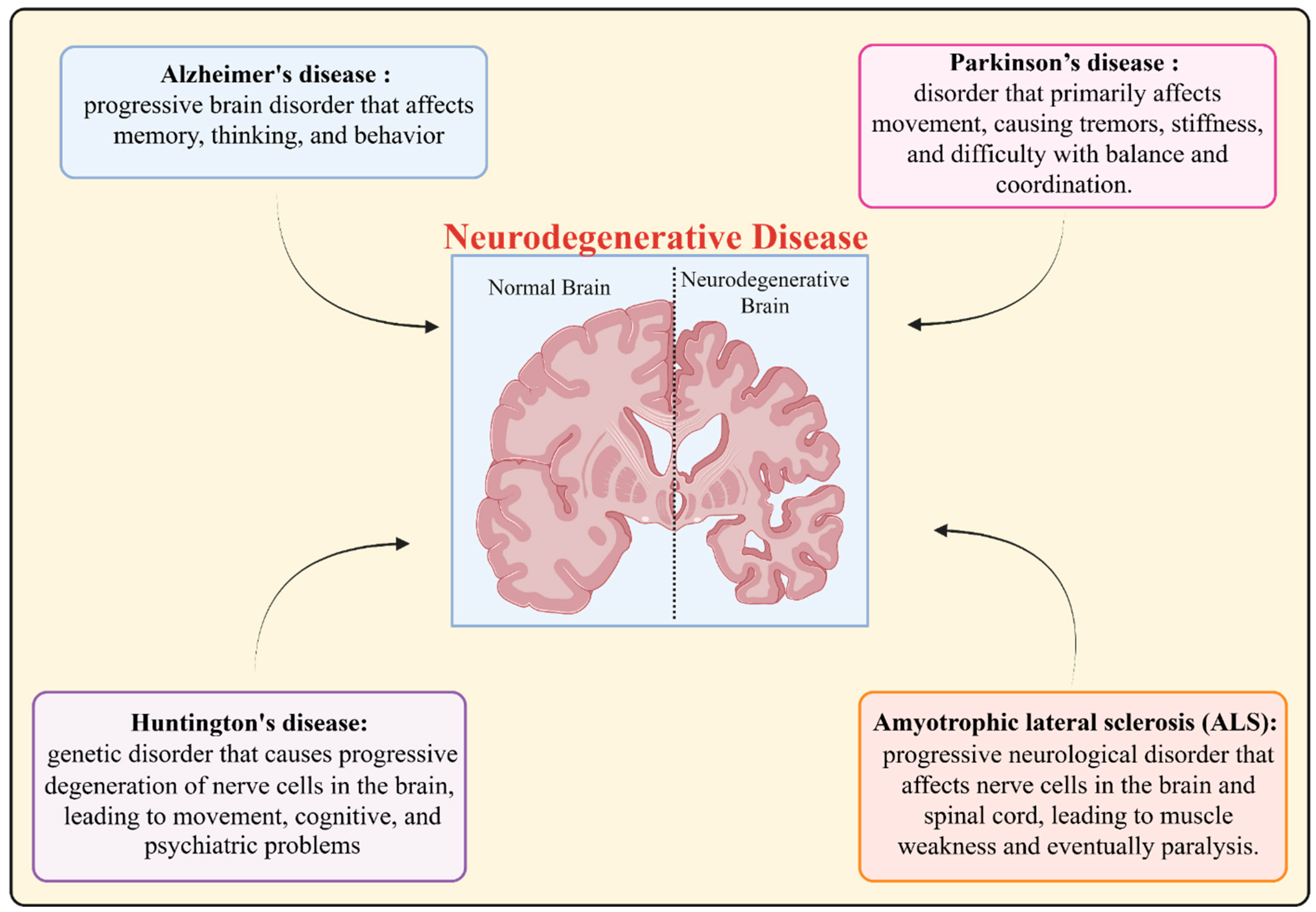
The illness states associated with neurodegeneration hallmark are collectively termed as neurodegenerative disorders (NDs) [17,18]. The most prevalent NDs are shown in Figure 1 [24,25,37].
(a)
Multiple sclerosis (MS): A condition in which the immune system assaults the coating that surrounds nerve fibers, which impairs the communication of the brain to other body parts.
(b)
Frontotemporal dementia (FTD): This is a state of mental illnesses that affect the frontal and temporal lobes of the brain over time, altering personality, behavior, and language.
(c)
Creutzfeldt–Jakob disease (CJD): A fatal, uncommon, degenerative brain illness brought on by prions

3. Autophagy: Mechanisms and Functions

The term autophagy comes from a combination of the word “auto”, meaning self, and “phagy”, meaning eating; thus, it basically means self-eating. An essential cellular practice known as autophagy clears away and recycles damaged or unnecessary parts of the cell to generate energy and the materials essential to operation, known as building blocks. This is a necessary process for cellular homeostasis. During autophagy, damaged organelles, forms of protein aggregates, and other waste in the cell are engulfed by double-membraned vesicles called autophagosomes. These autophagosomes then recycle and degrade their contents in the autolysosomes that develop when lysosomes and autophagosomes fuse together [38]. Chaperone-mediated autophagy (CMA) is a form of selective autophagy that preferentially targets particular proteins for degradation, in contrast to macroautophagy and microautophagy. Chaperone proteins identify and transport particular target proteins to lysosomes during CMA, where they are translocated into the lysosomal lumen for degradation [38,39].

Molecular Machinery of Autophagy

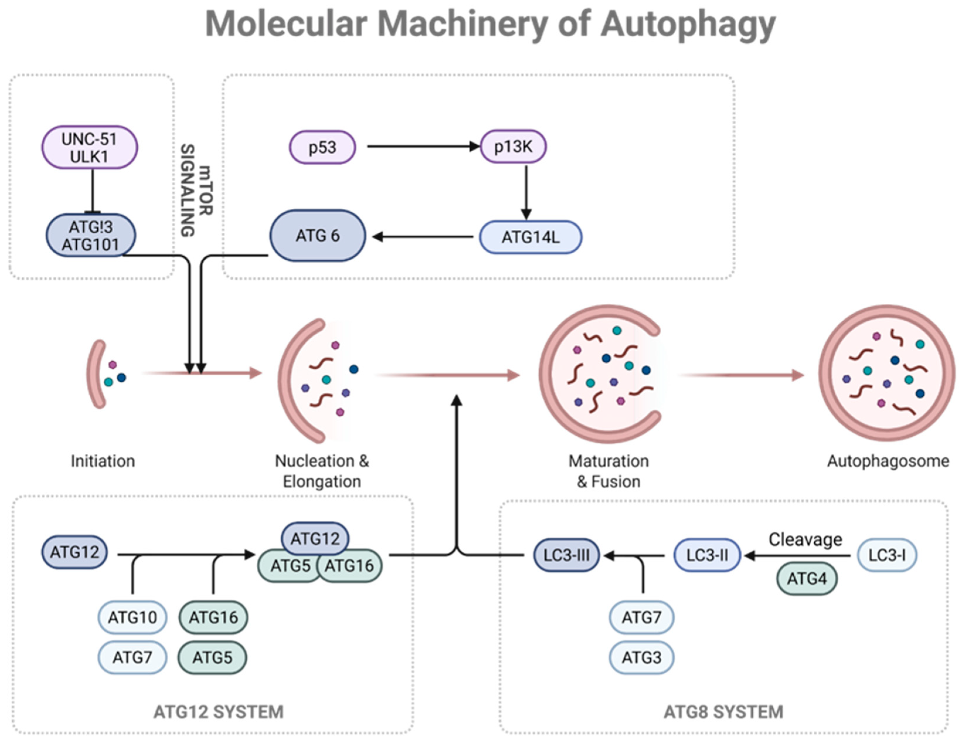
Autophagy is required to maintain cellular homeostasis, respond to stress, and regulate various physiological processes. The initial events of autophagy can be discussed as follows [40]:
The formation of the separation membrane, also referred to as the phagophore, is the first step of autophagy. A set of proteins called Atg (autophagy-related) proteins is unleashed in the initiation step. The key points of this process are as follows [41]: Unc-51-like autophagy-activating kinase 1 (ULK1) formation is needed. This complex consists of ULK1, FIP200 (focal adhesion kinase family-interacting protein of 200 kDa), ATG13, and ATG101. Among the many signaling pathways that regulate the ULK1 complex, the mTOR (mechanistic target of rapamycin) signaling pathway represses autophagy during nutrient-sufficient conditions. Vps34 complex, also referred to as class III phosphatidylinositol 3-kinase (PIP3K), comprises Vps34, Vps15, Beclin 1 (ATG6), and ATG14L. The complex leads to the production of phosphatidylinositol 3-phosphate (PI3P) to increase the development of the isolation membrane [18].
ULK1 complexes and PI3K complexes interact and facilitate nucleation by increasing the formation of the phagophore. Significant targets of the ULK1 complex are phosphorylated in the initiation of autophagy, and the PI3K complex forms PI3P in order to attract downstream effectors [42]. The nucleated phagophore then elongates and grows in size and ultimately engulfs the cytoplasmic contents to be digested. The cargoes, e.g., broken organelles as well as protein forms, are identified and specifically tagged for degradation. Cargo receptor autophagy receptors, e.g., p62/SQSTM1, NBR1, and OPTN, are particularly important in cargo recognition by recruiting cargo to the growing autophagosome membrane. The protruding phagophore eventually closes up to form a full-cycle autophagosome, a mature two-membrane vesicle. LC3 (microtubule-associated protein 1 light chain 3) can then undergo lipidation and be inserted into the autophagosomal membrane by other ATG proteins, including, but not limited to, ATG3, ATG4, ATG5, ATG7, and ATG12–ATG5–ATG16L1 complex, which is essential to the process.
Once they form, autophagosomes merge with lysosomes to form an autolysosome. This fusion involves proteins such as soluble N-ethylmaleimide-sensitive factor attachment protein receptors (SNAREs) and Rab GTPases. The material that is internalized is degraded by the lysosomal hydrolases of the autolysosome, and the degradation products are recycled back into the cytoplasm. Autophagic flux is attained in the process of autophagosome formation, autophagosome cargo engulfment, the creation of autolysosomes, and the degradation of autolysosomes. Maintaining the constant renewal of the cellular components is a crucial factor as well as the normal activity of autophagy. There are numerous signaling pathways that are involved in the process of autophagy regulation, with mTOR, AMPK, growth factor signaling, and cellular stress response being merely some examples of them (Figure 2). The number of diseases that include cancer, metabolic, and neurological disorders has been identified as being related to the dysregulation of autophagy. To develop new therapies that are able to target this process, it is essential to understand the molecular processes of autophagy [1,42,43].

4. Crosstalk Between Autophagy and UPS in Proteostasis

Neurodegenerative states are not only the result of cell-autonomous abnormalities in neurons but also due to the impaired crosstalk with glial cells. Autophagy and proteostasis are firmly controlled and regulated via crosstalk between neurons, astrocytes, microglia, and oligodendrocytes. The slight disruption of this cellular network can accelerate disease progression.
Astrocytes play a major role in neuronal health via neurotransmitter recycling, metabolic support, and degradation of toxic protein aggregates. The autophagy process in astrocytes helps in clearing the extracellular amyloid-β and tau proteins, which can contribute to the progression of Alzheimer’s disease. The deregulated autophagic functions of astrocytes exacerbate neuronal proteotoxic stress and synaptic dysfunction.
Microglia are the resident immune cells of the brain. These cells facilitate phagocytosis and synaptic degradation via autophagy–lysosome. In Parkinson’s disease, the dysfunctional autophagy process in microglia leads to the aggregation of α-synuclein and chronic neuroinflammation. The improvement in microglial autophagy has been found to be linked to the restoration of proteostasis and dampening inflammatory signaling.
Oligodendrocytes, along with their precursors, play a key role in the maintenance of myelin integrity and provide axonal support. Recent reports have established that dysfunctional autophagic functions in oligodendrocytes lead to demyelination and axonal degeneration, which are responsible for the progression of ALS and leukodystrophies.
Neuron–glia crosstalk is cellular communication supported with the help of extracellular vesicles, secreted cytokines, and metabolic coupling. Various pharmacological conditions resulting in dysfunctional autophagic functions of one cell are amplified in other cells, causing a feed-forward loop of proteostasis disruption. This emergent perception limits the viewpoint of autophagy and proteostasis as multicellular and network-level processes rather than neuron-centric pathways. Thus, integration of therapeutic strategies targeting neuron–glial crosstalk functions may yield more effective disease-modifying treatments.
Autophagy and UPS are not parallel pathways; they communicate bidirectionally.
  • The inhibition of UPS (e.g., by oxidative stress or proteasome mutations) leads to the activation of autophagy via upregulating HSF1-mediated autophagic genes (e.g., LC3, p62) and AMPK-dependent mTOR suppression.
  • The blockade of autophagy causes the overloading of UPS, as seen in ATG5 or ATG7 knockout mice, observed from the accumulation of ubiquitinated proteins before neuronal death.
  • The sharing adaptors, such as p62/SQSTM1, NBR1, OPTN, etc., contain both LC3-interacting motifs (LIR) and ubiquitin-binding domains (UBA), allowing them to deliver ubiquitinated cargo to both the proteasome and autophagosome simultaneously.
  • The autophagy mediated via chaperone (CMA) also plays a key role in UPS intersection via degradation of mutant or oxidized proteins, as demonstrated for mutant SOD1 in ALS and α-synuclein in PD.
  • Thus, it can be concluded that neurodegeneration takes place due to the compromise of both systems simultaneously, resulting in proteostasis collapse.

5. Cell-Type-Specific Autophagy–Proteostasis Interactions

By eliminating broken or improperly folded proteins, minimizing their buildup, and avoiding proteotoxicity, autophagy maintains cellular homeostasis. This is particularly important during times of cellular stress, such as oxidative stress or nutrient deprivation [44]. Autophagy also helps to clear dysfunctional or excess organelles, such as mitochondria (mitophagy) and peroxisomes (pexophagy), to maintain the proper functioning of the cell. The process ensures the continuous replacement of old and damaged organelles with new and functional ones [45]. Under conditions of nutrient limitation, autophagy becomes a crucial mechanism for cellular survival. It entails the breakdown of cellular constituents, such as proteins and organelles, into their fundamental building elements, which can then be recycled for energy production or the synthesis of crucial molecules. The immunological response to intracellular infections involves autophagy. The xenophagy mechanism hunts down and destroys invasive microbes, aiding in the body’s overall defense against illnesses and preserving cellular health [46]. It helps to eliminate unnecessary or unwanted cellular components, allowing cells to specialize and carry out specific functions effectively such as apoptosis (programmed cell death) and can determine the fate of a cell under certain circumstances. In some cases, autophagy promotes cell survival by recycling nutrients, while in others, it can contribute to cell death by excessive self-digestion and cellular metabolism, especially during fasting or nutrient deprivation. It helps to sustain cellular energy levels by breaking down and recycling cellular components to provide nutrients for ATP production [47,48].

5.1. Autophagy Dysregulation in Neurodegenerative Diseases

There is also a deposition of abnormal protein aggregates in AD, especially the beta-amyloid and tau, around the neurons in the brain. One of the suspected mechanisms involves autophagy dysfunction that would induce failure in clearing them, eventually resulting in their subsequent neurotoxicity. PD arises due to the loss of neurons in the brain, which produces dopamine. Autophagy destroys both misfolded proteins, related to the progression of PD, and damaged mitochondria. Maladaptive capacity of the mitochondria to maintain quality and accumulation of these neurotoxic protein aggregates may be a consequence of dysfunctional autophagy. HD is noted by the growth of mutant huntingtin protein clumps in brain cells. The lack of autophagy can contribute to the inability to eliminate these aggregates, causing the malfunctioning and death of neurons. ALS is characterized by the degeneration of motor neurons. It is clarified that autophagy degrades misfolded proteins associated with ALS. The existence of these proteins might be an accumulation factor for increased dysregulated autophagy, hence accelerating the degradation of motor neurons [18,49,50,51].

5.2. Proteostasis

Proteostasis refers to the intricate biological process responsible for maintaining protein homeostasis within living organisms. It is crucial for the proper functioning and survival of cells. Proteins are fundamental building blocks and perform diverse functions in cells, ranging from enzymatic activity, cell structure, signaling, and the regulation of gene expression [4]. Cells continuously synthesize, fold, assemble, and degrade proteins to maintain a balanced and functional proteome. However, numerous internal and external factors can disrupt this delicate balance, leading to protein misfolding, aggregation, and other forms of protein damage [23]. Some factors that can affect proteostasis include:
  • Extreme heat or cold can denature proteins, leading to loss of function or aggregation.
  • Oxidative stress: Reactive oxygen species can damage proteins and impair their functions.
  • Genetic mutations: Mutations in protein-coding genes can lead to misfolded or dysfunctional proteins.
  • Environmental toxins: Exposure to certain chemicals or toxins can disrupt proteostasis and lead to protein damage.
Age-related changes: Cells become less efficient in maintaining proteostasis as organisms age, leading to an accumulation of damaged proteins [4,52].
The ubiquitin–proteasome system is primarily responsible for the degradation of short-lived and misfolded proteins. In neurodegenerative conditions, the excessive production of abnormal proteins or direct inhibition of proteasomal activity leads to proteasome overload, resulting in the accumulation of toxic protein species. This impairment further disrupts cellular signaling and stress response pathways.
Autophagy plays a critical role in clearing long-lived proteins, protein aggregates, and damaged organelles. In neurodegenerative diseases, autophagic dysfunction may occur at multiple stages, including defective cargo recognition, impaired autophagosome formation, disrupted autophagosome–lysosome fusion, or lysosomal insufficiency. Such defects compromise aggregate clearance and exacerbate neuronal stress [49,51].
Molecular chaperones assist in protein folding and prevent aggregation under stress conditions. Reduced chaperone availability or functional impairment diminishes the cell’s capacity to refold misfolded proteins, thereby accelerating proteostasis collapse and neuronal vulnerability.
The Chaperone proteins also ensure that the newly synthesized proteins are folded in the right direction, as well as preventing the misfolding and aggregation of the proteins. The cell produces machinery, e.g., in the form of the proteasome and lysosomes, to degrade damaged or misfolded proteins. In it, damaged or superfluous cellular components, such as protein aggregates, are captured in autophagosomes and damaged during sequestration. The heat shock proteins are responsive to stress and thus can assist in refolding damaged proteins within the cells. In this system, ubiquitin molecules spot damaged proteins, marking them to be destroyed by the proteasome. The chaperone proteins act as guides in the process of the folding of other proteins, as well as preventing aggregation.

5.3. Protein Folding and Quality Control

  • Protein Folding: In cellular protein synthesis, there is a specific 3-dimensional protein structure that the protein molecule must adopt, its so-called native conformation, so that the protein is able to effectively perform its functions. Protein folding is a complex and complicated process that depends a lot on the interactions of the amino acid residues in the polypeptide chain. The folding of protein is heavily dependent on the environmental conditions, such as temperature, PH, and the availability of specific molecular chaperons [53].
  • Misfolding and Aggregation: Proteins might misfold sometimes because of genetic defects, cell stress, or environmental influences, despite the complex folding mechanisms. Mismatch proteins are not stable and more likely to create aggregates that can poison cells. Gradually, such aggregates can accumulate and lead to the development of several neurodegenerative disorders, such as AD, PD, and HD [54].
  • Molecular Chaperones: Molecular chaperones are used by cells to prevent misfolding, as well as aggregation of proteins. Molecular chaperones keep the right folding of newly synthesized, as well as the refolding of misfolded proteins, in the original form. They act as protein custodians and are essential for the maintenance of proteostasis [55].
  • Ubiquitin–Proteasome System: The system tags the misfolded or damaged proteins with ubiquitin molecules, which are recognized by a massive cellular protein complex called the proteasome, which destroys the proteins.
  • Autophagy: Autophagy may be induced in instances involving the formation of aggregates or other larger protein structures. Autophagy is the abduction of non-functional proteins into specialized vesicles known as autophagosomes. The autophagosomes further merge with the lysosomes and cause the degradation and recycling of proteins.
  • Endoplasmic Reticulum (ER) Quality Control: ER has the stringent quality checks, especially against the proteins that occur within the ER. With such a mechanism, in case proteins are misfolded, they are re-exported to the cytoplasm to be damaged or undergo ER-associated degradation (ERAD) [56,57].

5.4. Chaperone-Mediated Proteostasis

A type of specialized protein known as a chaperone plays a central role in assisting the orderly folding of newly synthesized proteins, as well as the refolding of misfolded or denatured proteins. These chaperones are necessary to allow the proteins to be correctly formed in the correct three-dimensional shape and inhibit the formation of non-functional protein aggregates [58].
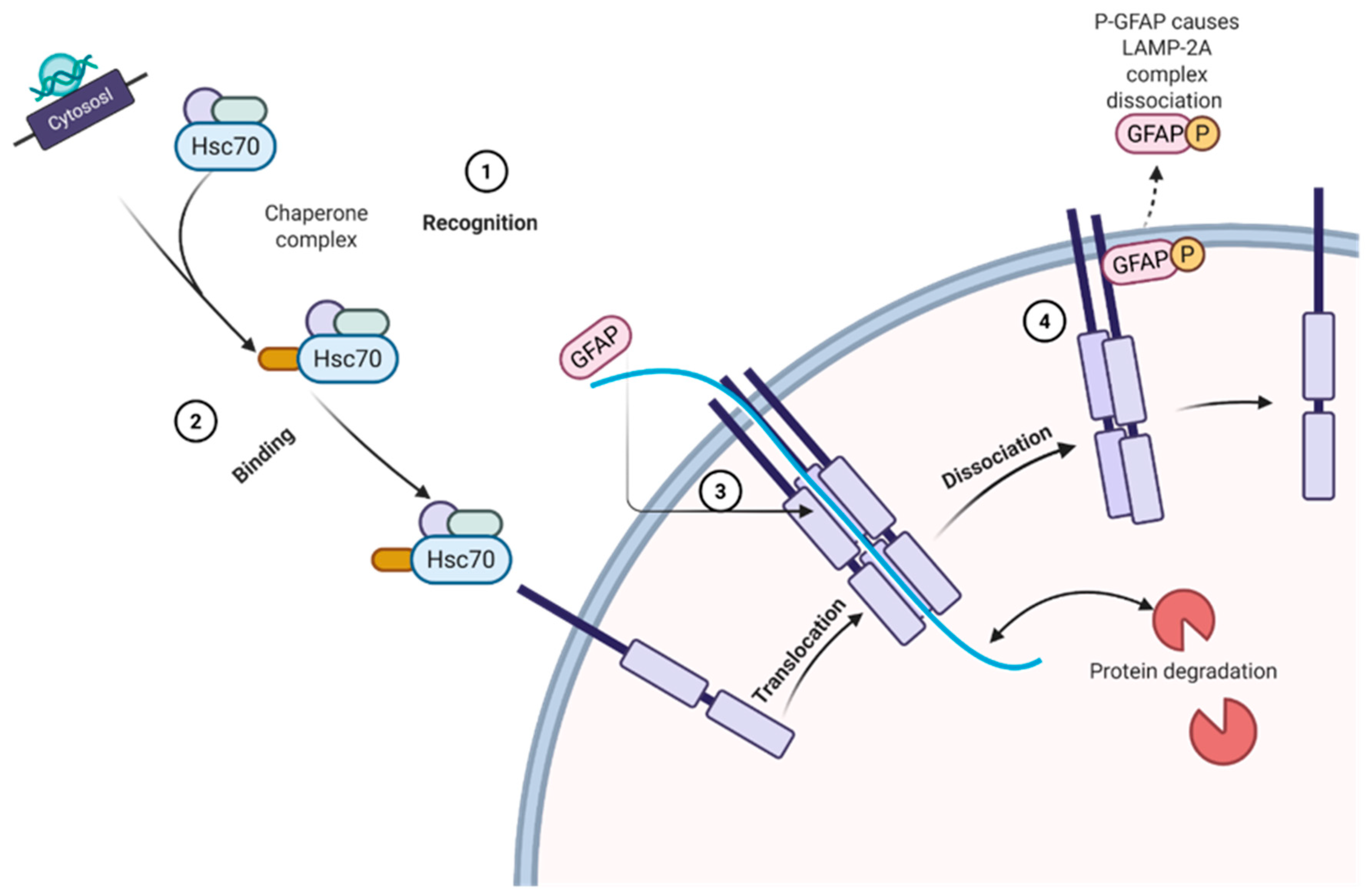
When the proteins are being produced within the cell, there is a chance that they can fail to acquire their proper three-dimensional form spontaneously. Chaperones assist the folding process as a means to ensure that the nascent protein attains its functional conformation. Chaperones also play a role in ensuring quality because they find the misfolded or denatured proteins (Figure 3). Failure in the proper folding of a protein results in a protein susceptible to aggregation that might lose its functionality, or even become a poison to the cell. To prohibit the creation of aggregates, chaperones identify these misfolded proteins and either refold or mark them to be broken down, resulting in the selective destruction of cytosolic proteins in lysosomes as a result of the process of chaperone-mediated autophagy. The involvement of this process is through a cytosolic chaperone called heat shock cognate 70 (Hsc70), since this chaperone identifies a targeting motif, which refers to the KFERQ-like motif found on the misfolded or undesired proteins. The entire complex of the Hsc70-chaperone-bound substrate is then detected by LAMP-2A-expressing lysosomes, after which the substrate protein enters the lysosomal lumen to be digested by lysosomal proteases. Cellular homeostasis and biological maintenance against the accumulation of harmful proteins and their aggregation may lead to a number of neurodegenerative diseases, including AD and PD. Dysregulation of proteostasis and chaperones has been implicated in many age-related diseases and proteinopathies. In general, chaperone-mediated proteostasis is a complex and critical process in which the cells protect the integrity of proteins and guarantee normal cell functioning and general well-being [58,59].

5.5. Ubiquitin–Proteasome System

Eukaryotic cells regulate the degradation of proteins by a unique process called the ubiquitin–proteasome system (UPS). It is thus also important in maintaining cellular homeostasis by selectively degrading undesirable or damaged proteins, regulating cell cycle progression, and controlling the abundance of several key proteins, which affect various cellular processes [60]. Ubiquitin is a small protein of 76 amino acid residues, very conserved in eukaryotes. This means that as a molecular tag, it can be attached to the target proteins to mark them to be destroyed. Ubiquitination is a cascade of enzyme reactions, mediated by the three classes of enzymes E1 (ubiquitin-activating enzymes), E2 (ubiquitin-conjugating enzymes), and E3 (ubiquitin ligases). Underlying all these is the formation of a high-energy thioester bond between the C-terminal glycine residue of ubiquitin and a cysteine residue of an E1 enzyme.
  • E2 Enzymes: Together with E1 enzymes, E2 enzymes transmit active ubiquitin from E1 to the substrate protein.
  • E3 Enzymes: The transfer of ubiquitin from the E2 enzyme to the target protein is made possible by E3 enzymes, which are essential for substrate specificity. The cell contains hundreds of different E3 ligases, each of which recognizes a particular substrate.
  • Proteasome: The huge protein complex known as the proteasome is in charge of actually breaking down ubiquitinated proteins. The 20S proteasome possesses proteolytic activity, which means it breaks down the target protein into smaller peptides by cleaving peptide links in it [61].

6. Disease-Specific Mechanisms

  • Ubiquitination: The target protein is recognized and marked for degradation by the addition of multiple ubiquitin molecules, forming a polyubiquitin chain.
  • Recognition and Unfolding: Ubiquitinated proteins are recognized by the 19S regulatory particles (also known as the 19S cap) of the proteasome. The 19S regulatory particle recognizes polyubiquitin and unfolds the substrate protein, making it accessible to the 20S proteasome.
  • Proteolytic Degradation: The unfolded protein is translocated into the central chamber of the 20S proteasome, where its peptide bonds are hydrolyzed by the proteolytic activity of the 20S core, generating short peptide fragments.
  • Peptide Release: The resulting peptides are released from the proteasome and further degraded by other peptidases to produce amino acids that can be recycled for the synthesis of new proteins [62,63].
The UPS is essential for several cellular functions, including controlling the cell cycle, immune system antigen presentation, DNA repair, and protein quality control. It has been suggested that a number of illnesses, including cancer and neurological disorders, are caused by dysregulation of the ubiquitin–proteasome system. The UPS is still being studied by researchers to better comprehend its complex operations and its therapeutic implications.

6.1. Role of Proteostasis in Neurodegeneration

Since it is intimately linked to the onset and course of various disorders, proteostasis plays a critical role in neurodegeneration. Key elements of proteostasis in neurodegeneration include the following [4,23,64]:
  • Protein misfolding and aggregation: Certain proteins have a propensity to misfold and collect, generating insoluble clumps, in neurodegenerative disorders. Examples of proteins that can congregate include amyloid-beta and tau proteins in Alzheimer’s disease, alpha-synuclein in Parkinson’s disease, huntingtin protein in Huntington’s disease, and SOD1 in ALS. The course of the illness may be aided by proteostasis processes failing to remove these aggregates.
  • Chaperones and folding machinery: A complex network of molecular chaperones and folding machinery is present in cells to aid in the correct folding and assembly of proteins. These chaperones may be overworked or defective in neurodegenerative illnesses, which causes an accumulation of misfolded proteins.
  • Autophagy and lysosomal degradation: A cellular process called autophagy aids in the removal of damaged or misfolded organelles and proteins through lysosomal breakdown. Impaired autophagy can prevent the removal of protein aggregates, causing neurodegeneration.
  • Ubiquitin–proteasome system: Unwanted or damaged proteins are destroyed by the ubiquitin–proteasome system. When this mechanism is dysfunctional, harmful proteins can build up and contribute to neurodegenerative processes.
  • Oxidative stress and proteostasis: Proteins can be harmed, and proteostasis pathways can be upset by oxidative stress, which develops when reactive oxygen species (ROS) and antioxidant defenses are out of balance. Additionally, by causing protein misfolding and aggregation, this oxidative damage can also damage cells.
  • Genetic factors: In some instances, mutations in genes encoding proteostasis-related proteins can enhance a person’s susceptibility to neurodegeneration. For instance, familial ALS is linked to mutations in the superoxide dismutase 1 (SOD1) gene.
Understanding the role of proteostasis in neurodegeneration is critical for developing potential therapeutic approaches. Targeting proteostasis pathways and enhancing protein quality control mechanisms may offer promising strategies to prevent or slow down the progression of neurodegenerative diseases. However, this remains an active area of research, and much more needs to be understood about the intricate mechanisms governing proteostasis in the context of neurodegeneration.

6.2. Interplay Between Autophagy and Proteostasis and Co-Regulation

Autophagy is necessary in order to remove the clumped and abnormally folded proteins that accumulate in the cell interiors. This tendency to aggregate is often the cause of the formation of toxic aggregates of proteinaceous material, which may be detrimental to the cell’s functioning. Such aggregates can be identified by autophagy and translocated into double-membraned sacs known as autophagosomes, after which they will be delivered to lysosomes to be destroyed. The mechanism results in the maintenance and restoration of proteostasis and the mitigation of proteotoxic stress [65]. Autophagy contributes to proteome quality control by selectively targeting faulty or misfolded proteins, damaged organelles, and pathogens for degradation. It cooperates with the chaperone-mediated autophagy (CMA) and endoplasmic reticulum-associated degradation (ERAD) process to detect and remove aberrant proteins, so that only folded and correctly functioning proteins survive within the cell [66]. Autophagic activity can also be influenced by the contribution of mechanisms that regulate proteostasis. An example would be that the emergence of misfolded proteins or intense proteotoxic tenacity could trigger the process of autophagy to protect against the damaged products. Proteostasis influences the autophagic process in a direct manner, controlling its induction and activity [67]. Autophagic activity can also be influenced by mechanisms of proteostasis. An example would be when misfolded proteins accumulate or grievous forms of proteotoxic pressure trigger the action of autophagy, which is a defensive mechanism that is in place to remove the damaged elements. A test has been performed on the direct effect of proteostasis in the autophagic process, where both induction and activity of the process are controlled [68]. Loss of autophagy and proteostasis efficacy during the aging process leads to the accumulation of misfolded and broken proteins and cellular dysfunction. Failure of autophagy can also lead to the amassing of protein aggregates and the worsening of proteostatic imbalances. The interplay among these mechanisms is of special significance in considering aging and age-associated neurodegenerative diseases [4].
Altogether, proteostasis and autophagy are two closely associated cellular processes, and their crosstalk and co-regulation ensure that protein quality is controlled effectively, protein aggregates are prevented, and that the cell is secure against proteotoxic stress. The elucidation of the complexity of the relationship between these processes is critical in establishing appropriate therapeutic approaches to target a variety of diseases linked to protein misfolding and aggregation.

6.3. Impairment and Role of Autophagy and Proteostasis in Neurodegenerative Diseases

The disruption of proteostasis and autophagy is an important event in the pathogenesis and progression of neurodegenerative diseases. In neurodegenerative diseases, notably AD, PD, HD, and ALS, misfolded and aggregated proteins accumulate within neurons. Some of the protein aggregates that are found in the diseases, and which are deemed to contribute to the neuronal abrasion and death, are beta-amyloid, tau, alpha-synuclein, huntingtin, and TDP-43 [69].
Autophagy plays a critical role in clearing these toxic protein aggregates and maintaining cellular health. However, in neurodegenerative diseases, autophagy becomes compromised, leading to reduced clearance of these protein aggregates. Several factors can contribute to impaired autophagy in these conditions [34,67,70]:
  • To break down the cargo, autophagy, the generation of lysosome-fusing autophagosomes is involved. Neurodegenerative diseases may restrict the stepping-stone of autophagosome–lysosome merger and the degradation of dangerous proteins due to the lack of functionality of the lysosomes.
  • A number of proteins and signaling pathways control autophagy. The malfunction or mutation of these regulators produces or interferes with the autophagic process, which in turn affects protein clearance.
  • Autophagy decreases with age, and because neurodegenerative diseases are more common in older people, this age-related decline in autophagy might be part of the pathogenesis of neurodegenerative diseases.

6.4. Proteostasis Imbalance

Protein misfolding and aggregation are prevented by maintaining adequate proteostasis. The proteostasis network is overloaded in neurodegenerative disorders, which causes an accumulation of improperly folded proteins [71]. Proteostasis imbalance is caused by a number of factors [72]:
  • Genetic mutations can result in the generation of misfolded or aggregation-prone proteins in several neurological disorders.
  • Oxidative stress, caused by an imbalance between free radicals and antioxidant defenses, can damage proteins and contribute to their misfolding.
  • Molecular chaperones aid in the folding of proteins. Protein misfolding and aggregation could result from dysfunctional chaperones.
  • Damaged and improperly folded proteins are destroyed by the proteasome. Protein buildup may result from the proteasome system being damaged.
(a)
Alzheimer’s Disease
AD is a neurodegenerative disorder that progresses and affects mainly memory and cognitive functions. Although there is still a lack of substantial knowledge about the exact origin of AD, there are some data that show that abnormal aggregation of proteins in the brain is also a significant factor in the pathophysiology of the disease. Two reasons behind the importance of engaging in the above biological processes, autophagy and proteostasis, in maintaining brain health and preventing the onset and further development of AD are considered to be the following [73].
In AD, Aβ oligomers directly inhibit proteasome peptidase activity, while hyperphosphorylated tau blocks autophagosome–lysosome fusion via the disruption of Rab7 and LAMP-2A expression. This dual hit impairs both UPS and autophagy, leading to intraneuronal Aβ and tau accumulation, synaptic loss, and cognitive decline. Restoring CMA or proteasome activity in mouse models reduces both plaques and tangles, confirming causality.
In Alzheimer’s disease, proteostasis dysfunction is prominently associated with the impaired clearance of amyloid-β peptides and hyperphosphorylated tau protein. Although autophagy is initiated in affected neurons, autophagosomes accumulate due to defective fusion with lysosomes and reduced lysosomal degradative capacity. Lysosomal enzyme insufficiency further limits the degradation of autophagic cargo, promoting intracellular accumulation of amyloidogenic peptides [71].
As applied to AD, autophagy plays an important role in eliminating the accumulated presence of two key proteinaceous masses of the AD-riddled brain, amyloid-beta plaques and tau tangles, and there is too much amyloid-beta protein with an abnormal aggregation accumulating externally to the neurons, forming the mass as plaques. It is autophagy that detects this and the amyloid-beta proteins and takes them to the lysosomes, where they are destroyed and recycled. Tau proteins are necessary for the stability and structural integrity of the neurons. In AD, tau proteins are hyperphosphorylated, thus leading to protein misfolding and aggregation of neurofibrillary tangles in neurons [74,75].
Additionally, tau pathology interferes with chaperone-mediated autophagy by disrupting lysosomal receptor dynamics, thereby impairing selective protein degradation. The combined failure of macroautophagy and chaperone-mediated autophagy results in synaptic dysfunction, mitochondrial damage, neuroinflammation, and progressive neuronal loss, highlighting the central role of proteostasis imbalance in the pathogenesis of Alzheimer’s disease.
Misfolded proteins of AD, such as amyloid-beta and tau, lead to an imbalance in proteostasis. In the case of imbalance, protein production and toxic aggregates accumulate, but eventually lead to the malfunction and death of the neurons. There are a number of research studies and drug development that have concentrated on autophagy enhancement and proteostasis, having the potential to treat AD. Drugs to induce or have the ability to restore autophagy to boost the removal of the toxic protein aggregates in AD are being studied. Chaperone proteins help to achieve the proper protein folding and exclude aggregation. AD may be treated by drugs that potentiate the effect of chaperones by restoring proteostasis. The lysosome is in charge of the degradation of proteins and cellular waste. Retrieving mines of toxic protein accumulation, lysosomal fixing may encourage the intervention of noxious protein agglomerations [75].
(b)
Parkinson’s Disease:
PD is a neurodegenerative disorder that leads to motor and non-motor symptoms because of the progressive loss of dopaminergic neurons in the substantia nigra region of the brain. The pathophysiology of PD remains poorly understood; however, evidence is growing that dysregulation of the autophagy and proteostasis system is a contributing factor [76].
Parkinson’s disease is characterized by the accumulation of α-synuclein-rich Lewy bodies, reflecting profound disturbances in proteostasis pathways. Misfolded α-synuclein oligomers directly inhibit proteasomal activity, reducing the clearance of damaged proteins and amplifying proteotoxic stress. Simultaneously, mutations in genes such as PINK1 and Parkin impair mitophagy, leading to defective removal of damaged mitochondria. The accumulation of dysfunctional mitochondria results in increased oxidative stress and energy failure, particularly affecting dopaminergic neurons, which are highly dependent on mitochondrial function. The convergence of UPS inhibition and defective mitophagy establishes a vicious cycle of proteostasis failure, ultimately driving dopaminergic neuron degeneration in Parkinson’s disease [75,76].
Mutant α-synuclein (A53T, A30P) blocks 20S proteasome gate opening and impairs LAMP-2A translocation, simultaneously disabling UPS and CMA. PINK1/Parkin-mediated mitophagy—a specialized autophagy pathway—is also compromised, leading to damaged mitochondria accumulation, ROS burst, and α-synuclein aggregation in a vicious feed-forward loop. Enhancing Parkin translocation or LAMP-2A expression rescues dopaminergic neurons in iPSC-derived midbrain cultures, establishing therapeutic proof-of-concept.
One of the defective protein aggregates that accumulate in PD due to the evidence of impaired autophagy is alpha-synuclein, a disease-specific protein. PD types that are familial include the mutations in genes such as Parkin, PINK1, and DJ-1. These mutations have been implicated in reduced autophagy and poor clearance of damaged proteins and the abolition of mitochondria. These autophagy mutations interfere with the autophagy process, leading to the buildup of toxic aggregates and oxidative stress, which will further lead to neuronal cell death. [77] The cause of proteostatic dysregulation in PD involves the aberrant accumulation of the alpha-synuclein protein. Overall, neurotoxicity and protein aggregation may occur in a vicious circle with alpha-synuclein aggregates misfolding and accumulating, clogging protein degradation pathways such as the ubiquitin–proteasome pathway and autophagy [78]. Since the autophagy and proteostasis mechanisms are known to be involved in PD, these cellular mechanisms have gained interest in exploring potential therapeutic interventions. Pro-autophagic or pro-proteasomal strategies to target the clearance of toxic protein aggregates to preserve neurons have been tested in pre-clinical models [79].
(c)
Huntington’s Disease
HD is a degenerative neurological disorder that occurs due to a mutation in the huntingtin (*HTT*) gene that leads to the production of the deadly protein called mutant huntingtin (mHTT). It makes the cells generate mHTT instead of normal *HTT*, due to which the cell has numerous malfunctions, and eventually, the neurodegeneration develops [80].
Huntington’s disease arises from polyglutamine expansion in the huntingtin protein, rendering it highly prone to misfolding and aggregation. Mutant huntingtin is inefficiently degraded by the ubiquitin–proteasome system, leading to proteasome overload and reduced degradation capacity for other regulatory proteins. This proteostatic burden disrupts normal cellular homeostasis and stress responses.
When it comes to HD, autophagy is more important since it prevents the spread of mutant huntingtin protein aggregates. Autophagy facilitates the degradation and digestion of these dangerous masses, thus preventing their deleterious impacts on the neurons. Another of these potentials, on the other hand, is that impaired autophagy may cause the accumulation of mHTT and worsen the pathology [81]. Genetic manipulation or pharmacological interventions can relieve the effects of HD-related symptoms in animal models. Thus, the enhancement of autophagy has been viewed as a potential treatment option for HD.
Mutant huntingtin (mHTT) contains expanded polyQ tracts that resist proteasomal degradation and sequester p62, preventing autophagosome formation. Additionally, mHTT disrupts dynein-mediated retrograde transport, trapping autophagosomes in distal axons and blocking lysosomal fusion. Consequently, both UPS and autophagy are paralyzed, leading to inclusion body formation and neuronal death. Trehalose or rapamycin restores autophagic flux and reduces mHTT aggregates in Q175 mouse striatum, validating the autophagy–proteasome axis as a drug target.
In the case of HD, the mutant huntingtin protein destabilizes proteostasis, resulting in the accumulation of misfolded and aggregated proteins, which are toxic to the neurons. The proteasome and the rest of the cellular machinery that interferes with proteostasis include the autophagy–lysosome system. HD affects both of these systems, and this is attributed to the fact that mHTT aggregates exist. This leads to reduced elimination of the misfolded proteins, hence leading to their accumulation and toxicity in the cell [82].
(d)
Amyotrophic Lateral Sclerosis
ALS is a neurological disorder and is referred to as Lou Gehrig’s disease that afflicts motor neurons in the spinal cord and brain, which lead to the loss of strength, loss of muscle control, and eventually death. The exact cause of ALS is not clear, but researchers have focused on a number of cell processes such as autophagy and proteostasis, which can influence the development and progression of ALS.
Amyotrophic lateral sclerosis is characterized by cytoplasmic aggregation of RNA-binding proteins such as TDP-43 and FUS, reflecting significant impairment of proteostasis mechanisms. Mutations in genes including C9orf72, SOD1, and OPTN disrupt autophagy initiation, endolysosomal trafficking, and the selective degradation of stress granules.
Mutant SOD1 and TDP-43 both inhibit proteasome activity and disrupt optineurin (OPTN)-dependent selective autophagy. OPTN loss-of-function mutations (e.g., E478G) abolish LC3 recruitment to ubiquitinated aggregates, while TBK1 mutations impair OPTN phosphorylation, leading to the accumulation of toxic TDP-43 oligomers in motor neurons. Double-knockout (OPTN/TBK1) mice develop ALS-like symptoms, confirming genetic evidence for autophagy–proteasome crosstalk failure as a core pathomechanism.
It is believed that the abnormal accumulation of toxic protein aggregates, including TDP-43 (TAR DNA-binding protein 43) and SOD1 (superoxide dismutase 1), the hallmark of clinical symptoms of ALS, is the consequence of flawed autophagy in this disorder. The inability to remove such toxic protein aggregates promotes their buildup, consequently causing neurotoxicity when the autophagy process is impaired. Improved autophagy has been demonstrated as beneficial in ALS models, and hence, augmentation of this pathway may be a feasible therapeutic intervention [83,84,85].
(e)
Other Neurodegenerative Diseases
  • Frontotemporal Dementia (FTD): The frontal and temporal lobes of the brain gradually degenerate in a set of illnesses known as FTD. Certain types of FTD, such as those brought on by mutations in the progranulin (GRN) gene, have been linked to impaired autophagy. The buildup of aberrant protein aggregates in the affected brain areas may be attributed to altered autophagy [86].
  • Spinocerebellar Ataxias (SCAs): SCAs are a class of inherited neurodegenerative diseases characterized by progressive ataxia and cerebellar degeneration. The aberrant protein aggregation in neurons is linked to a number of SCAs. The buildup of these harmful protein aggregates in SCAs may be influenced by dysfunctional autophagy [87].
  • Multiple System Atrophy (MSA): A combination of symptoms that resemble Parkinson’s disease and autonomic dysfunction characterize MSA, an uncommon neurodegenerative condition. Alpha-synuclein buildup in oligodendrocytes is a pathogenic feature of MSA. The aggregation of alpha-synuclein in MSA may be caused by the dysregulation of proteostasis and autophagy processes [88].
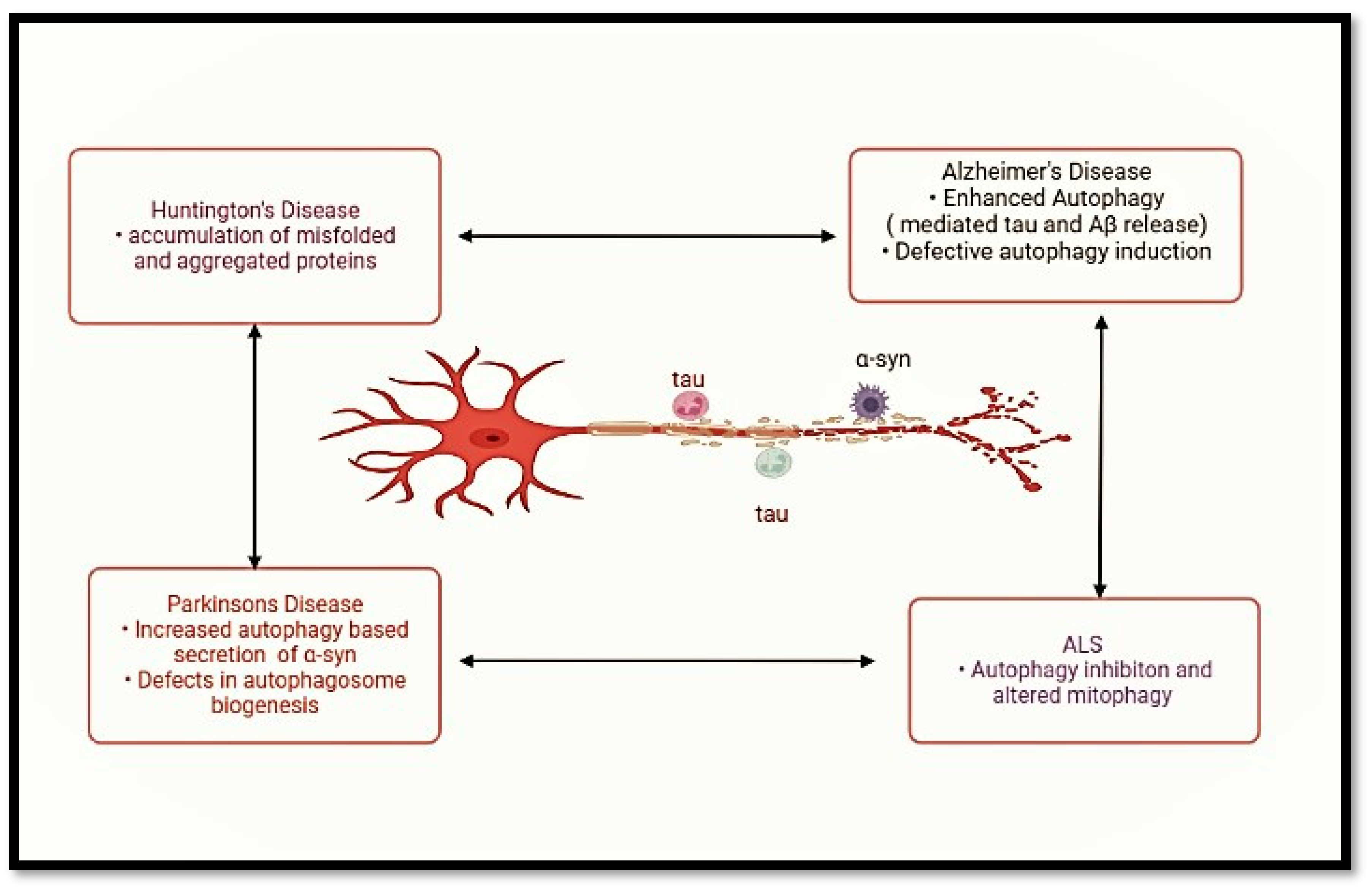
As summarized in Figure 4, autophagy, chaperone systems, ER quality control, and UPS are tightly integrated in neurons and glia to maintain proteostasis.

7. Therapeutic Strategies Targeting the Autophagy Proteasome Axis

There are drugs that may boost the start or development of the autophagy process. Examples of the common inducers of autophagy are rapamycin (sirolimus) and its derivatives (rapalogs), temsirolimus, and everolimus. Rapamycin and its analogues display their effects by inhibiting the mTOR (mammalian target of rapamycin) C pathway, which is a negative regulator of autophagy. This amplifies autophagosome production and the level of autophagic turnover. Autophagy and cellular metabolism are regulated by a protein kinase that is an energy sensor known as AMP-activated protein kinase (AMPK). Two mechanisms through which activating AMPK with medicines such as metformin or AICAR (5-aminoimidazole-4-carboxamide ribonucleotide) may induce autophagy include the inhibition of mTOR and the stimulation of ULK1, an essential initiator of autophagy.
Chaperone-mediated autophagy is one of the specific forms of autophagy, and it remains associated with the selective degradation of some proteins. Certain compounds like trehalose have also been proven to increase CMA and could be used as a treatment for some conditions. The examples of autophagy inhibitors are chloroquine, hydroxychloroquine, and 3-methyladenine, which increase lysosomal pH, disrupt fusion between autophagosomes and lysosomes, and autophagic clearance.
Autophagy may be selective (i.e., in certain instances it selects particular cargo to be degraded). A number of compounds have been found that regulate selective autophagy pathways. An example is the receptor protein p62/SQSTM1, which plays a part in biomarker-selective autophagy, and has been observed to be activation-enhancing by compounds such as verteporfin and fisetin [23,69].

7.1. Enhancing Proteostasis Pathways

Enhancing proteostasis pathways is the process of boosting pathways that sustain the level of proteins in a cell. Proteins are very crucial to cell survival and effective operation, and proteins are coordinated through proteostasis. Conditions such as aging, genetic mutations, environmental stressors, and disease status may disrupt proteostasis, giving rise to the build-up of misfolded or damaged proteins found in several neurodegenerative and other disorders [76].
To support proteostasis pathways, scientists and researchers search for different solutions that would contribute to a better control of protein quality and preserve a balance between proteins in the cell. These are some of the ways of improving proteostasis [89,90].
Chaperone-type proteins facilitate the normal folding of other proteins. The chaperones ensure that misfolded proteins are not accumulated as they encourage proper protein folding. A potential method that can withstand the harnessing of proteostasis is by modulating chaperone activity. The UPS acts to inflict the targeted destruction of misfolded or damaged proteins. An increase in UPS activity can help in the disposing of aberrant proteins and therefore lower the accumulation of such proteins.
As a response to cellular stress, heat shock proteins (HSPs) are synthesized, aiding in protein folding and refolding. Stimulating the heat shock response will supplement the cell capacity to overcome protein misfolding and aggregation, which may lead to the discovery of small molecules or drugs that specifically engage the proteostasis pathway by accelerating protein chaperones, stimulating protein clearance mechanisms or modulating other appropriate cellular processes. Genetic manipulation, such as by gene editing or RNA interference, can be used to overexpress or to “turn on” various proteostasis-relevant genes. There are other lifestyle drivers that may impact proteostasis pathways, including diet, exercise, and the ability to cope with stress. To keep protein homeostasis balanced, it is perhaps possible to adopt a healthy lifestyle.

7.2. Potential Therapeutic Strategies for Neurodegenerative Diseases

Other innovations are drugs that can attack particular pathways in the disease progression. As an example, drugs that inhibit acetylcholinesterase and block NMDA receptors are being used to treat Alzheimer’s in a bid to improve the functions of neurotransmitters.
Correction or replenishment of defective genes that cause neurodegenerative diseases: This has been promising in a few rare hereditary neurodegenerative diseases. This entails bringing healthy cells to inject them into the damaged areas of the brain in order to have healthy brain cells take the place of dead cells. The potential has been seen in stem cell research. The aim is to detect and find drugs that prevent damage to nerve cells and retard the course of the disease.
Anti-inflammatory Therapies: Decreasing inflammation of the brain since chronic inflammation has been found to contribute to the pathogenesis of a number of neurodegenerative disorders. The immune system of the body attacks and destroys misfolded proteins (e.g., beta-amyloid, alpha-synuclein) that cause the development of diseases based on the elimination of aberrant tau protein aggregation in the brain, which is common in Alzheimer’s and other tauopathies.
Sudden surgical stimulation of specific areas of the brain has been investigated in Parkinson’s disease and other movement disorders by implanting electric electrodes. Lifestyle advisements involve positive behaviors such as regular physical activity, healthy diet, intellectual stimulation, and social engagement, which can supposedly reduce the risk or possibly delay the occurrence of any neurodegenerative disease. Exploring the possibility of drugs with proven effectiveness in treating different ailments to work in treating neurodegenerative disorders. This further development of research aims to find reliable biomarkers to enable the diagnosis and monitoring of the progress of the disease, which can be used to develop specialized treatment. The use of a combination of treatment regimens that work on different fields of the disease at the same time could possibly increase the effectiveness of the treatment [91,92,93].

8. Challenges and Future Directions

The precise temporal and spatial regulation of autophagy and proteostasis in different cell types and disease stages remains an area of active investigation. Understanding these dynamic changes could offer valuable insights into disease progression and potential intervention points. The interactions between autophagy, proteostasis, and other cellular pathways involved in neurodegenerative diseases require further exploration.
For potential therapies targeting autophagy and proteostasis, long-term safety and efficacy profiles need to be established. Unanswered questions about possible off-target effects or unintended consequences must be addressed.
Investigating the potential benefits of combining autophagy and proteostasis modulators with other therapeutic approaches represents an avenue for future research. Synergistic effects might enhance treatment outcomes in neurodegenerative diseases. The identification of reliable biomarkers reflecting autophagy and proteostasis dysregulation in patients could aid in disease diagnosis, monitoring disease progression, and assessing treatment efficacy. With the emergence of powerful gene-editing technologies, ethical considerations related to modifying autophagy and proteostasis pathways must be carefully evaluated [94,95].
The emerging technology of single-cell analysis allows researchers to study autophagy and proteostasis at the individual cell level. This approach provides insights into cell-to-cell variability, identifying subpopulations with distinct autophagic and proteostatic profiles in neurodegenerative diseases. Single-cell techniques can also reveal dynamic changes during disease progression and treatment response. The revolutionary CRISPR-Cas9 gene-editing technology offers new opportunities to investigate the direct manipulation of autophagy and proteostasis-related genes. CRISPR can be used to modulate key regulators of these pathways, enabling a deeper understanding of their roles in disease and potential therapeutic targets. Advancements in mass spectrometry-based proteomics allow for a more comprehensive analysis of protein aggregates and post-translational modifications involved in neurodegeneration. Integrative proteomic approaches can identify novel disease-associated protein targets and further unravel the intricacies of proteostasis dysregulation. Artificial intelligence and machine learning algorithms are increasingly utilized to analyze large datasets in neurodegenerative diseases. These technologies help identify novel autophagy and proteostasis-related biomarkers, predict disease progression, and identify potential drug candidates more efficiently. The development of small compounds, gene treatments, and biologics that target the autophagy and proteostasis pathways is the main focus of current research. Novel therapeutic modulators aim to restore proper protein homeostasis, clear toxic aggregates, and promote neuroprotection in neurodegenerative diseases. Nanoparticle-based drug delivery systems offer precise and targeted delivery of autophagy and proteostasis modulators to the brain. These nanocarriers can improve therapeutic efficacy while minimizing off-target effects. Several autophagy-enhancing drugs are under investigation as potential treatments for neurodegenerative diseases. These drugs aim to stimulate the autophagic process, promoting the clearance of toxic protein aggregates [95,96,97].

Prospects for Therapeutic Advancements

The function of autophagy and proteostasis in neurodegenerative disorders identifies interesting opportunities for therapeutic developments that focus on these cellular systems. As researchers deepen their understanding of autophagy and proteostasis dysregulation in neurodegenerative disorders, several potential therapeutic strategies have emerged.
A potential method to improve the removal of disease-specific protein aggregates is to stimulate autophagy. Research is currently being conducted on nanoparticles and pharmaceuticals that enhance autophagosome–lysosome fusion or initiate autophagy. These treatments are designed to lessen the buildup of harmful protein aggregates that are linked to conditions like Alzheimer’s, Parkinson’s, and Huntington’s. Targeting the proteostasis network is another avenue for therapeutic advancements. Chaperone-based therapies and pharmacological proteostasis regulators are being explored to promote proper protein folding and prevent aggregation. These approaches may have broad applicability across multiple neurodegenerative diseases.
The advent of CRISPR-Cas9 gene-editing technology offers exciting possibilities for directly targeting key autophagy and proteostasis-related genes. Precise editing of these genes may restore proper cellular function and reduce protein aggregation, presenting a potential disease-modifying strategy. Given the multifaceted nature of neurodegenerative diseases, combination therapies may hold promise. Combining autophagy-enhancing drugs with proteostasis modulators or other disease-specific interventions could have synergistic effects and improve therapeutic outcomes. Nanoparticle-based drug delivery systems provide a promising means of targeting therapeutics to the brain. These nanocarriers can deliver autophagy and proteostasis modulators with enhanced precision, potentially reducing off-target effects and improving treatment efficacy.
Identifying reliable biomarkers that reflect autophagy and proteostasis dysregulation may aid in patient stratification and monitoring treatment response. The development of personalized medicine strategies that are suited to the demands of certain patients may be made easier by biomarkers.
Emerging research on the gut–brain axis suggests that modulating the gut microbiome may influence autophagy and proteostasis in the brain. Therapeutic interventions targeting the gut microbiome could offer innovative approaches to managing neurodegenerative diseases. Stem cell-derived neurons and brain organoids provide a valuable platform for drug screening and disease modeling. These models enable researchers to test autophagy and proteostasis modulators in a human-relevant context before advancing to clinical trials.
While these therapeutic advancements show great promise, several challenges and considerations remain. The blood–brain barrier must be crossed in order to deliver medications to the brain, and it must be managed in order to prevent adverse effects. All of these issues are crucial and must be taken care of. Long-term clinical trials and translational research are also required to confirm these strategies’ efficacy in people [78,94,98].

9. Conclusions

To conclude, multifaceted interactions between autophagy and proteostasis processes in the background of the neurodegenerative condition emphasize the significance of both processes to maintain cellular homeostasis and control of protein quality. Several studies have been conducted to find the pathogenic mechanisms that cause the debilitating and progressive nature of diseases such as Alzheimer’s, Parkinson’s, Huntington’s, and ALS. In this review article, we revealed the meaning of autophagy and the recycling system inside the cell, and proteostasis, the watchdog of the protein balance, in terms of neurodegenerative diseases. Autophagy proves to be a key process in the removal of disease-specific protein aggregates, whereas proteostasis is a vital mechanism in the realization of adequate protein folding and degradation. The deregulation of these processes has been closely related to the accumulation of harmful proteins, which is also characteristic of neurodegenerative disorders.
Insight into how problems with proteostasis and autophagy alter the processes of disease onset and progression provide us with valuable new data on drug development targets. The discovery of new targets to be used based on these cell mechanisms gives us hope of developing disease-modifying therapies, an opportunity that may be within the reach of millions of people suffering from these horrible diseases.
However, there are difficulties in the future. Creating effective treatments that can actually regulate the autophagy and proteostasis processes without disruption of other processes that are necessary in a cell is going to require thorough research and consideration. This research requires the further cooperation of all researchers, clinicians, and pharmaceutical industries to implement these discoveries into real therapeutic approaches. Much is on the way as the role of autophagy and proteostasis in neurodegeneration is increasingly being explored, providing a promising perspective of a brighter future. It is against a stronger comprehension of these cellular processes that we might achieve breakthrough interventions that would moderate disease progression and make the lives of the affected better.
To conclude, the review reminds us of how vital autophagy and proteostasis are in the complicated world of neurodegenerative diseases. As we continue to unravel the mysteries of these disorders, the search for new therapies allowing the use of the forces of recycling in cells and the correction of protein balance is the hope in the war against neurodegeneration. With diligence in research and resolute determination, we are on the edge of a future with new ways of treatment, which can be a source of alleviation and cure to patients and families afflicted with these inexorable illnesses.

Author Contributions

I.P. and N.B. wrote the original manuscript. T.G.S. and B.K. supervised and revised the final draft. All authors have read and agreed to the published version of the manuscript.

Funding

This research received no external funding.

Data Availability Statement

No new data was generated during this study.

Acknowledgments

The authors are thankful to the respective institutes for providing necessary infrastructure and facilities to carry out this work. The English language of the article was improved with Grammarly.

Conflicts of Interest

The authors declare no conflicts of interest related to this work.

References

  1. Checkoway, H.; Lundin, J.I.; Kelada, S.N. Neurodegenerative diseases. IARC Sci. Publ. 2011, 163, 407–419. [Google Scholar]
  2. Emard, J.-F.; Thouez, J.-P.; Gauvreau, D. Neurodegenerative diseases and risk factors: A literature review. Soc. Sci. Med. 1995, 40, 847–858. [Google Scholar] [CrossRef]
  3. Nixon, R.A. The role of autophagy in neurodegenerative disease. Nat. Med. 2013, 19, 983–997. [Google Scholar] [CrossRef]
  4. Kurtishi, A.; Rosen, B.; Patil, K.S.; Alves, G.W.; Møller, S.G. Cellular proteostasis in neurodegeneration. Mol. Neurobiol. 2019, 56, 3676–3689. [Google Scholar] [CrossRef]
  5. Basisty, N.; Meyer, J.G.; Schilling, B. Protein turnover in aging and longevity. Proteomics 2018, 18, 1700108. [Google Scholar] [CrossRef]
  6. Bonam, S.R.; Wang, F.; Muller, S. Lysosomes as a therapeutic target. Nat. Rev. Drug Discov. 2019, 18, 923–948. [Google Scholar] [CrossRef] [PubMed]
  7. Scrivo, A.; Bourdenx, M.; Pampliega, O.; Cuervo, A.M. Selective autophagy as a potential therapeutic target for neurodegenerative disorders. Lancet Neurol. 2018, 17, 802–815. [Google Scholar] [CrossRef]
  8. Wen, X.; Klionsky, D.J. At a glance: A history of autophagy and cancer. In Seminars in Cancer Biology; Elsevier: Amsterdam, The Netherlands, 2020; pp. 3–11. [Google Scholar]
  9. Suresh, S.N.; Verma, V.; Sateesh, S.; Clement, J.P.; Manjithaya, R. Neurodegenerative diseases: Model organisms, pathology and autophagy. J. Genet. 2018, 97, 679–701. [Google Scholar] [CrossRef]
  10. Komatsu, M.; Ueno, T.; Waguri, S.; Uchiyama, Y.; Kominami, E.; Tanaka, K. Constitutive autophagy: Vital role in clearance of unfavorable proteins in neurons. Cell Death Differ. 2007, 14, 887–894. [Google Scholar] [CrossRef] [PubMed]
  11. Metaxakis, A.; Ploumi, C.; Tavernarakis, N. Autophagy in age-associated neurodegeneration. Cells 2018, 7, 37. [Google Scholar] [CrossRef]
  12. Durães, F.; Pinto, M.; Sousa, E. Old drugs as new treatments for neurodegenerative diseases. Pharmaceuticals 2018, 11, 44. [Google Scholar] [CrossRef]
  13. Cai, Z.; Yan, L.-J. Rapamycin, autophagy, and Alzheimer’s disease. J. Biochem. Pharmacol. Res. 2013, 1, 84. [Google Scholar] [PubMed]
  14. Emanuele, E. Can trehalose prevent neurodegeneration? Insights from experimental studies. Curr. Drug Targets 2014, 15, 551–557. [Google Scholar] [CrossRef] [PubMed]
  15. Forlenza, O.V.; de Paula, V.J.; Machado-Vieira, R.; Diniz, B.S.; Gattaz, W.F. Does lithium prevent Alzheimer’s disease? Drugs Aging 2012, 29, 335–342. [Google Scholar] [CrossRef] [PubMed]
  16. Engelender, S.; Stefanis, L.; Oddo, S.; Bellucci, A. Can we treat neurodegenerative proteinopathies by enhancing protein degradation? Mov. Disord. 2022, 37, 1346–1359. [Google Scholar] [CrossRef]
  17. Suresh, S.N.; Chakravorty, A.; Giridharan, M.; Garimella, L.; Manjithaya, R. Pharmacological tools to modulate autophagy in neurodegenerative diseases. J. Mol. Biol. 2020, 432, 2822–2842. [Google Scholar] [CrossRef]
  18. Guo, F.; Liu, X.; Cai, H.; Le, W. Autophagy in neurodegenerative diseases: Pathogenesis and therapy. Brain Pathol. 2018, 28, 3–13. [Google Scholar] [CrossRef] [PubMed]
  19. Riancho, J.; Ruiz-Soto, M.; Berciano, M.T.; Berciano, J.; Lafarga, M. Neuroprotective effect of bexarotene in the SOD1G93A mouse model of amyotrophic lateral sclerosis. Front. Cell. Neurosci. 2015, 9, 250. [Google Scholar] [CrossRef]
  20. Caruso, G.; Fresta, C.G.; Musso, N.; Giambirtone, M.; Grasso, M.; Spampinato, S.F.; Merlo, S.; Drago, F.; Lazzarino, G.; Sortino, M.A.; et al. Carnosine prevents Aβ-induced oxidative stress and inflammation in microglial cells: A key role of TGF-β1. Cells 2019, 8, 64. [Google Scholar] [CrossRef]
  21. Van Bulck, M.; Sierra-Magro, A.; Alarcon-Gil, J.; Perez-Castillo, A.; Morales-Garcia, J.A. Novel Approaches for the Treatment of Alzheimer’s and Parkinson’s Disease. Int. J. Mol. Sci. 2019, 20, 719. [Google Scholar] [CrossRef]
  22. Hsieh, S.J.; Alexopoulou, Z.; Mehrotra, N.; Struyk, A.; Stoch, S.A. Neurodegenerative diseases: The value of early predictive end points. Clin. Pharmacol. Ther. 2022, 111, 835–839. [Google Scholar] [CrossRef]
  23. Yerbury, J.J.; Ooi, L.; Dillin, A.; Saunders, D.N.; Hatters, D.M.; Beart, P.M.; Cashman, N.R.; Wilson, M.R.; Ecroyd, H. Walking the tightrope: Proteostasis and neurodegenerative disease. J. Neurochem. 2016, 137, 489–505. [Google Scholar] [CrossRef] [PubMed]
  24. Erkkinen, M.G.; Kim, M.-O.; Geschwind, M.D. Clinical neurology and epidemiology of the major neurodegenerative diseases. Cold Spring Harb. Perspect. Biol. 2018, 10, a033118. [Google Scholar] [CrossRef] [PubMed]
  25. Dugger, B.N.; Dickson, D.W. Pathology of neurodegenerative diseases. Cold Spring Harb. Perspect. Biol. 2017, 9, a028035. [Google Scholar] [CrossRef]
  26. Kovacs, G.G. Concepts and classification of neurodegenerative diseases. In Handbook of Clinical Neurology; Elsevier: Amsterdam, The Netherlands, 2018; pp. 301–307. [Google Scholar]
  27. Du, M.; Yu, Y.; Wang, J.; Ji, C. Autophagy–Lysosome Pathway Dysfunction in Neurodegeneration and Cancer: Mechanisms and Therapeutic Opportunities. Int. J. Mol. Sci. 2025, 27, 366. [Google Scholar] [CrossRef]
  28. Prakash, D.; Bisht, D. Biomarkers for Proteostasis Disruption: An Overview. In Proteostasis: Investigating Molecular Dynamics in Neurodegenerative Disorders; Puranik, N., Ed.; Springer Nature: Singapore, 2025; pp. 163–184. [Google Scholar]
  29. Kwon, J.; Kim, J.; Kim, K.I. Crosstalk between endoplasmic reticulum stress response and autophagy in human diseases. Anim. Cells Syst. 2023, 27, 29–37. [Google Scholar] [CrossRef]
  30. Batool, S.; Raza, H.; Zaidi, J.; Riaz, S.; Hasan, S.; Syed, N.I. Synapse formation: From cellular and molecular mechanisms to neurodevelopmental and neurodegenerative disorders. J. Neurophysiol. 2019, 121, 1381–1397. [Google Scholar] [CrossRef] [PubMed]
  31. Kovacs, G.G. Molecular pathological classification of neurodegenerative diseases: Turning towards precision medicine. Int. J. Mol. Sci. 2016, 17, 189. [Google Scholar] [CrossRef]
  32. Peplow, P.V.; Martinez, B.; Gennarelli, T.A. Prevalence, needs, strategies, and risk factors for neurodegenerative diseases. In Neurodegenerative Diseases Biomarkers: Towards Translating Research to Clinical Practice; Springer: Berlin/Heidelberg, Germany, 2021; pp. 3–8. [Google Scholar]
  33. Kumar, R.R.; Singh, L.; Thakur, A.; Singh, S.; Kumar, B. Role of vitamins in neurodegenerative diseases: A review. CNS Neurol. Disord. Drug Target 2022, 21, 766–773. [Google Scholar] [CrossRef]
  34. Singh, S.; Chib, S.; Akhtar, J.; Kumar, B.; Chawla, P.A.; Bhatia, R. Paradigms and success stories of natural products in drug discovery against neurodegenerative disorders (NDDs). Curr. Neuropharmacol. 2024, 22, 992–1015. [Google Scholar] [CrossRef]
  35. Zahra, W.; Rai, S.N.; Birla, H.; Singh, S.S.; Dilnashin, H.; Rathore, A.S.; Singh, S.P. The global economic impact of neurodegenerative diseases: Opportunities and challenges. In Bioeconomy for Sustainable Development; Keswani, C., Ed.; Springer: Singapore, 2020; pp. 333–345. [Google Scholar]
  36. Maiese, K. Targeting molecules to medicine with mTOR, autophagy and neurodegenerative disorders. Br. J. Clin. Pharmacol. 2015, 82, 1245–1266. [Google Scholar] [CrossRef]
  37. Shang, H.; Zhao, X.; Zhang, X. Neurodegenerative diseases. In Pediatric Neuroimaging: Cases and Illustrations; Liu, H., Zhang, X., Eds.; Springer: Singapore, 2022; pp. 211–214. [Google Scholar]
  38. Badadani, M. Autophagy mechanism, regulation, functions, and disorders. Int. Scholarly Res. Notices 2012, 2012, 927064. [Google Scholar] [CrossRef]
  39. Wang, L.; Klionsky, D.J.; Shen, H.M. The emerging mechanisms and functions of microautophagy. Nat. Rev. Mol. Cell Biol. 2023, 24, 186–203. [Google Scholar] [CrossRef] [PubMed]
  40. Wang, Y.-T.; Lu, J.-H. Chaperone-mediated autophagy in neurodegenerative diseases: Molecular mechanisms and pharmacological opportunities. Cells 2022, 11, 2250. [Google Scholar] [CrossRef]
  41. Honda, S.; Arakawa, S.; Yamaguchi, H.; Torii, S.; Sakurai, H.T.; Tsujioka, M.; Murohashi, M.; Shimizu, S. Association between Atg5-independent alternative autophagy and neurodegenerative diseases. J. Mol. Biol. 2020, 432, 2622–2632. [Google Scholar] [CrossRef] [PubMed]
  42. Le, N.T.T.; Chang, L.; Kovlyagina, I.; Georgiou, P.; Safren, N.; Braunstein, K.E.; Kvarta, M.D.; Van Dyke, A.M.; LeGates, T.A.; Philips, T.; et al. Motor neuron disease, TDP-43 pathology, and memory deficits in mice expressing ALS–FTD-linked UBQLN2 mutations. Proc. Natl. Acad. Sci. USA 2016, 113, E7580–E7589. [Google Scholar] [CrossRef] [PubMed]
  43. Moore, A.S.; Holzbaur, E.L.F. Dynamic recruitment and activation of ALS-associated TBK1 with its target optineurin are required for efficient mitophagy. Proc. Natl. Acad. Sci. USA 2016, 113, E3349–E3358. [Google Scholar] [CrossRef]
  44. Awan, M.U.F.; Deng, Y. Role of autophagy and its significance in cellular homeostasis. App. Microbiol. Biotech. 2014, 98, 5319–5328. [Google Scholar] [CrossRef]
  45. Ryter, S.W.; Cloonan, S.M.; Choi, A.M.K. Autophagy: A critical regulator of cellular metabolism and homeostasis. Mol. Cell. 2013, 36, 7–16. [Google Scholar] [CrossRef]
  46. Smith, M.; Wilkinson, S. ER homeostasis and autophagy. Essays Biochem. 2017, 61, 625–635. [Google Scholar] [CrossRef]
  47. Morishita, H.; Mizushima, N. Diverse cellular roles of autophagy. Ann. Rev. Cell Develop. Biol. 2019, 35, 453–475. [Google Scholar] [CrossRef] [PubMed]
  48. Chun, Y.; Kim, J. Autophagy: An essential degradation program for cellular homeostasis and life. Cells 2018, 7, 278. [Google Scholar] [CrossRef] [PubMed]
  49. Maiese, K. Dysregulation of metabolic flexibility: The impact of mTOR on autophagy in neurodegenerative disease. Int. Rev. Neurobiol. 2020, 155, 1–35. [Google Scholar]
  50. Cortes, C.J.; La Spada, A.R. TFEB dysregulation as a driver of autophagy dysfunction in neurodegenerative disease: Molecular mechanisms, cellular processes, and emerging therapeutic opportunities. Neurobiol. Dis. 2019, 122, 83–93. [Google Scholar] [CrossRef] [PubMed]
  51. Cheung, Z.H.; Ip, N.Y. Autophagy deregulation in neurodegenerative diseases–recent advances and future perspectives. J. Neurochem. 2011, 118, 317–325. [Google Scholar] [CrossRef]
  52. Kaushik, S.; Cuervo, A.M. Proteostasis and aging. Nat. Med. 2015, 21, 1406–1415. [Google Scholar] [CrossRef]
  53. Díaz-Villanueva, J.F.; Díaz-Molina, R.; García-González, V. Protein folding and mechanisms of proteostasis. Int. J. Mol. Sci. 2015, 16, 17193–17230. [Google Scholar] [CrossRef]
  54. Papsdorf, K.; Richter, K. Protein folding, misfolding and quality control: The role of molecular chaperones. Essays Bbiochem. 2014, 56, 53–68. [Google Scholar]
  55. Kleizen, B.; Braakman, I. Protein folding and quality control in the endoplasmic reticulum. Curr. Opin. Cell Biol. 2004, 23, 464–475. [Google Scholar]
  56. Ferris, S.P.; Kodali, V.K.; Kaufman, R.J. Glycoprotein folding and quality-control mechanisms in protein-folding diseases. Dis. Model. Mech. 2014, 7, 331–341. [Google Scholar] [CrossRef]
  57. Phillips, B.P.; Miller, E.A. Membrane protein folding and quality control. Curr. Opin. Struct. Biol. 2021, 69, 50–54. [Google Scholar] [CrossRef] [PubMed]
  58. Bourdenx, M.; Gavathiotis, E.; Cuervo, A.M. Chaperone-mediated autophagy: A gatekeeper of neuronal proteostasis. Autophagy 2021, 17, 2040–2042. [Google Scholar] [CrossRef]
  59. Bourdenx, M.; Martín-Segura, A.; Scrivo, A.; Rodriguez-Navarro, J.A.; Kaushik, S.; Tasset, I.; Diaz, A.; Storm, N.J.; Xin, Q.; Juste, Y.R.; et al. Chaperone-mediated autophagy prevents collapse of the neuronal metastable proteome. Cell 2021, 184, 2696–2714. [Google Scholar] [CrossRef]
  60. Löw, P. The role of ubiquitin–proteasome system in ageing. Gen. Comp. Endocrinol. 2011, 172, 39–43. [Google Scholar] [CrossRef]
  61. Nandi, D.; Tahiliani, P.; Kumar, A.; Chandu, D. The ubiquitin-proteasome system. J. Biosci. 2006, 31, 137–155. [Google Scholar] [CrossRef]
  62. Zhao, L.; Zhao, J.; Zhong, K.; Tong, A.; Jia, D. Targeted protein degradation: Mechanisms, strategies and application. Sig. Transduct. Targeted Ther. 2022, 7, 113. [Google Scholar] [CrossRef]
  63. Hanna, J.; Guerra-Moreno, A.; Ang, J.; Micoogullari, Y. Protein degradation and the pathologic basis of disease. Am. J. Pathol. 2019, 189, 4–143. [Google Scholar] [CrossRef]
  64. Weber, J.J.; Taniguchi, H.; Sokolov, M. Proteostasis disruption in neurodegenerative disorders: Mechanisms and treatment strategies. Front. Mol. Neurosci. 2026, 19, 1796704. [Google Scholar] [CrossRef]
  65. Verma, R.; Bhatia, R.; Singh, G.; Kumar, B.; Mehan, S.; Monga, V. Design, synthesis and neuropharmacological evaluation of new 2,4-disubstituted-1,5-benzodiazepines as CNS active agents. Bioorganic Chem. 2020, 101, 104010. [Google Scholar] [CrossRef]
  66. Kumar, B.; Kumar, M.; Dwivedi, A.R.; Kumar, V. Synthesis, biological evaluation and molecular modeling studies of propargyl-containing 2,4,6-trisubstituted pyrimidine derivatives as potential anti-Parkinson agents. ChemMedChem 2018, 13, 705–712. [Google Scholar] [CrossRef] [PubMed]
  67. Liang, Y.; Sigrist, S. Autophagy and proteostasis in the control of synapse aging and disease. Curr. Opin. Neurobiol. 2018, 48, 113–121. [Google Scholar] [CrossRef]
  68. Robert, G.; Jacquel, A.; Auberger, P. Chaperone-mediated autophagy and its emerging role in hematological malignancies. Cells 2019, 8, 1260. [Google Scholar] [CrossRef]
  69. Tanaka, K.; Matsuda, N. Proteostasis and neurodegeneration: The roles of proteasomal degradation and autophagy. Biochim. Biophys. Acta 2014, 1843, 197–204. [Google Scholar] [CrossRef]
  70. Niforou, K.; Cheimonidou, C.; Trougakos, I.P. Molecular chaperones and proteostasis regulation during redox imbalance. Redox Biol. 2014, 2, 323–332. [Google Scholar] [CrossRef] [PubMed]
  71. Salwan, S.; Passi, I.; Kumar, B. Nano Spray Dryer as an Advancement in Preparation of Nanoformulations: An Editorial. Curr. Anal. Chem. 2022, 18, 1037–1039. [Google Scholar] [CrossRef]
  72. Anwal, L.; Chandakavate, S.; Lalitha, S.; Thilagasundari, M.K. A comprehensive review on Alzheimer’s disease. World J. Pharm. Pharm. Sci. 2021, 10, 1170–1185. [Google Scholar]
  73. Sohrabi, H.R.; Weinborn, M. Cognitive impairments in Alzheimer’s disease and other neurodegenerative diseases. In Neurodegeneration and Alzheimer’s Disease: The Role of Diabetes, Genetics, Hormones, and Lifestyle; John Wiley & Sons Ltd.: Hoboken, NJ, USA, 2019; pp. 267–290. [Google Scholar]
  74. Abubakar, M.B.; Sanusi, K.O.; Ugusman, A.; Mohamed, W.; Kamal, H.; Ibrahim, N.H.; Khoo, C.S.; Kumar, J. Alzheimer’s disease: An update and insights into pathophysiology. Front. Aging Neurosci. 2022, 14, 742408. [Google Scholar] [CrossRef]
  75. Monfared, A.A.T.; Byrnes, M.J.; White, L.A.; Zhang, Q. Alzheimer’s disease: Epidemiology and clinical progression. Neurol. Ther. 2022, 11, 553–569. [Google Scholar] [CrossRef] [PubMed]
  76. Kumar, L.; Malhotra, M.; Singh, A.P. Comprehensive Review on Parkinson’s Disease: Insights into Prevalence, Pathophysiology, Diagnosis, and Multifaceted Treatment Approaches. J. Drug Deliv. Ther. 2024, 14, 200–213. [Google Scholar] [CrossRef]
  77. Rana, A.Q.; Ahmed, U.S.; Chaudry, Z.M.; Vasan, S. Parkinson’s disease: A review of non-motor symptoms. Expert Rev. Neurother. 2015, 15, 549–562. [Google Scholar] [CrossRef]
  78. Kulkarni, A.; Preeti, K.; Pushpa, K.T.; Srivastava, S.; Singh, S.B.; Khatri, D.K. Proteostasis in Parkinson’s disease: Recent development and possible implication in diagnosis and therapeutics. Ageing Res. Rev. 2022, 84, 101816. [Google Scholar] [CrossRef]
  79. Vingtdeux, V.; Giliberto, L.; Zhao, H.; Chandakkar, P.; Wu, Q.; Simon, J.E.; Janle, E.M.; Lobo, J.; Ferruzzi, M.G.; Davies, P.; et al. AMP-activated protein kinase signaling activation by resveratrol modulates amyloid-β peptide metabolism. J. Biol. Chem. 2010, 285, 9100–9113. [Google Scholar] [CrossRef]
  80. Croce, K.R.; Yamamoto, A. A role for autophagy in Huntington’s disease. Neurobiol. Dis. 2019, 122, 16–22. [Google Scholar] [CrossRef]
  81. Martin, D.D.O.; Ladha, S.; Ehrnhoefer, D.E.; Hayden, M.R. Autophagy in Huntington disease and huntingtin in autophagy. Trends Neurosci. 2015, 38, 26–35. [Google Scholar] [CrossRef] [PubMed]
  82. Harding, R.J.; Tong, Y. Proteostasis in Huntington’s disease: Disease mechanisms and therapeutic opportunities. Acta Pharmacol. Sin. 2018, 39, 754–769. [Google Scholar] [CrossRef] [PubMed]
  83. Nguyen, D.K.H.; Thombre, R.; Wang, J. Autophagy as a common pathway in amyotrophic lateral sclerosis. Neurosci. Lett. 2019, 697, 34–48. [Google Scholar] [CrossRef]
  84. Bosco, D.A.; LaVoie, M.J.; Petsko, G.A.; Ringe, D. Proteostasis and movement disorders: Parkinson’s disease and amyotrophic lateral sclerosis. Cold Spring Harb. Perspect. Biol. 2011, 3, a007500. [Google Scholar] [CrossRef]
  85. Webster, C.P.; Smith, E.F.; Shaw, P.J.; De Vos, K.J. Protein homeostasis in amyotrophic lateral sclerosis: Therapeutic opportunities? Front. Mol. Neurosci. 2017, 10, 123. [Google Scholar] [CrossRef]
  86. Deng, Z.; Sheehan, P.; Chen, S.; Yue, Z. Is amyotrophic lateral sclerosis/frontotemporal dementia an autophagy disease? Mol. Neurodegener. 2017, 12, 90. [Google Scholar] [CrossRef] [PubMed]
  87. Marcelo, A.; Afonso, I.T.; Afonso-Reis, R.; Brito, D.V.; Costa, R.G.; Rosa, A.; Alves-Cruzeiro, J.; Ferreira, B.; Henriques, C.; Nobre, R.J.; et al. Autophagy in Spinocerebellar ataxia type 2, a dysregulated pathway, and a target for therapy. Cell Death Dis. 2021, 12, 1117. [Google Scholar] [CrossRef]
  88. Wakabayashi, K.; Tanji, K. Multiple system atrophy and autophagy. Clin. Neurol. 2014, 54, 966–968. [Google Scholar]
  89. Powers, E.T.; Morimoto, R.I.; Dillin, A.; Kelly, J.W.; Balch, W.E. Biological and chemical approaches to diseases of proteostasis deficiency. Annu. Rev. Biochem. 2009, 78, 959–991. [Google Scholar] [CrossRef]
  90. Taylor, R.C.; Dillin, A. Aging as an event of proteostasis collapse. Cold Spring Harb. Perspect. Biol. 2011, 3, a004440. [Google Scholar] [CrossRef] [PubMed]
  91. Kumar, B.; Thakur, A.; Dwivedi, A.R.; Kumar, R.; Kumar, V. Multi-target-directed ligands as an effective strategy for the treatment of Alzheimer’s disease. Curr. Med. Chem. 2022, 29, 1757–1803. [Google Scholar] [CrossRef]
  92. Kumar, B.; Kumar, N.; Thakur, A.; Kumar, V.; Kumar, R.; Kumar, V. A review on the arylpiperazine derivatives as potential therapeutics for the treatment of various neurological disorders. Curr. Drug Targets 2022, 23, 729–751. [Google Scholar] [CrossRef]
  93. Lamptey, R.N.L.; Chaulagain, B.; Trivedi, R.; Gothwal, A.; Layek, B.; Singh, J. A review of the common neurodegenerative disorders: Current therapeutic approaches and the potential role of nanotherapeutics. Int. J. Mol. Sci. 2022, 23, 1851. [Google Scholar] [CrossRef]
  94. Panwar, S.; Uniyal, P.; Kukreti, N.; Hashmi, A.; Verma, S.; Arya, A.; Joshi, G. Role of autophagy and proteostasis in neurodegenerative diseases: Exploring the therapeutic interventions. Chem. Biol. Drug Des. 2024, 103, e14515. [Google Scholar] [CrossRef]
  95. Zhang, C.; Li, J.; Tang, Q.; Li, L.; Cao, D. Targeting proteostasis for cancer therapy: Current advances, challenges, and future perspectives. Mol. Cancer 2025, 24, 265. [Google Scholar] [CrossRef]
  96. Ghosh, P.; Bera, A.; De, P. Current status, challenges and future directions in the treatment of neurodegenerative diseases by polymeric materials. J. Indian Chem. Soc. 2021, 98, 100011. [Google Scholar] [CrossRef]
  97. Le Guerroué, F.; Youle, R.J. Ubiquitin signaling in neurodegenerative diseases: An autophagy and proteasome perspective. Cell Death Differ. 2021, 28, 439–454. [Google Scholar] [CrossRef] [PubMed]
  98. Eroglu, E.; Harmanci, N. Emerging Molecular Targets in Neurodegenerative Disorders: New Avenues for Therapeutic Intervention. Basic Clin. Pharmacol. Toxicol. 2025, 137, e70107. [Google Scholar] [CrossRef]
Figure 1. Overview of neurodegenerative diseases.
Biophysica 06 00012 g001
Figure 2. Molecular machinery of autophagy. Autophagy initiation begins with the ULK1 complex (ULK1–ATG101), which is regulated by mTOR signaling. The class III PI3K complex controls nucleation. The ATG12–ATG5–ATG16 system and LC3 lipidation regulate elongation and closure. Fusion with lysosomes completes autophagososme maturation.
Biophysica 06 00012 g002
Figure 3. Chaperone-mediated autophagy (CMA) pathway. (1) Recognition of KFERQ-motif substrates by Hsc70; (2) docking at LAMP-2A; (3) translocation into the lysosome; (4) degradation. GFAP phosphorylation regulates LAMP-2A dynamics and CMA efficiency. Abbreviations: CMA, chaperone-mediated autophagy; Hsc70, heat-shock cognate 70; GFAP, glial fibrillary acidic protein; LAMP-2A, lysosome-associated membrane protein 2A.
Biophysica 06 00012 g003
Figure 4. Links between autophagy dysregulation and major neurodegenerative diseases. Alzheimer’s disease: defective induction and enhanced tau/Aβ release; Parkinson’s disease: α-synuclein secretion/aggregation and defective autophagosome biogenesis; Huntington’s disease: mutant huntingtin aggregation and impaired cargo recognition; ALS: impaired mitophagy and autophagy inhibition.
Biophysica 06 00012 g004
Table 1. Key proteins implicated in neurodegenerative diseases, genes, and associated disorders.
S. NoProteinGeneAssociated Disorders
1Amyloid-β (Aβ)APP (21q21.3)Alzheimer’s disease
2α-SynucleinSNCA (4q22)PD; MSA
3Prion protein (PrP)PRNP (20p13)Prion diseases (CJD etc.)
4Tau (MAPT)MAPT (17q21)AD; FTLD-tau
5TDP-43TARDBP (1p36)ALS; FTLD-TDP
6FUSFUS (16p11)ALS; FTLD-FUS
7HuntingtinHTT (4p16.3)Huntington’s disease
Abbreviations: AD, Alzheimer’s disease; PD, Parkinson’s disease; ALS, amyotrophic lateral sclerosis; FTLD, frontotemporal lobar degeneration; CJD, Creutzfeldt–Jakob disease; MSA, multiple system atrophy.
Disclaimer/Publisher’s Note: The statements, opinions and data contained in all publications are solely those of the individual author(s) and contributor(s) and not of MDPI and/or the editor(s). MDPI and/or the editor(s) disclaim responsibility for any injury to people or property resulting from any ideas, methods, instructions or products referred to in the content.