Article

Engineering Biomedical Problems to Detect Carcinomas: A Tomographic Impedance Approach

by
Filippo Laganà
1,†,
Danilo Prattico
2,*,†,
Domenico De Carlo
2,
Giuseppe Oliva
1,
Salvatore A. Pullano
1 and
Salvatore Calcagno
2,*
1
Department of Health Science, Magna Graecia University, I-88100 Catanzaro, Italy
2
DICEAM Department, Mediterranea University, I-89122 Reggio Calabria, Italy
*
Authors to whom correspondence should be addressed.
These authors contributed equally to this work.
Eng 2024, 5(3), 1594-1614; https://doi.org/10.3390/eng5030084
Submission received: 4 June 2024 / Revised: 22 July 2024 / Accepted: 23 July 2024 / Published: 25 July 2024
(This article belongs to the Special Issue Feature Papers in Eng 2024)

Abstract

:
Computed tomography (CT), magnetic resonance imaging (MRI), and radiography expose patients to electromagnetic fields (EMFs) and ionizing radiation. As an alternative, Electrical Impedance Tomography (EIT) offers a less EMF-influenced method for imaging by measuring superficial skin currents to provide a map of the body’s conductivity. EIT allows for functional monitoring of anatomical regions using low electromagnetic fields and minimal exposure times. This paper investigates the application of EIT for the morphological and functional assessment of tissues. Using the Finite Element Method (FEM) (Comsol 5.2), both two-dimensional and three-dimensional models and simulations of physiological and pathological tissues were developed to replicate EIT operations. The primary objective is to detect carcinoma by analysing the electrical impedance response to externally applied excitations. An eight-electrode tomograph was utilised for this purpose, specifically targeting epithelial tissue. The study allowed the characterisation of tomographs of any size and, therefore, the possibility to verify both their geometric profile and the ideal value of the excitation current to be delivered per second of the type of tissue to be analysed. Simulations were conducted to observe electrical impedance variations within a homogeneously modelled tissue and a carcinoma characterized by regular geometry. The outcomes demonstrated the potential of EIT as a viable technique for carcinoma detection, emphasizing its utility in medical diagnostics with reduced EMF exposure.

1. Introduction

Electrical Impedance Tomography (EIT) is a non-invasive imaging modality that exploits the varying electrical impedance properties of biological tissues [1]. EIT operates on the principle that different tissues exhibit distinct impedance responses when subjected to a small, alternating current, a variance attributed to the tissues’ intrinsic electrical conductivity and permittivity characteristics influenced by factors such as cellular structure, fluid content, and pathological alterations. The result is a map of the electrical conductivity of the analyzed area [2]. An EIT system typically consists of a series of surface electrodes arranged around the periphery of the region of interest. Adhesive electrodes are placed on the skin, and an alternating electric current, typically a few milliamps and within the range of 10 to 100 kHz—below the threshold of nerve stimulation—is applied between pairs of electrodes [3]. Voltage measurements are then collected using additional electrodes, requiring numerous stimulation patterns to gather comprehensive data. These measurements are processed through sophisticated reconstruction algorithms to generate cross-sectional images depicting the internal impedance distribution of the tissues. A primary advantage of the EIT is its ability to provide functional images in real-time, along with its ability to produce thousands of images per second. Its main limitation is its low spatial resolution because recordings are typically made by applying current to the organism or system under examination, using a series of electrodes, and measuring the voltage developed between other electrodes. The electric current usually flows through the sample by ionic conduction, and therefore, measurements can provide information on changes in ion mobility, such as viscosity and temperature. The impedance of ice, for example, is much higher than that of water, so EIT measurements can provide information on freezing and thawing. Unlike traditional imaging techniques such as CT and MRI, which predominantly offer anatomical details, EIT can monitor dynamic physiological processes, including respiration, cardiac function, and blood flow.
EIT systems employ the finite element method (FEM) for image reconstruction and visualisation, solving iterative algorithms to produce detailed images. Electrical Capacitance Tomography (ECT), a related technique, retrieves information on the distribution of contents within closed vessels, providing cross-sectional images, volume fraction measurements, and flow analysis through dielectric properties. The sensitivity of EIT to impedance variations renders it an effective tool for detecting pathological conditions [4]. For example, tumours or lesions that alter the normal impedance profile of tissues can be identified through EIT imaging. The technique shows promise in the early detection of conditions such as breast cancer, lung pathologies, and brain injuries. In EIT, the electrical current flows through ionic conductors, providing information about ion mobility, viscosity, and temperature changes. Data acquisition methods in EIT include the “Adjacent strategy” and the “Opposite Potentials Method” [5,6]. Despite its potential, EIT faces challenges, notably the lower spatial resolution of its images compared to other imaging modalities. This limitation arises from the ill-posed nature of the inverse problem involved in reconstructing impedance distributions from surface measurements. Enhancing image resolution and accuracy necessitates the development of advanced reconstruction algorithms and improved electrode configurations. The image reconstruction process begins with defining a physical model for the observations derived from equations linking the measurements of potentials, injected currents, and resistivity distribution based on Maxwell’s equations [7,8]. While the governing equation for the body’s interior remains constant across models, the boundary conditions differ. The interpretation of EIT images can be complex, requiring the integration of EIT data with other imaging modalities and clinical information to enhance diagnostic accuracy. Ongoing research aims to address these limitations and expand the clinical applications of EIT, demonstrating its potential as a valuable tool in medical diagnostics and patient care. In this paper, a comprehensive modeling of Electrical Impedance Tomography (EIT) is presented, examining the most widely used physical models. The research focuses on analysing epithelial tissue using EIT to identify any carcinomas present within it. Different mathematical formulations of EIT problem are possible, i.e., using integral boundary equations [9] usually solved using the Galerkin method [10,11] in conjunction with GMRES or BiCGSTAb methods [12], or differential models solved using the difference finite (DF) approach [13]. In this work, Finite Element Method (FEM) software (Version 5.2) is employed to model a cylindrical EIT system with eight electrodes. Subsequently, the electrical and magnetic properties of the tomograph and the tissue sample are meticulously configured. The analysis involves applying a potential difference across the electrodes, generating an induced current within the material immersed in a saline solution. The resultant impedance variations are then evaluated and analysed to detect abnormalities. In summary, Electrical Impedance Tomography offers a valuable, non-invasive approach for both functional and pathological imaging. Its ability to provide real-time monitoring and detect tissue abnormalities through impedance variations makes it a promising tool in medical diagnostics and patient care. Ongoing advancements in algorithm development, hardware design, and clinical integration are crucial to fully harness the potential of EIT in various medical applications. The present document is organised as follows: Section 2 lists similar works in which there are different points of view from the one presented in the paper. Section 3 discusses the EIT mathematical model and epithelial tissue, particularly the physical and mathematical characteristics. Section 4 describes the model implemented in the Comsol (Version 5.2) Multiphysics environment. Section 5 reports the results obtained from the processing of the experimental data. Finally, conclusions are drawn.

2. Related Works

Electrical impedance tomography (EIT) has emerged as a significant innovation that has revolutionised critical care [14]. Even still, compared to other imaging techniques, such as computerised thermography (TC), EIT has a lower reconstruction quality and needs more processing resources for medical applications [15]. However, in recent years, EIT has been widely used, with excellent results, thanks in part to the evolution of the tomographs, which have new features [16]. Over time, the information obtained has been combined with the patient’s vital signs automatically and in real-time and provides the doctor with timely and in-depth information even during the visit. Electrical impedance tomography, which has proven to be incredibly helpful and has been more readily incorporated into routine clinical practice, is primarily responsible for making this feasible. In the past, the EIT was not widely utilised in hospitals, and those who did thought its primary applications were in research and critical care. However, those very same researchers have demonstrated that EIT is not only useful in resuscitation but also in the operating room, directly contributing significantly at the patient’s bedside without requiring the patient to wait for data processing or the outcome of potentially lengthy investigations. The image reconstruction procedure is the main area of attention for EIT improvement. Through a complex process, the conductivity distribution of the body is described by coherent pictures created from recorded voltage variations. Complex methods developed to solve the inverse issue of determining internal conductivity from surface voltage measurements are used to perform this task. Due to the small number of electrodes and the complicated three-dimensional structure of the human anatomy, these algorithms handle the challenges presented by the limited data that are accessible [17]. Numerous factors, including the number and configuration of measuring electrodes, measurement precision, applied voltage and injected current patterns, and measurement accuracy, impact the quality of the EIT image reconstruction [18]. Small intrusive spikes are used to circumvent the high-impedance area of the stratum corneum in a minimally invasive electrical impedance-based approach to assess the electrical impedance of the skin [19]. This method involves inserting tiny, intrusive spikes into the targeted skin layer to measure changes associated with disease [20]. This technique’s ability to identify malignancies linked to the skin’s deeper layers is one of its main advantages. Based on our investigation of the literature, we have categorised minimally invasive approaches into three categories: electrical impedance tomography (EIT), electrical impedance scanning, and minimally invasive electrical impedance spectroscopy. Using a variety of potential methods, EIT has been suggested as a legitimate way to determine the patient’s ideal PEEP [21,22]. Therefore, electrical impedance measurement plays an important role in showing morphological changes related to the growth of the cancerous skin lesion [23].
Multi-frequency impedance spectra are used to detect the electrical bio-impedance of different skin lesions [24,25]. To improve the signal-to-noise ratio, impedance is measured using an impedance spectrometer between 1 kHz and 1 MHz of various skin tumours, including melanoma. By applying a small AC voltage and comparing the measured current with the voltage, the impedance between two electrodes is measured.
Impedance variation is used to detect skin cancer using information about the shape, structure and orientation of cells, the integrity of cell membranes, the relative properties of intra- and extra-cellular fluids and ionic composition. Electrical impedance helps differentiate cancerous from non-cancerous cells in the range of 1 kHz to 2.5 MHz [26]. Bio-impedance spectroscopy is also available in portable form and is used to monitor the physiological system [27]. The latest advances in electrical bio-impedance approaches for skin cancer diagnosis are shown in Table 1.
After outlining some research on EIT to overcome the problem of detecting skin cancer and smaller lesions on highly vascularised body regions, the paper focuses in Section 3 on the materials and methodologies applied to the study in this paper.

3. Materials and Methods

In the EIT reconstruction problem, the initial step involves constructing a physical model for the observations. This requires deriving equations that establish relationships between the measured voltages, injected currents and the resistivity distribution. These equations are based on Maxwell’s fundamental equations of electromagnetism. While the equation governing the interior of the body remains consistent across all EIT models, the boundary conditions differ. This section will present and discuss the various physical models that are most commonly employed in EIT.

3.1. EIT Mathematical Model

EIT involves encircling the body with a series of electrodes, typically numbering 16 or 32. Small alternating currents are applied to these electrodes at frequencies ranging from 10 kHz to 100 kHz. The resulting voltages are then measured, either between adjacent electrodes or relative to a reference electrode. The current can be applied between pairs of electrodes or distributed among all the electrodes [31]. Considering the human body as a conductor through which electric charges flow, these charges, according to Biot-Savart’s law, generate an induced magnetic field. Therefore, the study of Maxwell’s equations becomes critical in understanding these phenomena [32]. The human body contains billions of free ions and complex proteins, such as haemoglobin, circulating in the blood.
Consequently, it is reasonable to apply the laws of electromagnetism to the human body [33]. For instance, the resistance of the human body is approximately 600 Ohms, and the brain exhibits a potential difference of 25 millivolts [34]. Additionally, the neurons maintain a potential difference of 70 millivolts between the outside of the myelin sheath and the inside of the axon [35]. In our analysis, the human body is considered a non-homogeneous medium. Consequently, Maxwell’s equations can be expressed in the following form:
∇ × E = −∂B/∂t
∇ × H = J + ∂D/∂t
Here, E represents the electric field, H the magnetic field, B the magnetic induction, D the electric displacement field, and J the current density. In the phasor domino E ~ = Eejωt and B ~ = Bet. In addition, in an isotropic linear medium, D = εE, B = µH, J = σE are also valid, where ε is the permittivity, μ is the permeability and σ is the conductivity of the medium.
In Electrical Impedance Tomography (EIT), bodies are typically approximated as isotropic. Given that the current injection is sinusoidal, the fields can be represented as (3):
E = E ~ e j w t ,   B = B ~ e j w t
In EIT, current sources are represented by Js. Thus, the current density J can be decomposed into two components: J0 = σE, the Ohmic current, and Js, the source current. Consequently, the Equations are (4) and (5):
× E = j ω μ H
× H = J s + ( σ + j ω ε ) E
These modified Maxwell’s equations can be simplified under certain approximations. For instance, considering static conditions, where the induced electric field from magnetic induction is negligible, we have (6):
E = u A t
If the magnetic vector potential A is neglected, this approximation is valid if (7):
ω μ σ L c 1 + ω ε σ 1
where Lc represents the characteristic length scale of the most significant distance variations, thus justifying the neglect of magnetic induction effects.
Another common approximation in EIT is the neglect of capacitive effects, which is valid if (8):
ω ε σ 1
With these approximations, Maxwell’s equations for a linear, isotropic, and quasi-static medium become (9) and (10):
E = u
× H = J s + σ E
Taking the divergence of both sides and substituting, we obtain the simplified form (11):
· σ u = 0
within the body for EIT, assuming no internal sources (Js = 0).

3.2. Boundary Conditions

Considering the scenario depicted in Figure 1, a small cylindrical volume element is positioned on the surface of an object, with its top and bottom surfaces nearly parallel to the boundary.
Integrating the Equation (12) over a volume τ is obtained:
· σ E = · J s
τ · σ E   d τ = τ · J s d τ
and using the divergence theorem, we have (14):
S σ E · ν   d S = S ν · J s dS
where S is the contour of τ and ν is the normal. When the volume τ → 0, the top and bottom of the cylinder coincide. Since Js = 0 inside the object and, on the other hand, E = 0 outside the object, the Equation is
σ E · ν i n s i d e = ν · J o u t s i d e
Considering E = −∇u, the boundary condition comes from
σ u ν = J s · ν = j
where j is the negative normal component of Js.

3.3. Epithelial Tissue

The skin comprises three primary layers: the epidermis, dermis, and hypodermis, each with a heterogeneous structure and distinct electrodermal activity [36]. Understanding the electrical conductivity and permittivity of these layers in response to a stimulus provides insights into various biological processes. Research has shown that specific pathological conditions, such as tumours, lead to significant variations in electrical conductivity and permittivity compared to healthy tissue. However, the dielectric properties of tumours exhibit substantial variability and cannot be generalised.
For the purposes of this study, we model the skin as a homogeneous layer in terms of its electrical characteristics. At low frequencies (up to 100 kHz), the ionic conductivity is considered to be 2 S/m, with the extra-cellular fluid comprising approximately 10% of solid tissues [37,38,39]. This results in a low-frequency conductivity of about 0.14 S/m. At frequencies between 100 kHz and 300 MHz, the cellular membrane acts as a capacitor in short-circuit, allowing us to apply mixture theory by considering the proteins contained within the cells.
At frequencies above 300 MHz, three phenomena are observed: interfacial polarisation between electrolytes in solution and proteins, which are poorly conductive (as described by the Maxwell-Wagner relaxation model) with a relaxation frequency around 300 MHz; losses due to small polar molecules such as amino acids or polar side chains of proteins, which have relaxation frequencies greater than 100 MHz; and pure water relaxation, occurring at a frequency of approximately 20 GHz. For frequencies much lower than the critical frequency (f << fc), we can make certain assumptions regarding the electrical properties of the skin, as described in Equation (17).
Σ = [2πf2 (εsε)ε0/fc]/[1 + (f/fc)2]
Additionally, the conductivity of tissues and proteins in solution includes the contribution of dipolar relaxation of bound water, known as δ-type relaxation, with a critical frequency (fc) approximately an order of magnitude smaller than that of free water. Therefore, the total conductivity is defined by three main contributions: (a) ionic conductivity, (b) relaxation due to bound water, and (c) relaxation due to free water. Conductivity can be represented as the sum of a dipolar and an ionic term (18):
σ = [ 2 π f 2 ε S ε   ε 0 / f c ] 1 + f f c 2 + σ s
where εs, ε, and fc pertain to the dipolar contribution of pure water, and σS represents ionic conductivity. Assuming non-conductive proteins, we can rewrite the Equation for conductivity derived from mixture theory in the case of σi << σa as follows in Equation (19).
Considering a proportional coefficient k that scales the conductivity contributions can be written as follows:
σ k ( 1 ρ ) σ a 1 + ρ / 2 + 9 ρ σ i ( 2 + ρ ) 2
where k is the proportional coefficient, σi is the conductivity contribution of the protein-water bound system, and ρ is the volume fraction of protein dissolved in the solution. The addition of k allows the entire expression to be scaled up to account for factors such as tissue-specific properties and experimental conditions that influence conductivity measurements. By increasing the frequency, the membrane, schematised with a capacitor, can be considered a short circuit, and the theory of mixtures can then be applied, considering the proteins contained inside the cell [40]. The difference with dispersion is that the effect of ionic conductivity is less influential than that due to the presence of proteins in the solution. At frequencies above 300 MHz, the ionic conductivity curve shows little variation as the frequency varies and can, therefore, be considered almost constant. At high frequencies, losses can occur due to small polar molecules, such as amino acids in solution, or due to the polar side chains of proteins. At low frequencies, the denominator approaches one and remains the only square dependency of the numerator. The asymptotic trend tends to be a constant value, as predictable from physical considerations on mobility. For tissues with high water content, in the frequency range between 3 and 5 GHz, this increase in conductivity is of the same order of magnitude as ionic. The proposed study addresses the complex field of biological tissue transplantation, a transformative area in modern medicine that has revolutionised the treatment of several serious diseases. For successful transplantation, the harvested tissues must be thoroughly analysed to ensure the absence of malignant cells [41]. Previous research on the electrical properties of biological tissues has demonstrated that the electrical impedance of malignant tissues is significantly different from that of normal tissues or benign tumours. This study aims to demonstrate the effectiveness of Electrical Impedance Tomography (EIT) in detecting carcinogenic tissues within other tissue types. In EIT, an electric current is injected through a pair of electrodes, and the resulting variation in impedance across the tissue, with and without carcinoma, is measured. The electrodes are arranged in a circular configuration around a container filled with a saline solution to maintain the viability of the tissue. The following section will discuss the development of the Finite Element Method (FEM) model and the results obtained from the study.

4. Model Realisation in COMSOL-Multiphysics®

The goal of Section 4 is to analyse a sample of epithelial tissue through an electrical impedance tomograph. The tomograph must be able to detect the presence of carcinomas inside the tissue through the FEM simulation software (Version 5.2). Tissues taken and subsequently transplanted into the body of patients must be analysed to ensure that they do not have tumour cells in them. Studies on the electrical properties of biological tissues have shown that the electrical impedance in malignant tissues is significantly different from that of normal tissues or benign tumours [42]. With EIT, an electric current is injected through a pair of electrodes and then the change in impedance on the tissue with and without carcinoma is evaluated. The electrodes are placed circularly on a vessel containing a saline solution to preserve the tissue. This section is organised according to a standardised structure. The initial experimental step involves varying the electrical parameters of the tomograph, such as the thickness and type of skin. Subsequently, the electrical properties of the carcinoma are defined. This section is organised according to a standardised structure. The initial experimental step involves varying the electrical parameters of the tomograph, such as the thickness and type of skin. Subsequently, the electrical properties of the carcinoma are defined. EIT systems generally estimate the distribution of transverse sections of an object by conducting measurements under specified boundary conditions. These non-invasive measurements are sensitive to the electrical properties of the examined objects. To this end, the model, developed in the Comsol Multiphysics® (Version 5.2) environment, simulates the tomograph by realising its cylindrical geometry (both two-dimensional initially and three-dimensionally subsequently), the eight electrodes to which to apply the potential values and the geometry of the tissue to be analysed. The aim is to understand how the model of the impedance electric tomograph facilitates the detection of carcinomas within the epithelial tissue. The study takes into account the dominant equations for both 2D and 3D geometry modelling using four numerically tested models:
2D tomograph model analysing a tissue sample without carcinoma;
2D tomograph model analysing a tissue sample with the presence of carcinoma;
3D tomograph model analysing a tissue sample without carcinoma;
3D tomograph model analysing a tissue sample with the presence of carcinoma.
Two main configurations are considered for the electrical impedance tomograph:
Adjacent potential configuration;
Opposite potential configuration.
The thickness of the epithelial tissue ranges from a minimum of 0.5 mm (cornea) to a maximum of 4 mm (nape of the neck). The potential values considered range from 0.05 V to a maximum of 10 V, which are within the tolerable limits for the type of tissue under examination. In the multi-physics domain, the coupling between different phenomena extends beyond electromagnetic interactions to include couplings with acoustics, structural mechanics, and variations in material properties. Due to the impossibility of creating a mesh over an infinite volume, it is necessary to define a finite volume for discretisation and calculation of the solution. This is achieved by placing the x–y plane at z = 0.
The subsequent step involves modelling the tissue sample for analysis. The skin tissue, characterised by its irregular shape and distinct edges, was considered. The presence of carcinoma within the skin was then simulated.
The values for conductivity and permittivity were obtained through studies [43,44,45] that developed software to calculate the electrical parameters of tissues at various frequencies. By initiating the simulation calculations, a series of electromagnetic parameters are generated, which can then be applied to our model for further analysis.
The primary electrical measurement considered in this study is the variation in the electrical impedance of the tissue sample. Given that the tissue size is an order of magnitude smaller than that of the tomograph, the initial step involves scaling down the tomograph accordingly. This adjustment ensures that the simulation accurately reflects the conditions relevant to the tissue under examination. The geometry shown in Figure 2 is implemented to represent the ideal tomograph model.
The geometry of the circle has a length (x) of 0.13 m and a height (y) of 0.1 m; the base is the centre, and the angle of rotation is 0°. It is now necessary to draw the electrodes by modelling them as points, so one by one, we position the points, representing the electrodes on the circle as shown in Figure 2a.
It is important to initially set the grid of points with a step of 0.005 m for the X- and Y-axes so that the electrodes can be positioned, as far as possible, in diametrically opposite positions. The next step is to prepare the tissue sample to be analysed. To make the simulation more real, let us consider the irregular shape of the skin tissue, which usually has obvious edges. Subsequently, inserting a small circle will simulate the presence of carcinoma on the skin tissue. Once the geometry of the system is complete, we move on to set the physical data for the various objects. Based on these considerations, it is essential to set the physical parameters for the sample by incorporating the established values for conductivity and permittivity, as summarised in Table 2.
The analysis was performed on a sample of skin tissue, considering that our device works with a mains frequency of 50 [Hz]. The various potential areas are outlined on both the tomograph and the sample [46].
The next step is to suppress the sub-domain of the tomograph because the order of magnitude of the sample is much smaller, and we cannot show the electrical measurements well. At the same time, we have to show the electrical impedance variation of the sample, which is the main objective of the paper. The distribution of the flow lines of the electric field, shown in Figure 3, is interesting to better understand how much we can exploit the software to represent the electrical quantities on the sample we are interested in.
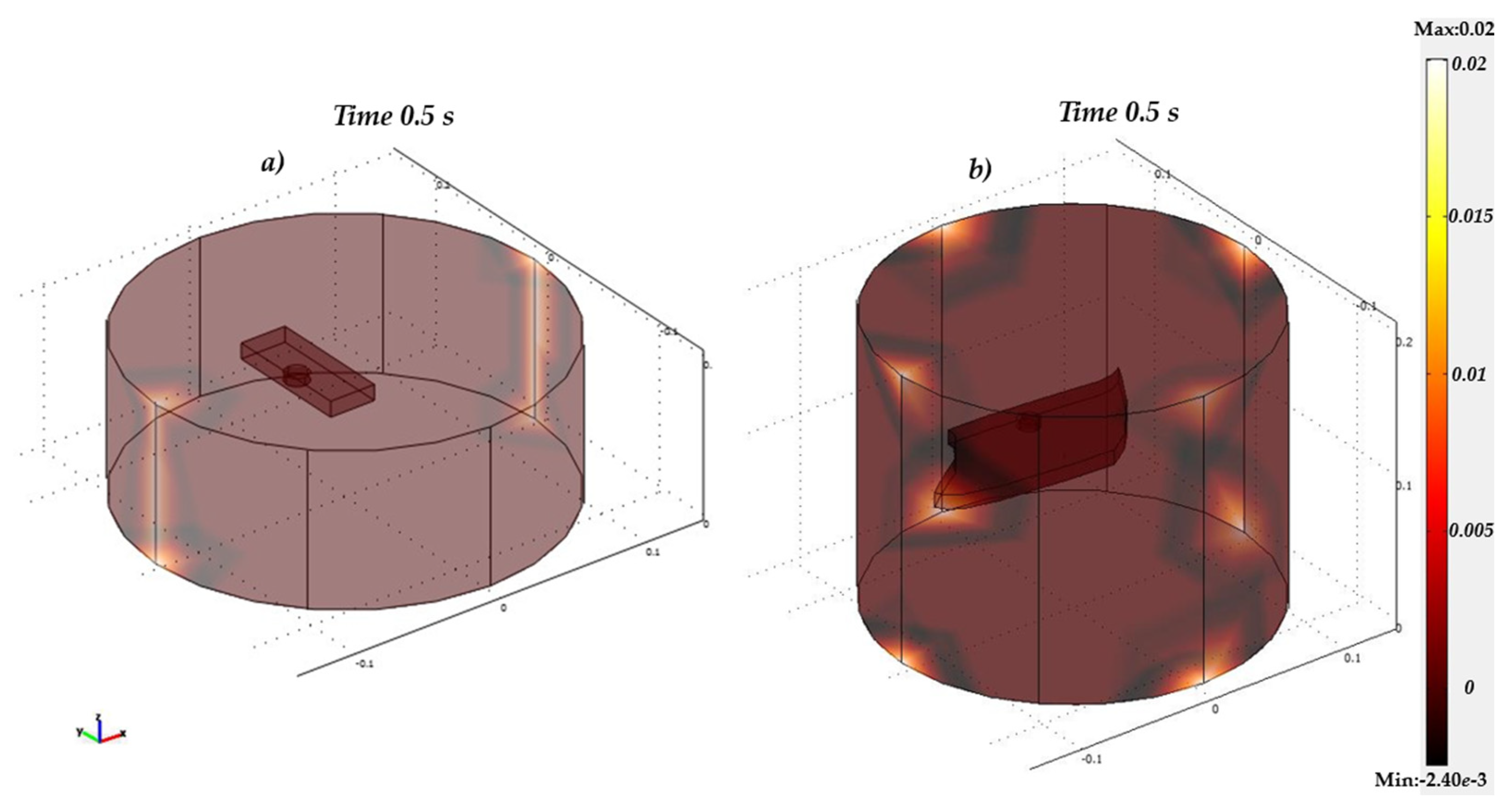
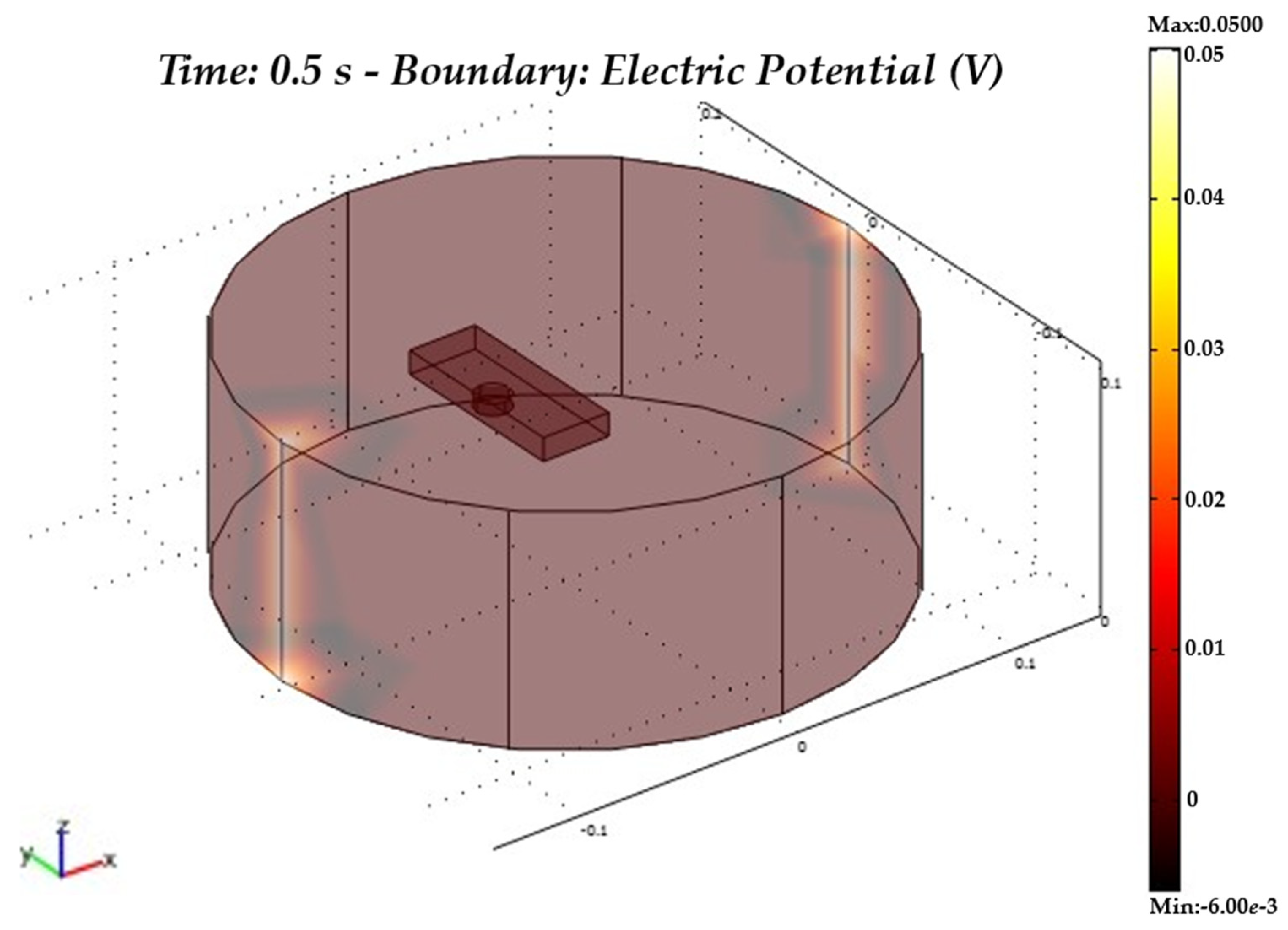
The two-dimensional problem described so far was subsequently realised with a three-dimensional approach in order to make the simulation of the proposed model as real as possible (Figure 3).
In the next section, the results of the three-dimensional simulations of the tomograph will be analysed in detail, varying its electrical characteristics appropriately in order to obtain the best visualisation of the impedance variation for the detection of the tumour cell. Other parameters (potentials, dimensions, etc.) will also be modified in order to obtain results that are exhaustive for the purpose of the paper.

5. Results

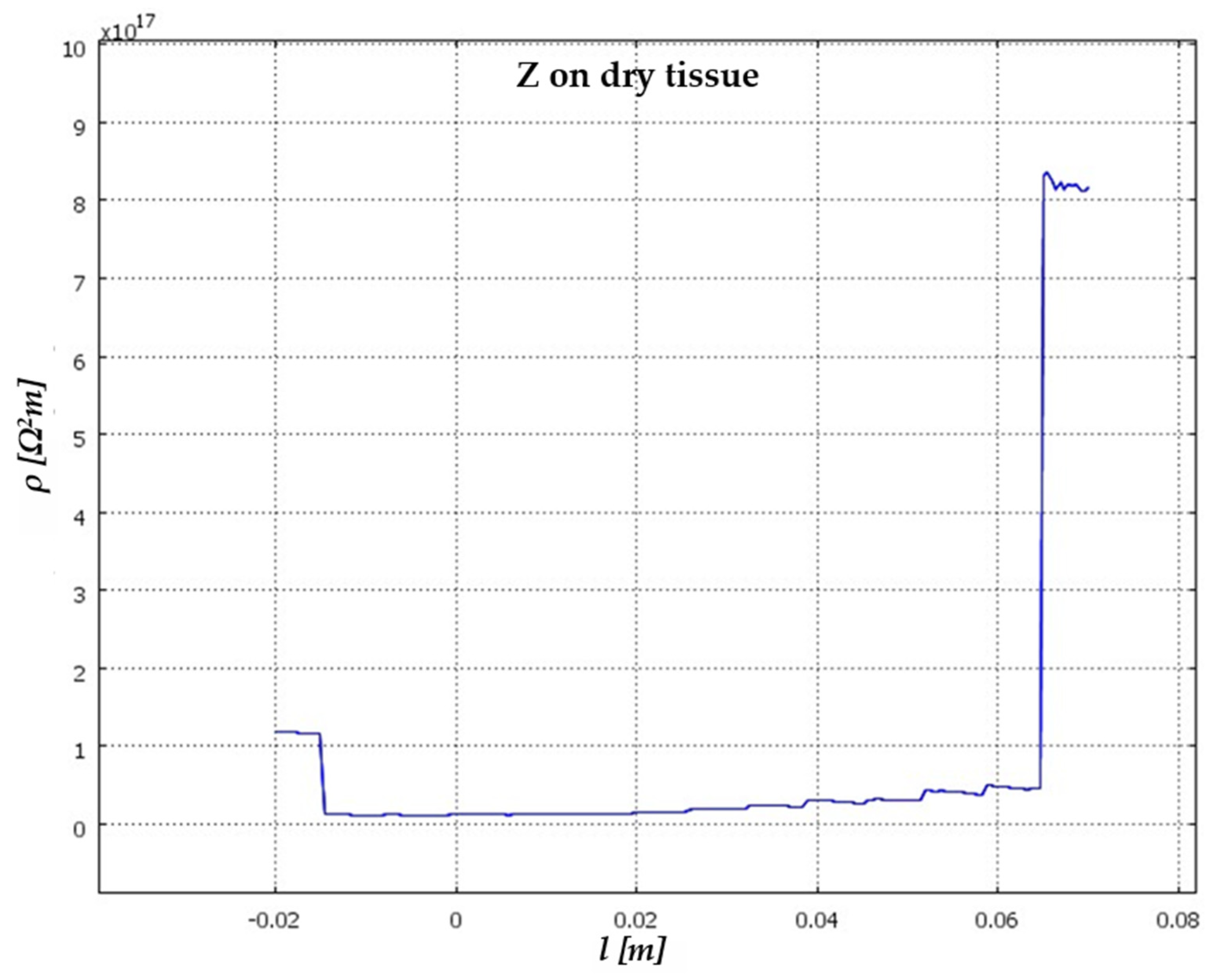
The final part presents the obtained results, which enable the evaluation of the most effective configuration for carcinoma detection. By varying the electrical parameters of the tomograph, the thickness and/or type of skin, and, finally, by providing the electrical properties of the carcinoma, we illustrate the results obtained, allowing us to assess which configuration may be most effective in detecting the carcinoma itself. In general, electrical tomography systems provide an estimate of the transverse distribution of an object by making measurements with appropriate boundary conditions. These measurements, which are non-invasive in nature, are sensitive to the electrical properties of the objects under examination. Modelling the system in two dimensions, as covered extensively in the study [46], studied a simple model of skin tissue. From Figure 4, it can be seen that the impedance variation occurs with an abrupt jump when it encounters the edges of the tissue at its actual position within our tomograph at various instants of solution time.
The trend of the impedance changes on the tissue, set appropriately on the plotting parameters, is shown in Figure 4. Specifically, the y-axis shows the surface resistivity, which is represented as a function of the intrinsic properties and geometry of the investigated material. The impedance Z of a tissue can be described as a function of frequency f, taking into account the resistive (R) and reactive (X) components. For dry tissues, the resistive component R is significant due to the lower moisture content. The relationship between voltage V, current I and impedance Z is given by Ohm’s law. Applying this law to dry tissue, Z will primarily reflect a higher resistance. Resistance changes its behaviour as the length of the current flowing through the tissue is indicated on the x-axis.
The impedance variation determined considers the frequency constant and changes as the surface resistivity varies. For dry tissue, this paper uses a simple model in which the impedance of the tissue, comprising a resistive and a capacitive component, changes as a constant current flow through the medium considered (the skin) as the surface resistivity changes.
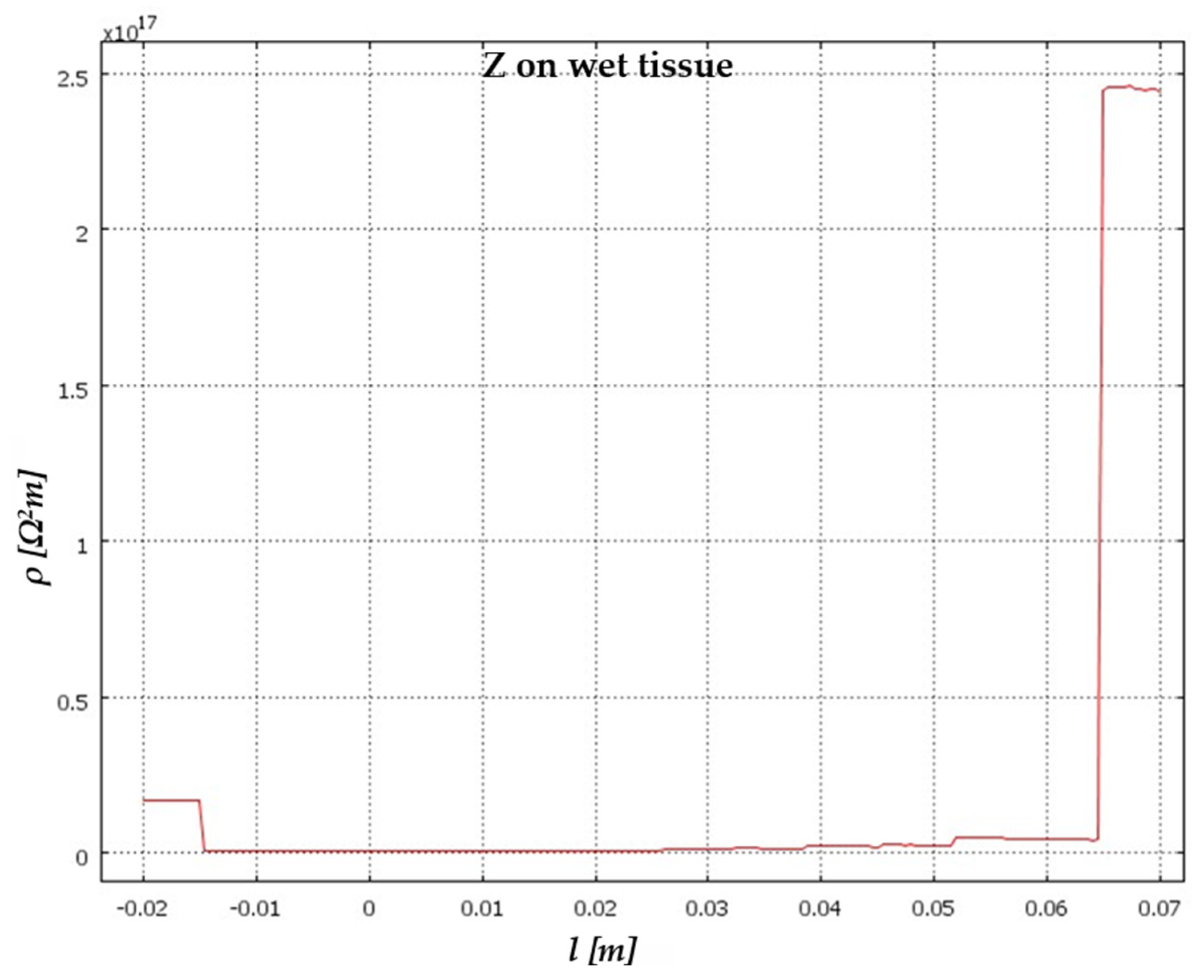
Maintaining the same settings for the tomograph, we proceed to vary the tissue type by considering wet tissue. The electrical and geometrical parameters for this wet tissue are provided in Table 3.
The results obtained by restarting the calculation simulation are shown in Figure 5.
Comparing the two results obtained (Figure 6) highlights the significant differences in impedance values between the tissue types. This initial result is crucial for distinguishing between various tissue types, as it allows for their identification based on a range of pre-established impedance values.
In comparing the variations in electrical impedance between the two types of epithelial tissue, this study also focused on the analysis of a sample of epithelial tissue containing a carcinoma. In these cases, the thickness is significantly reduced, and the conductivity is significantly higher. Electrosurgery and radiotherapy are effective alternatives for this type of cancer, particularly in the case of large carcinomas, but they can damage the lungs and cause major respiratory symptoms, such as dyspnea (difficulty breathing), coughing, and even pneumonia [47,48]. Carcinomas exhibit conductivity values that are 6–7 times greater than those of normal tissue, attributable to the higher water content within the malignant tissue [49]. Table 4 summarises the values adopted for this new simulation, reflecting the distinct electrical properties of carcinomatous tissue.
The biological attributes of moistened skin tissue in the presence of carcinoma manifest distinct electrical properties, encompassing alterations in conductivity, permittivity, and other pertinent parameters compared to healthy tissue. These modifications are pivotal for the identification and diagnosis of pathological conditions such as carcinoma. Moreover, the distinctive electrical signatures exhibited by carcinoma tissue offer valuable insights into medical imaging techniques and diagnostic methodologies.
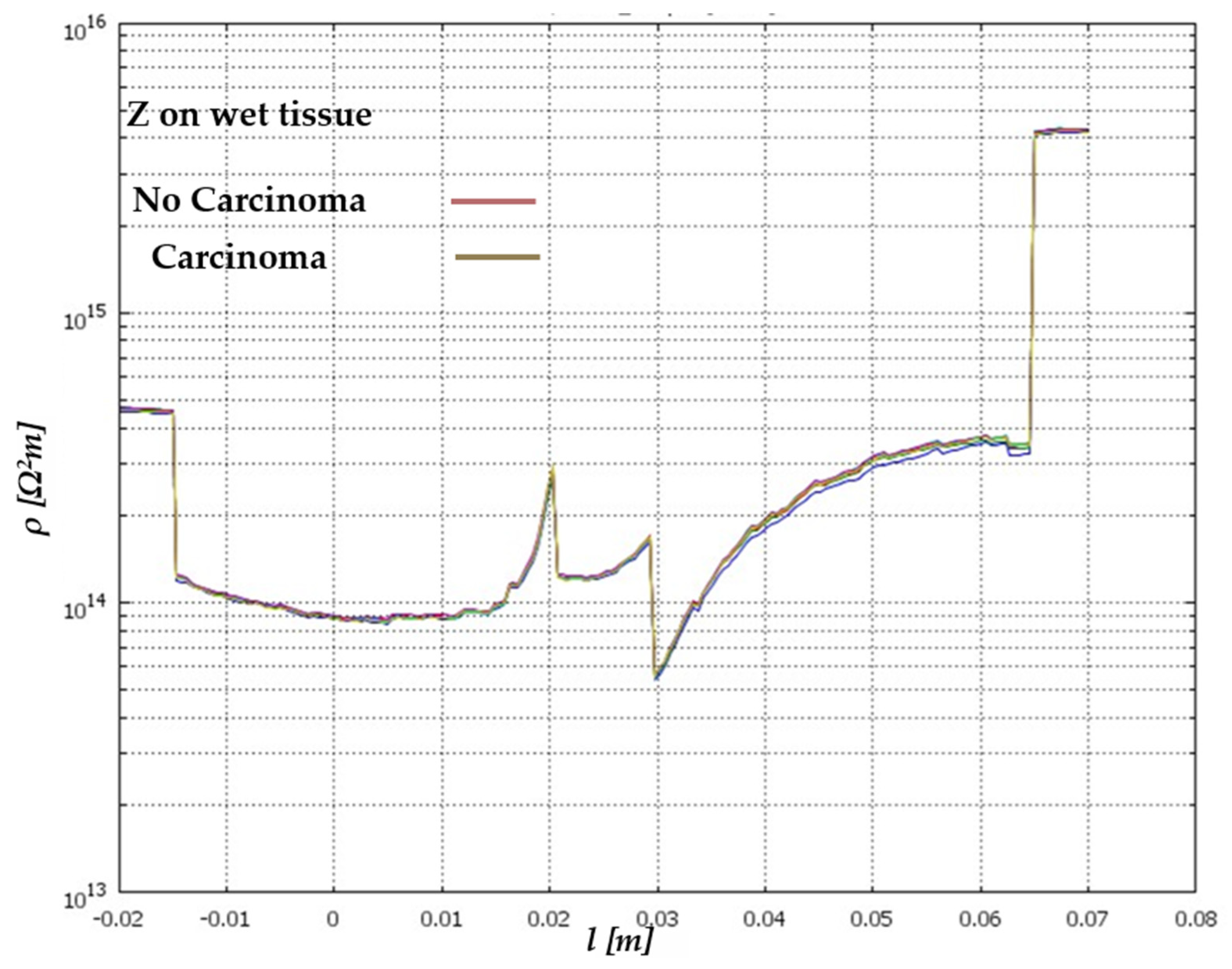
The solution depicted in Figure 7 illustrates how, despite the analysis of tissue with a heightened water concentration akin to that of the tumour, it remains feasible to visibly and quantitatively pinpoint the carcinoma within its precise location within the examined tissue.
The biological attributes of moistened skin tissue in the presence of carcinoma manifest distinct electrical properties, encompassing alterations in conductivity, permittivity, and other pertinent parameters compared to healthy tissue. These modifications are pivotal for the identification and diagnosis of pathological conditions such as carcinoma.
Moreover, the distinctive electrical signatures exhibited by carcinoma tissue offer valuable insights into medical imaging techniques and diagnostic methodologies. The solution (Figure 7—brown curve) shows how, despite the analysis of tissue with a high-water concentration similar to that of the tumour, it is possible to visually and quantitatively identify the carcinoma in its precise position within the examined tissue. The two-dimensional model successfully demonstrated how tomographic analysis, coupled with variations in electrical impedance across biological tissues, serves as an effective means for detecting cancerous cells within tissues intended for human transplantation. The two-dimensional model was able to show how this tomographic analysis, linked to the variation of electrical impedance on biological tissues, can be considered an effective tool for detecting cancer cells in tissues to be transplanted into humans [46]. We thus also wanted to develop the study on a three-dimensional model to try to make our modelling even more real. The first step is to set the parameters shown in Table 5. Initially, employing the adjacent strategy and deactivating the tomograph, the solution is computed.
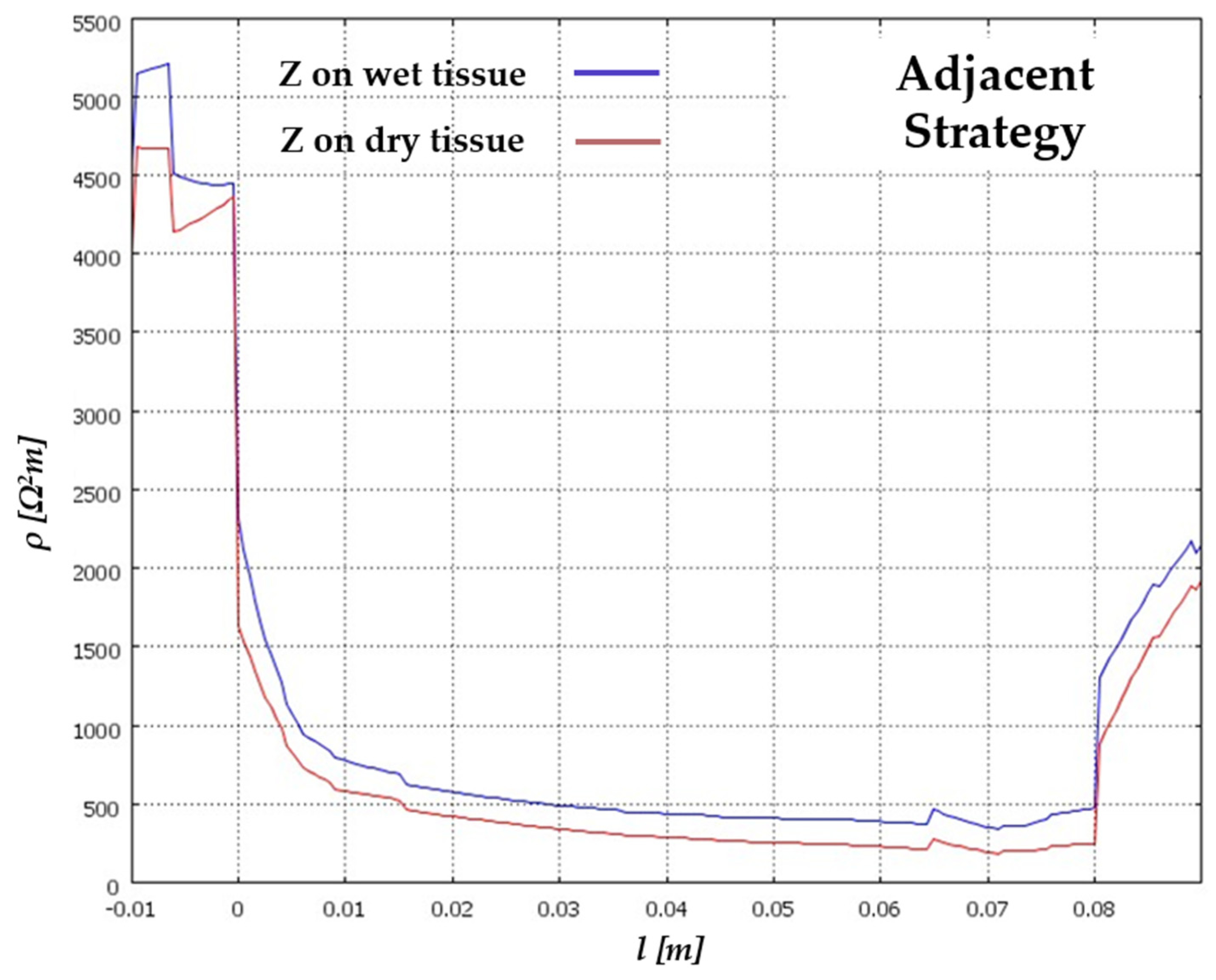
By repeating the simulation for both tissue types, the alterations in electrical impedance are depicted in Figure 8. This comparative analysis provides valuable insights into the distinct electrical properties exhibited by different tissue compositions, particularly in the context of detecting pathological conditions such as carcinoma.
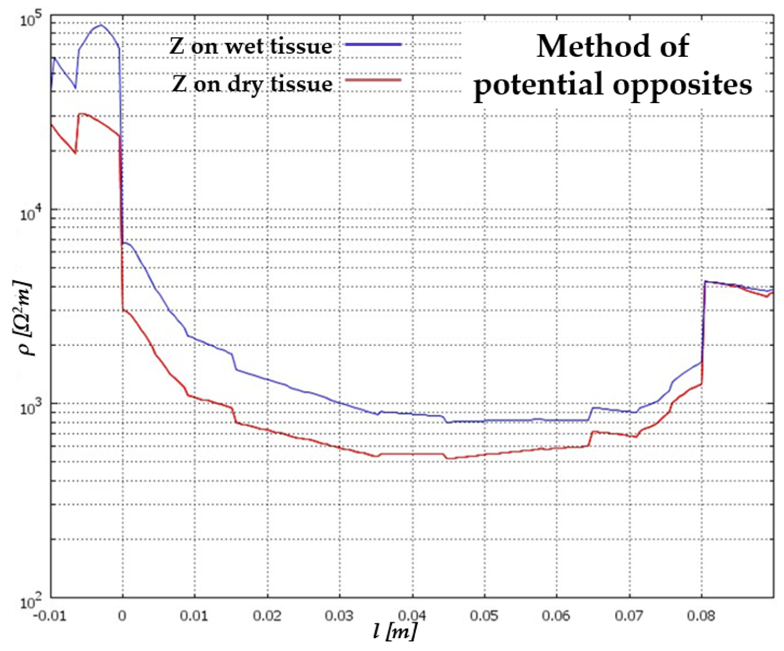
Once more, the efficacy of the tomograph is evident, clearly delineating the alteration in electrical impedance between the two tissue types. Notably, the values depicted on the Y-axis exhibit a considerable increase compared to the two-dimensional scenario. This discrepancy is attributed to the three-dimensional implementation, wherein the potential is uniformly applied along a line, contrasting with the two-dimensional model where the potential was localised to a point. This observation underscores the importance of considering the dimensionality of the model when interpreting impedance variations in biological tissues. The configuration dictated by the method of opposing potentials is shown in Figure 9.
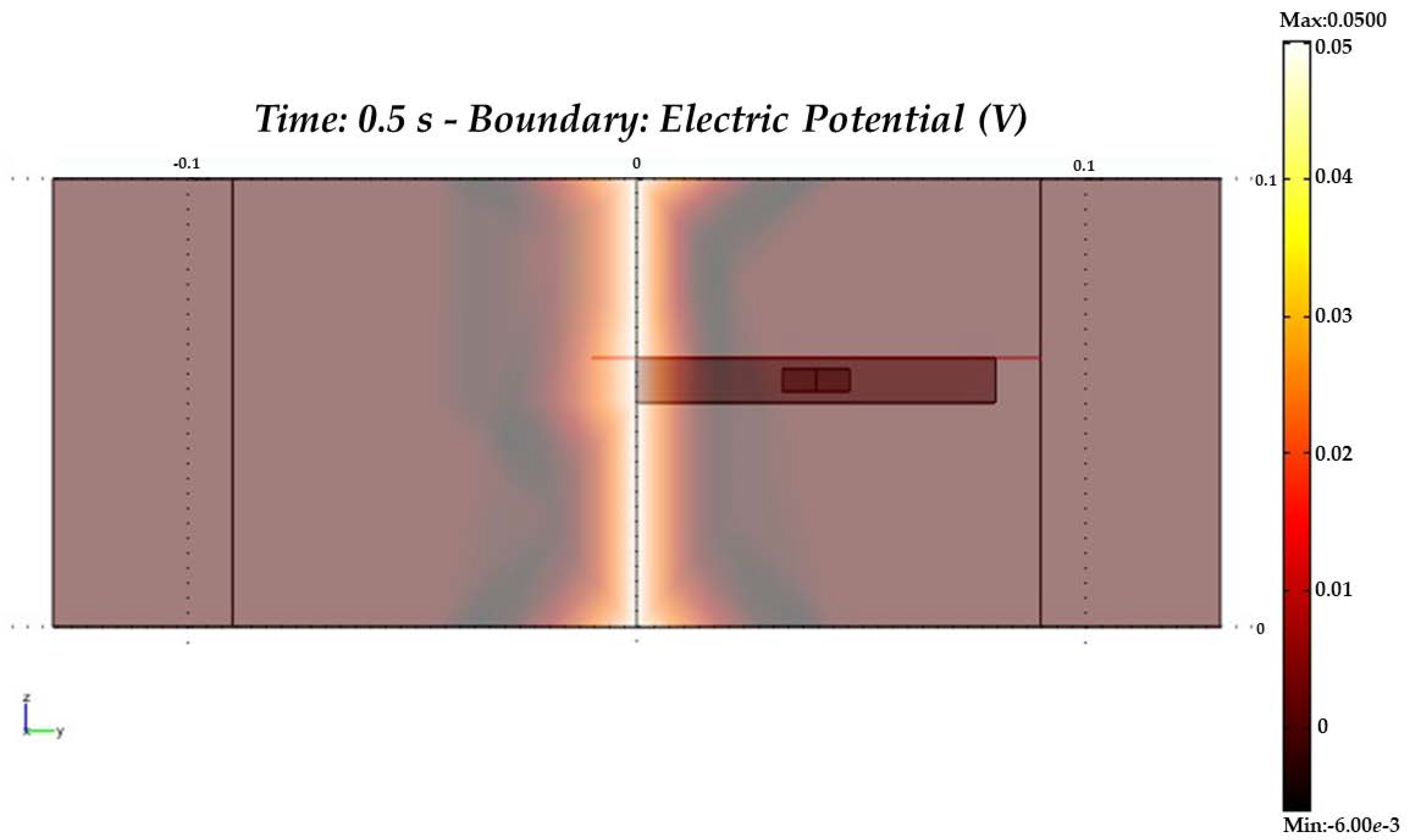
Once more, employing the method of opposite potentials enhances our ability to observe the fluctuations in electrical impedance between the two tissue types. This approach provides greater clarity in discerning the differences in electrical properties between the tissues, facilitating the identification of pathological conditions such as carcinoma. The three-dimensional depiction of the carcinoma is geometrically approximated as a cylinder, with the method of adjacent potentials being employed once more for analysis. This approach allows for a comprehensive evaluation of the electrical impedance variations within the tissue, offering insights into the presence and characteristics of pathological conditions such as carcinoma. To better observe the cross-section we refer to for our analysis, we consider the Y–Z representation of the model, as shown in Figure 10.
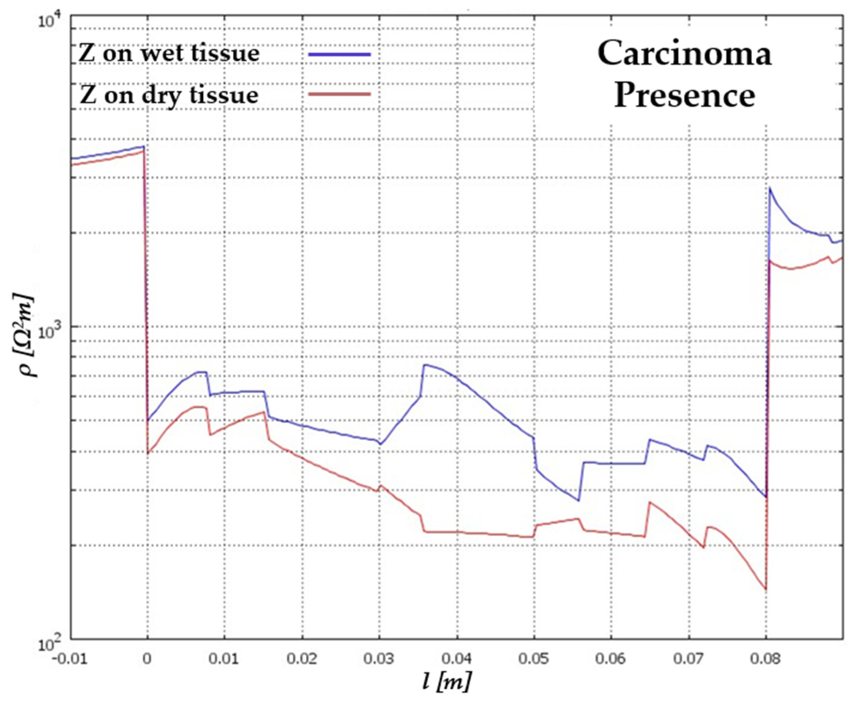
By contrasting a tissue exhibiting a higher water concentration with one affected by carcinoma, the presence of carcinoma can be discerned at the same location as the drier tissue, as depicted in Figure 11.
The analysis shown in Figure 12 highlights the distinct electrical signatures associated with carcinoma within biological tissues, facilitating their identification through non-invasive imaging techniques.
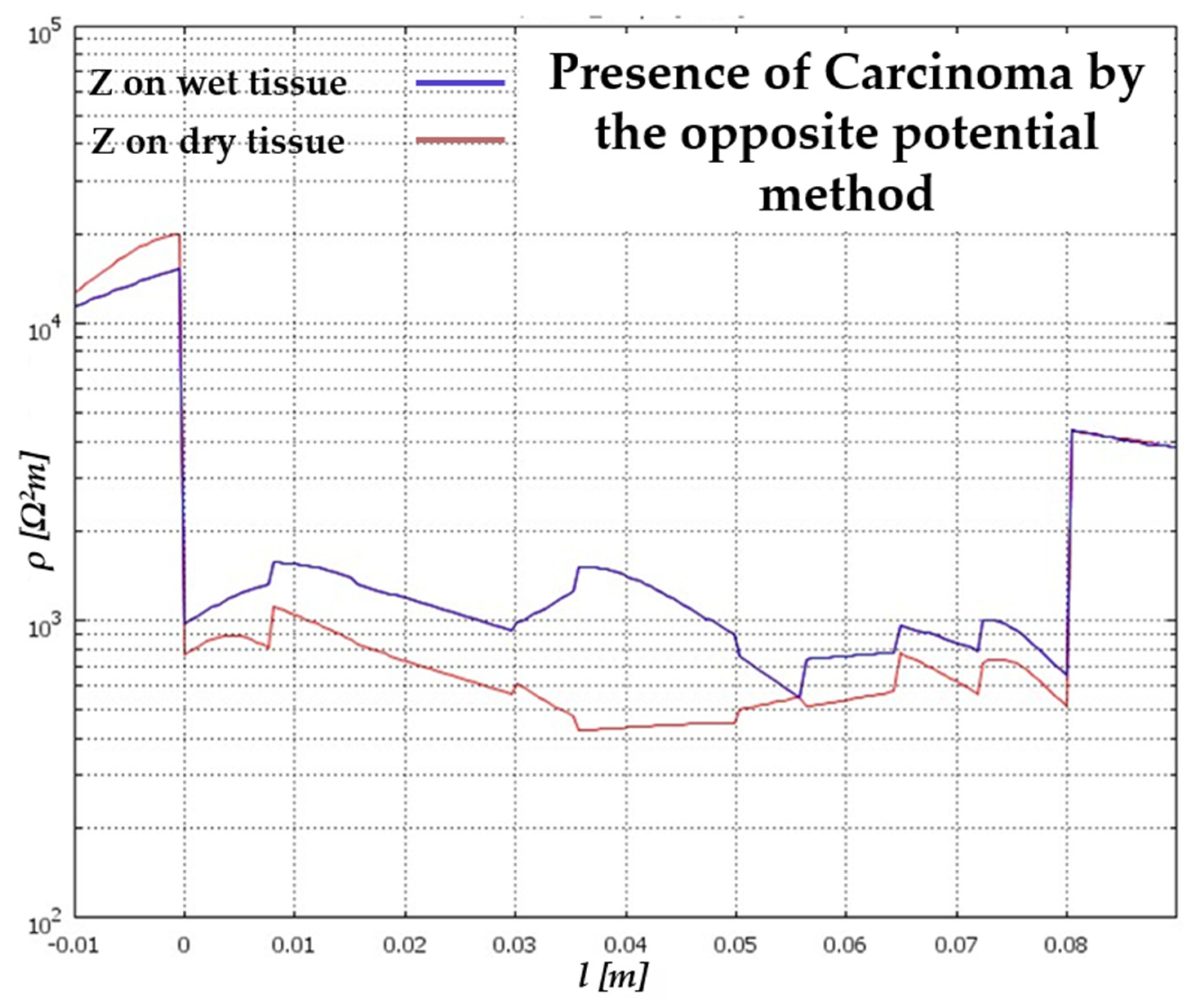
This observation underscores the potential of electrical impedance tomography in detecting pathological conditions within biological tissues, irrespective of variations in moisture content. The comparison between the two tissue types reveals the pattern of electrical impedance variation in epithelial tissue with the presence of carcinoma, derived using the method of opposite potentials (Figure 13). Even for the three-dimensional case, as was legitimate to expect from the two-dimensional (2D) analysis, the method of potential opposites turns out to be significantly more sensitive and effective than the adjacent potential method. This analysis highlights the distinct electrical signatures associated with carcinoma within biological tissues, facilitating their identification through non-invasive imaging techniques.
Furthermore, in the three-dimensional scenario, consistent with the observations from the two-dimensional analysis, the method employing opposite potentials demonstrates notably higher sensitivity and efficacy compared to the adjacent potential’s method, as evident in Figure 13. These simulations conducted via electrical impedance tomography underscore the potential for future practical experimentation, offering several advantages:
Simplified implementation of the instrumentation due to the accessibility and cost-effectiveness of materials utilised;
Immediate detection of carcinoma presence and concurrent localization within the examined tissue;
Non-invasive analysis, ensuring no harm to the tissue being scrutinised;
Convenient electrical measurement facilitated by sensors or other cost-effective devices.

6. Conclusions

The advancement of non-invasive diagnostic techniques for detecting carcinomas is experiencing rapid growth, primarily driven by technological advancements in both hardware and software support systems. This study specifically focused on analyzing epithelial tissue using electrical impedance tomography (EIT) to identify any carcinomas present within it. The approach involved detecting carcinoma through its response to externally applied excitation generated by a potential applied through an experimental tomograph, facilitating the determination of changes in electrical impedance. It is worth pointing out that the paper proposed is the evolution of the model already implemented in 2D by the research group in a previous work. The results produced in this paper are obtained by considering variations in electrical impedance in homogeneous tissue without simulating the cellular structure entirely to avoid computational complexity. For this reason, the results obtained are limited to a limited number of data points. However, the implemented equations govern the approach for the modelling of 2D and three-dimensional (3D) geometries through four numerically tested models, specifically a 2D tomograph model that analyses a tissue sample without carcinoma; a 2D tomograph model that analyses a tissue sample with carcinoma; a 3D tomograph model that analyses a tissue sample without carcinoma; and a 3D tomograph model that analyses a tissue sample with carcinoma. Furthermore, for each model, the two main configurations, i.e., the adjacent potential and the opposite potential, were considered for the electrical impedance tomograph. It is essential to note that the simulation considered electrical impedance variations in the tissue homogeneously without simulating the intricate cellular structure to avoid computational complexity. Additionally, both the tissue and carcinoma were modelled with regular geometry during the simulation phase, simplifying the mesh calculation process. The selection of the 8-electrode tomograph, after multiple simulations, aimed to achieve adequate resolution in measuring impedance variation. Contact impedance between electrodes and skin is a critical factor that significantly influences the quality and reliability of acquired biopotential signals. This paper recognises that high contact impedance can introduce noise and artefacts, thus compromising the accuracy of measurements. In the proposed method, to mitigate the impact of contact impedance, changes in electrical impedance on epithelial tissue are detected with point values by performing a longitudinal scan in a direction coincident with one of the tissue axes for carcinoma detection of an unstratified sample. However, employing a greater number of electrodes could yield more meaningful measurements, a possibility warranting further investigation for enhancing carcinoma detection efficacy. This consideration is important because it provides reassuring answers and opens up new fields of research for the creation of EIT devices that improve the visual process to detect skin cancer. High-tech imaging devices can provide additional accurate data to help doctors monitor and manage specific patients. Real-world application poses challenges not captured by software simulations, such as non-uniform stratified sample structures and instrumentation-induced noise during the measurement phase, necessitating consideration. Regarding the experimental phase, conducting numerous tests suggested potential refinements for practical device implementation, which could be further optimized in future developments. In the simulation phase, the software package allows tomography of any size to be characterised, with the possibility of verifying both the geometric profile and the ideal value of the excitation current to be delivered according to the type of tissue to be analysed. In electrical impedance tomography (EIT), the choice between the use of voltage and current sources is crucial and has significant implications for the method of data acquisition, the quality of reconstructed images, and the accuracy of impedance measurements. The implemented model uses a constant current source, such that it remains unchanged with respect to the size of the skin under examination. This is particularly useful when considering carcinoma on the skin, as the tumour involves impedance variability, as demonstrated by the results obtained. To achieve high levels of accuracy, it is necessary to use very precise and stable current control circuits to ensure that the current delivered is exactly as desired. Finally, prolonged use of high currents can cause polarisation of the electrodes, altering the accuracy of measurements. Future endeavors may involve modifying the model to analyze transplantable organs adjusting geometric, electrical, and biological characteristics accordingly. This iterative approach holds promise for advancing diagnostic capabilities in medical settings.

Author Contributions

Conceptualization, F.L. and G.O.; methodology, D.D.C. and S.A.P.; software, D.P. and F.L.; validation, D.D.C. and F.L.; formal analysis, F.L. and D.P.; investigation, S.C., D.D.C. and F.L.; resources, D.D.C. and F.L.; data curation, D.D.C. and F.L.; writing—original draft preparation, D.P.; writing—review and editing, G.O. and S.A.P.; supervision, S.A.P. and S.C. All authors have read and agreed to the published version of the manuscript.

Funding

This work was supported by both the BATS Laboratory, Department of Health Science ‘Magna Graecia’, Catanzaro, Italy, and the NdT&E Laboratory, Department DICEAM ‘Mediterranea’, Reggio Calabria, Italy.

Informed Consent Statement

Not applicable.

Data Availability Statement

The data are contained within this article.

Conflicts of Interest

The authors declare no conflicts of interest.

References

  1. Wu, Y.; Hanzaee, F.F.; Jiang, D.; Bayford, R.H.; Demosthenous, A. Electrical Impedance Tomography for Biomedical Applications: Circuits and Systems Review. IEEE Open J. Circuits Syst. 2021, 2, 380–397. [Google Scholar] [CrossRef]
  2. Padilha Leitzke, J.; Zangl, H. A review on electrical impedance tomography spectroscopy. Sensors 2020, 20, 5160. [Google Scholar] [CrossRef] [PubMed]
  3. Seçkin, A.Ç.; Ateş, B.; Seçkin, M. Review on Wearable Technology in sports: Concepts, Challenges and opportunities. Appl. Sci. 2023, 13, 10399. [Google Scholar] [CrossRef]
  4. Ke, X.Y.; Hou, W.; Huang, Q. Advances in electrical impedance tomography-based brain imaging. Mil. Med. Res. 2022, 9, 10. [Google Scholar] [CrossRef] [PubMed]
  5. Adam, E.E.B.; Babikir, E. Survey on medical imaging of electrical impedance tomography (EIT) by variable current pattern methods. J. IoT Soc. Mob. Anal. Cloud 2021, 3, 82–95. [Google Scholar]
  6. Zhang, H.; Kalra, A.; Lowe, A.; Yu, Y.; Anand, G. A hydrogel-based electronic skin for touch detection using electrical impedance tomography. Sensors 2023, 23, 1571. [Google Scholar] [CrossRef] [PubMed]
  7. Li, Y.; Wang, N.; Fan, L.F.; Wang, Y.Q.; Zhao, P.F.; Huang, L.; Wang, Z.Y. Imaging spatial-temporal distribution of moisture in intact maize ear. Measurement 2022, 202, 111809. [Google Scholar] [CrossRef]
  8. Khan, T.A.; Ling, S.H.; Rizvi, A.A. Optimisation of electrical Impedance tomography image reconstruction error using heuristic algorithms. Artif. Intell. Rev. 2023, 56, 15079–15099. [Google Scholar] [CrossRef]
  9. Tyni, T.; Stinchcombe, A.R.; Alexakis, S. A boundary integral equation method for the complete electrode model in electrical impedance tomography with tests on experimental data. SIAM J. Imaging Sci. 2024, 17, 672–705. [Google Scholar] [CrossRef]
  10. Angiulli, G.; Versaci, M.; Calcagno, S. Computation of the cutoff wavenumbers of metallic waveguides with symmetries by using a nonlinear eigenproblem formulation: A group theoretical approach. Mathematics 2020, 8, 489. [Google Scholar] [CrossRef]
  11. Jain, H.; Isaacson, D.; Edic, P.M.; Newell, J.C. Electrical impedance tomography of complex conductivity distributions with noncircular boundary. IEEE Trans. Biomed. Eng. 1997, 44, 1051–1060. [Google Scholar] [CrossRef] [PubMed]
  12. Angiulli, G.; Cacciola, M.; Calcagno, S.; De Carlo, D.; Morabito, C.F.; Sgró, A.; Versaci, M. A numerical study on the performances of the flexible BiCGStab to solve the discretized E-field integral equation. Int. J. Appl. Electromagn. Mech. 2014, 46, 547–553. [Google Scholar] [CrossRef]
  13. Kulkarni, R.; Boverman, G.; Isaacson, D.; Saulnier, G.; Newell, J.C. Layered model for breasts in electrical impedance tomography. In Proceedings of the 2007 29th Annual International Conference of the IEEE Engineering in Medicine and Biology Society, Lyon, France, 22–26 August 2007; pp. 4150–4153. [Google Scholar]
  14. Cappellini, I.; Campagnola, L.; Consales, G. Electrical Impedance Tomography, Artificial Intelligence, and Variable Ventilation: Transforming Respiratory Monitoring and Treatment in Critical Care. J. Pers. Med. 2024, 14, 677. [Google Scholar] [CrossRef]
  15. Pennati, F.; Angelucci, A.; Morelli, L.; Bardini, S.; Barzanti, E.; Cavallini, F.; Conelli, A.; Di Federico, G.; Paganelli, C.; Aliverti, A. Electrical Impedance Tomography: From the Traditional Design to the Novel Frontier of Wearables. Sensors 2023, 23, 1182. [Google Scholar] [CrossRef] [PubMed]
  16. Singh, R.K.; Nayak, N.P.; Behl, T.; Arora, R.; Anwer, M.K.; Gulati, M.; Bungau, S.G.; Brisc, M.C. Exploring the Intersection of Geophysics and Diagnostic Imaging in the Health Sciences. Diagnostics 2024, 14, 139. [Google Scholar] [CrossRef]
  17. Bianco, M.G.; Quattrone, A.; Sarica, A.; Vescio, B.; Buonocore, J.; Vaccaro, M.G.; Aracri, F.; Calomino, C.; Gramigna, V.; Quattrone, A. Cortical atrophy distinguishes idiopathic normal pressure hydrocephalus from progressive supranuclear palsy: A machine learning approach. Park. Relat. Disord. 2022, 103, 7–14. [Google Scholar]
  18. Tang, M.; Wang, W.; Wheeler, J.; McCormick, M.; Dong, X. The number of electrodes and basis functions in EIT image reconstruction. Physiol. Meas. 2002, 23, 129–140. [Google Scholar] [CrossRef]
  19. Sarac, E.; Meiwes, A.; Eigentler, T.K.; Forchhammer, S.; Kofler, L.; Häfner, H.M.; Garbe, C. Diagnostic accuracy of electrical impedance spectroscopy in non-melanoma skin cancer. Acta Derm. Venereol. 2020, 100, adv00328. [Google Scholar] [CrossRef]
  20. Braun, R.P.; Mangana, J.; Goldinger, S.; French, L.; Dummer, R.; Marghoob, A.A. Electrical Impedance Spectroscopy in Skin Cancer Diagnosis. Dermatol. Clin. 2017, 35, 489–493. [Google Scholar] [CrossRef]
  21. Becher, T.; Buchholz, V.; Hassel, D.; Meinel, T.; Schädler, D.; Frerichs, I.; Weiler, N. Individualization of PEEP and tidal in ARDS patients with electrical impedance tomography: A pilot feasibility study. Ann. Intensive Care 2021, 11, 89. [Google Scholar] [CrossRef]
  22. Zhao, Z.; Steinmann, D.; Frerichs, I.; Guttmann, J.; Möller, K. PEEP titration guided by ventilation homogeneity: A feasibility study using electrical impedance tomography. Crit. Care 2010, 14, R8. [Google Scholar] [CrossRef]
  23. Glickman, Y.A.; Filo, O.; David, M.; Yayon, A.; Topaz, M.; Zamir, B.; Kenan, G. Electrical impedance scanning: A new approach to skin cancer diagnosis. Ski. Res. Technol. 2003, 9, 262–268. [Google Scholar] [CrossRef]
  24. Aberg, P.; Nicander, I.; Hansson, J.; Geladi, P.; Holmgren, U.; Ollmar, S. Skin cancer identification using multifrequency electrical impedance—A potential screening tool. IEEE Trans. Biomed. Eng. 2004, 51, 2097–2102. [Google Scholar] [CrossRef]
  25. Åberg, P.; Birgersson, U.; Elsner, P.; Mohr, P.; Ollmar, S. Electrical impedance spectroscopy and the diagnostic accuracy for malignant melanoma. Exp. Dermatol. 2011, 20, 648–652. [Google Scholar] [CrossRef]
  26. Mohr, P.; Birgersson, U.; Berking, C.; Henderson, C.; Trefzer, U.; Kemeny, L.; Paoli, J. Electrical impedance spectroscopy as a potential adjunct diagnostic tool for cutaneous melanoma. Ski. Res. Technol. 2013, 19, 75–83. [Google Scholar] [CrossRef]
  27. Yang, Y.; Wang, J.; Yu, G.; Niu, F.; He, P. Design and preliminary evaluation of a portable device for the measurement of bioimpedance spectroscopy. Physiol. Meas. 2006, 27, 1293. [Google Scholar] [CrossRef]
  28. Åberg, P.; Geladi, P.; Nicander, I.; Hansson, J.; Holmgren, U.; Ollmar, S. Non-invasive and microinvasive electrical impedance spectra of skin cancer—A comparison between two techniques. Ski. Res. Technol. 2005, 11, 281–286. [Google Scholar] [CrossRef]
  29. Narayanamurthy, V.; Padmapriya, P.; Noorasafrin, A.; Pooja, B.; Hema, K.; Nithyakalyani, K.; Samsuri, F. Skin cancer detection using non-invasive techniques. RSC Adv. 2018, 8, 28095–28130. [Google Scholar] [CrossRef]
  30. Kamat, D.K.; Chavan, A.P.; Patil, P.M. Bio-impedance measurement system for analysis of skin diseases. Int. J. Appl. Innov. Eng. Manag. 2014, 3, 92–96. [Google Scholar]
  31. Zhao, Z.; Zhu, H.; Li, X.; Sun, L.; He, F.; Chung, J.E.; Xie, C. Ultraflexible electrode arrays for months-long high-density electrophysiological mapping of thousands of neurons in rodents. Nat. Biomed. Eng. 2023, 7, 520–532. [Google Scholar] [CrossRef] [PubMed]
  32. Eisenberg, R.S. Maxwell equations without a polarization field, Using a paradigm from biophysics. Entropy 2021, 23, 172. [Google Scholar] [CrossRef]
  33. Abdul-Al, M.; Amar, A.S.; Elfergani, I.; Littlehales, R.; Ojaroudi Parchin, N.; Al-Yasir, Y.; Abd-Alhameed, R.A. Wireless electromagnetic radiation assessment based on the specific absorption rate (SAR): A review case study. Electronics 2022, 11, 511. [Google Scholar] [CrossRef]
  34. Wust, P.; Stein, U.; Ghadjar, P. Non-thermal membrane effects of electromagnetic fields and therapeutic applications in oncology. Int. J. Hyperth. 2021, 38, 715–731. [Google Scholar] [CrossRef]
  35. Mazuir, E.; Fricker, D.; Sol-Foulon, N. Neuron–oligodendrocyte communication in myelination of cortical GABAergic cells. Life 2021, 11, 216. [Google Scholar] [CrossRef]
  36. Wu, H.; Zhou, W.; Yang, Y.; Jia, J.; Bagnaninchi, P. Exploring the potential of electrical impedance tomography for tissue engineering applications. Materials 2018, 11, 930. [Google Scholar] [CrossRef]
  37. Yilmaz, T.; Ates Alkan, F. In vivo dielectric properties of healthy and benign rat mammary tissues from 500 MHz to 18 GHz. Sensors 2020, 20, 2214. [Google Scholar] [CrossRef]
  38. Laganà, F.; Britti, D.; Fiorillo, A.S.; Pullano, S.A. New Surface Electrical Charge Detection System for Ecology and Insect Monitoring. In Proceedings of the 2023 IEEE International Workshop on Biomedical Applications, Technologies and Sensors, BATS 2023—Proceedings, Catanzaro, Italy, 28–29 September 2023. [Google Scholar]
  39. Di Meo, S.; Bonello, J.; Farhat, I.; Farrugia, L.; Pasian, M.; Camilleri Podesta, M.T.; Sammut, C.V. The variability of dielectric permittivity of biological tissues with water content. J. Electromagn. Waves Appl. 2022, 36, 48–68. [Google Scholar] [CrossRef]
  40. Seweryn, A. Interactions between surfactants and the skin—Theory and practice. Adv. Colloid Interface Sci. 2018, 256, 242–255. [Google Scholar] [CrossRef]
  41. Pullano, S.A.; Greco, M.; Islam, S.K.; Fiorillo, A.S. Thermoelectrical Characterization of Cells Using a Pyroelectric Sensor. In Proceedings of the 2023 IEEE SENSORS, Vienna, Austria, 29 October–1 November 2023. [Google Scholar]
  42. Tang, J.; Lu, M.; Xie, Y.; Yin, W. A Novel Efficient FEM Thin shell model for bio-impedance analysis. Biosensors 2020, 10, 69. [Google Scholar] [CrossRef]
  43. Pullano, S.A.; Greco, M.; Foti, D.P.; Brunetti, A.; Fiorillo, A.S. Dual Pyroelectric Sensor for Thermal Characterization of Cell Lines. In Proceedings of the 2021 IEEE Sensors Applications Symposium (SAS), Virtual Conference, Sundsvall, Sweden, 23–25 August 2021. [Google Scholar]
  44. Laganà, F.; De Carlo, D.; Calcagno, S.; Pullano, S.A.; Critello, D.; Falcone, F.; Fiorillo, A.S. Computational model of cell deformation under fluid flow-based rolling. In Proceedings of the 7th IEEE International Conference on E-Health and Bioengineering, EHB 2019, Iasi, Romania, 21–23 November 2019. [Google Scholar]
  45. Lee, S.; Ozkavukcu, S.; Ku, S.Y. Current and future perspectives for improving ovarian tissue cryopreservation and transplantation outcomes for cancer patients. Reprod. Sci. 2021, 28, 1746–1758. [Google Scholar] [CrossRef]
  46. Laganà, F.; De Carlo, D.; Calcagno, S.; Oliva, G.; Pullano, S.A.; Fiorillo, A.S. Modeling of Electrical Impedance Tomography for Carcinoma Detection. In Proceedings of the 10th E-Health and Bioengineering Conference EHB 2022, Virtual Conference, Iasi, Romania, 17–18 November 2022. [Google Scholar] [CrossRef]
  47. Menniti, M.; Oliva, G.; Laganà, F.; Bianco, M.G.; Fiorillo, A.S.; Pullano, S.A. Portable Non-Invasive Ventilator for Homecare and Patients Monitoring System. In Proceedings of the 2023 IEEE International Symposium on Medical Measurements and Applications (MeMeA), Jeju, Republic of Korea, 14–16 June 2023; pp. 1–5. [Google Scholar] [CrossRef]
  48. Menniti, M.; Laganà, F.; Oliva, G.; Bianco, M.; Fiorillo, A.S.; Pullano, S.A. Development of Non-Invasive Ventilator for Homecare and Patient Monitoring System. Electronics 2024, 13, 790. [Google Scholar] [CrossRef]
  49. Vaupel, P.; Piazena, H. Hyperhydration of cancers: A characteristic biophysical trait strongly increasing O2, CO2, glucose and lactate diffusivities, and improving thermophysical properties of solid malignancies. In International Society on Oxygen Transport to Tissue; Springer International Publishing: Cham, Switzerland, 2022; pp. 135–145. [Google Scholar]
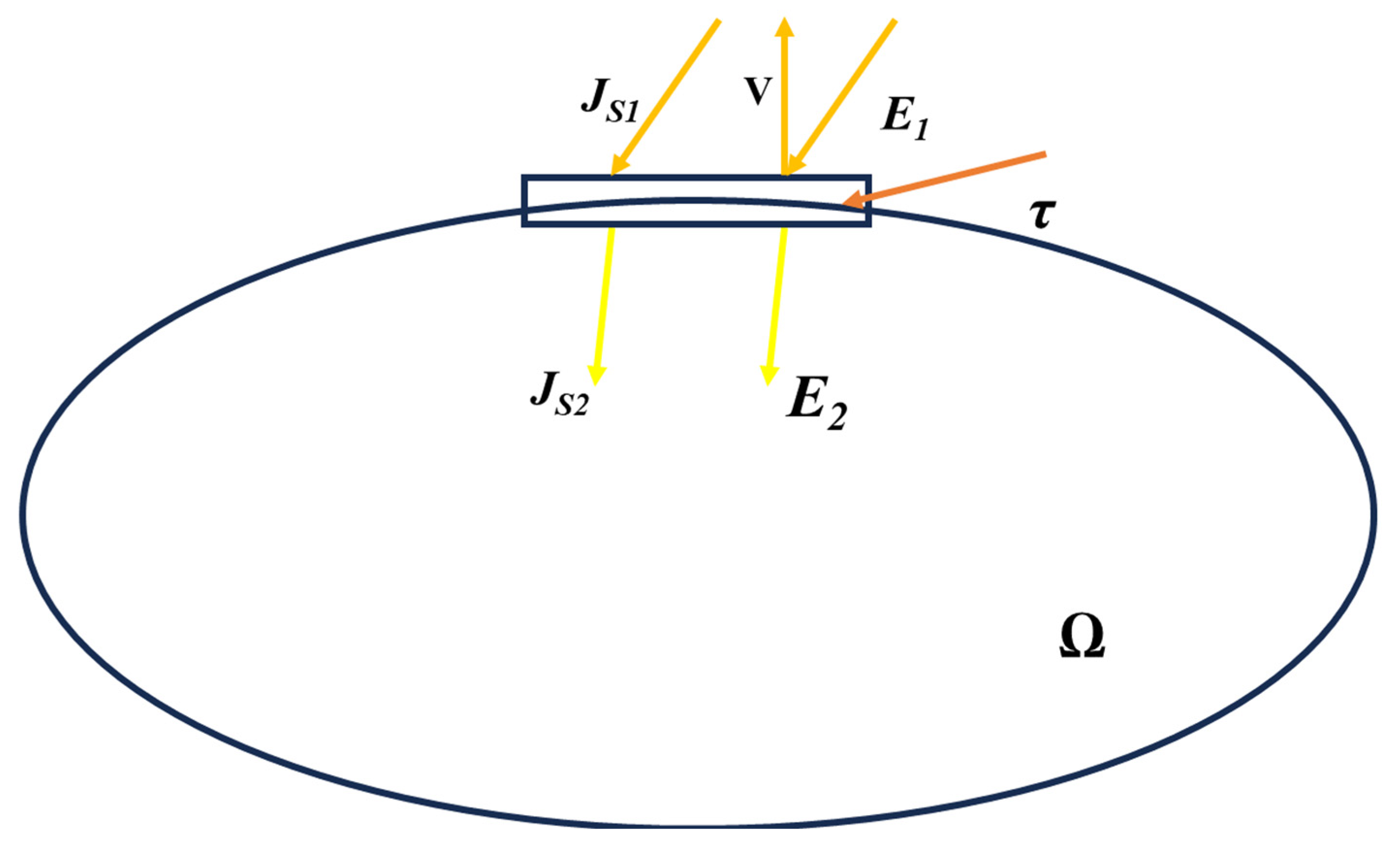
Figure 1. Determination of circumference conditions. Js1 and Js2 are the current densities outside and inside the object, respectively. E1 and E2 are the corresponding electrical fields.
Eng 05 00084 g001
Figure 2. Geometry: (a) Realisation of electrodes through point modelling; (b) modelling of irregularly shaped skin tissue and carcinoma.
Eng 05 00084 g002
Figure 3. Three-dimensional (3D) model: (a) Electrical potential trend on the tomograph and sample; (b) time-change of the electric potential.
Eng 05 00084 g003
Figure 4. Cartesian representation of electrical impedance variation on dry tissue.
Eng 05 00084 g004
Figure 5. Cartesian representation of electrical impedance variation on wet tissue.
Eng 05 00084 g005
Figure 6. Comparison of electrical impedance variations between the two types of tissues considering a potential difference of 1 V. DRY = BLUE − WET = RED.
Eng 05 00084 g006
Figure 7. Change in electrical impedance on wet tissue: The red curve represents the change in electrical impedance on skin without carcinoma; the brown curve represents the change in electrical impedance on skin with carcinoma.
Eng 05 00084 g007
Figure 8. Electrical impedance variation on epithelial tissue obtained by the method of adjacent potentials.
Eng 05 00084 g008
Figure 9. Variation of electrical impedance on the two epithelial tissues obtained by the method of opposite potentials.
Eng 05 00084 g009
Figure 10. Y–Z plane of the cross-section for calculating the surface electrical impedance variation on epithelial tissue with the presence of carcinoma.
Eng 05 00084 g010
Figure 11. Variation of electrical impedance on epithelial tissue with the presence of carcinoma obtained by the method of the opposite potentials.
Eng 05 00084 g011
Figure 12. Superficial electrical impedance variation on epithelial tissue with the presence of carcinoma on wet tissue (blue curve) and on dry tissue (red curve).
Eng 05 00084 g012
Figure 13. Electrical impedance variation on epithelial tissue with the presence of carcinoma was obtained by the method of opposite potentials on wet tissue (blue curve) and on dry tissue (red curve).
Eng 05 00084 g013
Table 1. Electrical bio-impedance techniques for skin cancer diagnosis.
Signal UsedDescriptionMeritsDemeritsReference
1 kHz and 1000 kHzDistinguishes the skin cancer from the benign lesions using multi-frequency impedance spectraThe result obtained is more accurate than conventional methodsDistinguishing the tumours takes more time, and false results may also be obtained[24]
1–1000 kHzCompares the detection of skin cancer by a non-invasive probe and micro-invasive electrode system, whose surface is furnished with tiny spikes which get penetrated to the stratum corneumThe electrode system produces a better resultMinimally invasive technique[28]
1 kHz and 1 MHzDescribes the method for detecting skin cancer using electric impedance. The electric impedance of the biological system decreases with the increase in frequencyHigh resolutionMultivariate and the impedance is complex[29]
1 kHz to 2.5 MHzAccuracy of electrical impedance to classify malignant melanoma from benign tumour by automated classification algorithmAccuracy is highVarious algorithm is needed for the classification of skin cancer[25]
1–100 kHzNon-invasive approach for detecting the presence of skin lesions by measuring the impedance changeLow-cost and portableElectrodes are used, which cause discomfort[30]
1 kHz to 2.5 MHzEIS algorithm is used on lesions to differentiate normal skin from abnormal lesionsHigh resolutionAn experienced physician is required[20]
20 kHz to 1 MHzA portable bio-impedance system is used to diagnose skin cancer based on the magnitude ratio and phase detection methodAct as a great tool for monitoring the physiological conditions of the biological systemHigh cost[27]
Table 2. Characteristics of biological tissues dry skin.
Thickness [m]Applied Potential [V]Conductivity [S/m]PermittivityFrequency [Hz]
0.0050.050.0002113650
Table 3. Characteristics of biological tissues wet skin.
Thickness [m]Applied Potential [V]Conductivity [S/m]PermittivityFrequency [Hz]
0.0050.050.0002113650
Table 4. Characteristics of biological tissues wet skin in the presence of carcinoma.
Type of TissueThickness [m]Applied Potential [V]Conductivity [S/m]PermittivityFrequency [Hz]
Wet0.030.050.004271951,27450
Cancer0.00010.050.0013150
Table 5. Electrical and geometric parameters three-dimensional model.
Type of TissueDimensions XYZ [m]Applied
Potential [V]
Conductivity [S/m]PermittivityFrequency [Hz]
Dry0.08 × 0.03 × 0.010.050.0002113650
Wet0.08 × 0.03 × 0.010.050.004271951,27450
Disclaimer/Publisher’s Note: The statements, opinions and data contained in all publications are solely those of the individual author(s) and contributor(s) and not of MDPI and/or the editor(s). MDPI and/or the editor(s) disclaim responsibility for any injury to people or property resulting from any ideas, methods, instructions or products referred to in the content.