From Imaging to Instrument: A CT-Based Classification of the Nasofrontal Beak and Frontal Ostium Clearance with Prospective Surgical Correlation

 , , , and
Abstract
1. Introduction
2. Materials and Methods
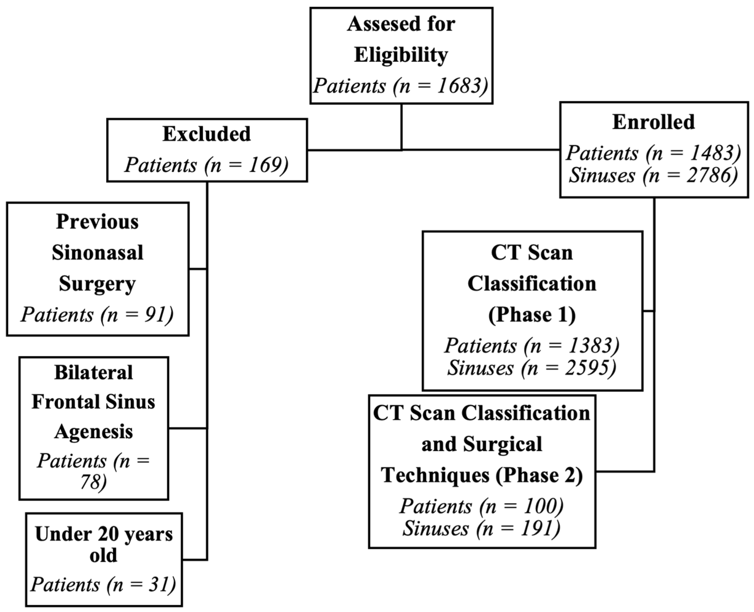
2.1. Study Design and Participants
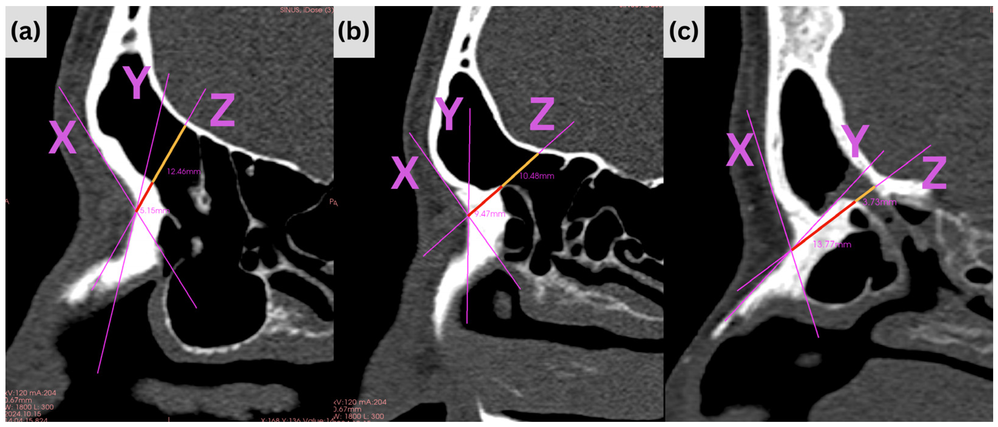
2.2. CT Acquisition and Measurement Protocol
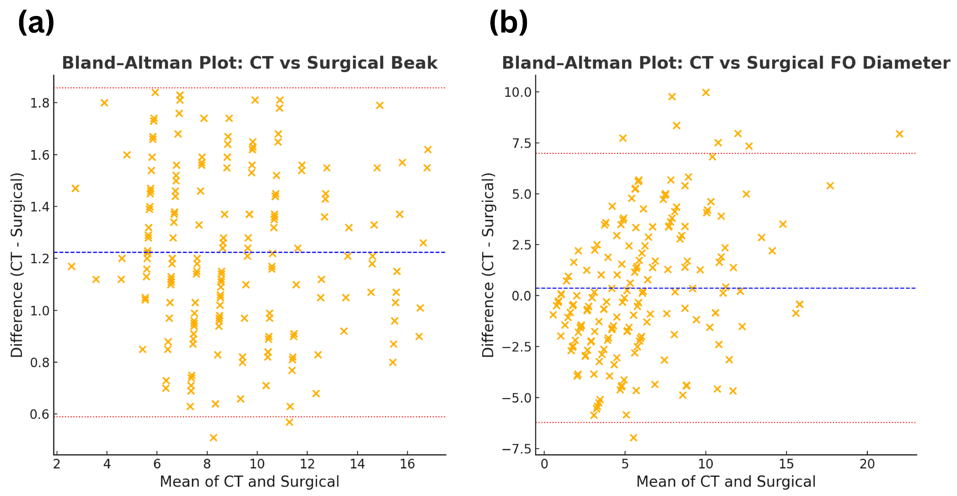
2.3. Method Equivalence Analysis
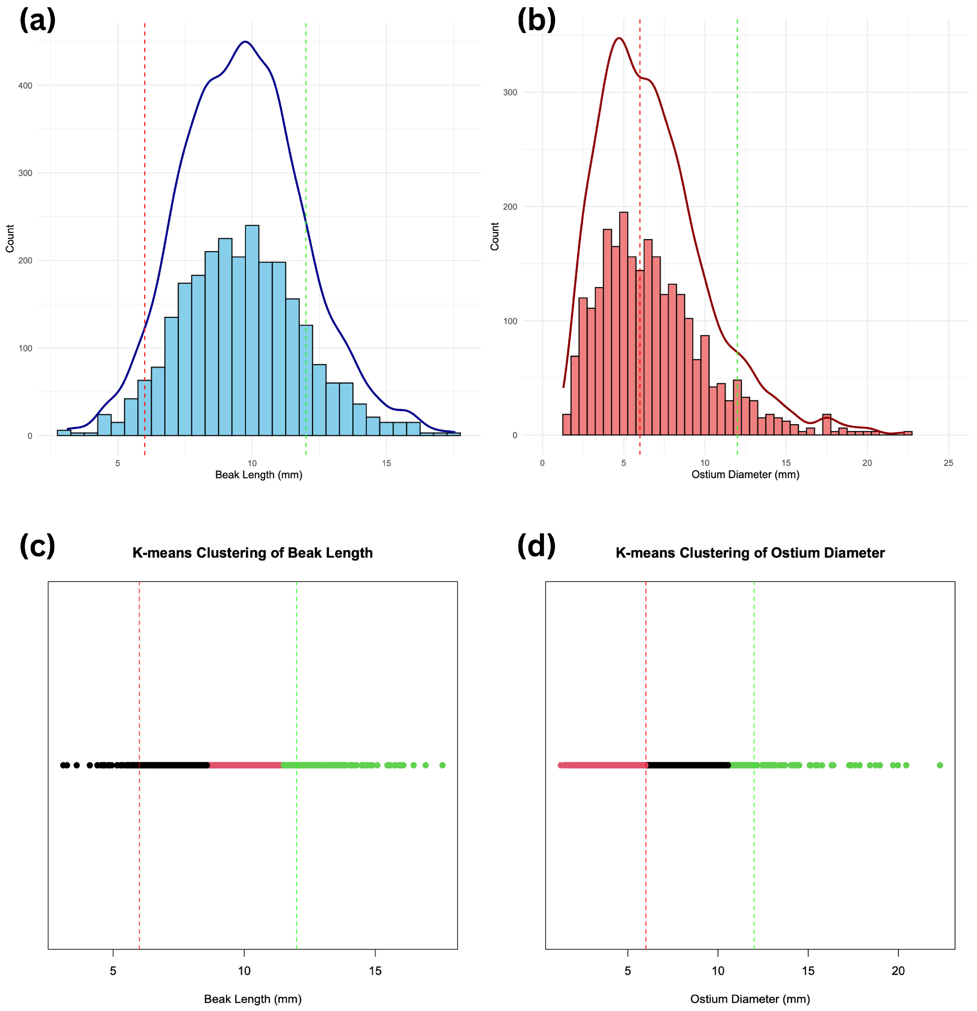
2.4. CT Classification Development
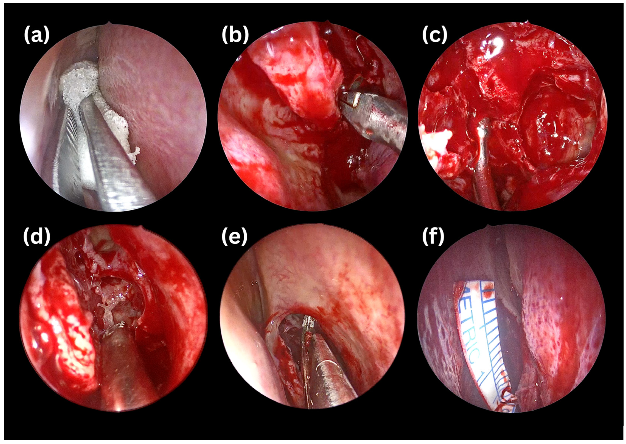
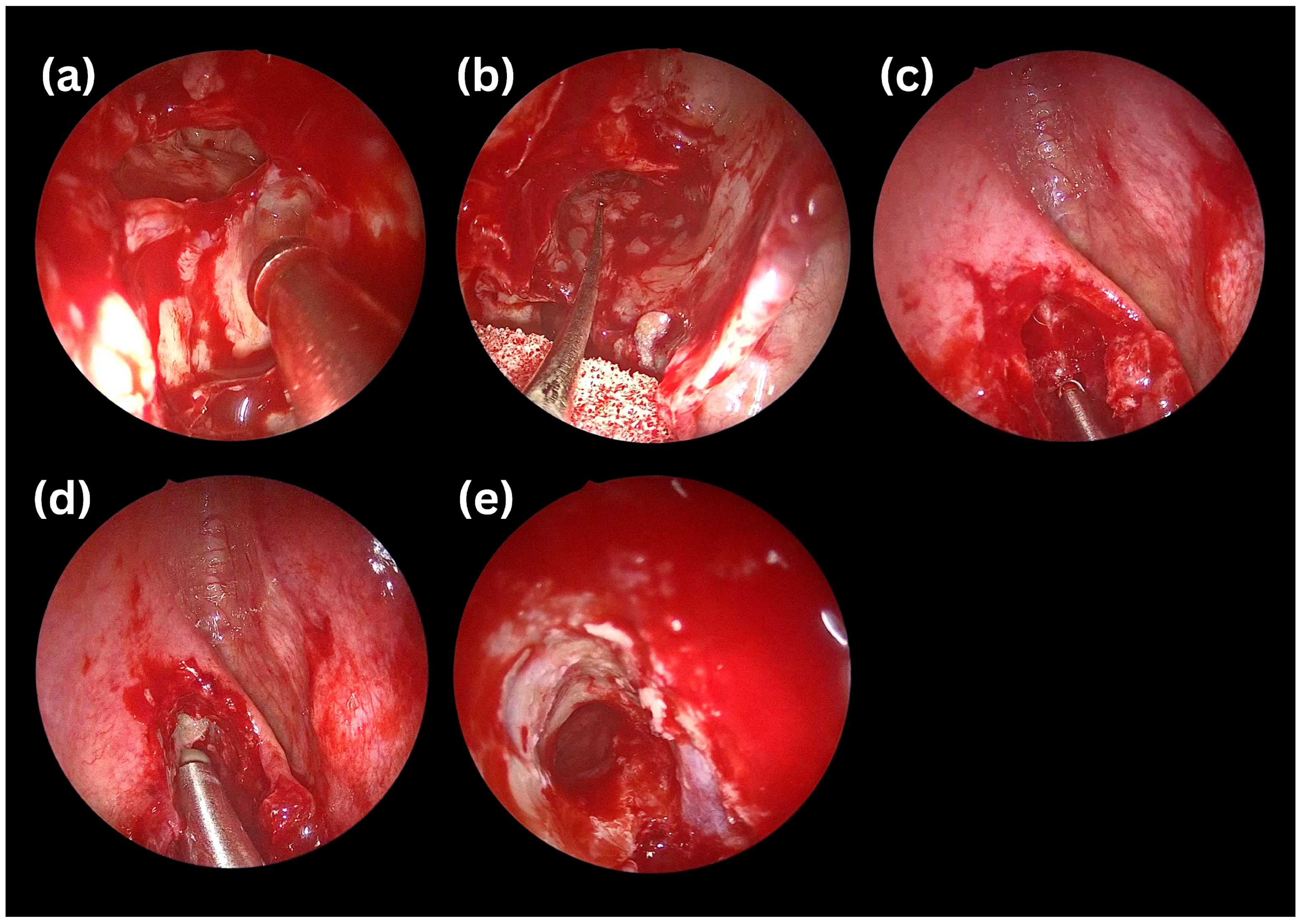
2.5. Surgical Intervention
2.6. Interobserver Agreement and Statistical Analysis
3. Results
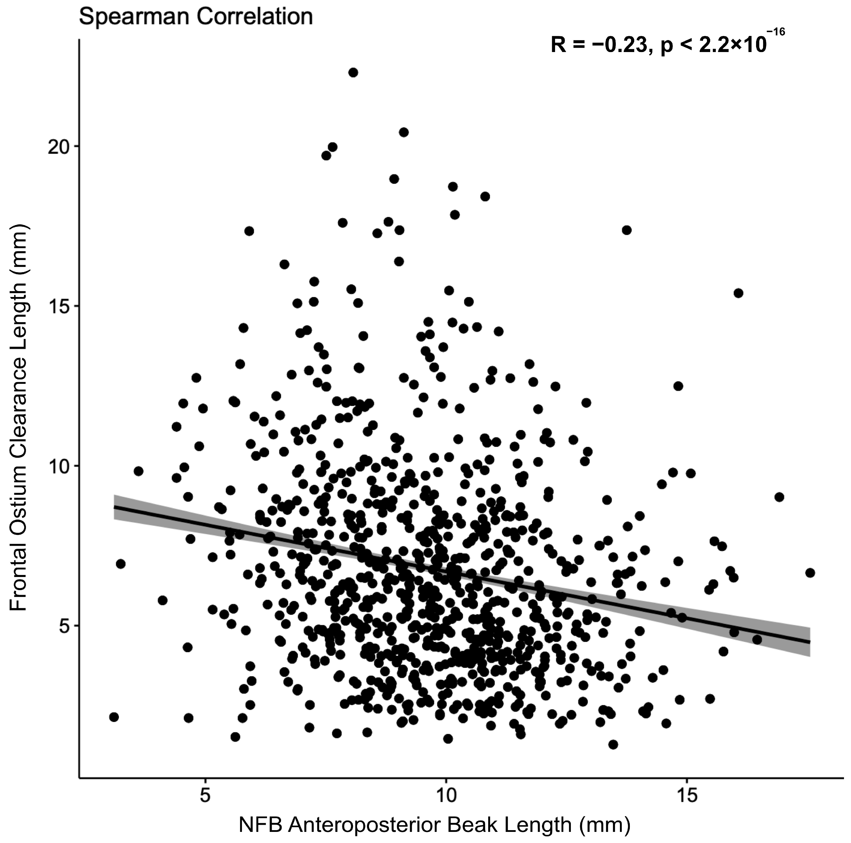
3.1. Findings from Phase 1
3.2. Findings from Phase 2
4. Discussion
5. Conclusions
Author Contributions
Funding
Institutional Review Board Statement
Informed Consent Statement
Data Availability Statement
Conflicts of Interest
Abbreviations
| CI | Confidence Interval |
| CSF | Cerebrospinal Fluid |
| CT | Computed Tomography |
| DICOM | Digital Imaging and Communications in Medicine |
| ESS | Endoscopic Sinus Surgery |
| FO | Frontal Ostium |
| FOC | Frontal Ostium Clearance |
| FOG | Frontal Ostium Grade |
| ICC | Intraclass Correlation Coefficient |
| IFAC | International Frontal Sinus Anatomy Classification |
| IQR | Interquartile Range |
| MPR | Multiplanar Reconstruction |
| NFB | Nasofrontal Beak |
References
- Marino, M.J.; McCoul, E.D. Frontal sinus surgery: The state of the art. Int. J. Head Neck Surg. 2016, 7, 5–12. [Google Scholar] [CrossRef] [Scilit]
- De Castro Dantas Braz, R.E.F.; Toro, M.D.C.; Sakuma, E.T.I.; Machado, V.S.B.; Sakano, E. International Frontal Sinus Anatomy Classification (IFAC): Evaluation of frontoethmoidal cells prevalence in a Brazilian population. Braz. J. Otorhinolaryngol. 2023, 89, 101309. [Google Scholar] [CrossRef] [Scilit] [PubMed]
- Figueroa, R.E.; Sullivan, J. Radiologic anatomy of the frontal sinus. In The Frontal Sinus; Springer: Berlin/Heidelberg, Germany, 2005; pp. 7–20. [Google Scholar] [CrossRef] [Scilit] [PubMed]
- Lobo, D.R.; Artiles, J.V.; Ospina, J.A. Atlas of Frontal Sinus Surgery; Springer: Cham, Switzerland, 2022. [Google Scholar] [CrossRef] [Scilit]
- Noller, M.; Fischer, J.L.; Gudis, D.A.; Riley, C.A. The Draf III procedure: A review of indications and techniques. World J. Otorhinolaryngol. Head Neck Surg. 2022, 8, 1–7. [Google Scholar] [CrossRef] [Scilit] [PubMed]
- Kumar, A.; Ademá-Alcover, J.M.; Alobid, I.; Amine, M.A.; Anand, V.K.; Battaglia, P.; Berens, A.M.; Bernal-Sprekelsen, M.; Campbell, R.G.; Capoccioni, G.M. Chapter 4 Frontal Sinus and Draf Approaches. In Endoscopic Approaches to the Paranasal Sinuses and Skull Base; Georg Thieme Verlag: Stuttgart, Germany, 2017. [Google Scholar] [CrossRef] [Scilit]
- Sansoni, E.R.; Sacks, R.; Harvey, R.J. Frontal Sinusotomy—DRAF III. In Atlas of Endoscopic Sinus and Skull Base Surgery; Elsevier: Amsterdam, The Netherlands, 2019; pp. 93–100.e1. [Google Scholar] [CrossRef] [Scilit]
- Weber, R.; Draf, W.; Kratzsch, B.; Hosemann, W.; Schaefer, S.D. Modern concepts of frontal sinus surgery. Laryngoscope 2001, 111, 137–146. [Google Scholar] [CrossRef] [Scilit] [PubMed]
- Moore, K.; Ross, A. Frontal sinus development and juvenile age estimation. Anat. Rec. 2017, 300, 1609–1617. [Google Scholar] [CrossRef] [Scilit] [PubMed]
- Lai, W.S.; Yang, P.L.; Lee, C.H.; Lin, Y.Y.; Chu, Y.H.; Wang, C.H.; Wang, H.; Shih, C.-P. The association of frontal recess anatomy and mucosal disease on the presence of chronic frontal sinusitis: A computed tomographic analysis. Rhinology 2014, 52, 208–214. [Google Scholar] [CrossRef] [Scilit] [PubMed]
- Tubbs, R.S.; Elton, S.; Salter, G.; Blount, J.P.; Grabb, P.A.; Oakes, W.J. Superficial surgical landmarks for the frontal sinus. J. Neurosurg. 2002, 96, 320–322. [Google Scholar] [CrossRef] [Scilit] [PubMed]
- Lieberman, S.M. Anatomical landmarks in revision sinus surgery and advanced nasal polyposis. Oper. Tech. Otolaryngol. Head Neck Surg. 2014, 25, 149–155. [Google Scholar] [CrossRef] [Scilit]
- Baudoin, T.; Gregurić, T.; Bacan, F.; Jelavić, B.; Geber, G.; Košec, A. A systematic review of common landmarks in navigated endoscopic sinus surgery (NESS). Comput. Assist. Surg. 2021, 26, 77–84. [Google Scholar] [CrossRef] [Scilit] [PubMed]
- Dassi, C.S.; Demarco, F.R.; Mangussi-Gomes, J.; Weber, R.; Balsalobre, L.; Stamm, A.C. The frontal sinus and frontal recess: Anatomical, radiological and surgical concepts. Int. Arch. Otorhinolaryngol. 2020, 24, e364–e375. [Google Scholar] [CrossRef] [Scilit] [PubMed]
- Gheriani, H.; Al-Salman, R.; Habib, A.R.; Javer, A. Frontal ostium grade (FOG): A new computer tomography grading system for endoscopic frontal sinus surgery. Otolaryngol. Head Neck Surg. 2020, 163, 611–617. [Google Scholar] [CrossRef] [Scilit] [PubMed]
- Makihara, S.; Kariya, S.; Okano, M.; Naito, T.; Uraguchi, K.; Matsumoto, J.; Noda, Y.; Nishizaki, K. The relationship between the width of the frontal recess and the frontal recess cells in Japanese patients. Clin. Med. Insights Ear Nose Throat 2019, 12, 1179550619884946. [Google Scholar] [CrossRef] [Scilit] [PubMed]
- Craig, J.R.; Petrov, D.; Khalili, S.; Brooks, S.G.; Lee, J.Y.K.; Adappa, N.D.; Palmer, J.N. The nasofrontal beak: A consistent landmark for superior septectomy during Draf III drill out. Am. J. Rhinol. Allergy 2016, 30, 230–234. [Google Scholar] [CrossRef] [Scilit] [PubMed]
- Park, S.S.; Yoon, B.N.; Cho, K.S.; Roh, H.J. Pneumatization pattern of the frontal recess: Relationship of the anterior-to-posterior length of frontal isthmus and/or frontal recess with the volume of agger nasi cell. Clin. Exp. Otorhinolaryngol. 2010, 3, 76. [Google Scholar] [CrossRef] [Scilit] [PubMed]
- Wormald, P.J. Surgery of the frontal recess and frontal sinus. Rhinology 2005, 43, 82–85. [Google Scholar] [PubMed]
- Burkart, C.M.; Zimmer, L.A. Endoscopic modified Lothrop procedure: A radiographic anatomic study. Laryngoscope 2010, 121, 442–445. [Google Scholar] [CrossRef] [Scilit] [PubMed]
- Jacobs, J.B.; Lebowitz, R.A.; Sorin, A.; Hariri, S.; Holliday, R. Preoperative sagittal CT evaluation of the frontal recess. Am. J. Rhinol. 2000, 14, 33–38. [Google Scholar] [CrossRef] [Scilit] [PubMed]
- Dubin, M.G.; Kuhn, F.A. Endoscopic modified Lothrop (Draf III) with frontal sinus punches. Laryngoscope 2005, 115, 1702–1703. [Google Scholar] [CrossRef] [Scilit] [PubMed]
- Nishiike, S.; Shibata, D.; Yoda, S.; Murata, J. Endoscopic modified Lothrop procedure with frontal sinus punch. Nihon Bika Gakkai Kaishi 2008, 47, 126–130. [Google Scholar] [CrossRef] [Scilit]
- Turri-Zanoni, M.; Battaglia, P.; Bignami, M.; Castelnuovo, P.; Arosio, A.D. Comprehensive access strategies to the frontal sinus. Curr. Opin. Otolaryngol. Head Neck Surg. 2023, 31, 57–64. [Google Scholar] [CrossRef] [Scilit] [PubMed]




| Nasofrontal Beak Anteroposterior Length | Frontal Ostium Clearance Length | ||
|---|---|---|---|
| Class A | ≤6 mm | Class A | ≥12 mm |
| Class B | 6–12 mm | Class B | 6–12 mm |
| Class C | ≥12 mm | Class C | ≤6 mm |
| Surgical Techniques (common pairs §) | |||
| Class A NFB, Class A FOC: The NFB was left unaltered; the operation continued based on pathology. | |||
| Class B NFB, Class B FOC: The posterior two-thirds of the NFB was shortened using a frontal sinus punch (70°, small). | |||
| Class C NFB, Class C FOC: A straight or curved (45°) drill was used to remove the posterior two-thirds and part of the anterior one-third of the NFB. | |||
| Variables | Phase 1—n (%) | Phase 2—n (%) | Total |
|---|---|---|---|
| Sex | |||
| Male | 828 (59.9%) | 57 (57.0%) | 885 (59.7%) |
| Female | 555 (40.1%) | 43 (43.0%) | 598 (40.3%) |
| Frontal Sinus Side | |||
| Right | 1305 (50.3%) | 98 (51.3%) | 1403 (50.4%) |
| Left | 1290 (49.7%) | 93 (48.7%) | 1383 (49.6%) |
| NFB Length Classes | |||
| Class A (≤6 mm) | 123 (4.7%) | 10 (5.2%) | 133 (4.8%) |
| Class B (6–12 mm) | 2100 (80.9%) | 146 (76.4%) | 2246 (80.6%) |
| Class C (≥12 mm) | 372 (14.3%) | 35 (18.4%) | 407 (14.6%) |
| FOC Length Classes | |||
| Class A (≥12 mm) | 198 (7.6%) | 23 (12.0%) | 221 (7.9%) |
| Class B (6–12 mm) | 1182 (45.5%) | 68 (35.6%) | 1250 (44.9%) |
| Class C (≤6 mm) | 1215 (46.8%) | 100 (52.4%) | 1315 (47.2%) |
| Male | Female | p-Value | ||
|---|---|---|---|---|
| NFB Classification | Class A | 63 (51.2%) | 60 (48.8%) | <0.001 |
| Class B | 1222 (58.2%) | 878 (41.8%) | ||
| Class C | 273 (73.4%) | 99 (26.6%) | ||
| FOC Classification | Class A | 100 (50.5%) | 98 (49.5%) | 0.017 |
| Class B | 717 (60.7%) | 465 (39.3%) | ||
| Class C | 741 (61.0%) | 474 (39.0%) |
| Surgical Class A | Surgical Class B | p-Value | ||
|---|---|---|---|---|
| CT Classification | Class A | 10 (100%) | 0 (0%) | <0.001 |
| Class B | 49 (33.6%) | 97 (66.4%) | ||
| Class C | 0 (0.0%) | 12 (34.3%) * |
| Surgical Class A | Surgical Class B | p-Value | ||
|---|---|---|---|---|
| CT Classification | Class A | 85 (85%) | 15 (15%) | <0.001 |
| Class B | 39 (57.4%) | 25 (36.8%) | ||
| Class C | 3 (13.0%) | 13 (56.5%) |
| CT Class | Sensitivity | Specificity | PPV | NPV | n (CT) |
|---|---|---|---|---|---|
| Class A (≤6 mm) | 100.0% | 72.9% | 16.9% | 100.0% | 10 |
| Class B (6–12 mm) | 66.4% | 73.3% | 89.0% | 40.2% | 146 |
| Class C (≥12 mm) | 65.7% | 100.0% | 100.0% | 92.9% | 35 |
| CT Class | Sensitivity | Specificity | PPV | NPV | n (CT) |
|---|---|---|---|---|---|
| Class A (≥12 mm) | 30.4% | 97.6% | 63.6% | 91.1% | 23 |
| Class B (6–12 mm) | 36.8% | 77.2% | 47.2% | 68.8% | 68 |
| Class C (≤6 mm) | 85.0% | 53.8% | 66.9% | 76.6% | 100 |
Disclaimer/Publisher’s Note: The statements, opinions and data contained in all publications are solely those of the individual author(s) and contributor(s) and not of MDPI and/or the editor(s). MDPI and/or the editor(s) disclaim responsibility for any injury to people or property resulting from any ideas, methods, instructions or products referred to in the content. |
© 2026 by the authors. Licensee MDPI, Basel, Switzerland. This article is an open access article distributed under the terms and conditions of the Creative Commons Attribution (CC BY) license.
Share and Cite
Omer, G.L.; Ali, S.S.; Turri-Zanoni, M.; Battaglia, P.; Dallan, I.; Gravina, A.; De Donato, G.; Castelnuovo, P.; Casiano, R.R.; Di Girolamo, S. From Imaging to Instrument: A CT-Based Classification of the Nasofrontal Beak and Frontal Ostium Clearance with Prospective Surgical Correlation. Surgeries 2026, 7, 86. https://doi.org/10.3390/surgeries7030086
Omer GL, Ali SS, Turri-Zanoni M, Battaglia P, Dallan I, Gravina A, De Donato G, Castelnuovo P, Casiano RR, Di Girolamo S. From Imaging to Instrument: A CT-Based Classification of the Nasofrontal Beak and Frontal Ostium Clearance with Prospective Surgical Correlation. Surgeries. 2026; 7(3):86. https://doi.org/10.3390/surgeries7030086
Chicago/Turabian StyleOmer, Goran Latif, Sahand Soran Ali, Mario Turri-Zanoni, Paolo Battaglia, Iacopo Dallan, Andrea Gravina, Giuseppe De Donato, Paolo Castelnuovo, Roy R. Casiano, and Stefano Di Girolamo. 2026. "From Imaging to Instrument: A CT-Based Classification of the Nasofrontal Beak and Frontal Ostium Clearance with Prospective Surgical Correlation" Surgeries 7, no. 3: 86. https://doi.org/10.3390/surgeries7030086
APA StyleOmer, G. L., Ali, S. S., Turri-Zanoni, M., Battaglia, P., Dallan, I., Gravina, A., De Donato, G., Castelnuovo, P., Casiano, R. R., & Di Girolamo, S. (2026). From Imaging to Instrument: A CT-Based Classification of the Nasofrontal Beak and Frontal Ostium Clearance with Prospective Surgical Correlation. Surgeries, 7(3), 86. https://doi.org/10.3390/surgeries7030086