Article

A New Mini-Open Technique for the Treatment of Chronic Distal Radioulnar Joint Instability: Surgical Technique and Early Outcomes

1
Departmental Faculty of Medicine and Surgery, Unicamillus-Saint Camillus International University of Health Sciences, Via di Sant’Alessandro 8, 00131 Rome, Italy
2
Department of Orthopaedics and Traumatology, Ospedale San Martino—Belluno, ULSS 1 Dolomiti, Viale Europa 22, 32100 Belluno, Italy
3
Department of Orthopaedics and Traumatology, Fondazione Policlinico Universitario A. Gemelli IRCCS, Largo Agostino Gemelli 8, 00168 Rome, Italy
*
Author to whom correspondence should be addressed.
Surgeries 2026, 7(3), 87; https://doi.org/10.3390/surgeries7030087
Submission received: 24 May 2026 / Revised: 29 June 2026 / Accepted: 10 July 2026 / Published: 15 July 2026
(This article belongs to the Section Hand Surgery and Research)

Abstract

Background: Chronic distal radioulnar joint (DRUJ) instability secondary to triangular fibrocartilage complex (TFCC) injury remains challenging to treat. This study describes a novel mini-open dorsal technique for TFCC reconstruction and reports preliminary clinical outcomes. Methods: Six patients with chronic symptomatic DRUJ instability underwent mini-open dorsal TFCC reconstruction using a dorsal capsular flap fixed to the ulnar fovea with a 2 mm suture anchor. Clinical outcomes included DRUJ stability, forearm range of motion, pain (VAS), QuickDASH, and PRWHE scores. Results: At final follow-up, clinical DRUJ stability was restored in all patients, with full forearm rotation. Mean QuickDASH improved from 61.0 to 6.5, mean PRWHE improved from 58.7 to 8.0, and mean VAS pain score improved from 7.5 to 0.83. No complications or recurrences were observed. Conclusions: This mini-open dorsal technique was feasible in this small case series and was associated with encouraging early clinical outcomes and restoration of clinical DRUJ stability. Larger prospective studies with longer follow-up are required before definitive conclusions can be drawn.

1. Introduction

The distal radioulnar joint (DRUJ) plays a pivotal role in wrist biomechanics, acting as the primary movement that enables forearm pronation and supination through rotation of the radius around a relatively fixed ulna [1]. Owing to its limited osseous congruence, DRUJ stability relies predominantly on soft-tissue restraints [2,3], particularly the triangular fibrocartilage complex (TFCC) and its deep foveal attachments [4,5]. Disruption of these stabilizing structures—whether due to acute trauma, degenerative changes, or alterations in ulnar variance—leads to abnormal radioulnar kinematics, increased joint translation, and ulnocarpal overload, ultimately resulting in pain, weakness, and functional impairment [1,6]. Acute DRUJ instability most commonly follows traumatic TFCC injuries and distal radius fractures [7,8,9,10], whereas chronic instability typically develops when these lesions are unrecognized or inadequately treated, allowing persistent abnormal motion and progressive dysfunction [6,11]. Because restoration of the deep foveal attachment of the TFCC is essential for re-establishing normal DRUJ mechanics, surgical management aims to anatomically reconstruct or substitute the deficient stabilizing structures [12]. While arthroscopic and open techniques remain established options for foveal TFCC repair [13,14], each presents inherent limitations. Arthroscopic procedures preserve soft tissues but require specialized equipment and advanced surgical expertise, whereas conventional open techniques provide direct visualization of the foveal insertion at the expense of greater soft-tissue dissection [15,16]. More recently, hybrid and minimally invasive techniques have been proposed to combine the advantages of both approaches, including arthroscopy-assisted repair with dorsal capsular imbrication. Building on the same concept of preserving soft tissues while restoring foveal stability, we developed a mini-open dorsal technique that provides direct access to the ulnar fovea through a limited exposure without the need for wrist arthroscopy. The aim of this Technical Note is to describe the surgical technique and report our preliminary clinical experience in a small consecutive case series.

2. Materials and Methods

2.1. Study Design and Patient Selection

A retrospective consecutive case series was performed at a single institution. All six patients underwent the same mini-open dorsal procedure for chronic distal radioulnar joint (DRUJ) instability performed by the senior author. Inclusion criteria were: clinical diagnosis of symptomatic DRUJ instability with ulnar-sided wrist pain and functional limitation; clinical evidence of DRUJ instability on physical examination (ballottement test), recorded preoperatively and postoperatively as a stability grade. Exclusion criteria were: prior ipsilateral wrist surgery; concomitant fractures or major carpal ligament injuries requiring separate reconstruction; advanced DRUJ arthritis requiring salvage procedures; revision TFCC/DRUJ stabilization procedures. All patients underwent standardized clinical examination including DRUJ ballottement testing and assessment of forearm rotation. Imaging work-up followed an institutional protocol consistent with expert consensus: conventional radiographs as first-line; MRI for TFCC evaluation; and (when required) MR arthrography/CT arthrography specifically to assess suspected foveal (Palmer 1B) lesions [17].

2.2. Surgical Technique

2.2.1. Patient Positioning and Preparation

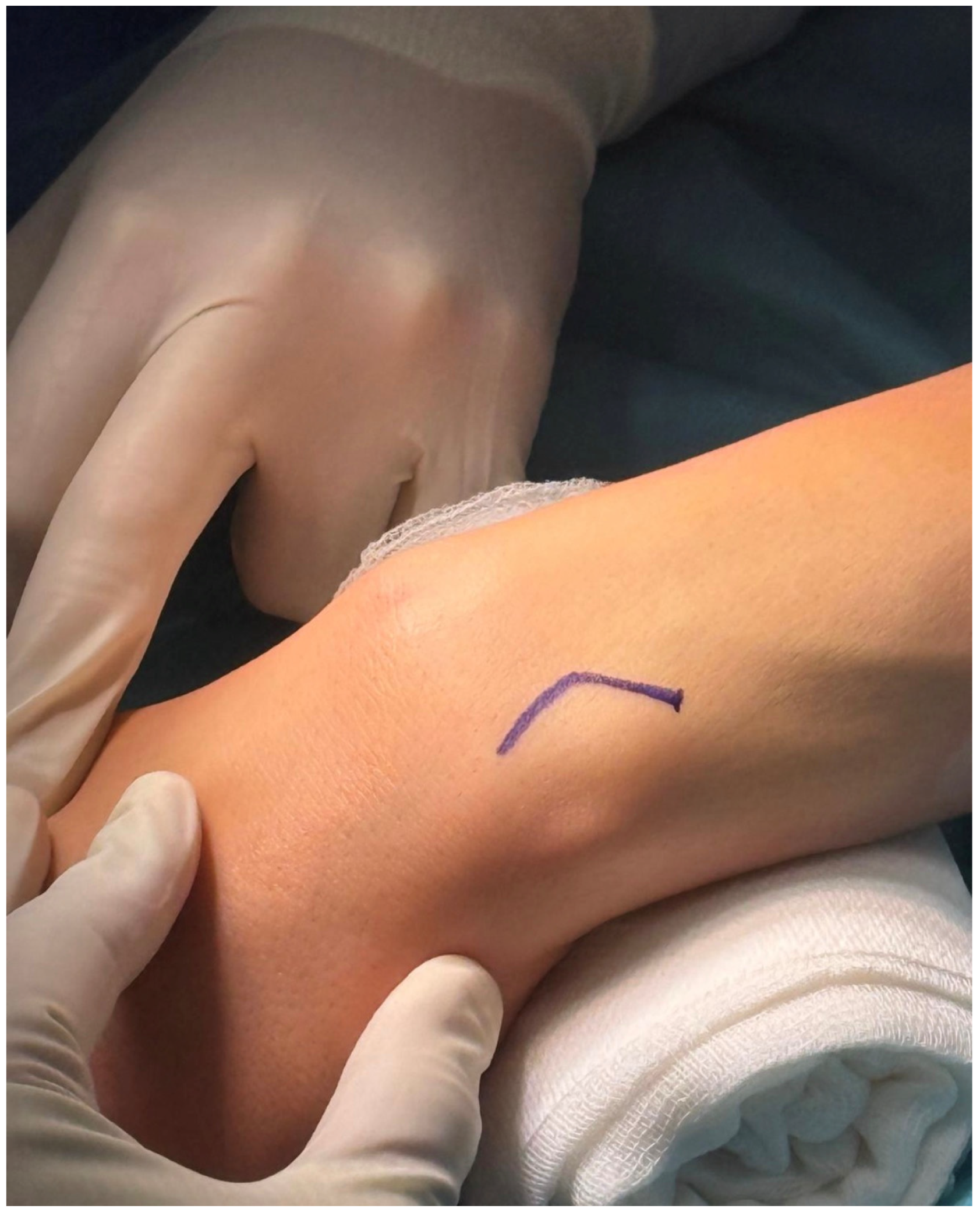
The patient is placed supine with the affected upper limb positioned on a hand table. The wrist is supported on a small sterile roll with the dorsal aspect facing upward (Figure 1). Care is taken to protect all bony prominences. Prophylactic antibiotics are administered prior to incision. After standard skin preparation and sterile draping, a pneumatic tourniquet is applied at the proximal arm to provide a bloodless field.

2.2.2. Surgical Approach

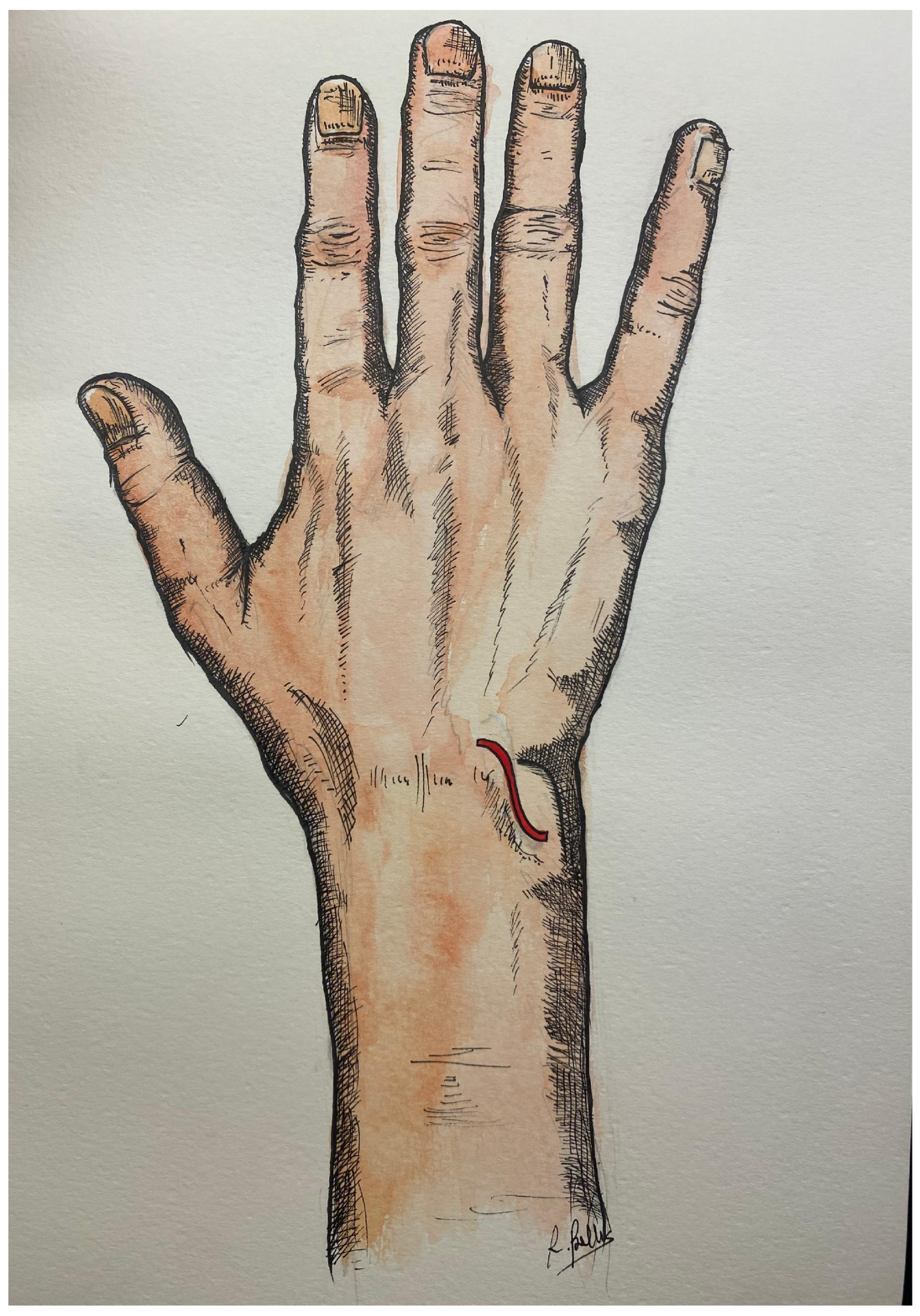
A 3 cm bayonet-shaped incision is performed over the distal radioulnar joint (DRUJ), parallel to the longitudinal axis of the ulna, centered between the dorsal ulnar border and the wrist joint line (Figure 2).
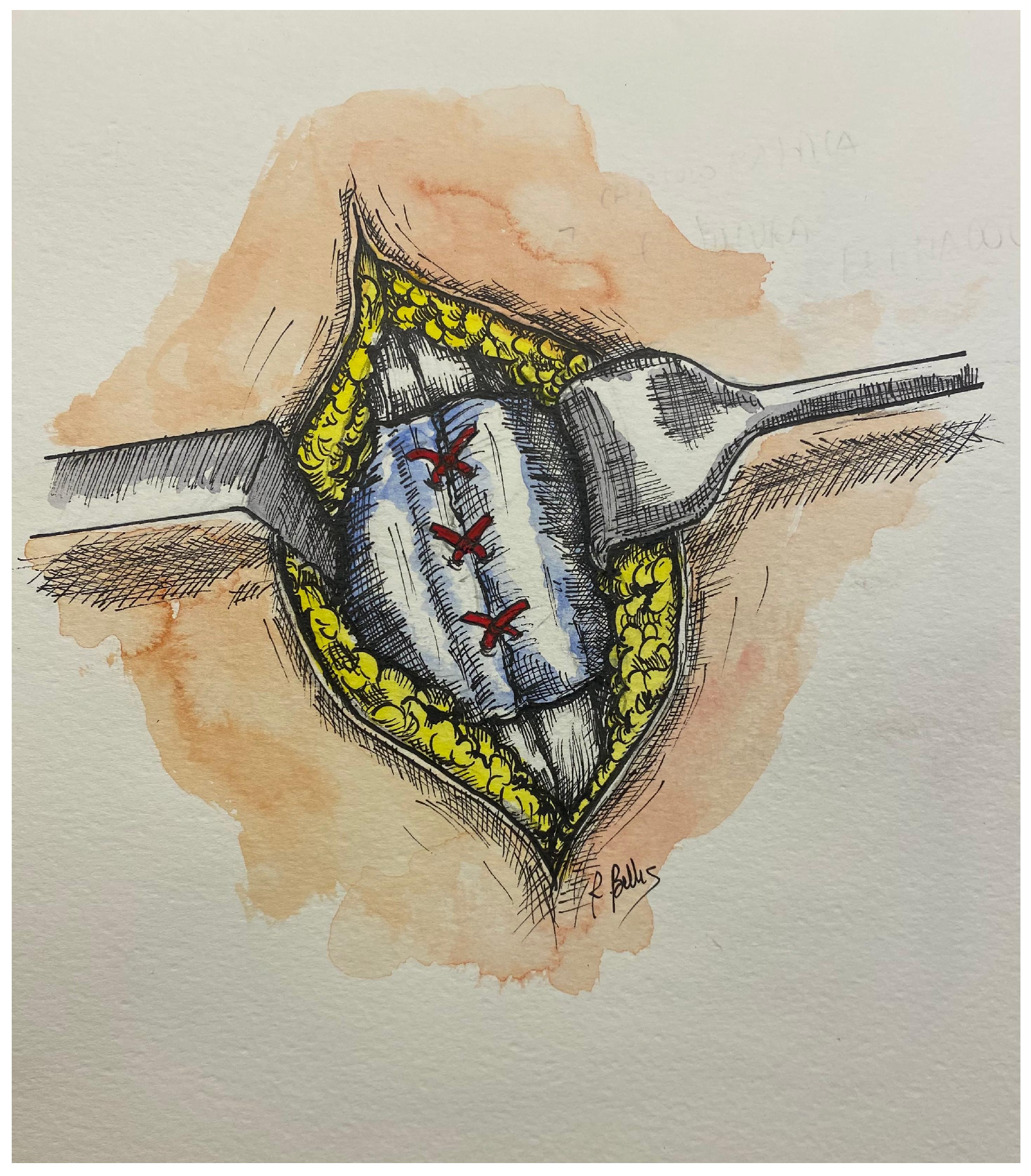
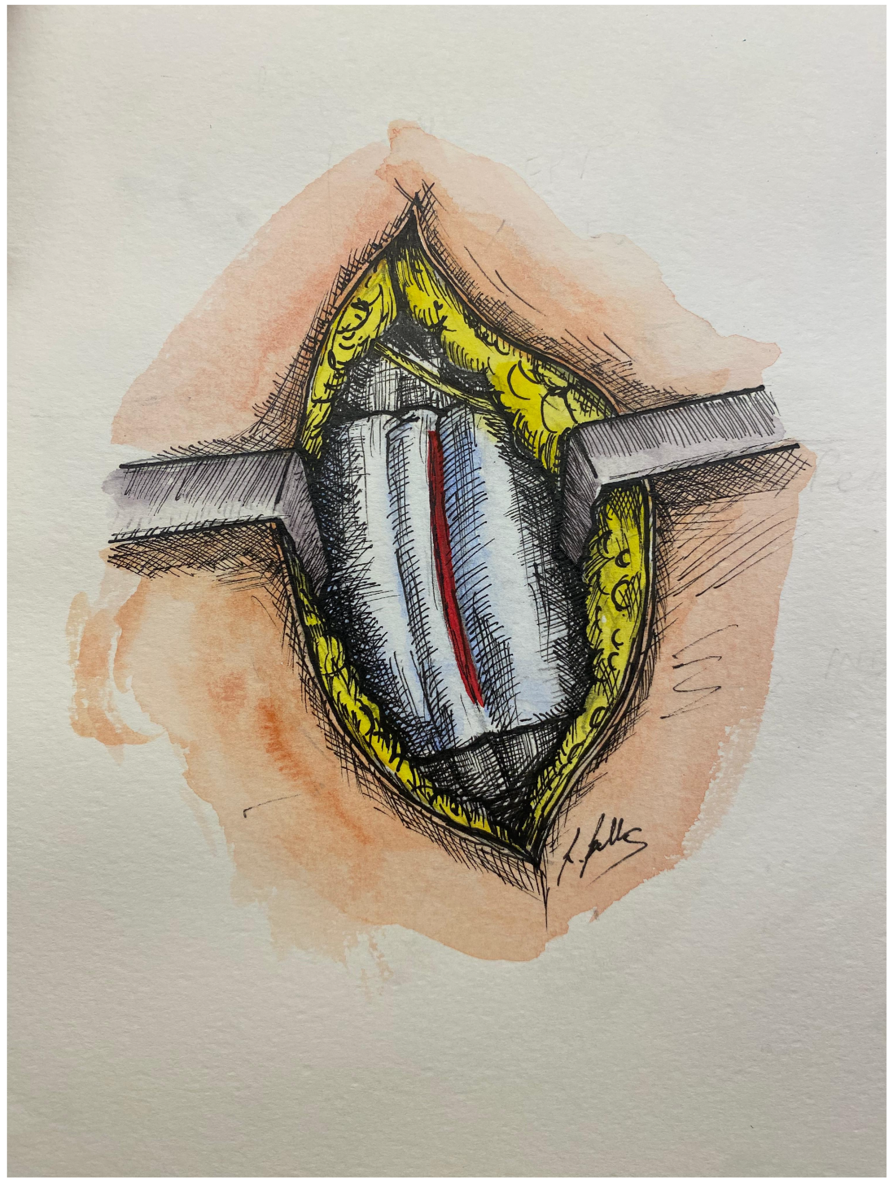
Following skin incision, careful dissection is carried through the subcutaneous tissue with identification and protection of the dorsal sensory branches of the ulnar nerve and the dorsal ulnar vein [11]. Meticulous hemostasis is achieved. The fascia is incised, and the extensor retinaculum is exposed and divided longitudinally (Figure 3).
The extensor digiti minimi (EDM) compartment is identified and gently retracted radially [18]. The dorsal capsule of the DRUJ is then exposed and incised longitudinally to access the joint.

2.2.3. Intra-Articular Exposure and Assessment

Direct visualization of the triangular fibrocartilage complex (TFCC) and distal radioulnar ligaments is obtained [1]. The lesion typically involves the foveal insertion of the deep radioulnar ligament complex.
The ulnar fovea is clearly identified as the key anatomical landmark [4,5], together with the base of the ulnar styloid and the dorsal and palmar radioulnar ligaments. Care is taken to preserve surrounding neurovascular structures and the EDM tendon.
This exposure allows direct assessment of the TFCC lesion, particularly the integrity of the foveal insertion, which is essential for DRUJ stability [12].

2.2.4. Reconstruction and Stabilization

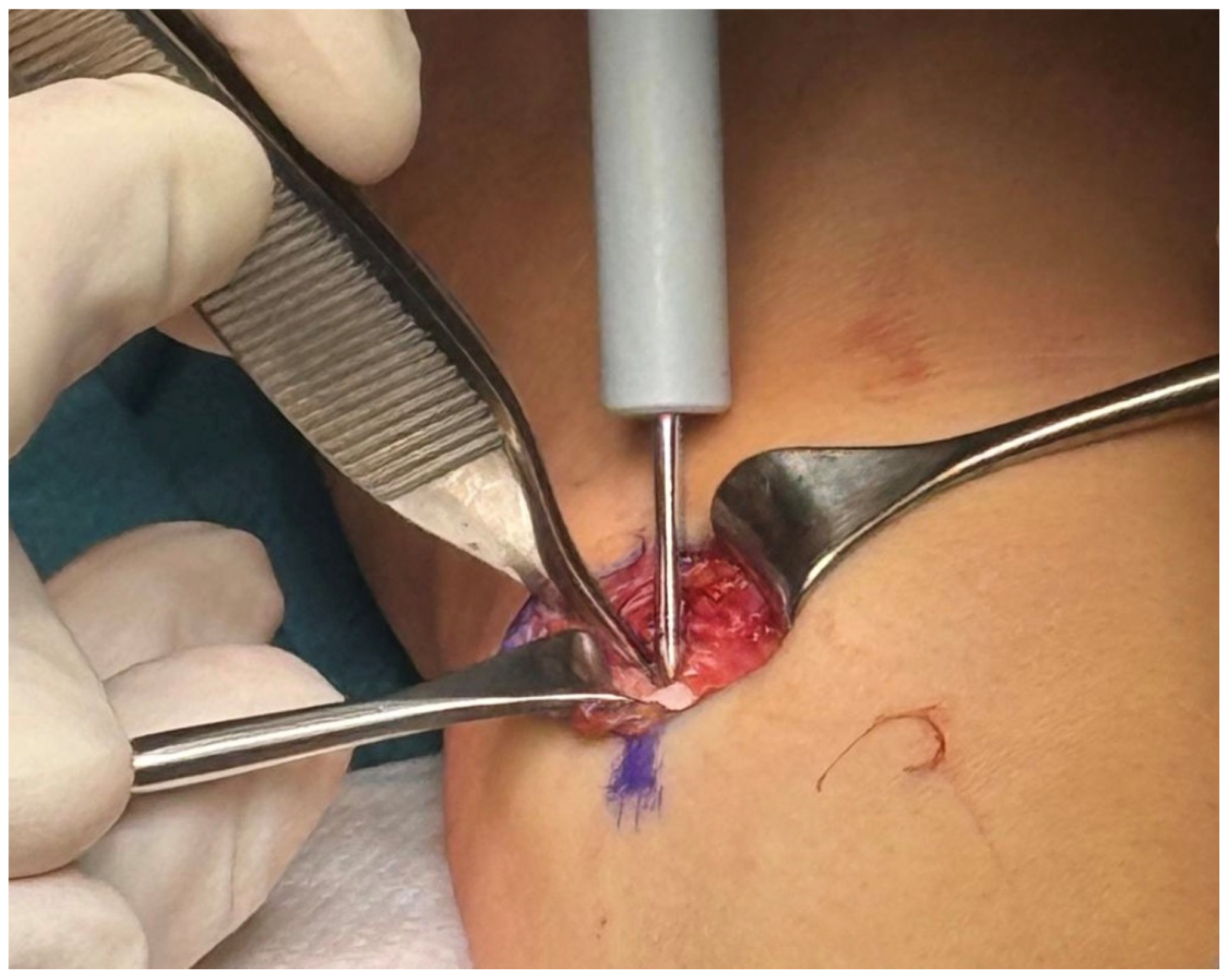
Following reduction in the DRUJ performed with the forearm in neutral rotation, the ulnar fovea is directly identified and prepared [19,20,21] using a handheld awl before insertion of the 2 mm Mikai-Medartis® (Medartis AG, Basel, Switzerland) suture anchor loaded with FiberWire® (Arthrex Inc., Naples, FL, USA) (Figure 4).
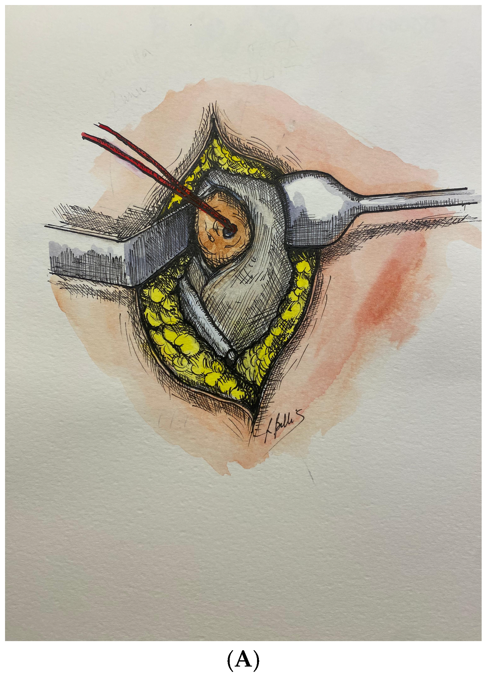
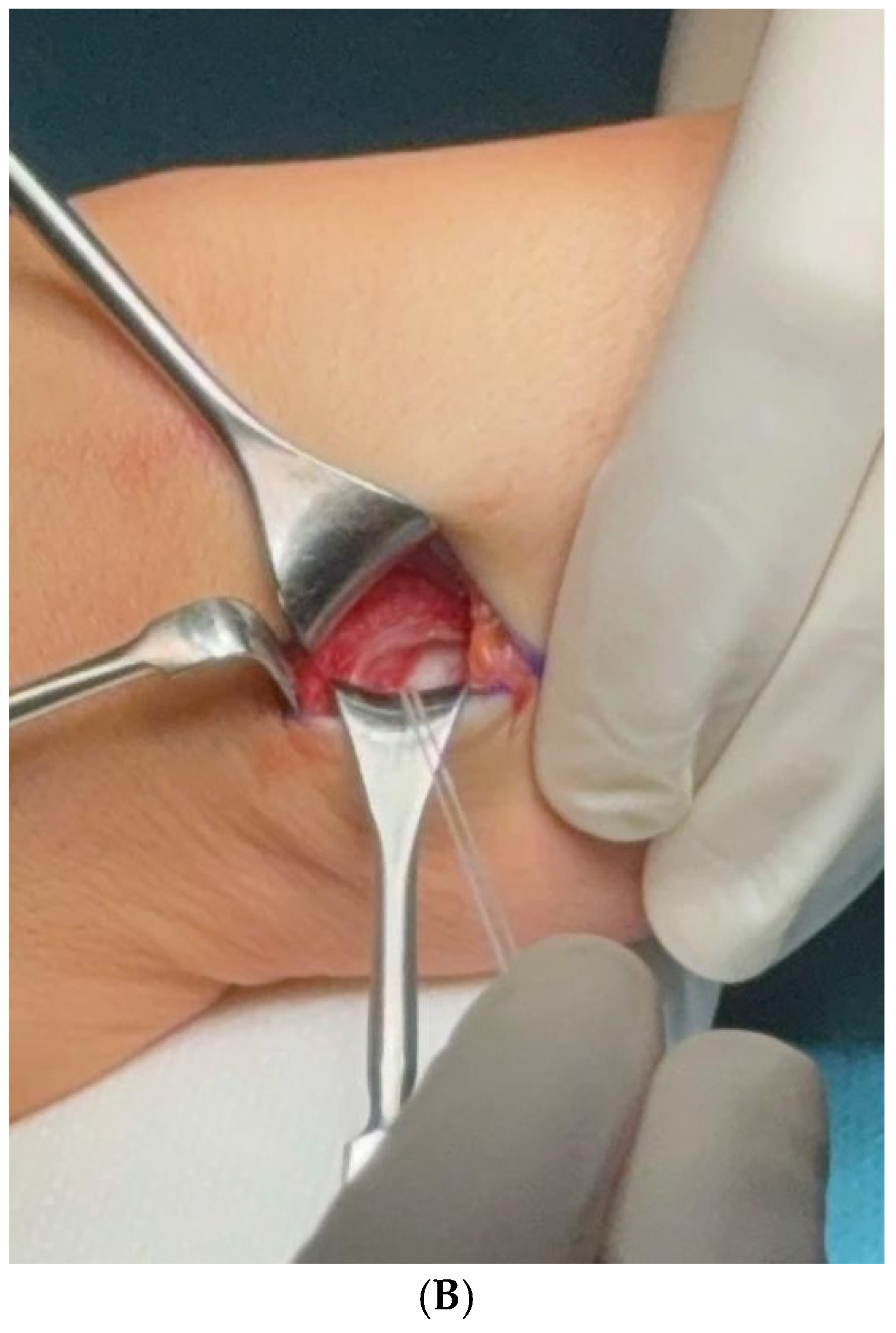
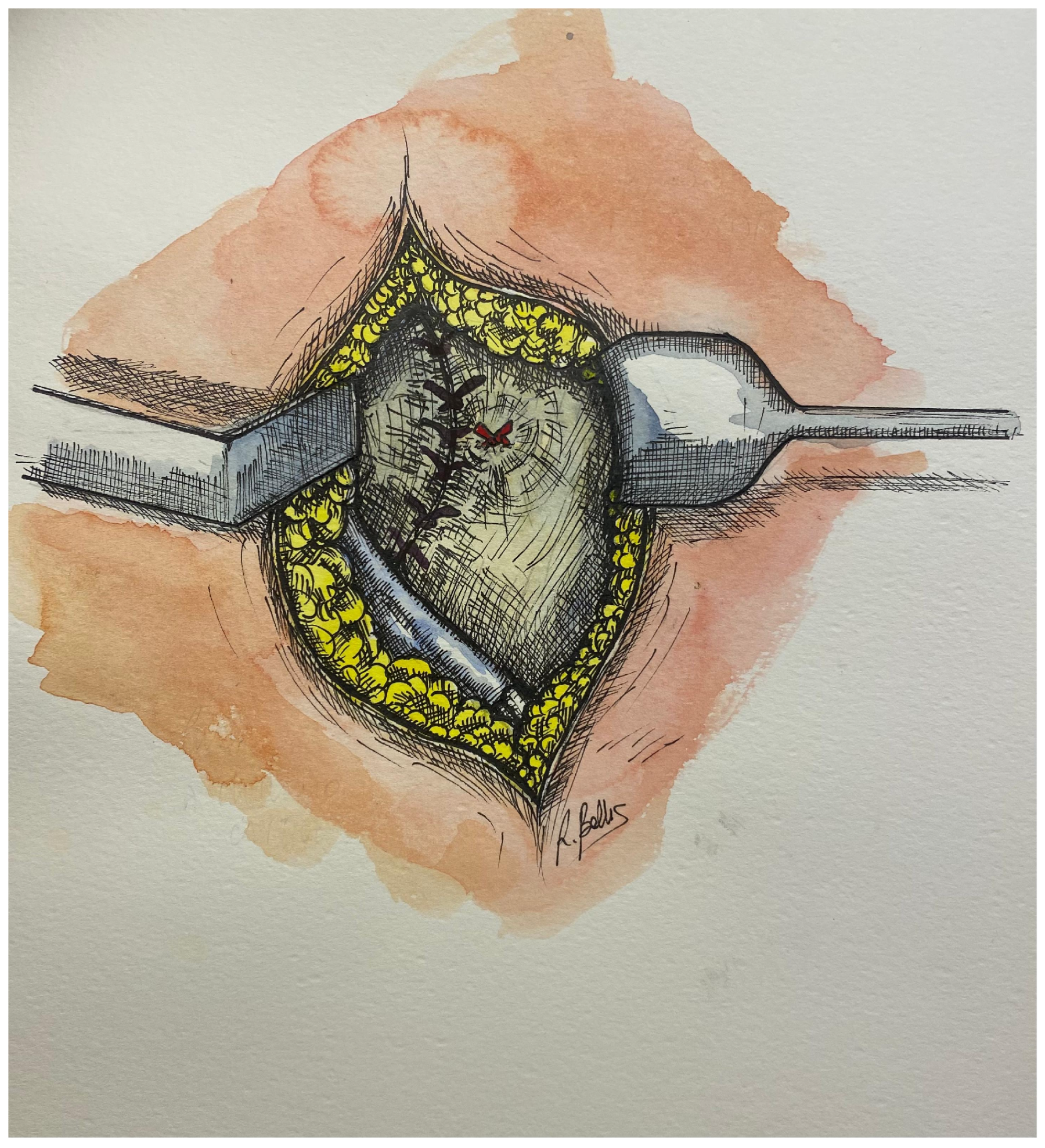
A radially based dorsal capsular flap is fashioned by elevating the dorsal capsule from its ulnar attachment while preserving its radial base. It is then advanced and rotated approximately 30° over the ulnar head and then tensioned before being secured to the anchor (Figure 5A,B).
Capsular retensioning is completed with interrupted 3-0 Vicryl sutures, resulting in a stable reconstruction of the dorsal soft-tissue restraints of the distal radioulnar joint (Figure 6).
Following flap advancement and fixation, the final reconstruction restores soft-tissue continuity over the foveal region and stabilizes the DRUJ. Intraoperative stability was assessed by forearm pronation–supination and DRUJ ballottement testing [11]. Adequate reduction and restoration of stability are confirmed prior to closure.

2.2.5. Closure

Copious irrigation is performed, and hemostasis is verified. Capsuloplasty is completed with interrupted sutures. The extensor retinaculum is repaired with care to avoid excessive tension (Figure 7).
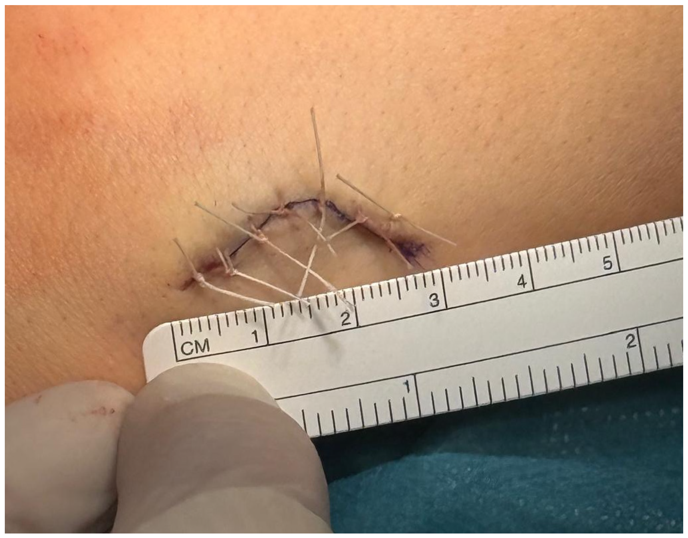
Layered closure of the subcutaneous tissue and skin is performed using absorbable sutures. Final wound closure demonstrates the limited extent of the mini-open dorsal approach (Figure 8).
A sterile dressing is applied, followed by immobilization with a single sugar tong splint to prevent forearm rotation during the initial healing phase.

2.2.6. Postoperative Rehabilitation

Postoperatively, the wrist and forearm are immobilized in a sugar-tong splint with the forearm maintained in a neutral position for three weeks to protect the repair and prevent forearm rotation. After splint removal, patients undergo a supervised rehabilitation program consisting of three physiotherapy sessions per week for one month. Rehabilitation focuses on active-assisted and active range-of-motion exercises to restore wrist flexion-extension and forearm pronation-supination. Adjunctive ultrasound therapy is also administered according to the institutional rehabilitation protocol. Return to activities of daily living and light occupational activities is generally allowed shortly after surgery according to patient tolerance, while unrestricted sports activities are deferred for at least four months.

2.3. Outcome Measures

Clinical outcomes were assessed at the final follow-up through physical examination and patient-reported outcome measures. DRUJ stability was evaluated clinically using the ballottement test and assessment of forearm pronation–supination. Functional outcome was assessed using the QuickDASH questionnaire and the Patient-Rated Wrist/Hand Evaluation (PRWHE). Pain intensity was recorded using a 10-point visual analogue scale (VAS). Range of motion, postoperative complications, recurrent instability, and the need for revision surgery were also documented.

2.4. Statistical Analysis

Given the descriptive nature of this retrospective case series and the limited sample size, data were summarized descriptively using mean (range) or median (range), as appropriate. No comparative statistical analysis was performed.

3. Results

Six consecutive patients (4 men, 2 women) underwent mini-open dorsal TFCC reconstruction for chronic DRUJ instability. Mean age at surgery was 36.7 years (range, 27–47 years), and median follow-up was 13 months (range, 12–22 months). Symptom onset was traumatic in four patients (66.7%) and overuse-related in two (33.3%). All patients presented with ulnar-sided wrist pain, while four (66.7%) also reported functional limitation. At final follow-up, all patients demonstrated restoration of clinical DRUJ stability and full forearm pronation–supination. No complications, reoperations, or recurrent instability were observed. Functional outcomes improved substantially following surgery. Mean QuickDASH improved from 61.0 preoperatively to 6.5 at final follow-up. Mean PRWHE improved from 58.7 (range, 45–70) preoperatively to 8.0, while mean VAS pain score improved from 7.5 (range, 6–9) preoperatively to 0.83. Four patients reported VAS ≤ 1 at final follow-up. Although minor inter-patient variability in PROMs was observed, clinical outcomes were consistently favorable across the cohort, with restored stability, preserved range of motion, and low residual symptom burden.

4. Discussion

This case series suggests that a mini-open dorsal approach with controlled foveal exposure and suture-anchor fixation of a dorsal capsular flap can provide consistent clinical stabilization of chronic DRUJ instability while preserving forearm rotation. In this small consecutive series, restoration of clinical DRUJ stability and full forearm rotation was observed in all treated patients at short-term follow-up. In chronic DRUJ instability-where tissue quality may be compromised and reliable restoration of the foveal attachment can be technically demanding –these results support the feasibility of a limited dorsal exposure that enables direct control of the foveal target while aiming to reduce the morbidity typically associated with more extensive open approaches. Recent comparative clinical data indicate that outcomes and failure rates after arthroscopic versus open TFCC foveal repair can be comparable [22], supporting the concept that durable results may depend more on restoration of the foveal stabilizers than on the access route itself. Similarly, Yeh et al. reported favorable mid-term outcomes using an arthroscopy-assisted repair combined with dorsal capsular imbrication [23]. Unlike that technique, our approach does not require wrist arthroscopy and instead relies on a limited dorsal exposure to achieve direct visualization of the foveal insertion while minimizing soft-tissue dissection. In that context, our mini-open technique is positioned as a hybrid strategy: it prioritizes direct control of the foveal target while limiting the exposure typically required in traditional open reconstructions. Although no direct comparison can be made from the present case series, the early clinical outcomes observed are broadly consistent with those reported after both open and arthroscopic foveal repair techniques. The principal advantage of the proposed technique lies not in demonstrating superior outcomes but in providing a simplified hybrid approach that combines direct foveal visualization with limited soft-tissue dissection.
Although the proposed mini-open technique is technically less demanding than arthroscopic repair, a learning curve is still expected, particularly during the identification of the ulnar fovea and the preparation and tensioning of the dorsal capsular flap. In addition, the choice of surgical technique should be individualized according to the pattern and chronicity of the TFCC lesion. Arthroscopic techniques may be particularly advantageous for selected acute peripheral tears and may facilitate earlier functional recovery, whereas mini-open or open foveal repair may be preferable in chronic foveal avulsions associated with DRUJ instability requiring direct visualization and secure foveal fixation. Mid-long- term outcome data following arthroscopically assisted transosseous TFCC refixation show good-to excellent clinical results despite some loss of DRUJ stability over time [24], emphasizing the importance of longer follow-up and standardized stability assessment even when PROMs are favorable. This consideration applies equally to the present technique and supports the need for continued surveillance beyond the short-to-mid-term window. A distinguishing feature of the described procedure is the use of a dorsal capsular flap secured with a suture anchor at the foveal region. An RCT evaluating combined repair strategies for TFCC tears with DRUJ instability reported improved grip strength when a capsular repair/augmentation component was added to transosseous repair [25], suggesting a potential functional advantage of addressing capsular restraint in addition to the primary TFCC fixation. Editorial commentary has similarly framed combined transosseous and capsular repair as a step toward more complete joint stabilization [26]. Although objective grip strength was not assessed, the low disability scores and preserved forearm rotation observed in this series are compatible with the rationale for incorporating capsular augmentation. Although these observations are encouraging, they should be interpreted cautiously given the limited sample size and the descriptive nature of this study. Suture- anchor fixation is widely used in foveal reattachment and has been directly compared with transosseous constructs in arthroscopic-assisted settings [19]. Additional comparative clinical work has evaluated anchor repair against capsular suture variants with minimum 2-year follow-up 6, and a recent systematic review summarized the effectiveness of anchor-based and transosseous suture techniques in arthroscopic foveal repair [27]. In the present study, the principal technical contribution is less the anchor itself than its integration into a reproducible mini-open exposure and dorsal capsular flap construct aimed at stabilizing the DRUJ while limiting soft-tissue disruption. Recurrent instability symptoms after TFCC repair can relate to patient-specific factors such as generalized laxity [28]. A retrospective series of 120 patients found a higher recurrence rate in those with generalized ligamentous laxity (17.7%) compared with those without (5.8%) [29]. Although no recurrences occurred in our small cohort, future studies of this technique should systematically capture laxity metrics (e.g., Beighton score) to better define indications and recurrence risk [28,29]. The present study has several limitations that should be considered when interpreting the findings. First, this is a retrospective case series including a small cohort of six patients with a relatively short follow-up. Second, no control group was available for comparison. Third, DRUJ stability was assessed clinically using standard physical examination, without adjunctive objective or imaging-based measurements, and grip strength was not routinely recorded. Nevertheless, the study was primarily conceived as a Technical Note aimed at describing a reproducible surgical technique, with the clinical outcomes serving as an initial assessment of its feasibility and early performance. Therefore, the encouraging results observed in this preliminary experience should be interpreted with appropriate caution. Future prospective studies with larger cohorts, longer follow-up, and standardized objective and imaging-based outcome measures will be important to further evaluate the reproducibility and durability of this technique.

5. Conclusions

This Technical Note describes a mini-open approach that provides controlled foveal exposure through limited dorsal access. In this preliminary experience, the technique proved feasible and was associated with encouraging early clinical outcomes. Further prospective studies are needed to confirm its reproducibility, durability, and long-term clinical effectiveness.

Author Contributions

Conceptualization, M.G. and R.D.V.; methodology, C.M.; validation, R.B.; formal analysis, R.B.; investigation, C.M.; data curation, A.P.; writing—original draft preparation, C.M.; writing—review and editing, A.P.; visualization, R.B.; supervision, M.G. All authors have read and agreed to the published version of the manuscript.

Funding

This research received no external funding.

Institutional Review Board Statement

This study was conducted in accordance with the principles of the Declaration of Helsinki. Formal ethical approval was not required because this retrospective study involved exclusively the analysis of fully anonymized clinical data collected during routine clinical practice, without any additional interventions or procedures performed for research purposes and without the processing of identifiable personal data. The study was conducted in accordance with Regulation (EU) 2016/679 (General Data Protection Regulation, GDPR) and the Italian Personal Data Protection Code (Legislative Decree No. 196/2003, as amended by Legislative Decree No. 101/2018), governing the processing of anonymized personal data for scientific research.

Informed Consent Statement

Written informed consent for the surgical procedure was obtained from all subjects involved in the study as part of routine clinical care. Due to the retrospective nature of the study, the use of fully anonymized clinical data, and the absence of identifiable patient information, additional informed consent for study participation was not required, in accordance with Article 110 of the Italian Privacy Code (Legislative Decree No. 196/2003, as amended) and Regulation (EU) 2016/679 (GDPR).

Data Availability Statement

The data presented in this study are available on request from the corresponding author due to privacy and ethical restrictions.

Conflicts of Interest

The authors declare no conflicts of interest.

References

  1. Flores, D.V.; Umpire, D.F.; Rakhra, K.S.; Jibri, Z.; Belmar, G.A.S. Distal Radioulnar Joint: Normal Anatomy, Imaging of Common Disorders, and Injury Classification. RadioGraphics 2023, 43, e220109. [Google Scholar] [CrossRef] [Scilit] [PubMed]
  2. Spies, C.K.; Langer, M.; Müller, L.P.; Oppermann, J.; Unglaub, F. Distal radioulnar joint instability: Current concepts of treatment. Arch. Orthop. Trauma Surg. 2020, 140, 639–650. [Google Scholar] [CrossRef] [Scilit] [PubMed]
  3. Haugstvedt, J.R.; Langer, M.F.; Berger, R.A. Distal radioulnar joint: Functional anatomy, including pathomechanics. J. Hand Surg. (Eur. Vol.) 2017, 42, 338–345. [Google Scholar] [CrossRef] [Scilit] [PubMed]
  4. Shin, W.-J.; Kim, J.-P.; Yang, H.-M.; Lee, E.-Y.; Go, J.-H.; Heo, K. Topographical Anatomy of the Distal Ulna Attachment of the Radioulnar Ligament. J. Hand Surg. Am. 2017, 42, 517–524. [Google Scholar] [CrossRef] [Scilit] [PubMed]
  5. Zhao, X.; Sun, J.; Duan, F.; Xin, F.; Shi, L.L.; Yu, T. Qualitative and Quantitative Anatomy of the Deep Radioulnar Ligaments’ Insertion on Ulna: Cadaveric, Histologic, and MRI Study. J. Hand Surg. Am. 2024, 49, 377.e1–377.e9. [Google Scholar] [CrossRef] [Scilit] [PubMed]
  6. Crowe, C.S.; Kakar, S. Structurally intact and functionally incompetent foveal triangular fibrocartilage complex injuries. Bone Jt. J. 2023, 105-B, 5–10. [Google Scholar] [CrossRef] [Scilit] [PubMed]
  7. Giddins, G. The distal radioulnar joint after distal radial fractures: When and how do we need to treat pain, stiffness or instability? J. Hand Surg. (Eur. Vol.) 2023, 48, 230–245. [Google Scholar] [CrossRef] [Scilit] [PubMed]
  8. Rubio, F. Distal Radioulnar Joint, Distal Ulna Injury, and Lunate Facet Considerations in Distal Radius Fractures. J. Orthop. Trauma 2021, 35, s11–s16. [Google Scholar] [CrossRef] [Scilit] [PubMed]
  9. Kim, K.W.; Lee, C.H.; Choi, J.H.; Ahn, J.M.; Gong, H.S. Distal radius fracture with concomitant ulnar styloid fracture: Does distal radioulnar joint stability depend on the location of the ulnar styloid fracture? Arch. Orthop. Trauma Surg. 2021, 143, 839–845. [Google Scholar] [CrossRef] [Scilit] [PubMed]
  10. Li, C.; Kong, L.; Shi, X.; Zhang, Z.; Lu, J.; Zhang, B. Predictive factors of distal radioulnar joint instability after surgical treatment of distal radius fractures. Medicine 2023, 102, e36505. [Google Scholar] [CrossRef] [Scilit] [PubMed]
  11. Marès, O.; Bosch, C. Distal radioulnar joint instability: Diagnosis and treatment of acute and chronic lesions. Orthop. Traumatol. Surg. Res. 2023, 109, 103465. [Google Scholar] [CrossRef] [Scilit] [PubMed]
  12. Crowe, M.M.; Martin, J.T.; Grier, A.J.; Spritzer, C.E.; Richard, M.J.; Ruch, D.S. In Vivo Mechanical Function of the Distal Radial Ulnar Ligaments During Rotation of the Wrist. J. Hand Surg. Am. 2020, 45, 1012–1021. [Google Scholar] [CrossRef] [Scilit] [PubMed]
  13. Srinivasan, R.C.; Shrouder-Henry, J.J.; Richard, M.J.; Ruch, D.S. Open and arthroscopic triangular fibrocartilage complex (TFCC) repair. J. Am. Acad. Orthop. Surg. 2021, 29, 518–525. [Google Scholar] [CrossRef] [Scilit] [PubMed]
  14. Park, Y.-C.; Shin, S.-C.; Kang, H.-J.; Jeon, S.-Y.; Song, J.-H.; Kim, J.-S. Arthroscopic Foveal Repair of the Triangular Fibrocartilage Complex Improved the Clinical Outcomes in Patients with Persistent Symptomatic Distal Radio-Ulnar Joint Instability After Plate Fixation of Distal Radius Fractures: Minimum 2-Year Follow-Up. Arthrosc. J. Arthrosc. Relat. Surg. 2022, 38, 1146–1153.e1. [Google Scholar] [CrossRef] [Scilit] [PubMed]
  15. de Vries, E.N.; Walenkamp, M.M.J.; Mulders, M.A.M.; Dijkman, C.D.; Strackee, S.D.; Schep, N.W.L. Minimally invasive stabilization of the distal radioulnar joint: A cadaveric study. J. Hand Surg. (Eur. Vol.) 2017, 42, 363–369. [Google Scholar] [CrossRef] [Scilit] [PubMed]
  16. Filius, A.; Zuidam, J.M.; Jaquet, J.B.; Slijper, H.P.; Coert, J.H. Modification of the use of the extensor retinaculum for reducible distal radioulnar joint instability: Technique and results. J. Hand Surg. (Eur. Vol.) 2017, 42, 839–845. [Google Scholar] [CrossRef] [Scilit] [PubMed]
  17. Cerezal, L.; Del Piñal, F.; Atzei, A.; Schmitt, R.; Becce, F.; Klich, M.; Bień, M.; de Jonge, M.C.; Teh, J.; Boutin, R.D.; et al. Interdisciplinary consensus statements on imaging of DRUJ instability and TFCC injuries. Eur. Radiol. 2023, 39, 6322–6338. [Google Scholar] [CrossRef] [Scilit]
  18. Iwamoto, A.; Morris, R.P.; Andersen, C.; Patterson, R.M.; Viegas, S.F. An Anatomic and Biomechanic Study of the Wrist Extensor Retinaculum Septa and Tendon Compartments. J. Hand Surg. Am. 2006, 31, 896–903. [Google Scholar] [CrossRef] [Scilit] [PubMed]
  19. Afifi, A.; Abdel-Ati, E.A.; Abdel-Wahed, M.; Moharram, A.N. Arthroscopic-Assisted Foveal Reattachment of Triangular Fibrocartilage Complex Tears with Distal Radioulnar Joint Instability: A Comparison of Suture Anchors and Transosseous Sutures. J. Hand Surg. Am. 2022, 47, 507–516. [Google Scholar] [CrossRef] [Scilit] [PubMed]
  20. Lo, I.-N.; Chen, K.-J.; Yin, C.-Y.; Huang, H.-K.; Wang, J.-P.; Huang, Y.-C. Comparing the Outcomes of Suture Anchor Repair and Rein-Type Capsular Suture for Triangular Fibrocartilage Complex Foveal Tears with a Minimum 2-Year Follow-Up. J. Hand Surg. Am. 2024, 49, 321–328. [Google Scholar] [CrossRef] [Scilit] [PubMed]
  21. Yeh, K.-T.; Wu, W.-T.; Wang, J.-H.; Shih, J.-T. Arthroscopic foveal repair with suture anchors for traumatic tears of the triangular fibrocartilage complex. BMC Musculoskelet. Disord. 2022, 23, 634. [Google Scholar] [CrossRef] [Scilit] [PubMed]
  22. Lee, S.W.; Hong, J.J.; Sung, S.-Y.; Park, T.-H.; Kim, J.-S. Clinical Outcomes and Failure Rate of Triangular Fibrocartilage Complex Foveal Repair Were Comparable between Arthroscopic and Open Techniques. J. Clin. Med. 2024, 13, 2766. [Google Scholar] [CrossRef] [Scilit] [PubMed]
  23. Yeh, C.-W.; Hsu, C.-E.; Ho, T.-Y.; Wei, B.; Wang, W.-C.; Chiu, Y.-C. Midterm Results of Arthroscopy-Assisted “Tent Form” Triangular Fibrocartilage Complex Repair With Dorsal Distal Radioulnar Joint Capsule Imbrication for Posttraumatic Chronic Distal Radioulnar Joint Instability. Arthrosc. J. Arthrosc. Relat. Surg. 2022, 38, 1846–1856. [Google Scholar] [CrossRef] [Scilit] [PubMed]
  24. Thalhammer, G.; Haider, T.; Lauffer, M.; Tünnerhoff, H.-G. Mid- and Long-Term Outcome After Arthroscopically Assisted Transosseous Triangular Fibrocartilage Complex Refixation—Good to Excellent Results in Spite of Some Loss of Stability of the Distal Radioulnar Joint. Arthrosc. J. Arthrosc. Relat. Surg. 2021, 37, 1458–1466. [Google Scholar] [CrossRef] [Scilit] [PubMed]
  25. Liao, L.; Xiong, F.; Gu, F.; Fang, X.; Ying, Q.; Pan, X.; Mi, J. Combined Transosseous and Capsular Repair Improves Grip Strength in Triangular Fibrocartilage Complex Tears: A Randomized Controlled Trial. Arthrosc. J. Arthrosc. Relat. Surg. 2025, 41, 4505–4514. [Google Scholar] [CrossRef] [Scilit] [PubMed]
  26. Nuelle, J.A.V. Editorial Commentary: Combined Transosseous and Capsular Repair for Triangular Fibrocartilage Complex Tears with Distal Radioulnar Joint Instability—A Step Toward Comprehensive Joint Stabilization. Arthrosc. J. Arthrosc. Relat. Surg. 2025, 41, 4515–4516. [Google Scholar] [CrossRef] [Scilit] [PubMed]
  27. Ma, H.-H.; Wang, J.-P.; Yang, C.-Y. Effectiveness of suture anchor and transosseous suture technique in arthroscopic foveal repair of the triangular fibrocartilage complex: A systematic review. J. Orthop. Surg. Res. 2024, 19, 72. [Google Scholar] [CrossRef] [Scilit] [PubMed]
  28. Kim, J.; Kim, K.; Lee, S.; Jeon, S.; Yang, H.; Choi, Y. Generalized Joint Laxity Increases the Risk of Recurrence of Distal Radioulnar Joint Instability after Arthroscopic Foveal Repair of the Triangular Fibrocartilage Complex. Arthroscopy 2025, 41, 2309–2316. [Google Scholar] [CrossRef] [Scilit] [PubMed]
  29. Thalhammer, G.; Tünnerhoff, H.-G. Editorial Commentary: Symptomatic Recurrent Instability of the Distal Radioulnar Joint After Triangular Fibrocartilage Complex Repair in Patients With Ulnar-Sided Wrist Pain May Be Related to Generalized Laxity. Arthrosc. J. Arthrosc. Relat. Surg. 2025, 41, 2317–2319. [Google Scholar] [CrossRef] [Scilit] [PubMed]
Figure 1. Patient positioning and skin marking. The patient is positioned supine with the affected upper limb resting on a hand table. The wrist is supported on a sterile roll with the dorsal aspect facing upward, and the planned bayonet-shaped incision is marked over the dorsal distal radioulnar joint.
Surgeries 07 00087 g001
Figure 2. Surface landmarks and skin incision of the mini-open dorsal approach to the distal radioulnar joint (DRUJ). A 3 cm bayonet-shaped incision is centered over the DRUJ and aligned with the dorsal ulnar border to provide limited but controlled surgical access.
Surgeries 07 00087 g002
Figure 3. Deep exposure of the mini-open dorsal approach. The dorsal sensory branch of the ulnar nerve is identified and protected (superior aspect), while the extensor retinaculum is longitudinally incised (red line) to expose the dorsal capsule of the DRUJ.
Surgeries 07 00087 g003
Figure 4. Following reduction in the DRUJ, the ulnar fovea is directly identified and prepared using a handheld awl before insertion of the 2 mm suture anchor.
Surgeries 07 00087 g004
Figure 5. Foveal fixation during mini-open dorsal TFCC reconstruction. (A) Schematic illustration of foveal fixation following reduction in the distal radioulnar joint, showing advancement of the dorsal capsular flap and anchor placement at the ulnar foveal region. (B) Corresponding intraoperative view demonstrating direct foveal exposure through the mini-open dorsal approach.
Surgeries 07 00087 g005aSurgeries 07 00087 g005b
Figure 6. Completed dorsal capsular flap reconstruction following foveal anchor fixation. The final construct provides soft-tissue stabilization of the distal radioulnar joint through limited dorsal exposure.
Surgeries 07 00087 g006
Figure 7. Closure of the extensor retinaculum following completion of the dorsal capsular flap reconstruction. The retinaculum is repaired without excessive tension to restore the extensor compartment while maintaining adequate soft-tissue coverage.
Surgeries 07 00087 g007
Figure 8. Final skin closure following mini-open dorsal TFCC reconstruction. The limited dorsal incision measures approximately 3 cm, highlighting the minimally invasive nature of the approach.
Surgeries 07 00087 g008
Disclaimer/Publisher’s Note: The statements, opinions and data contained in all publications are solely those of the individual author(s) and contributor(s) and not of MDPI and/or the editor(s). MDPI and/or the editor(s) disclaim responsibility for any injury to people or property resulting from any ideas, methods, instructions or products referred to in the content.