Systematic Review

The Meek Micrografting Technique in Burn Wound Reconstruction: A Systematic Review of Technical Evolution and Clinical Outcomes


by
Alexandru-Hristo Amarandei
1,2,
Dan Cristian Moraru
1,2,*,
Stefana Avadanei-Luca
1,2,
Malek Benamor
1,2,
Andrei-Nicolae Gologan
1,2,
Mihai-Codrin Constantinescu
1,2 and
Mihaela Pertea
1,2
1
Grigore T. Popa University of Medicine and Pharmacy, 700115 Iasi, Romania
2
Department of Plastic Surgery and Reconstructive Microsurgery, “Sf. Spiridon” Emergency County Hospital, 700111 Iasi, Romania
*
Author to whom correspondence should be addressed.
Surgeries 2026, 7(3), 108; https://doi.org/10.3390/surgeries7030108
Submission received: 30 July 2026 / Revised: 15 September 2026 / Accepted: 18 September 2026 / Published: 20 September 2026

Abstract

Background: The Meek micrografting technique remains an important option in burn reconstruction, particularly when donor sites are limited. By expanding a small split-thickness skin graft into a regularly spaced micrograft array, the method can achieve expansion ratios well beyond those of conventional meshed grafting. This systematic review aims to synthesize the clinical evidence on the Meek technique, its technical evolution since 1958, and its performance relative to mesh grafting across the full spectrum of burn severity. Methods: A systematic review of the surgical literature was conducted according to PRISMA 2020 recommendations. Searches were performed in PubMed, Scopus, and Web of Science from 1958 to 2026, using combinations of the terms Meek technique, Meek micrografting, microdermagrafting, burns, wound coverage, and skin graft expansion. Eligible studies included clinical outcome reports, comparative analyses with mesh grafting, randomized controlled trials, and technical modifications of the Meek method. Results: The search yielded 412 records, of which 15 unique publications met inclusion criteria and were synthesized, including a 2025 intra-patient randomized controlled trial conducted in a mixed wound population. Reports documented the evolution of the Meek technique from its original description in 1958 to contemporary refinements, while clinical studies reported expansion ratios of 1:2 to 1:9 and variable graft-take estimates, often around or above 80% in extensive burns. At the 1:2–1:3 expansion ratios studied, however, the only completed randomized controlled trial found mesh grafting superior to Meek micrografting for graft take and short-term scar quality, indicating that the relative advantage of Meek is most consistent in donor-site-limited injuries requiring high expansion ratios rather than a universal benefit. Conclusions: The Meek technique remains a reproducible and valuable strategy for burn reconstruction when donor sites are critically limited, but current randomized evidence does not support its routine use over mesh grafting within the population and expansion ratios studied, and the available evidence does not establish a universal TBSA threshold for selecting between the two techniques. Future research should prioritize randomized comparisons across a wider range of burn sizes and standardized long-term scar outcome reporting.

1. Introduction

Severe burn injuries remain a major global health challenge. According to the World Health Organization (WHO), burns cause an estimated 180,000 deaths each year, with the majority occurring in low- and middle-income countries. Non-fatal burns represent a leading cause of morbidity, often resulting in prolonged hospitalizations, permanent disabilities, and psychosocial consequences [1]. Each year, approximately 11 million people require medical attention for burn injuries worldwide [2]. In patients with extensive total body surface area (TBSA) involvement, limited donor sites pose one of the most critical barriers to successful reconstruction. Conventional split-thickness and meshed grafts remain standard approaches, providing expansion ratios typically between 1:1 and 1:3. However, these methods are inadequate in cases of extensive burns where skin reserves are critically reduced [3].
The Meek micrografting technique provides an important alternative in such contexts. First described by C.P. Meek in 1958 and reintroduced in the 1990s with technical refinements, the method allows expansion ratios of up to 1:9 while maintaining good graft take and rapid epithelialization. Unlike meshed grafts, which produce large interstices requiring prolonged secondary healing, Meek grafts consist of evenly distributed micro-islands that epithelialize centrifugally, leading to faster wound closure and more homogeneous coverage [4]. The technique is particularly advantageous in situations with severely limited donor areas, pediatric populations, and patients with high TBSA burns [4,5].
Several studies have underlined the reliability and reproducibility of the method, emphasizing its superior efficiency when donor sites are scarce [6]. Other authors have highlighted additional benefits, such as reduced contracture formation, better aesthetic outcomes, and improved functional recovery, especially when combined with dermal substitutes in complex reconstructions [4,6]. Furthermore, Meek micrografting facilitates staged procedures, as its modular design allows easy integration with adjunctive therapies, including negative-pressure wound therapy or bioengineered skin substitutes [4,6].
Recent systematic reviews and meta-analyses have begun to consolidate the evidence regarding skin grafting techniques [6]. For instance, Haug et al. [7] evaluated wound coverage techniques specifically in massive burns (≥50% TBSA), while Almujaydil et al. [8] provided a broader meta-analysis of micrografting technologies across general surgical specialties. However, these prior syntheses either focus exclusively on the extremes of burn severity or encompass general surgical applications outside of specific burn care. This systematic review expands upon these works by integrating five recent clinical studies published between 2024 and 2026 that were not included in the aforementioned meta-analyses. Most notably, this includes the first completed intra-patient randomized controlled trial comparing Meek and mesh grafting [3], alongside recent clinical cohorts and technical updates [9,10,11,12]. By incorporating these latest findings, this review aims to synthesize the clinical evidence on the Meek technique, its technical evolution since 1958, and its comparative performance across the full spectrum of burn severity.
This study reviews the technical execution and evidence-based outcomes of the Meek technique in burn reconstruction, tracing its evolution from 1958 to the present, and situates its comparative performance against mesh grafting in light of the most recent randomized controlled trial data.

2. Materials and Methods

A systematic review of the surgical literature was conducted according to PRISMA 2020 recommendations. Searches were performed in PubMed, Scopus, and Web of Science. The final search was executed on 15 August 2026. The search strategy utilized the following Boolean operators and terms: (“Meek technique” OR “Meek micrograft*” OR “microdermagraft*”) AND (“burn*” OR “wound coverage” OR “skin graft expansion”). No date, language, publication-type, or other limits were applied at the search stage; the included literature spanned 1958 through the search date. The complete search strategy for each database, including all Boolean operators, search fields, limits, and exact record counts, is provided in Supplementary Table S2. The searches retrieved 132 records from PubMed, 171 from Scopus, and 109 from Web of Science (412 records in total before deduplication).
Eligibility criteria were defined separately for the two prespecified synthesis domains. For the technical-evolution synthesis (Table 1), eligible publications were landmark historical and technical reports; narrative technical reviews could be used only for this domain and contributed no patient outcome data. For the clinical outcome synthesis (Table 2), eligible publications were primary clinical outcome reports, including comparative studies and randomized trials, provided that Meek-related outcome data were extractable. Systematic reviews and meta-analyses were excluded from both quantitative tables and were retained only for contextual discussion. Mixed-etiology studies that included non-burn wounds were retained only when burn-specific data could be extracted; otherwise, their findings were labelled as indirect evidence. Studies of adjunctive combinations, such as Meek with platelet-rich plasma, cultured epithelial autografts, or enzymatic debridement, were retained as contextual evidence unless Meek-related clinical outcomes could be separated and met the clinical-outcome criteria. Eligible studies included adult or pediatric burn populations and reports in any language when extractable data were available. Published conference supplements were eligible only when they provided unique quantitative clinical or resource-use data not duplicated in a full-length report.
Exclusion criteria were case reports without quantitative outcome data, completely non-burn indications for which burn data could not be separated, duplicate reports from the same cohort, and studies that did not provide extractable Meek-related clinical or technical information. Systematic reviews and meta-analyses were not used as sources of patient-level outcome data; they were retained only for contextual discussion. During the title and abstract screening phase, 257 records were excluded: 139 were not related to burn injuries or to the Meek technique, 58 addressed non-surgical management of burns, 41 were non-clinical or in vitro studies, and 19 were editorials, letters, or commentaries without extractable primary data. The distribution of these exclusions, together with the reasons and counts for the 32 reports excluded after full-text assessment, is reported in the PRISMA flow diagram (Figure 1). While previous meta-analyses were excluded from the primary quantitative extraction table to prevent double-counting of patients, they were retained for contextual discussion.
Study selection and data collection: Title and abstract screening and full-text eligibility assessment were performed independently by two reviewers (M.B. and A.H.A.) using the prespecified criteria. Disagreements were resolved by discussion or consultation with a third senior reviewer (M.P.). No automation tools were used. Data extraction was then performed independently by the same two reviewers using a standardized extraction form. Any discrepancies were resolved through discussion or by consultation with a third senior reviewer (M.P.). Extraction focused on two main domains. First, publications describing the development and evolution of the Meek technique were analyzed for methodological refinements and their impact on clinical practice. Second, clinical studies were reviewed with attention to study design, patient population, total body surface area (TBSA) involvement, expansion ratios, graft take rates, epithelialization times, complications, and long-term outcomes.
Risk of Bias Assessment: The risk of bias of the included clinical studies was assessed independently by two reviewers. The Cochrane Risk of Bias 2 (RoB 2) tool was utilized for the randomized controlled trial, while the Methodological Index for Non-Randomized Studies (MINORS) was applied to the observational and retrospective cohorts, with every MINORS item scored for each study (0 = not reported; 1 = reported but inadequate; 2 = reported and adequate). Purely historical and technical reports without extractable clinical outcomes were not appraised with a formal instrument, as no risk-of-bias tool is applicable to publications that contribute no outcome data. The two previous systematic reviews retained for contextual discussion [6,7] were additionally appraised with AMSTAR-2, without contributing outcome data to the synthesis. Disagreements were resolved by consensus. The complete study-level assessments are provided in Supplementary Table S3, and the resulting quality judgements were used explicitly to weight the interpretation of the synthesized findings, as reported in the Section 3 and Section 4.
Narrative synthesis and scope of extraction: Because the included studies differed substantially in design, population, comparator, expansion ratio, outcome definition, and follow-up, no quantitative meta-analysis was performed. Results were synthesized narratively and organized by evidence domain, burn size, expansion ratio, comparator, study design, and outcome. No formal reporting-bias test, sensitivity analysis, or certainty-of-evidence grading was performed because the review did not undertake a quantitative synthesis. While operational factors such as the technical learning curve, specific equipment requirements (e.g., Meek-machine availability), and procedural costs represent well-recognized challenges associated with the Meek technique, they were not systematically compared in this review. This exclusion was based on the profound heterogeneity in healthcare systems, institutional resources, and temporal economic changes across the 68-year span of the included literature, which precludes reliable and standardized comparisons of these metrics.
The identification, screening, eligibility, and inclusion process is summarized in a PRISMA flow diagram in the Section 3 (Figure 1). This review was prospectively registered with PROSPERO (CRD420261456370) and adheres to the PRISMA 2020 guidelines (Supplementary Table S1).

3. Results

The systematic search across PubMed, Scopus, and Web of Science, updated through August 2026, yielded 412 records (PubMed, n = 132; Scopus, n = 171; Web of Science, n = 109). After removal of duplicates (n = 108), 304 titles and abstracts were screened. Of these, 257 records were excluded at the title and abstract stage: not related to burn injuries or the Meek technique (n = 139), non-surgical management of burns (n = 58), non-clinical or in vitro studies (n = 41), and editorials, letters, or commentaries without extractable primary data (n = 19). A total of 47 full-text articles were assessed for eligibility, of which 32 were excluded (reviews without a primary historical or technical contribution, n = 14; insufficient methodological detail, n = 10; not related to burns or the Meek technique, n = 4; secondary systematic reviews excluded to avoid double-counting of patients, n = 4). Ultimately, 15 unique publications met the inclusion criteria and were incorporated into the synthesis (Figure 1). To prevent double-counting of patients, these 15 unique publications were strictly categorized based on their primary evidence type: purely historical/technical reports (n = 3) and primary clinical outcome studies (n = 12), including the intra-patient randomized controlled trial. Previous systematic reviews were deliberately excluded from the primary quantitative extraction table to avoid patient overlap, but were retained for contextual discussion.
To provide a clearer overview of the methodological and historical trajectory of the Meek technique, Table 1 summarizes the key milestones in its evolution, from the original description in 1958 to recent innovations and refinements. This table highlights how the technique has progressively shifted from an experimental concept to a reproducible and widely applied method in burn surgery.
Building on this historical trajectory, Table 2 summarizes primary clinical outcome studies evaluating Meek micrografting, including study design, population, expansion ratio, graft take, and key reported outcomes.
Across the primary clinical studies included in Table 2, Meek micrografting was associated with wound coverage in patients with extensive burns. Reported expansion ratios ranged from 1:2 in smaller wounds to 1:9 in massive burns, and graft-take estimates varied across study designs and assessment times, often around or above 80% but with lower values also reported. In severe burns, specialized adaptations such as two-stage approaches or the addition of platelet-rich plasma were associated with improved graft survival and faster healing [10,19]. For the publication by Wilson et al. [11], only the primary case-series data were extracted for Table 2; the accompanying narrative evidence review was not part of the quantitative synthesis.
However, comparative outcomes varied depending on the burn size and comparator. In a pediatric comparative study, Meek achieved higher graft take than mesh grafting [5,21]. Evidence in massive burns is largely observational or derived from pooled heterogeneous studies [7,22] and should not be presented as a direct randomized comparison. In the randomized trial, conducted in a mixed wound population at matched 1:2–1:3 expansion ratios [3], standard mesh grafting demonstrated significantly higher graft take (87% vs. 79%, p = 0.003) and superior short-term observer- and patient-rated scar quality. Complications across the primary studies were generally manageable, with infection and delayed epithelialization being the primary causes of partial graft failure.
With respect to methodological quality, the randomized controlled trial [3] was judged at low risk of bias for randomization and selective reporting, the latter supported by its prospectively published protocol [23]. There were some concerns regarding deviations from intended interventions, missing outcome data, and outcome measurement because full blinding was not feasible; the overall RoB 2 judgement was therefore “some concerns”. The trial included a mixed wound population, including non-burn wounds, and its overall estimates were therefore treated as indirect evidence for the burn-specific synthesis. Among the non-randomized studies, the six non-comparative series [9,11,15,16,17,20] reached a median MINORS score of 8 of 16 (range 7–9), and the five comparative non-randomized studies [5,10,12,18,19] a median of 14 of 24 (range 14–19); the recurrent weaknesses were retrospective data collection, the absence of blinded endpoint assessment, and the lack of a prospective sample-size calculation (Supplementary Table S3). The three historical and technical reports [4,13,14] contributed no outcome data and were not formally appraised. This quality gradient was carried into the synthesis: the comparison of Meek and mesh grafting at the lower expansion ratios studied rests on randomized evidence, whereas the findings in extensive burns derive from observational studies at moderate risk of bias and are interpreted with corresponding caution in the Discussion.

4. Discussion

This systematic review found that Meek micrografting was associated with wound closure in extensive burns, with reported expansion ratios up to 1:9 and variable graft-take estimates [5,9,11,17,18,19]. Based on the comparative evidence, the advantage attributed to the Meek technique over standard mesh grafting in massive burns is generally explained by its geometric and biological design. By uniformly distributing precisely cut micro-islands across a pre-folded carrier, the technique is designed to provide a consistently short distance for centrifugal epithelialization [4,24]. This allows for faster and more homogeneous wound closure compared to the wide, irregular interstices of high-ratio meshed grafts, which are prone to delayed healing and desiccation. Consequently, in severely burned patients, this rapid epithelialization may translate into shorter operative times, a reduced number of required operations, and accelerated clinical recovery [7,21].
The reported graft-take and epithelialization findings across the included cohorts [5,9,11,17,18,19] are biologically consistent with the mechanical design of the technique. Furthermore, these benefits appear to translate into improved scar quality and reduced donor-site morbidity when compared to conventional partial-thickness or microskin grafting [20,25]. The strongest quantitative support for Meek’s role in the most severe injuries comes from a 2024 systematic review and meta-analysis by Haug et al., which evaluated massive burns (≥50% TBSA) over a 17-year window [7]. Their findings highlighted a possible superiority of the Meek technique specifically in terms of reducing the number of required operations and hospital length of stay, with the authors suggesting that combining Meek with cultured epidermal autografts (CEAs) may capture the advantages of both approaches. This is the most direct, quantitatively pooled evidence available for the population defined by the inclusion scope of that meta-analysis (≥50% TBSA), and it converges with the individual series synthesized here [11,15,19].
Despite these advantages in morbidity, resource utilization, and length of stay, current evidence remains insufficient to conclusively demonstrate that the Meek technique directly reduces overall mortality in patients with extensive burns. While varying mortality rates among coverage techniques exist, robust comparative data directly linking Meek micrografting to an independent survival benefit is currently lacking, highlighting an area that requires adequately powered prospective research. Beyond wound outcomes, an early adopting center reported substantial reductions in operative time and healthcare costs after implementing Meek micrografting as part of its major burns protocol [22]. Cost is a recurring, if under-quantified, theme across this literature: the specialized Meek equipment and additional preparation time make the technique more resource-intensive per operation than simple mesh grafting, such that its economic case rests on avoiding repeat procedures and shortening length of stay in donor-site-limited patients rather than on lower per-procedure cost [7,11]. Complications such as delayed epithelialization, infection, and hypertrophic scarring were reported, but cross-study comparison was limited by inconsistent outcome definitions. Taken together, these findings support Meek micrografting as a useful option when donor sites are critically limited and expansion needs are high.
This favorable picture, however, is drawn almost entirely from retrospective and observational series in large burns. The only completed randomized controlled trial to date (Rijpma et al. [3]), an intra-patient comparison of Meek and mesh grafting at matched 1:2–1:3 expansion ratios in a mixed wound population, found the opposite: mesh grafting demonstrated superior graft take and better short-term scar quality. Notably, this trial was conducted according to a prospectively published study protocol [23], which strengthens confidence in its pre-specified outcomes and reduces the risk of selective reporting—a methodological standard that most of the retrospective literature synthesized above cannot meet. This finding reframes the earlier literature considerably: the potential benefit of Meek micrografting appears most relevant to extensive, donor-site-limited burns rather than representing a universal advantage over mesh grafting [26]. Although donor-site area was smaller overall with Meek and patient preference favored Meek in the small exploratory 1:3 subgroup, these findings were exploratory and should not be interpreted as evidence of treatment superiority; the subgroup was small and was not powered for such a comparison. Therefore, the clinical interpretation should reflect the complex interaction between burn size, required expansion ratio, and donor-site availability, rather than primarily categorizing the evidence into “large” versus “small” burns [3]. Furthermore, a critical factor when interpreting direct comparisons between both techniques is the difference between nominal and actual expansion ratios. As demonstrated by Kamolz et al., meshed grafts generally fail to achieve their stated theoretical expansion in clinical practice [27]. Consequently, a nominal 1:3 mesh and a 1:3 Meek graft do not represent equivalent effective expansion. Comparisons using identical nominal ratios may inherently favor mesh grafting for graft take because the actual expansion achieved is lower. Conversely, one of the major clinical advantages of Meek is precisely its ability to achieve a predictable and greater effective expansion, thereby significantly reducing the required donor-site area—an outcome of paramount importance in severely burned patients [27]. Compared to the previous systematic review by Rijpma et al. (2022) [6], which meticulously examined the Meek literature and highlighted the predominantly observational nature of the evidence, this updated synthesis integrates the newly available randomized controlled trial data [3] alongside recent cohorts [9,10,11,12]. While this broadens the comparative evidence base, the overall methodological quality remains heterogeneous, indicating that recommendations must weigh both numerical outcomes and the inherent risk of bias in retrospective designs. Recent retrospective studies have explored Meek use in smaller burns, including patients in whom donor-site morbidity is a particular concern, but they do not establish a definitive TBSA threshold for routine use [11,24]. Further randomized trials across a broader range of burn sizes, and with longer follow-up, are needed before firm recommendations can be made.
A further avenue that may help resolve this size-dependent trade-off, rather than simply documenting it, is combination therapy. Beyond platelet-rich plasma, which improved healing speed and scar quality when combined with Meek in a 2024 case–control series [10], recent work has explored pairing Meek with cultured epithelial autografts, with early data suggesting improved graft take when the two are combined rather than used alone [25]. Similarly, a preliminary series combining selective bromelain-based enzymatic debridement with modified Meek grafting in extensive burns reported that selective, dermis-preserving debridement appeared to improve subsequent scar quality after micrografting [26]. These combination approaches point toward a more general principle: rather than treating Meek and mesh as competing defaults, the emerging literature increasingly frames the choice of grafting technique, wound-bed preparation, and regenerative adjunct as a single, size- and severity-stratified decision.
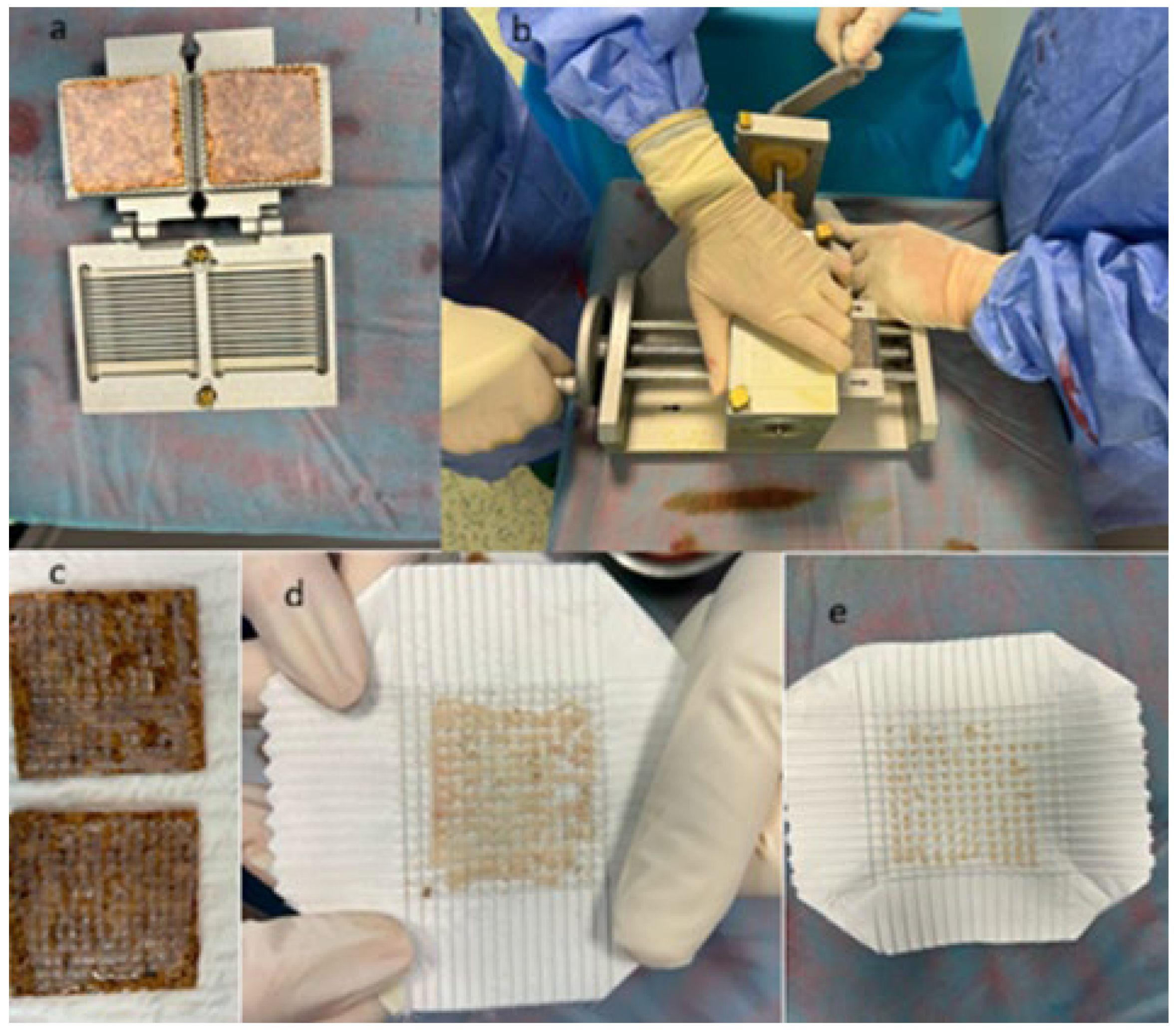
The Meek micrografting technique achieves its expansion by cutting a finite sheet of donor skin into small, evenly spaced islands that are mounted on a folded carrier and stretched to a predetermined ratio, maximizing the surface area that can be covered from a limited donor site (Figure 2).
Taken together, the available evidence positions Meek micrografting as a valuable but situational tool: best supported—mainly by observational cohorts and by meta-analytic data in massive burns—for extensive, donor-site-limited burns, whereas the only completed randomized trial does not support its routine use as a default substitute for mesh grafting within the population and expansion ratios studied. Future refinements, including bioengineered carriers, regenerative adjuncts such as platelet-rich plasma [10], and cell-based combinations [25,26], may further extend the range of indications for the technique, provided they are evaluated in adequately powered comparative trials that, unlike most of the current evidence base, are prospectively registered and span a wider range of burn sizes [23].
Given the size and heterogeneity of the current evidence base, the synthesized data allow provisional clinical considerations rather than firm recommendations. Meek micrografting may be useful in selected extensive or donor-site-limited burns in which high expansion ratios are required, but the available evidence does not establish a universal TBSA threshold for selecting Meek over mesh grafting. Current randomized evidence does not support routine substitution of Meek for mesh grafting where lower expansion ratios suffice, within the population and expansion ratios studied. In selected patients, including those in whom donor-site morbidity is a particular concern, Meek may remain a reasonable option, but this interpretation rests largely on observational evidence and a single mixed-etiology randomized trial and should be confirmed in larger comparative studies.

5. Limitations

This systematic review should be interpreted in light of several limitations. First, the available clinical evidence on the Meek technique, while steadily increasing, remains limited by small sample sizes and heterogeneous study designs; most included studies were retrospective or observational, and only one randomized intra-patient trial has been completed to date, which constrains the overall level of evidence and the applicability of its findings across the full range of burn sizes. Second, outcome reporting across studies was inconsistent: graft take and expansion ratios were almost universally documented, but data on long-term functional recovery, scar quality, and patient-reported outcomes were less standardized, limiting the possibility of formal meta-analysis. Third, although no language restrictions were applied and every effort was made to capture the complete body of relevant literature through comprehensive searches across multiple databases, outcome data from non-English publications were extracted from English abstracts or translated full texts, which may have limited the granularity of the information available for synthesis. Finally, the inclusion of original intraoperative photographs was intended to illustrate the technical execution of the Meek method; these images have pedagogical value but do not constitute new clinical outcome data. In addition, the randomized evidence included a mixed wound population, so its overall estimates cannot be assumed to apply exclusively to burns.
Despite these limitations, the synthesis provides an updated overview of both the technical evolution and comparative clinical performance of the Meek technique.

6. Conclusions

The Meek technique remains a valuable option in burn reconstruction, enabling limited donor skin to provide coverage in selected patients with extensive injuries. Its technical evolution, from an experimental 1950s dermatome system to standardized carriers, has made it a reproducible method for situations in which donor sites are critically limited.
At the same time, the first completed randomized controlled trial shows that this advantage does not extend to the lower expansion ratios it examined, where mesh grafting performed better on graft take and short-term scar quality. Although donor-site area was smaller overall with Meek and patient preference favored Meek in the small exploratory 1:3 subgroup, these findings were exploratory and should not be interpreted as evidence of treatment superiority. The available evidence does not establish a universal TBSA threshold for selecting Meek over mesh grafting, and Meek micrografting should therefore be regarded as an indication-specific tool rather than a categorical alternative. Importantly, direct clinical comparisons are limited when using identical nominal expansion ratios, as the actual expansion achieved with mesh is substantially lower than its theoretical value.
Looking ahead, refinements such as bioengineered carriers and regenerative adjuncts may further extend the range of indications for the technique, but additional randomized trials across a broader range of burn sizes, with standardized long-term scar and functional outcome measures, are needed to define its optimal role relative to mesh grafting. Future research should not only compare Meek versus mesh grafting across different burn sizes but should also investigate clinically equivalent effective expansion ratios (e.g., mesh 1:3 versus Meek 1:2). Such adequately powered, randomized studies must evaluate comprehensive metrics, including graft take, donor-site area, donor-site morbidity, the need for repeated harvesting, and standardized long-term patient-reported outcomes.

Supplementary Materials

The following supporting information can be downloaded at: https://www.mdpi.com/article/10.3390/surgeries7030108/s1, Table S1: PRISMA 2020 checklist [28]. Table S2: Complete database-specific search strategies. Table S3: Risk-of-bias/methodological quality assessment.

Author Contributions

Conceptualization, M.B., M.P. and D.C.M.; methodology, M.B. and A.-H.A.; software, S.A.-L. and A.-H.A.; validation, M.P., M.-C.C. and S.A.-L.; formal analysis, M.B.; investigation, D.C.M., M.B. and A.-N.G.; resources, M.B. and D.C.M.; data curation, M.P.; writing—original draft preparation, M.B. and M.P.; writing—review and editing, M.B. and M.P.; visualization, D.C.M. and M.-C.C.; supervision, M.P.; project administration, M.B., A.-N.G. and M.P. All authors have read and agreed to the published version of the manuscript.

Funding

This research received no external funding.

Institutional Review Board Statement

Not applicable. The original intraoperative photographs provided in Figure 2 depict only surgical equipment and materials (the Meek device and carriers) taken by the authors; no human subjects or patient-identifiable features are present.

Informed Consent Statement

Not applicable.

Data Availability Statement

Data extracted from the included studies are presented in Table 1 and Table 2, Tables S2 and S3. Additional extraction materials are available from the corresponding author on reasonable request.

Conflicts of Interest

The authors declare no conflicts of interest.

Abbreviations

The following abbreviations are used in this manuscript:
TBSATotal body surface area
PRISMAPreferred Reporting Items for Systematic Reviews and Meta-Analyses
WHOWorld Health Organization
CEACultured Epidermal Autografts
RCTRandomized Controlled Trial

References

  1. World Health Organization. Burns—Fact Sheet. 2018. Available online: https://www.who.int/news-room/fact-sheets/detail/burns (accessed on 26 September 2025).
  2. Peck, M.D. Epidemiology of burns throughout the world. Part I: Distribution and risk factors. Burns 2011, 37, 1087–1100. [Google Scholar] [CrossRef] [Scilit] [PubMed]
  3. Rijpma, D.; Claes, K.; Pijpe, A.; Hoeksema, H.; De Decker, I.; Verbelen, J.; Stoop, M.; De Mey, K.; Hoste, F.; van Zuijlen, P.; et al. Wound and short-term scar outcomes of Meek micrografting versus mesh grafting: An intra-patient randomized controlled trial. Eur. Burn J. 2025, 6, 26, Correction in Eur. Burn J. 2025, 6, 39. https://doi.org/10.3390/ebj6030039.. [Google Scholar] [CrossRef] [Scilit] [PubMed]
  4. Quintero, E.C.; Machado, J.F.E.; Robles, R.A.D. Meek micrografting history, indications, technique, physiology and experience: A review article. J. Wound Care 2018, 27, S12–S18. [Google Scholar] [CrossRef] [Scilit] [PubMed]
  5. Noureldin, M.A.; Said, T.A.; Makeen, K.; Kadry, H.M. Comparative study between skin micrografting (Meek technique) and meshed skin grafts in paediatric burns. Burns 2022, 48, 1632–1644. [Google Scholar] [CrossRef] [Scilit] [PubMed]
  6. Rijpma, D.; Claes, K.; Hoeksema, H.; De Decker, I.; Verbelen, J.; Monstrey, S.; Pijpe, A.; van Zuijlen, P.; Meij-de Vries, A. The Meek micrograft technique for burns; review on its outcomes: Searching for the superior skin grafting technique. Burns 2022, 48, 1287–1300. [Google Scholar] [CrossRef] [Scilit] [PubMed]
  7. Haug, V.; Tapking, C.; Panayi, A.C.; Veeramani, A.; Knoedler, S.; Tchiloemba, B.; Abdulrazzak, O.; Kadakia, N.; Bonaventura, B.; Culnan, D.; et al. Outcome comparison of the most commonly employed wound coverage techniques in patients with massive burns ≥50% TBSA—A systematic review and meta-analysis. Burns 2024, 50, 107210. [Google Scholar] [CrossRef] [Scilit] [PubMed]
  8. Almujaydil, R.; Yan, Y.; Kuswandani, S.; Alotaibi, F.; Suvan, J.; Nguyen, L.; D’Aiuto, F. Systematic review and meta-analysis of the use of micrografting technology in humans. J. Int. Med. Res. 2025, 53, 3000605251337859. [Google Scholar] [CrossRef] [Scilit] [PubMed]
  9. Tapking, C.; Panayi, A.; Haug, V.; Palackic, A.; Houschyar, K.S.; Claes, K.E.Y.; Kuepper, S.; Vollbach, F.; Kneser, U.; Hundeshagen, G. Use of the modified Meek technique for the coverage of extensive burn wounds. Burns 2024, 50, 1003–1010. [Google Scholar] [CrossRef] [Scilit] [PubMed]
  10. Zhang, T.; Liu, Z.; Wang, C.; Zhao, J.; Wei, D.; Xu, Y. Clinical effects of Meek skin grafting combined with platelet-rich plasma in repairing extensive deep burn wounds. Zhonghua Shao Shang Za Zhi 2024, 40, 1150–1157. [Google Scholar] [CrossRef] [Scilit]
  11. Wilson, E.; Grant, M.; Fleet, M.; Lee, M.; Jones, I. Exploring the emerging role of Meek micrografting in healing smaller burns: A case series and evidence review. Plast. Aesthet. Res. 2025, 12, 8. [Google Scholar] [CrossRef] [Scilit]
  12. López-Contreras, A.C.; Ferreira Aparicio, F.E.; Ramírez, E.D.; Elnecave-Olaiz, A.; Santiago-Ruiz, L.; Rodríguez Mendieta, C.A. The Meek technique versus partial-thickness skin graft in major burns patients: Experience in the National Center of Research and Care of Burns Patients in Mexico City. Int. J. Res. Med. Sci. 2026, 14, 2334–2338. [Google Scholar] [CrossRef] [Scilit]
  13. Meek, C.P. Successful microdermagrafting using the Meek-Wall microdermatome. Am. J. Surg. 1958, 96, 557–558. [Google Scholar] [CrossRef] [Scilit] [PubMed]
  14. Kreis, R.W.; Mackie, D.P.; Vloemans, A.W.; Hermans, R.P.; Hoekstra, M.J. Widely expanded postage stamp skin grafts using a modified Meek technique in combination with an allograft overlay. Burns 1993, 19, 142–145. [Google Scholar] [CrossRef] [Scilit] [PubMed]
  15. Munasinghe, N.; Wasiak, J.; Ives, A.; Cleland, H.; Lo, C.H. Retrospective review of a tertiary adult burn centre’s experience with modified Meek grafting. Burn. Trauma 2016, 4, 6. [Google Scholar] [CrossRef] [Scilit] [PubMed]
  16. Almodumeegh, A.; Heidekrueger, P.I.; Ninkovic, M.; Rubenbauer, J.; Hadjipanayi, E.; Broer, P.N. The MEEK technique: 10-year experience at a tertiary burn centre. Int. Wound J. 2017, 14, 601–605. [Google Scholar] [CrossRef] [Scilit] [PubMed]
  17. Lee, S.Z.; Halim, A.S.; Wan Sulaiman, W.A.; Mat Saad, A.Z. Outcome of the modified Meek technique in the management of major pediatric burns. Ann. Plast. Surg. 2018, 81, 295–301. [Google Scholar] [CrossRef] [Scilit] [PubMed]
  18. Lee, S.Z.; Halim, A.S. Superior long term functional and scar outcome of Meek micrografting compared to conventional split thickness skin grafting in the management of burns. Burns 2019, 45, 1386–1400. [Google Scholar] [CrossRef] [Scilit] [PubMed]
  19. Hu, G.; Zhang, P.; Chen, Y.; Yuan, Z.; Song, H. Efficacy of two-stage Meek micrografting in patients with severe burns. J. Burn Care Res. 2022, 43, 1081–1085. [Google Scholar] [CrossRef] [Scilit] [PubMed]
  20. Wang, Z.Y.; He, C.P.; Luo, X.L.; Wang, F.S. Clinical application of Meek skin grafting technique. Nan Fang Yi Ke Da Xue Xue Bao 2006, 26, 678–682. (In Chinese) [Google Scholar] [PubMed]
  21. Dahmardehei, M.; Vaghardoost, R.; Saboury, M.; Zarei, H.; Saboury, S.; Molaei, M.; Seyyedi, J.; Maleknejad, A. Comparison of modified Meek technique with standard mesh method in patients with third degree burns. World J. Plast. Surg. 2020, 9, 267–273. [Google Scholar] [CrossRef] [Scilit] [PubMed]
  22. Kok, Y.O.; Chong, S.J.; Liang, W.H.; Tan, B.K.; Tan, K.C. Revolutionizing major burns management with micrografting—Improved healthcare costs, time and burns resources. Plast. Reconstr. Surg. 2015, 136, 64. [Google Scholar] [CrossRef] [Scilit]
  23. Rijpma, D.; Pijpe, A.; Claes, K.; Hoeksema, H.; De Decker, I.; Verbelen, J.; van Zuijlen, P.; Monstrey, S.; Meij-de Vries, A. Outcomes of Meek micrografting versus mesh grafting on deep dermal and full thickness (burn) wounds: Study protocol for an intra-patient randomized controlled trial. PLoS ONE 2023, 18, e0281347. [Google Scholar] [CrossRef] [Scilit] [PubMed]
  24. Mishra, A.; Rabiee, S.; Opel, S.; Jones, I. Applying the modified Meek technique to heal smaller burns: A retrospective review. Burn. Open 2022, 6, 120–124. [Google Scholar] [CrossRef] [Scilit]
  25. Craft-Coffman, B.; Homsombath, B.; Cramer, C.; Hassan, Z.; Fagan, S.; Lack, K.; Wilson, J. CEA graft take after combining with a modified MEEK procedure. Burn. Open 2024, 8, 23–28. [Google Scholar] [CrossRef] [Scilit]
  26. Minic, J.; Vigato, E.; Shoham, Y.; Lavagnolo, U.; Governa, M. Selective enzymatic debridement and modified Meek technique in the treatment of extensive burns: Preliminary observations. Health Sci. Rep. 2024, 7, e1829. [Google Scholar] [CrossRef] [Scilit] [PubMed]
  27. Kamolz, L.P.; Schintler, M.; Parvizi, D.; Selig, H.; Lumenta, D.B. The real expansion rate of meshers and micrografts: Things we should keep in mind. Ann. Burn. Fire Disasters 2013, 26, 26–29. [Google Scholar] [PubMed] [PubMed Central]
  28. Page, M.J.; McKenzie, J.E.; Bossuyt, P.M.; Boutron, I.; Hoffmann, T.C.; Mulrow, C.D.; Shamseer, L.; Tetzlaff, J.M.; Akl, E.A.; Brennan, S.E.; et al. The PRISMA 2020 statement: An updated guideline for reporting systematic reviews. BMJ 2021, 372, n71. [Google Scholar] [CrossRef] [Scilit] [PubMed]
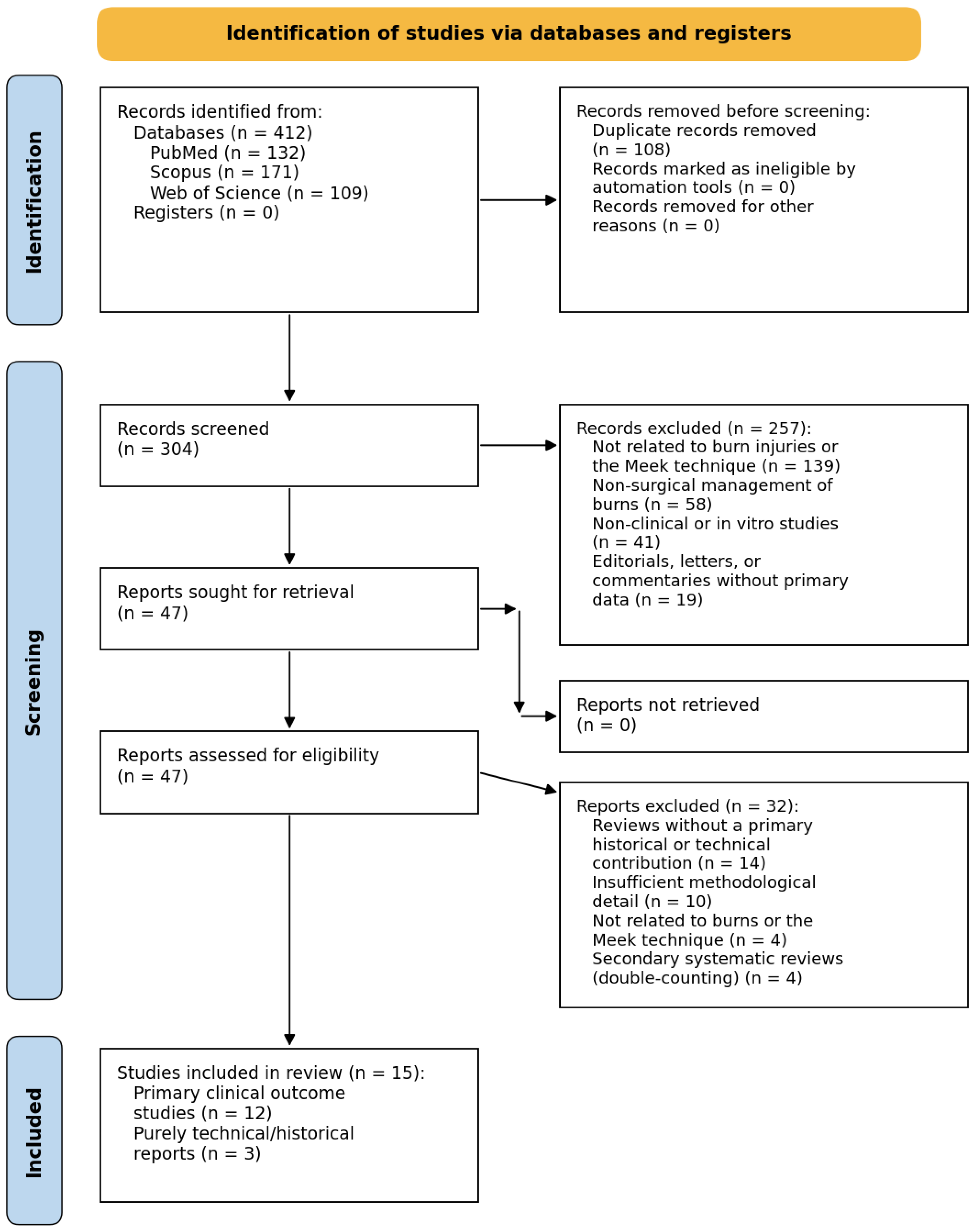
Figure 1. PRISMA flow diagram.
Surgeries 07 00108 g001
Figure 2. The principle and application of the Meek micrografting technique: (a) split-thickness skin grafts are mounted onto the cork carrier; (b,c) the carrier is cut into multiple small, regularly spaced micrografts in both directions in the Meek machine; (d,e) expansion of the pre-folded polyester gauze by pulling on the edges, first the ribbed ones and then the smooth ones.
Surgeries 07 00108 g002
Table 1. The evolution of the technique from an experimental concept to a reproducible method widely applied in burn surgery.
PeriodAuthorsKey DevelopmentImpact
1950s–1980sMeek CP et al. (1958) [13]First description of “microdermagrafting” using the Meek-Wall dermatome. Skin cut into 3 × 3 mm islands on cork plates, achieving expansion up to 1:9.Highly innovative for massive burns with scarce donor sites, but the device was complex and time-consuming; simpler mesh-grafting methods were subsequently more widely adopted.
1990sKreis RW et al. (1993) [14]Reintroduction of the Meek method with redesigned prefabricated carriers and disposable blades (HumecaR system).Simplified and standardized the technique, making it reproducible and clinically feasible.
2010sQuintero EC et al. (2018) [4]Comprehensive review of technique, physiology, and indications.Consolidated Meek as an evidence-based method in reconstructive burn surgery.
2020sNoureldin MA et al. (2022) [5]; Rijpma D et al. (2025) [3]; Tapking C et al. (2024) [9]Pediatric comparative study, first completed intra-patient randomized controlled trial (mesh superior at the lower expansion ratios studied), and modified Meek technique for extensive burns.Reported variable graft-take outcomes, extended applications to extensive burns, and comparative evidence in which mesh performed better in the randomized study at the lower expansion ratios examined.
mm—millimeters.
Table 2. Clinical characteristics and reported outcomes of primary studies evaluating Meek micrografting.
AuthorsCountryStudy DesignSample SizeTBSA/Expansion/Graft TakeKey Findings
1.Munasinghe et al. (2016) [15]AustraliaRetrospective review1157%/expansion 1:6/graft take ~87%Most grafted areas healed without regrafting. Infection was the main cause of partial graft failure. The study concluded that the Meek technique is useful for patients with extensive burns and limited donor sites.
2.Almodumeegh A et al. (2017) [16]GermanyCase series6765%/expansion 1:6–1:9/graft viability 60–90%Patients required on average 2.2 grafting operations. The mean hospital stay was 27 days. Graft viability ranged from 60–90% at day 7–10. Infections occurred in five patients, and seven patients died due to the severity of burns.
3.Lee SZ et al. (2018) [17]MalaysiaCase series (pediatric)1235%/expansion 1:6/graft take 82.3%Pediatric patients treated with the modified Meek technique achieved reliable wound closure, reduced contracture, and acceptable scar quality at follow-up.
4.Lee SZ et al. (2019) [18]MalaysiaComparative study (Meek vs. split-thickness graft)43~27%/expansion 1:6/graft take ~82%Patients in the Meek group had superior long-term scar quality (pigmentation, pliability, height) compared with split-thickness grafts, while functional outcomes were comparable.
5.Noureldin MA et al. (2022) [5]EgyptComparative study (Meek vs. mesh)30 pediatric20–60%/expansion 1:6/graft take 84.3% (Meek) vs. 71.5% (mesh)Meek achieved significantly higher graft take, required smaller donor areas, and resulted in faster wound coverage compared with mesh grafting in children.
6.Hu G et al. (2022) [19]ChinaRetrospective (two-stage vs. one-stage Meek)127Severe burns > 70% in many cases/expansion 1:9/graft take improved in two-stage groupThe two-stage Meek approach improved graft survival and reduced complications such as hypoproteinemia and hypoalbuminemia compared with single-stage Meek, while hospital stay and cost were similar.
7.Rijpma D. et al. (2025) [3]The Netherlands and Belgium; mixed wound cohortIntra-patient randomized controlled trial7010 ± 10% TBSA/expansion 1:2–1:3/graft take 79 ± 25% (Meek) vs. 87 ± 19% (mesh)The trial found significantly lower graft take with Meek (p = 0.003) and better short-term observer- and patient-rated scar outcomes with mesh at the expansion ratios studied. Although donor-site area was smaller overall with Meek and patient preference favored Meek in the small exploratory 1:3 subgroup, these findings were exploratory and were not powered to establish treatment superiority. Because the cohort included non-burn wounds, the findings were treated as indirect evidence for burn reconstruction.
8.Tapking C. et al. (2024) [9]GermanyCase series7360%/expansion up to 1:9/graft take 75.8 ± 14.7% at day 10Pre-treatment of wound beds improved graft take, while older age was associated with reduced take. The authors confirmed the reliability of the modified Meek technique for extensive burns.
9.Zhang T. et al. (2024) [10]ChinaCase–control (Meek ± platelet-rich plasma)44>50%/expansion 1:9/graft take ~85%Patients treated with Meek combined with platelet-rich plasma showed faster healing and better scar quality compared with those treated with Meek alone.
10.Wilson E. et al. (2025) [11]UKCase series64Mixed burn sizes/expansion 1:6/graft take >80%Over 13 years of follow-up, average time to 95% wound healing was 76 days. The mean hospital stay was 64 days, mortality rate was 4.6%, and scar quality was acceptable with a median Patient and Observer Scar Assessment Scale score of 5.
11.Wang ZY et al. (2006) [20]ChinaRetrospective case series14Expansion 1:4–1:6/graft take The Meek technique achieved reliable wound closure in extensive deep burns, demonstrating effective expansion and good clinical outcomes for patients with limited donor sites.
12.López-Contreras AC et al. (2026) [12]MexicoRetrospective cohort (Meek vs. partial-thickness graft)38Expansion 1:3–1:6/graft take comparable to conventional graftingIn a major Latin American burn center, Meek micrografting achieved wound coverage outcomes comparable to conventional partial-thickness skin grafting, with reduced donor-site area required per patient.
Disclaimer/Publisher’s Note: The statements, opinions and data contained in all publications are solely those of the individual author(s) and contributor(s) and not of MDPI and/or the editor(s). MDPI and/or the editor(s) disclaim responsibility for any injury to people or property resulting from any ideas, methods, instructions or products referred to in the content.