Article

Less Is More for Non-Dislocated Femoral Neck Fractures: Similar Results for Two versus Three Cannulated Hip Screws

Department of Trauma Surgery, Meander Medical Center, 3813 TZ Amersfoort, The Netherlands
*
Author to whom correspondence should be addressed.
Surgeries 2023, 4(4), 493-502; https://doi.org/10.3390/surgeries4040048
Submission received: 21 June 2023 / Revised: 5 September 2023 / Accepted: 19 September 2023 / Published: 22 September 2023

Abstract

:
Cannulated hip screws (CHS) can be used for the minimally invasive fixation of non-dislocated femoral neck fractures. Usually, three screws are inserted. This study aims to determine whether fixation by two CHS leads to similar results as fixation by three CHS. Since January 2019, all patients with an indication for internal fixation by CHS were treated with two CHS and followed prospectively. Results were compared to an equal-sized control group of patients who underwent fixation by three CHS (before 2019). The primary outcome was reoperation, while the secondary outcome was screw dislocation. Since January 2019, 50 patients were treated by two CHS. Of these, 14 patients (28%) underwent reoperation versus 13 patients (26%) in the control group (p = 1.000). Reoperations included screw replacement, hemiarthroplasty, and total hip prosthesis. Three major reasons for reoperation were pain due to osteosynthesis material (n = 15), coxarthrosis (n = 4), and screw cut out (n = 3). Six weeks postoperative X-rays showed a screw dislocation of 2 mm for the two CHS group and 1 mm for the three CHS group (p = 0.330). Clinical outcomes were very similar between the groups. The overall results were good; however, the reoperation rate varied from 26 to 28%. The majority of reoperations were screw replacements. Screw dislocation seems to be more prominent in patients treated with two screws (2 mm versus 1 mm). Fixation by two cannulated hip screws is an acceptable treatment method for non-dislocated femoral neck fractures, and the insertion of a third screw does not lead to superior clinical results.

1. Introduction

Hip fractures represent a significant global health issue, with an estimated incidence of 1.26 million cases worldwide in 1990. It is projected that by the year 2050, this number will quadruple [1]. Apart from the high morbidity and mortality [2,3], hip fractures contribute substantially health care costs. Per-patient costs amount to EUR 27.500 in the first year alone [4].
Numerous risk factors contribute to the incidence of femoral neck fractures, including female gender, low bone density, and hypertension [5,6,7].
Over the years, researchers have sought to obtain insight into the optimal treatment method for proximal femur fractures. Surgical treatment options include internal fixation, hemiarthroplasty, and total hip arthroplasty [8]. The optimal surgical treatment method depends on case specific factors including fracture type, age, and comorbidity of the patient [9], and conservative treatment might also be beneficial in some cases [10].
Choice of treatment method relies predominantly on fracture type. The Garden classification is the most commonly utilized system to classify femoral neck fractures [11]. The system classifies femoral neck fractures based on the degree of displacement and fracture pattern, as seen on anterior to posterior (AP) radiographs. Garden Class I and II fractures are often considered stable and may be amenable to internal fixation techniques. In contrast, Garden Class III and IV fractures are typically unstable and require more extensive surgical interventions such as arthroplasty or internal fixation with additional adjuncts to achieve adequate stability [12].
For non-dislocated femoral neck fractures, current guidelines indicate internal fixation through cannulated hip screws (CHS). Screws are inserted following the “three-point principle”, by inserting three cannulated screws in an inverted triangular pattern [13,14,15,16,17] (Figure 1). This method shows excellent results in the management of Garden I and II femoral neck fractures [18]. However, the question arises as to whether it is necessary to insert three screws, or if two screws can yield comparable clinical results.
In biomechanical studies, three CHS fixation can bear significantly higher weight than fixation with two CHS [19,20,21]. Yet, the clinical relevance of this increased weight-bearing capacity remains uncertain. In cases where the femoral neck could not accommodate three CHS due to its diameter, the insertion of two CHS has been justified [22]. Previous studies have shown that two screws provide adequate fixation [23,24,25]. Fixation with two screws offers several advantages, such as reduced surgery time, decreased operative irradiation, lower costs, and a less technically demanding procedure [26]. Moreover, radiological evidence suggests that union occurs more frequently in two-screw fixation than in three-screw fixation [27]. Despite these findings, there is a scarcity of literature on the clinical outcomes associated with fixation using two CHS. Therefore, the primary objective of this study is to investigate whether fixation of non-dislocated femoral neck fractures (Garden I and II) using two CHS yields similar results as fixation by three CHS. Screw displacement was considered as a secondary outcome parameter.

2. Materials and Methods

2.1. Study Design

This study was conducted at the Meander Medical Center, Amersfoort, The Netherlands. This study was approved by the local ethics committee and performed in accordance with the ethical standards (study number TWO 20-063). The duration of the study extended from 1 January 2019 to December 2020. Based on the literature, our trauma department decided to change their treatment strategy in January 2019 for patients with non-dislocated femoral neck fractures. Instead of inserting three cannulated screws, Garden 1 or 2 femoral neck fractures were fixated with two screws without patient selection. All patients entered a prospectively maintained database to carefully evaluate this change in practice. All patients who underwent fixation by two CHS until December 2020 were included in this study. Controls with three CHS were identified in consecutive order until a corresponding number of patients was obtained. Patients with dislocated femoral neck fractures (Garden III and IV) were excluded, as well as patients who were lost to follow-up (who did not visit the outpatient clinic).

2.2. Surgical Technique

A dose of 2 g cefazolin is given between 30 and 60 min preoperatively as antimicrobial prophylaxis. Surgery is performed under a supine position, with application of traction and internal rotation of the hip. Before incision, reduction of the fracture is verified using X-ray guidance. A 3 cm incision is made, starting at the flair of the great trochanter and extending distally. The fascia lata is split, and the vastus lateralis is held aside to reveal the femoral cortex.
Kirschner (K-)wires are placed under X-ray guidance for optimal positioning of the screws in two directions. The first K-wire is placed inferiorly, over the calcar, right in the middle of the femoral cortex. The second K-wire is placed superior to the first and more posterior. The length of the K-wires is measured to determine screw lengths. The cannulated 6.5 mm screws are placed over the K-wires, starting with the posterior screw to prevent varus deformity. After placement of both screws, positioning is checked by X-ray in both directions. For the positioning of the screws in two CHS fixation, see Figure 2. The fascia lata is closed with an absorbable continuous suture. The skin is closed with a non-absorbable suture. Directly after operation, a physiotherapist is consulted to support patients in mobilizing. Postoperative prophylaxis for venous thromboembolism is carried out based on established protocols, taking into consideration the patient’s current anticoagulant status. The administration of low-molecular-weight heparins (LMWH) is the prevailing method used for this purpose. Clinical rehabilitation usually takes five days, depending on the patient’s age and comorbidities. Patients are instructed to mobilize at 50% weight bearing with crutches or a walker. If the clinical situation allows so, patients are discharged home. If not, patients are transferred to a rehabilitation facility. Sutures will be removed by the general practitioner, two weeks after surgery. Six weeks postoperatively, patients return to the outpatient clinic for radiological and clinical follow-up. Patients are instructed to contact the hospital if any adverse events happen.

2.3. Data Collection

Data on patients’ demographics, fracture type, screw length, and screw translocation were obtained through electronic patient files. Renal function was categorized by the 2012 Kidney Disease Outcomes Quality Initiative (KDIGO) Chronic Kidney Disease (CKD) classification [28]. Fracture types were classified using the Garden Classification [12].
The intervention group included all patients who underwent fixation by two CHS between January 2019 and December 2020. The control group existed of an equal number of patients who underwent fixation by three CHS. The primary outcome parameter was reoperation and included screw replacement, conversion to hemiarthroplasty (HA), and conversion to total hip prosthesis (THP). A secondary outcome parameter was screw displacement. This was defined by lateral protrusion into soft tissue. Screw displacement was calculated six weeks postoperatively, using radiographic measurements in correlation with the actual screw lengths. For patients who underwent reoperation, screw displacement was measured once more, before reoperation. Follow-up duration was registered as full months up until the date of data extraction from the electronic patient’s files. Patients who died before the six weeks outpatient check-up were excluded (loss to follow-up). For patients who died after the check-up, mortality was registered, and follow-up duration was calculated until the date of death.

2.4. Statistical Analysis

Statistical analysis was performed using the computing environment R (R Studio Team; 2021). Categorical data are shown as numbers with percentages, and significant differences were calculated through the chi-square test or Fisher exact test as appropriate. For continuous data, normality of distribution was assessed using quantile-quantile (Q-Q) plots. In the case of normal distribution, categorical data are shown as the mean with range. In the case of non-normal distribution, categorical data are shown as the median with interquartile range (IQR). Significant differences were calculated using Student’s t-test or Mann–Whitney U-test, respectively. For all analyses, a two-sided p-value < 0.05 was used as the threshold of statistical significance.

3. Results

Between January 2019 and December 2020, a total of 53 patients underwent fixation of a femoral neck fracture with two CHS. Of these, three were lost to follow-up. A corresponding number of 50 control patients with non-dislocated femoral neck fractures who were treated with three CHS were identified in subsequent order. In total, 100 patients were included for analysis (Figure 3).
The average age was 71 years and ranged from 29 to 97 years. The majority of the sample (74%) was female. Distribution over American Society of Anesthesiologists (ASA) classes was as follows. Seventeen patients were ASA 1, 44 were ASA 2, 36 were ASA 3, and three were ASA 4. Kidney function was G1 for 29 patients, G2 for 52 patients, G3a for 10 patients, G3b for six patients, G4 for two patients, and G5 for one patient. The average time until patients visited the outpatient clinic was seven weeks. The majority (69%) suffered a Garden I type fracture, and the other 31% suffered a Garden II type fracture. Sixteen percent used a walking aid for mobilizing before the trauma. There were no significant differences in baseline characteristics between the intervention group and the control group (Table 1). Examples of postoperative radiographs are provided in Figure 4 and Figure 5.
In total, 27 patients underwent reoperation after a median of six months. In the two and three CHS groups, reoperation rates did not differ (28% and 26% respectively, p = 1.000). Reasons for reoperations in the two CHS group were complaints of pain due to osteosynthesis material (OSM) in seven patients (50%), coxarthrosis in two (14%), screw cut-out in two (14%), avascular necrosis (AVN) in one (7%), screw dislocation in one (7%), and revision to HA due to a fall in one (7%).
For the three CHS group, reasons for reoperation were complaints of pain due to OSM in eight patients (62%), screw cut-out in one (8%), coxarthrosis in two (15%), infection in one (8%) and non-union in one (8%, Table 2). This was the only patient from the total cohort who suffered a non-union. In all other patients, radiological union was confirmed after six weeks. A detailed description of each reoperation is provided in Table 3. This table demonstrates that for some patients, dislocation of screws progressed over time.
In the total cohort, 33 patients were older than 80 years. Of them, seven (21%) underwent reoperation compared to 20 patients (30%) below the age of 80. This difference was not significant (p = 0.499). The type of reoperation differed significantly between these groups (p = 0.005). Of patients above the age of 80 years who underwent reoperation, conversion to HA was performed in four subjects (57%), screw replacement in one subject (14%), and conversion to THP in two subjects (29%). For patients under the age of 80, one subject (5%) underwent conversion to HA, 14 subjects (70%) had screw replacement, and five subjects (25%) underwent conversion to THP.
The lateral protrusion of the screws outside the lateral cortex was 2 mm for the two CHS group and 1 mm for the three CHS group six weeks postoperatively (p = 0.330). The mean follow-up duration differed significantly, being 26 months for the two CHS group and 45 months for the three CHS group (p < 0.001, Table 2).

4. Discussion

The main objective of this study was to determine whether the fixation of non-dislocated femoral neck fractures (Garden I and II) by two CHS leads to similar results as fixation by three CHS. The most important finding of our study was the number of reoperations after two versus three CHS. We did not find a clinically significant difference between these two surgical techniques (28% versus 26%, p = 1.000). In terms of reoperation, infection, and screw dislocation, there were no differences in outcome between fixation by two or by three CHS. Also, no higher reoperation rate was seen in patients older than 80 years of age.
This is the first prospectively maintained database that investigated reoperation rates between non-displaced femoral neck fractures for two CHS versus three CHS. One randomized controlled trial has been performed comparing two versus three CHS. This study reported a significantly higher frequency of radiological union in two CHS fixation [27]. This study, however, did not mention the clinical outcomes or reoperation rates. One retrospective study did not find a significant difference in radiological outcome between two and three CHS [15]. In two studies, the clinical outcome was investigated for two CHS fixation. Both studies reported low rates of complications, indicating that two CHS suffice in the fixation of non-displaced femoral neck fractures [23,26]. Yet, these studies had no control group.
Biomechanical studies have examined the maximum force that both types of fixation can resist. In cadaveric or synthetic bones, femoral neck fractures were created and fixated with two or three CHS. Three CHS fixation showed greater resistance to mechanical loading in all studies [19,20,21]. This result is not surprising. The value of this ability to bear more load, however, seems insignificant for clinical practice, as seen in the present study.
Although no significant clinical difference was observed between these two groups in our study, our overall rate of reoperations was found to be higher compared to the literature. A recent systematic review by Kim et al. focused on internal fixation with cannulated screws for non-displaced femoral neck fractures in patients over 60 years of age [29]. They reported a reoperation rate of 15%, with 12% of cases requiring conversion to arthroplasty. Unfortunately, the review did not specify whether fixation was performed using two or three CHS. Studies specifically investigating fixation by two CHS or comparing two versus three CHS are limited. The available literature indicates reoperation rates ranging from 5% to 10% [23,26,30].
In the present study, the overall reoperation rate was notably higher at 27%. The majority of reoperations were due to the number of screw replacements (15 out of 27 reoperations). It would be worthwhile to explore whether the utilization of hydroxyapatite-coated screws could help reduce this high reoperation rate. Previous research on lateral femoral fractures has shown improved osteointegration with hydroxyapatite-coated screws compared to conventional screws [31]. This might decrease screw loosening and, thus, the need for screw replacement. Therefore, investigating the potential benefits of hydroxyapatite-coated screws in femoral neck fractures could be an interesting avenue for future research. When screw replacement was not counted as reoperation, our reoperation rates compare to previously described literature, as conversion to arthroplasty was performed in 12% of cases.
Complications after CHS fixation include non-union and AVN. In our study both non-union and AVN occurred once (1% for the total cohort). This is lower than the rate reported in literature. Non-union rates after fixation with two CHS vary between 0 and 22%, and AVN occurs in between 0 and 6% of cases [23,24,26,30].
Lateral protrusion for the total cohort showed a limited amount of screw dislocation, being 1 mm. It seems contradictory that the majority of reoperations in the current study were screw replacements due to pain of the OSM, since dislocation was minimal. Therefore, dislocation was measured again before reoperation. Some patients showed a progression in dislocation over time. This suggests that, although radiological union was confirmed for all but one of the patients, fractures may not be completely healed after six weeks. This might indicate the need for another visit to the outpatient clinic at six months postoperatively.
The three CHS group had a significantly longer follow-up duration (45 months compared to 26 months for the two CHS group). This was due to the fact that those patients underwent surgery between 2017 and 2019, because all CHS surgeries since 2019 were performed using two CHS. Since most reoperations for non-displaced fractures after internal fixation occur within the first year [32], we chose a follow-up duration of at least a year for all inclusions. Still, the longer follow-up duration for the three CHS group might explain the higher rate of conversion to THP.
This study knows several limitations. First and foremost, the study was not a randomized controlled trial. To minimalize the risk of bias, no patient selection took place, and all patients were included. Second, follow-up duration was counted to the date of data extraction through the electronic patient files and not to the date of the last visit to the outpatient clinic. Patients were instructed to contact the outpatient clinic in case of an adverse event. However, there is a possibility that patients went to a private clinic, without contacting the outpatient clinic. Consequently, the number of reoperations may have been underestimated. Moreover, the duration of surgery was not registered in this study. The expected advantage of reducing surgery time, therefore, cannot be confirmed by this study. This was a conscious decision since our hospital is a teaching hospital. Surgeries are partially performed by residents, inevitably resulting in prolonged surgery time and, therefore, skewed results.

5. Conclusions

In the current study, an evaluation of a prospectively maintained database was performed to compare clinical outcomes of non-displaced femoral neck fractures fixated with either two or three CHS. Our findings revealed that patients who underwent fixation with two CHS had comparable reoperation rates to those who underwent fixation with three CHS. These results suggest that the clinical outcomes achieved with both fixation methods were similar. However, there are advantages of two CHS fixation, although we did not investigate them in the current study. These advantages include reduced operation time, decreased irradiation, and a less demanding technique. Therefore, it can be concluded that two CHS fixation may be the preferable method. This study highlights that the choice for both two and three CHS is justified. Yet, there are potential benefits of utilizing two CHS for the treatment of non-displaced femoral neck fractures, offering a promising alternative to the traditional three CHS fixation approach.

Author Contributions

Conceptualization, W.K., G.v.O. and L.H.; methodology, H.S., W.K., G.v.O. and L.H.; software, H.S.; validation, H.S.; formal analysis, H.S.; investigation, H.S. and W.K.; resources, G.v.O., P.v.K., T.T. and W.K.; data curation, H.S., W.K., G.v.O., P.v.K., T.T. and L.H.; writing—original draft preparation, H.S.; writing—review and editing, W.K. and L.H.; visualization, H.S.; supervision, W.K., G.v.O., P.v.K. and T.T.; project administration, H.S. All authors have read and agreed to the published version of the manuscript.

Funding

This research received no external funding.

Institutional Review Board Statement

The study was conducted in accordance with the Declaration of Helsinki and approved by the Ethics Committee of Meander Medical Center.

Informed Consent Statement

Informed consent was obtained from all subjects.

Data Availability Statement

Data is contained within the article.

Acknowledgments

We wish to thank Matthijs de Haan for providing us with the figures.

Conflicts of Interest

The authors declare no conflict of interest.

References

  1. Gullberg, B.; Johnell, O.; Kanis, J.A. World-wide projections for hip fracture. Osteoporos. Int. 1997, 7, 407–413. [Google Scholar] [CrossRef] [PubMed]
  2. Lutnick, E.; Kang, J.; Freccero, D.M. Surgical treatment of femoral neck fractures: A brief review. Geriatrics 2020, 5, 22. [Google Scholar] [CrossRef] [PubMed]
  3. Sakaki, M.H.; Oliveira, A.R.; Coelho, F.F.; Leme, L.E.G.; Suzuki, I.; Amatuzzi, M.M. Estudo da mortalidade na fratura do fêmur proximal em idosos. Acta Ortopédica Bras. 2004, 12, 242–249. [Google Scholar] [CrossRef]
  4. Kanters, T.A.; van de Ree, C.L.P.; de Jongh, M.A.C.; Gosens, T.; Hakkaart-van Roijen, L. Burden of illness of hip fractures in elderly Dutch patients. Arch. Osteoporos. 2020, 15, 11. [Google Scholar] [CrossRef] [PubMed]
  5. Notarnicola, A.; Tafuri, S.; Maccagnano, G.; Moretti, L.; Moretti, B. Frequency of hypertension in hospitalized population with osteoporotic fractures: Epidemiological retrospective analysis of Hospital Discharge Data in the Apulian database for the period 2006–2010. Eur. J. Inflamm. 2017, 15, 53–56. [Google Scholar] [CrossRef]
  6. Cummings, S.R.; Black, D.M.; Nevitt, M.C.; Browner, W.; Cauley, J.; Ensrud, K.; Genant, H.K.; Palermo, L.; Scott, J.; Vogt, T.M. Bone density at various sites for prediction of hip fractures. The Study of Osteoporotic Fractures Research Group. Lancet 1993, 341, 72–75. [Google Scholar] [CrossRef] [PubMed]
  7. Kannus, P.; Parkkari, J.; Sievänen, H.; Heinonen, A.; Vuori, I.; Järvinen, M. Epidemiology of hip fractures. Bone 1996, 18 (Suppl. S1), 57S–63S. [Google Scholar] [CrossRef]
  8. Florschutz, A.V.; Langford, J.R.; Haidukewych, G.J.; Koval, K.J. Femoral neck fractures: Current management. J. Orthop. Trauma 2015, 29, 121–129. [Google Scholar] [CrossRef]
  9. Marks, R. Hip fracture epidemiological trends, outcomes, and risk factors, 1970–2009. Int. J. Gen. Med. 2010, 3, 1–17. [Google Scholar] [CrossRef]
  10. Raaymakers, E.L.F.B.; Marti, R.K. Non-operative treatment of impacted femoral neck fractures. A prospective study of 170 cases. J. Bone Jt. Surg. Ser. B 1991, 73, 950–954. [Google Scholar] [CrossRef]
  11. Garden, R.S. Low-angle fixation in fractures of the femoral neck. J. Bone Jt. Surg. Br. Vol. 1961, 43, 647–663. [Google Scholar] [CrossRef]
  12. Kazley, J.M.; Banerjee, S.; Abousayed, M.M.; Rosenbaum, A.J. Classifications in brief: Garden classification of femoral neck fractures. Clin. Orthop. Relat. Res. 2018, 476, 441–445. [Google Scholar] [CrossRef] [PubMed]
  13. Bray, T.J. Femoral neck fracture fixation: Clinical decision making. Clin. Orthop. Relat. Res. 1997, 339, 20–31. [Google Scholar] [CrossRef] [PubMed]
  14. Wheeless, C.R.; Nunley, J.A.; Urbaniak, J.R. Wheeless’ Textbook of Orthopaedics; Data Trace Internet Publishing, LLC: Brooklandville, MD, USA, 2016; Available online: https://www.wheelessonline.com/ (accessed on 6 December 2022).
  15. Richtlijnendatabase, Behandeling Niet-Gedislokeerde Femurfractuur. Available online: https://richtlijnendatabase.nl/richtlijn/proximale_femurfracturen/niet-gedislokeerde_collum_femoris_fractuur/behandeling_niet-gedislokeerde_femurfractuur.html (accessed on 6 December 2022).
  16. Orthobullets. Femoral Neck Fractures ORIF with Cannulated Screws. Available online: https://www.orthobullets.com/general/12370/femoral-neck-fractures-orif-with-cannulated-screws (accessed on 6 December 2022).
  17. AO Surgery Reference. Impacted or Nondisplaced Subcapital Femoral Neck Fractures; Cannulated Screws. Available online: https://surgeryreference.aofoundation.org/orthopedic-trauma/adult-trauma/proximal-femur/femoral-neck-fracture-subcapital-impacted-or-nondisplaced/cannulated-screws (accessed on 6 December 2022).
  18. Chen, W.C.; Yu, S.W.; Tseng, I.C.; Su, J.Y.; Tu, Y.K.; Chen, W.J. Treatment of undisplaced femoral neck fractures in the elderly. J. Trauma Inj. Infect. Crit. Care 2005, 58, 1035–1039. [Google Scholar] [CrossRef]
  19. Mansur, H.; Alvarez, R.; Freitas, A.; Gonçalves, C.B.; Ramos, M.R.F. Biomechanical analysis of femoral neck fracture fixation in synthetic bone. Acta Ortop. Bras. 2018, 26, 162–165. [Google Scholar] [CrossRef]
  20. Maurer, S.G.; Wright, K.E.; Kummer, F.J.; Zuckerman, J.D.; Koval, K.J. Two or three screws for fixation of femoral neck fractures? Am. J. Orthop. 2003, 32, 438–442. [Google Scholar]
  21. Husby, T.; Alho, A.; Rønningen, H. Stability of femoral neck osteosynthesis: Comparison of fixation methods in cadavers. Acta Orthop. 1989, 60, 299–302. [Google Scholar] [CrossRef] [PubMed]
  22. Khoo, C.; Haseeb, A.; Ajit Singh, V. Cannulated Screw Fixation For Femoral neck Fractures: A 5-year experience In A Single Institution. Malays. Orthop. J. 2014, 8, 14–21. [Google Scholar] [CrossRef]
  23. Krastman, P.; van den Bent, R.P.; Krijnen, P.; Schipper, I.B. Two cannulated hip screws for femoral neck fractures: Treatment of choice or asking for trouble? Arch. Orthop. Trauma Surg. 2006, 126, 297–303. [Google Scholar] [CrossRef]
  24. Basile, R.; Pepicelli, G.R.; Takata, E.T. Osteosynthesis Of Femoral Neck Fractures: Two Or Three Screws? Rev. Bras. De Ortop. 2012, 47, 165–168. [Google Scholar] [CrossRef]
  25. Lagerby, M.; Asplund, S.; Ringqvist, I. Cannulated screws for fixation of femoral neck fractures: No difference between Uppsala screws and Richards screws in a randomized prospective study of 268 cases. Acta Orthop. Scand. 1998, 69, 387–391. [Google Scholar] [CrossRef] [PubMed]
  26. Xarchas, K.C.; Staikos, C.D.; Pelekas, S.; Vogiatzaki, T.; Kazakos, K.J.; Verettas, D.A. Are Two Screws Enough for Fixation of Femoral Neck Fractures? A Case Series and Review of the Literature. Open Orthop. J. 2007, 1, 4–8. [Google Scholar] [CrossRef] [PubMed]
  27. Khalid, M.U.; Waqas, M.; Akhtar, M.; Nadeem, R.D.; Javed, M.B.; Gillani, S.F.U.H.S. Radiological outcome of fracture of neck-of-femur treated with two versus three cannulated screws fixation in adults. J. Coll. Physicians Surg. Pak. 2019, 29, 1062–1066. [Google Scholar] [CrossRef] [PubMed]
  28. Neild, G.H. Chronic renal failure. In The Scientific Basis of Urology, 2nd ed.; CRC Press: Boca Raton, FL, USA, 2004; pp. 257–264. [Google Scholar] [CrossRef]
  29. Kim, S.-J.; Park, H.-S.; Lee, D.-W. Complications after internal screw fixation of nondisplaced femoral neck fractures in elderly patients: A systematic review. Acta Orthop. Traumatol. Turc. 2020, 54, 337–343. [Google Scholar] [CrossRef]
  30. Bjørgul, K.; Reikerås, O. Outcome of undisplaced and moderately displaced femoral neck fractures: A prospective study of 466 patients treated by internal fixation. Acta Orthop. 2007, 78, 498–504. [Google Scholar] [CrossRef]
  31. Pesce, V.; Maccagnano, G.; Vicenti, G.; Notarnicola, A.; Moretti, L.; Tafuri, S.; Vanni, D.; Salini, V.; Moretti, B. The effect of hydroxyapatite coated screw in the lateral fragility fractures of the femur. A prospective randomized clinical study. J. Biol. Regul. Homeost. Agents 2014, 28, 125–132. [Google Scholar]
  32. Murphy, D.K.; Randell, T.; Brennan, K.L.; Probe, R.A.; Brennan, M.L. Treatment and displacement affect the reoperation rate for femoral neck fracture. Clin. Orthop. Relat. Res. 2013, 471, 2691–2702. [Google Scholar] [CrossRef]
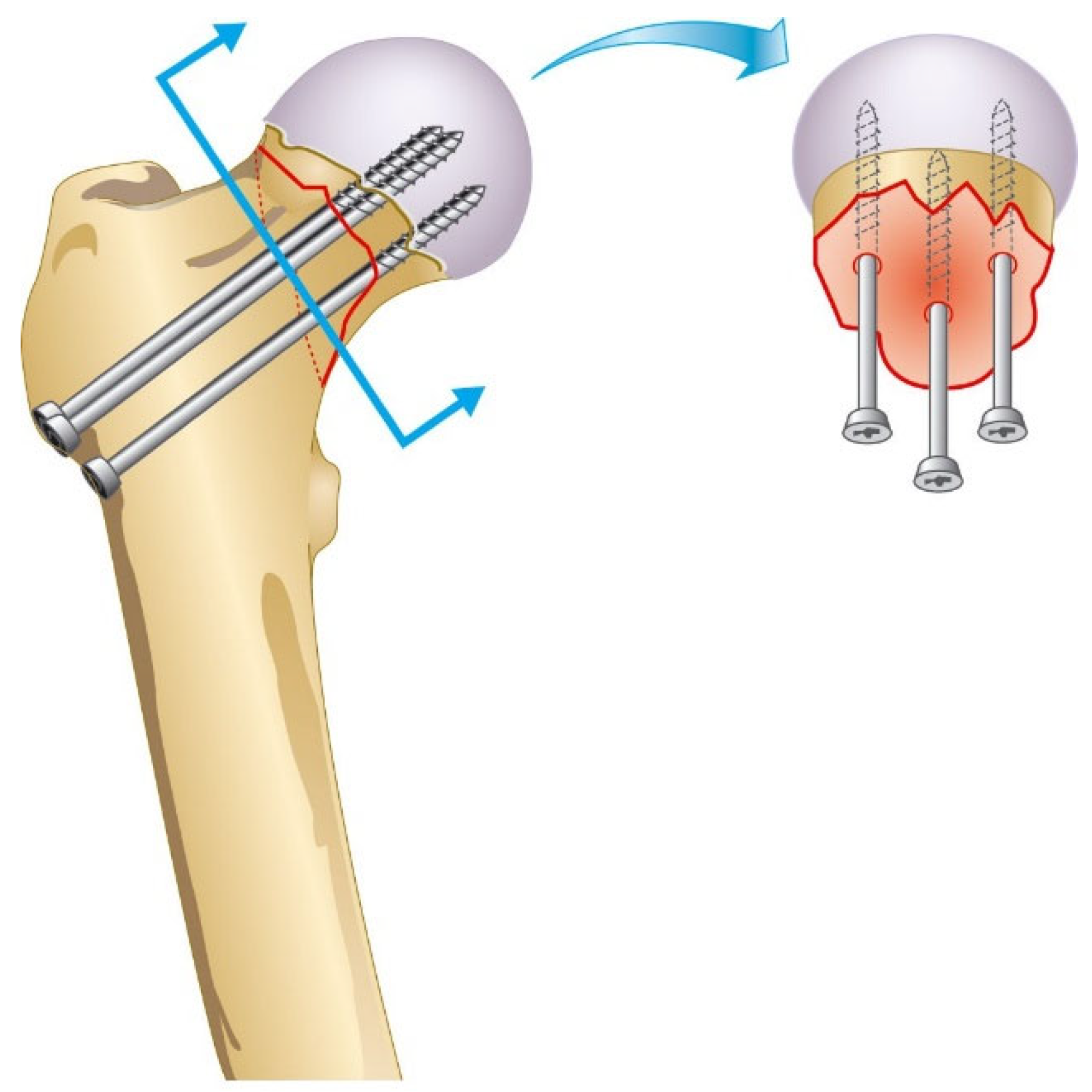
Figure 1. Triangular insertion pattern of three cannulated hip screws.
Surgeries 04 00048 g001
Figure 2. Insertion pattern of two cannulated hip screws.
Surgeries 04 00048 g002
Figure 3. Flowchart depicting inclusion of study subjects. CHS: cannulated hip screws. * Between January 2019 and December 2020.
Surgeries 04 00048 g003
Figure 4. Radiographs of cannulated hip screw fixation with two screws. (A) Preoperative radiograph. (B) Perioperative radiograph. (C) Anteroposterior radiograph at 6-week follow-up. (D) Lateral radiograph at 6-week follow-up.
Surgeries 04 00048 g004
Figure 5. Radiographs of cannulated hip screw fixation with three screws. (A) Preoperative radiograph. (B) Perioperative radiograph. (C) Anteroposterior radiograph at 6-week follow-up. (D) Lateral radiograph at 6-week follow-up.
Surgeries 04 00048 g005
Table 1. Baseline characteristics a.
Overall
(n = 100)
2 CHS
(n = 50)
3 CHS
(n = 50)
p
Age, mean (range)71 (29–97)72 (29–91)70 (38–97)0.427
Female, n (%) 74 (74)35 (70)39 (78)0.494
ASA-classification, n (%)117 (17)6 (12)11 (22)0.299
244 (44)21 (42)23 (46)
336 (36)22 (44)14 (28)
43 (3)1 (2)2 (4)
Kidney function class, n (%)G129 (29)13 (26)16 (32)0.326
G252 (52)27 (54)25 (50)
G3a10 (10)7 (14)3 (6)
G3b6 (6)1 (2)5 (10)
G42 (2)1 (2)1 (2)
G5000
Visit outpatient clinic a, mean (SD)7 (2)6 (2)7 (1)0.452
Garden, n (%)I69 (69)36 (72)33 (66)0.665
II31 (31)14 (28)17 (34)
Walking aid, n (%)Unknown5 (5)5 (10)00.070
Yes16 (16)8 (16)8 (16)
No79 (79)37 (74)42 (84)
Abbreviations: CHS, cannulated hip screw; SD, standard deviation; ASA, American Society of Anesthesiologists. a: in weeks.
Table 2. Treatment methods and outcomes a.
Overall
(n = 100)
2 CHS
(n = 50)
3 CHS
(n = 50)
p
Reoperation, n (%) 27 (27)14 (28)13 (26)1.000
Reoperation reason, n (%)Pain OSM15 (56)7 (50)8 (62)0.615
Coxarthrosis4 (15)2 (14)2 (15)
Screw cut out3 (11)2 (14)1 (8)
AVN1 (4)1 (7)0
Screw dislocation1 (4)1 (7)0
Fall1 (4)1 (7)0
Infection1 (4)01 (8)
Non-union1 (4)01 (8)
Reoperation type b, n (%)Screw replacement15 (56)7 (50)8 (62)0.042 *
HA5 (19)5 (36)0
THP7 (26)2 (14)5 (39)
Time to reoperation c, median [IQR]6 [3–10]4 [2–7]7 [5–11]0.157
Lateral protrusion d, median [IQR]1 [0–5]2 [0–5]1 [0–4]0.330
Mortality, n (%) 7 (7)3 (6)4 (8)1.000
Follow up c, mean (SD) 35 (15)26 (9)45 (12)<0.001 *
Abbreviations: CHS, cannulated hip screw; OSM, osteosynthesis material; AVN, avascular necrosis; HA, hemiarthroplasty; THP, total hip prosthesis; IQR, interquartile range; SD, standard deviation. a: due to rounding off, percentages might not add up to 100%. b: shown as percentage of all reoperations. c: in complete months. d: in millimeters, measured six weeks postoperatively. * Statistically significant difference (p < 0.05).
Table 3. Description of reoperations.
CaseCHSAgeSexASALateral Protrusion aLateral Protrusion bType of ReoperationReason of ReoperationMortalityTime to Reoperation c
1282F38 mm8 mmTHPCoxarthrosisNo25
2281F26 mm6 mmHAScrew cut outNo1
3276F32 mm5 mmScrew replacementPain complaintsNo4
4278F32 mm6 mmTHPCoxarthrosisNo5
5240M26 mm12 mmScrew replacementPain complaintsNo6
6268F25 mm5 mmScrew replacementPain complaintsNo7
7270F27 mm10 mmScrew replacementPain complaintsNo13
8256F113 mm17 mmScrew replacementPain complaintsNo3
9253F10 mm0 mmScrew replacementPain complaintsNo15
10271F34 mm5 mmHAFallYes2
11282M310 mm15 mmScrew replacementPain complaintsNo5
12286F31 mm11 mmHAAVNNo2
13289M35 mm9 mmHAScrew dislocationNo1
14287M413 mm15 mmHAScrew cut outYes1
15362F21 mm1 mmScrew replacementPain complaintsNo7
16366F41 mm6 mmScrew replacementPain complaintsNo8
17378F11 mm7 mmScrew replacementPain complaintsNo35
18378F38 mm10 mmScrew replacementPain complaintsNo7
19374F20 mm0 mmScrew replacementPain complaintsNo11
20389F36 mm12 mmTHPScrew cut outNo4
21366F24 mm5 mmTHPCoxarthrosisNo32
22344F116 mm19 mmScrew replacementPain complaintsNo10
23369F10 mm4 mmScrew replacementInfectionNo1
24373F210 mm13 mmTHPPain complaintsNo5
25375F28 mm12 mmScrew replacementPain complaintsNo1
26376F20 mm4 mmTHPCoxarthrosisNo12
27359F20 mm0 mmTHPNonunionNo6
Abbreviations: CHS, cannulated hip screw; ASA, American Society of Anesthesiologists; AVN, avascular necrosis of the femoral head. a: measured six weeks postoperatively. b: measured before reoperation. c: in complete months.
Disclaimer/Publisher’s Note: The statements, opinions and data contained in all publications are solely those of the individual author(s) and contributor(s) and not of MDPI and/or the editor(s). MDPI and/or the editor(s) disclaim responsibility for any injury to people or property resulting from any ideas, methods, instructions or products referred to in the content.