Article

Tannins from Acacia mearnsii De Wild as a Sustainable Alternative for the Development of Latent Fingerprints

by
Danielle Tapia Bueno
1,2,
Amanda Fonseca Leitzke
1,3,
Rayane Braga Martins
1,
Daisa Hakbart Bonemann
1,2,
Emanuel Gomes Bertizzolo
4,
Gabrielly Quartieri Sejanes
1,
Juliana Porciúncula da Silva
1,
Lucas Minghini Gonçalves
2,
Neftali Lenin Villarreal Carreno
2 and
Claudio Martin Pereira de Pereira
1,2,3,*
1
Center of Chemical, Pharmaceutical and Food Sciences, Innovation and Solutions in Chemistry Laboratory, Federal University of Pelotas, Eliseu Maciel St., s/n, Pelotas 96900-010, RS, Brazil
2
Postgraduate Program in Materials Science and Engineering, Technology Development Center, Federal University of Pelotas, Pelotas 96010-000, RS, Brazil
3
Postgraduate Program in Biotechnology, Federal University of Pelotas, Pelotas 96000-010, RS, Brazil
4
Department of Chemical Engineering, The University of Western Australia, Perth 6009, Australia
*
Author to whom correspondence should be addressed.
Organics 2025, 6(2), 27; https://doi.org/10.3390/org6020027
Submission received: 6 February 2025 / Revised: 2 June 2025 / Accepted: 12 June 2025 / Published: 18 June 2025

Abstract

:
Papilloscopy, the science of human identification through fingerprints, has seen notable advancements in developing less toxic latent fingerprint developers (LFDs), especially from natural feedstock. Tannins, the second most abundant natural polyphenol, present a potential eco-friendly and cost-effective alternative, with no record of their use as LFDs in the existing literature. This study characterized four types of tannins from black wattle, using Fourier Transform Infrared Spectroscopy, revealing key functional groups like C=O, C=C, and O–H. Ultraviolet–visible absorption spectra showed similar behaviors for all tannins, indicating phenolic and benzenoid structures. Energy-dispersive X-ray Spectroscopy identified high concentrations of chlorine, sodium, potassium, and sulfur, naturally found in biomass and soil. Finally, elements in significant concentrations, such as sodium, potassium, iron, zinc, and copper, were found through the incineration of the spent bark. On the basis of these findings, the tannin with the highest potential for LFD was selected. Combining this tannin with spent bark ash resulted in a composite whose performance was evaluated using different methods, including depletion studies, tests with various donors, and assessments on different surfaces. The results demonstrated that this combination significantly enhanced the material’s efficiency by integrating organic and inorganic properties, which improved visual contrast and powder adhesion.

1. Introduction

Fingerprints, when found at crime scenes, are considered circumstantial and indisputable evidence in forensic investigations and the criminal justice system due to their uniqueness, permanence, and immutability [1]. The study of fingerprints, or papilloscopy, is based on Locard’s exchange principle, which states that every contact leaves a trace. This principle highlights the forensic value of fingerprints, as they often retain exogenous compounds such as drugs or explosives, in addition to their distinct physical patterns [2]. Ridge patterns in fingerprints are permanent and can be used to individualize or exclude suspects in an investigation. The transfer of materials from the papillary ridges during contact provides a means to link individuals to objects or crime scenes [3]. Latent fingerprints (LFPs), which are invisible to the naked eye, are the most commonly found type at crime scenes. Specialized techniques are required to develop and visualize them [4]. These methodologies can be broadly categorized into physical and chemical techniques, depending on the type of surface and the conditions of the latent fingerprint [5]. Development involves the use of chemical or physical processes to interact with skin secretions and reveal the unique patterns of papillary ridges. Popular techniques include applying fine powders that adhere to the ridges and using optical or chemical methods such as ultraviolet light or cyanoacrylate fuming [4].
Challenges such as the toxicity and low contrast of conventional powders used in dusting techniques have encouraged the search for safer and more effective alternatives. These powders often have limitations on certain surfaces and can pose health risks to professionals exposed to them over prolonged periods [6]. Such factors emphasize the need to develop fingerprint developers with low or no toxicity and greater adherence to the diverse surfaces where latent fingerprints may be found [2,7]. In this context, the combination of organic and inorganic compounds has proven effective in improving fingerprint visualization through coloration or fluorescence. These approaches also ensure a strong affinity with substances commonly present in fingerprints, such as amino acids, proteins, fatty acids, and metal ions [8]. Continuous improvements in LFP development methods ensure their vital role in suspect identification and crime resolution. Furthermore, the literature reports various classes of organic compounds investigated as promising LFP developers, as in the work carried out by our research group [2,4,5,6,8,9,10,11,12,13,14,15,16,17]. Within this context, interest has emerged in investigating polyphenols as LFP developers.
Polyphenols represent a group of widely distributed and prevalent substances in plants, found in all vegetative organs such as flowers and fruits. There are several types of polyphenols, encompassing a broad range of molecules that share the characteristic of having at least one aromatic ring with one or more hydroxyl groups, in addition to other substituents [18]. This diversity ranges from simple molecules, such as phenolic acids, to complex and highly polymerized compounds like tannins, which stand out due to their intense pigmentation [19]. Tannins represent a natural, economical, and renewable biomass, extracted from various parts of plants and trees, such as seeds, fruits, roots, and bark. Their versatility is evidenced in applications ranging from leather manufacturing and wood additives to wine, beer, fruit juices, and cement superplasticizers, as well as in the medical and pharmaceutical industries [20]. In addition to that, this class of compounds have been studied as a bio-renewable and non-metallic flocculant for primary treatment in the water and wastewater industry [21,22]. Tannin is the second most abundant source of polyphenols, after lignin, and can be extracted from the bark of several forest species, including Acacia mearnsii, Schinopsis balansae, S. lorentzii, Stryphnodendron adstringens, Pinus radiata, Pinus oocarpa, Eucalyptus sp., Byrsonima verbascifolia Rich., Araucaria angustifolia, and Anadenanthera peregrina [23].
Acacia mearnsii De Wild, popularly known as black wattle or mimosa due to its dark green leaves in adulthood, is an exotic species in Brazil, originally from Australia [24]. In Brazil, black wattle is the main source of tannin, extensively cultivated in the southern region due to favorable climatic conditions. Additionally, black wattle stands out as a highly sustainable option, driven by its rapid growth cycle, reaching maturity in just seven years, and the high tannin concentration in its bark, which ranges from 30 to 45% [23]. Tannins can be categorized into two distinct groups, namely hydrolyzable and condensed (as shown in Figure 1a and Figure 1b, respectively) tannins, showing significant differences in their chemical structures and properties. Condensed tannins, in particular, are formed by flavonoid oligomers/polymers with varying degrees of polymerization. Their units are generally linked at C4–C6 or C4–C8, forming chains of different lengths. Black wattle bark tannin has a structure composed of oligomerized or polymerized flavan-3-ol monomeric units [25].
The bark of commercially cultivated black wattle trees undergoes a process of harvesting, hot water extraction, and subsequent spray-drying to obtain powdered acacia extract. Alternatively, it can be concentrated to form a solid or block product during cooling. These chemically unmodified acacia extracts are known in the market as acacia extract and are identified as A. mearnsii ext. by the European Chemicals Agency (EC No. 272-777-6; CAS No. 68911-60-4) [26]. As a result, the tannin extraction industry generates large volumes of waste, known as exhausted black wattle bark. According to the Brazilian Institute of Geography and Statistics (IBGE), more than 200,000 tons of this waste was produced in 2021. Classified as a lignocellulosic material, this waste is often associated with significant environmental issues due to improper management, with its primary application being energy generation [27]. In this context, exploring alternatives to enhance waste valorization of such by-products as a sustainable raw material source is essential.
This study explored, for the first time, the potential of tannins extracted from Acacia mearnsii De Wild as a developer for latent fingerprints. Additionally, a composite was developed using the extracted tannin and the ashes from the exhausted bark, which was subjected to a series of tests to assess its effectiveness in revealing latent fingerprints, reinforcing the full utilization of this lignocellulosic waste.

2. Materials and Methods

2.1. Sample Acquisition

Tannins from Acacia mearnsii De Wild, in powdered form, were kindly provided by Tanac SA (Montenegro, RS, Brazil). Four types of tannin samples (MTH and MT with CAS number 68911-60-4, and SH and SG with CAS number: 85029-52-3) were provided, as shown in Figure 2. All tannins are produced within the plant (Acacia mearnsii) through a specific precursor in a biosynthetic process. The biosynthetic process of the MT, MTH, SG, and SH tannins within the plant, as well as the meaning of these abbreviations, are not disclosed in this study due to restrictions related to the company’s intellectual property protection. The extraction of these tannins from the bark occurs in an aqueous medium, applying temperature and pressure. Subsequently, the extracted liquid is concentrated in an evaporator to remove water and then stored in tanks until the desired product is determined. The spent bark of Acacia mearnsii De Wild was also supplied by the same company.

2.2. Ash Production

To obtain ashes from each tannin, 3 g of the samples was weighed in pre-calcined porcelain crucibles. The samples were then placed in a muffle furnace at 550 °C for approximately 4 h or until the ashes turned white. After cooling in a silica desiccator, the samples were weighed, and the ash content was calculated by the difference between the initial and final mass after incineration [28].

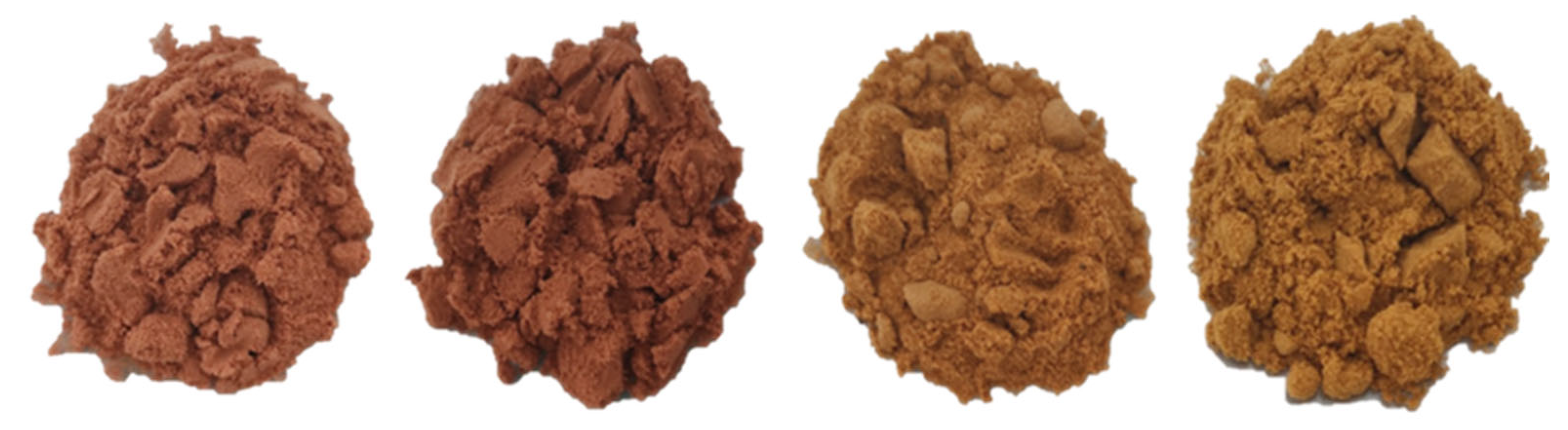
2.3. Composite Preparation

To formulate the composite made of MT tannin/ashes from spent bark in a 50:50 ratio (w/w), the powders were ground in a glass mortar with a pestle until a homogeneous mixture was obtained [2].

2.4. Characterization
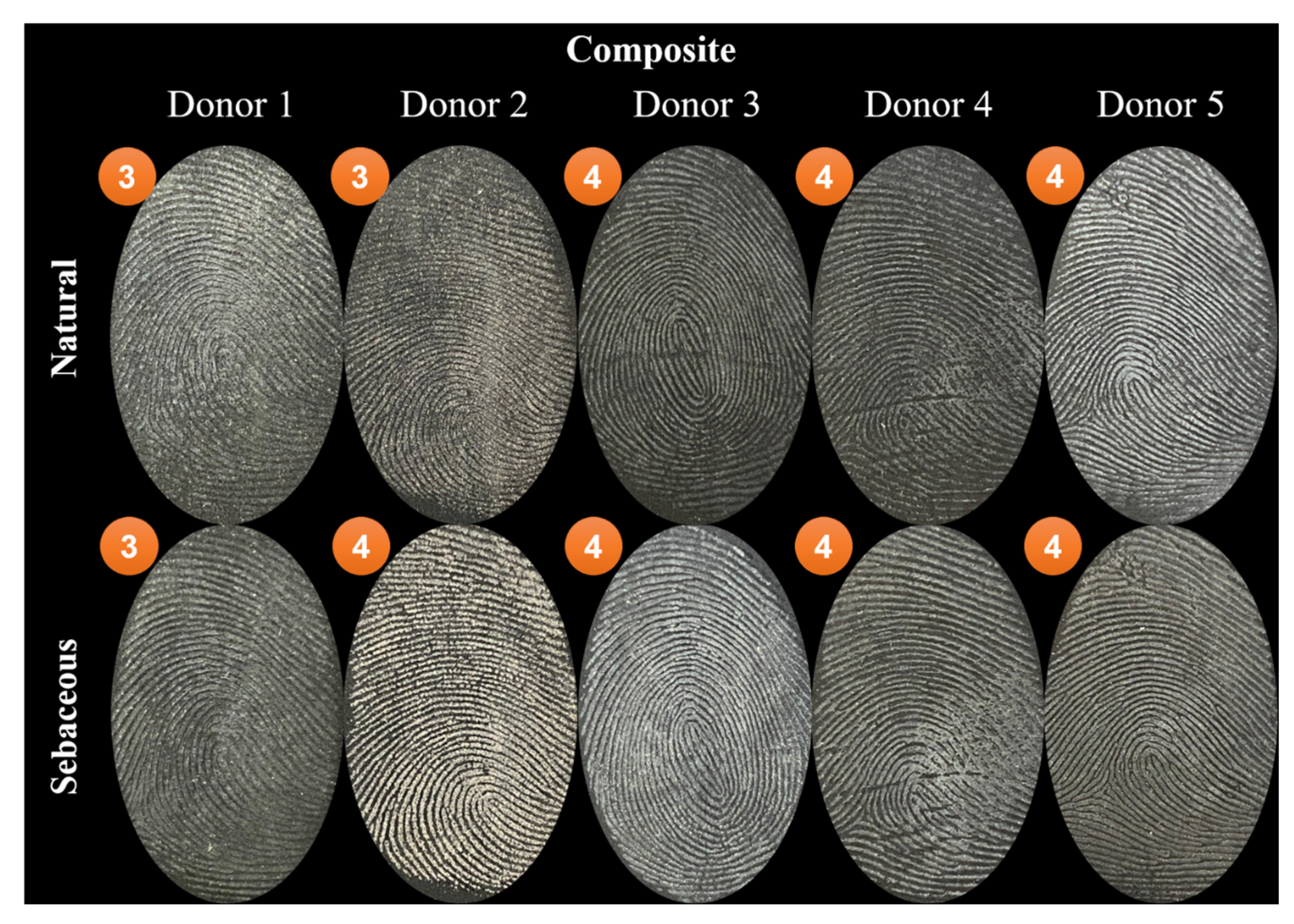
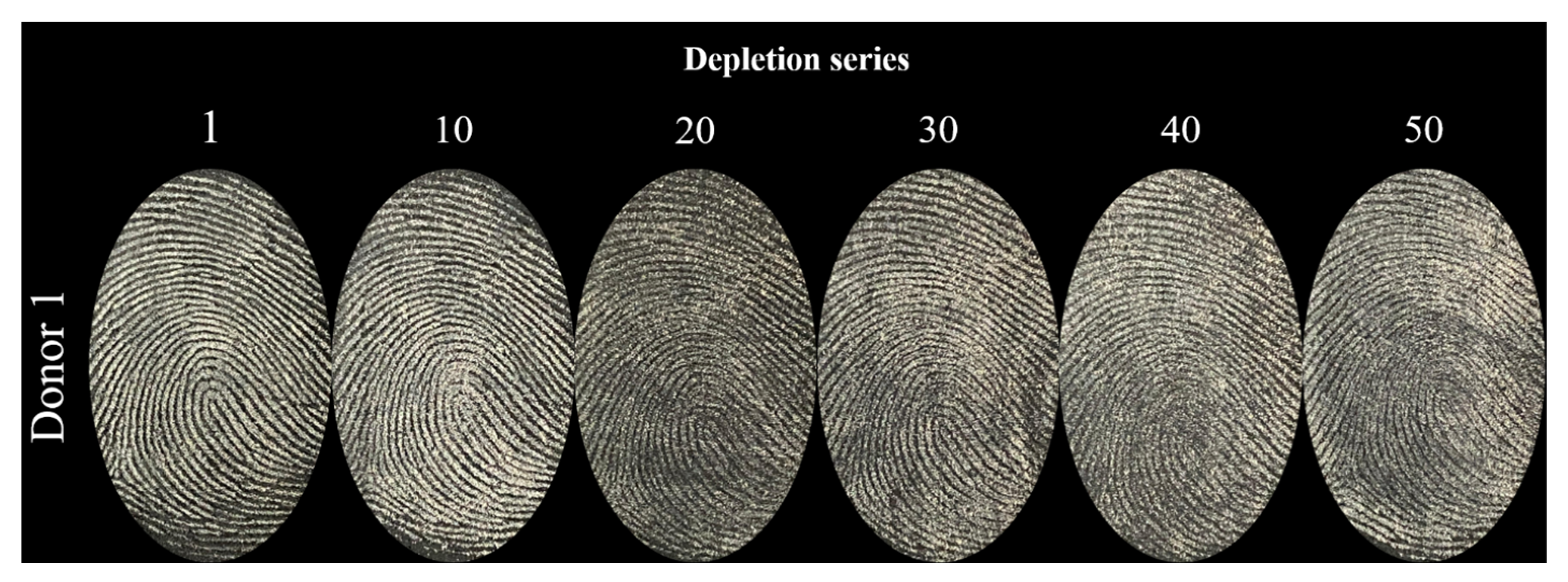
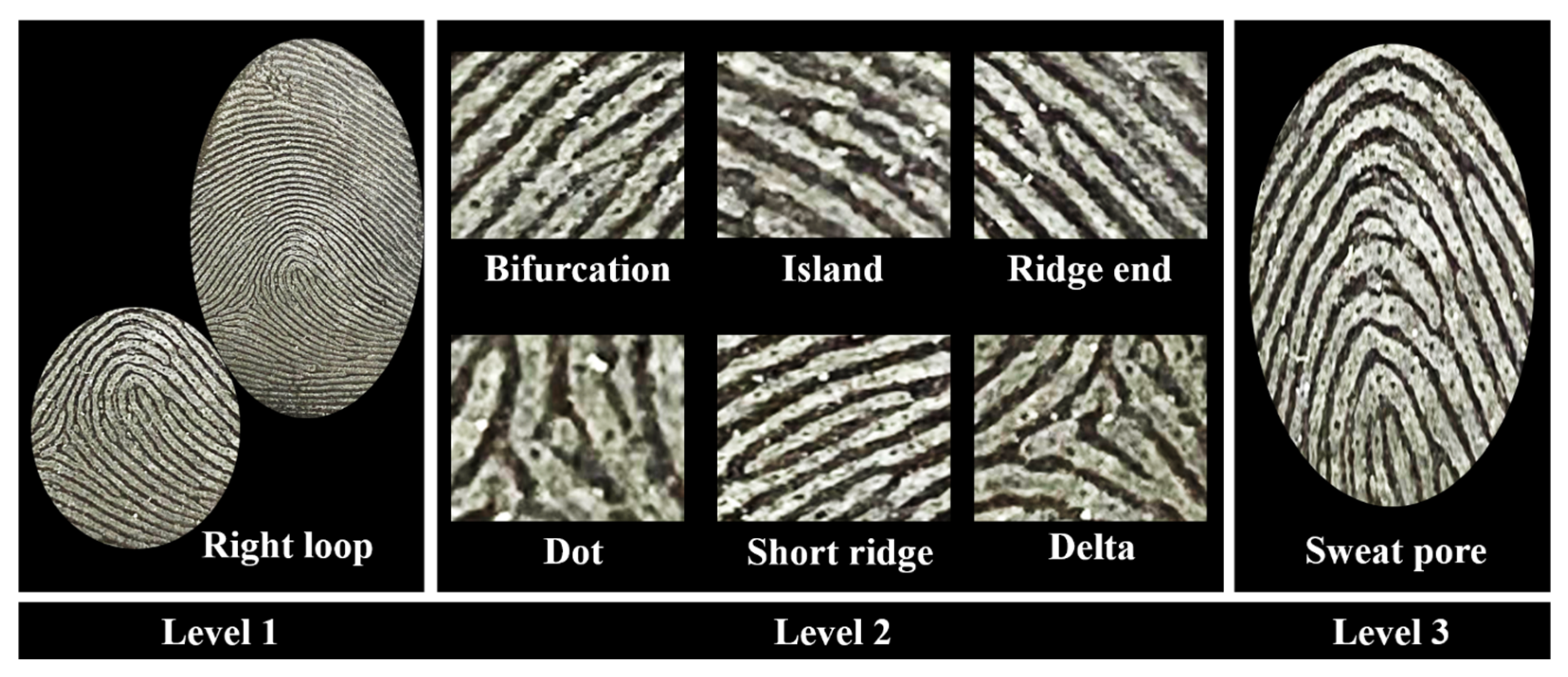
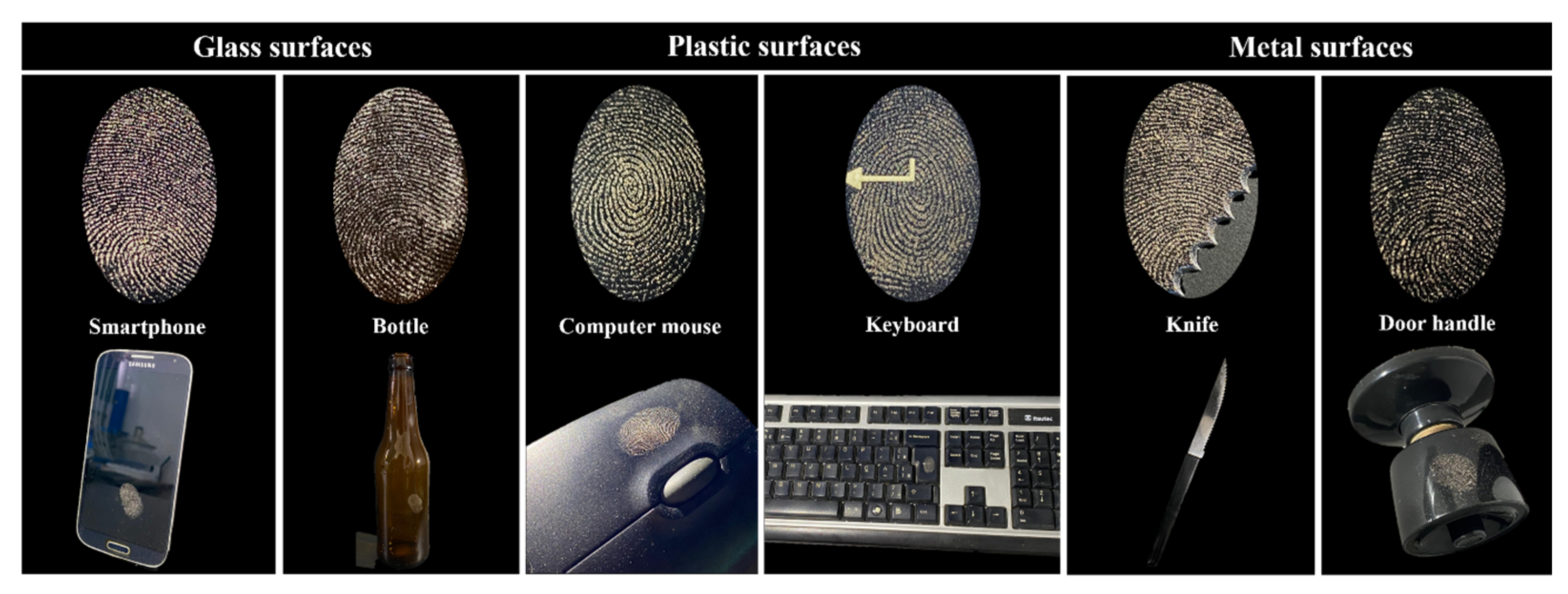
Ultraviolet–visible spectroscopy (UV–vis) was conducted using an LGS53 instrument (Monza, Italy—BEL Engineering). The samples were diluted in water to a concentration of 50 ppm (w/v) with the help of an ultrasonic bath. Fourier Transform Infrared Spectroscopy (FTIR) analyses were performed using a Shimadzu instrument (model SPIRIT) in ATR mode, scanning the wavenumber range of 4000 to 500 cm−1. For Energy-Dispersive X-ray Spectroscopy (EDS), the solid samples were analyzed using an EDS-720 X-ray energy-dispersive spectrometer (Shimadzu). Finally, for determining metals in the ash samples, aliquots were weighed and subsequently solubilized in nitric acid diluted in a 1:4 ratio. The elements chrome (Cr), copper (Cu), iron (Fe), potassium (K), manganese (Mn), sodium (Na), nickel (Ni), and zinc (Zn) were determined using a flame atomic absorption spectrometer (FAAS), while K and Na were analyzed using a flame atomic emission spectrometer (FAES) by an AAnalyst 200 model (Perkin Elmer, Shelton, CT, USA), equipped with a hollow cathode lamp for each analyte (Lumina, Perkin Elmer, Shelton, CT, USA) and a deuterium arc lamp for background correction. Na and K were measured in atomic emission mode. Acetylene gas (Linde, Barueri, SP, Brazil) was used as the fuel and compressed air as the oxidant. The atomizer height was set at 0.5 cm, and the acetylene gas flow rate was 2.5 L min−1 for all analytes.

2.5. Papilloscopy

The powder technique was used to develop fingerprints, employing specific brushes (132LBW and CFB100, purchased from Sirchie®, Youngsville, LA, USA). Developed fingerprints were photographed using a Canon EOS Rebel T6 digital camera (18 MP), equipped with a close-up +4 lens (58 mm), positioned at a distance of 9 cm with an f/5.6 aperture and an automatic ISO speed. To enhance visualization, three white LED lamps were used for brightness optimization, with a black background placed under the fingerprints. The captured images were processed in Adobe Photoshop. The surfaces used for fingerprint deposition and development were cleaned with water and neutral soap after photography. All captured photos were deleted after the publication of this work.
The method by Pacheco et al. [13] was used for depositing natural and sebaceous fingerprints. For simulating natural fingerprint deposition, donors washed their hands with water and neutral soap and then performed their routine activities for 30 min. For sebaceous deposition, the donor rubbed their thumb on the facial areas, including the forehead and nose, followed by rubbing with the fingertips to enhance the oily components in the fingerprint. During deposition, the applied pressure was subjectively firm (moderate pressure), with a contact time of 3 to 5 s. Fingerprints were developed after 24 h. Evaluations included comparisons among the composite, a standard Orange® powder (purchased from Sirchie®), and pure tannin.
Latent fingerprints were assessed using the classification proposed by Sears et al. [29] (Table 1). This evaluation was conducted by five independent analysts, all holding bachelor’s degrees in forensic chemistry from the Federal University of Pelotas. The final score, displayed to the left of the corresponding image in each figure, was determined by the most frequently assigned score among the five analysts.

3. Results and Discussion

3.1. Chemical Characterization

Figure 3a,b shows the FTIR-ATR spectra of the four types of tannins. A spectral range of 2800 to 3600 cm−1 in the spectra can be attributed to the significant extent of intermolecular interactions in tannin–tannin, tannin–water, and water–water structures [30]. The band at 1725 cm−1 is attributed to the stretching of C=O bonds due to tannin impurities, which may include residual hydrolyzed tannins or reagents such as formic or gallic acid [30,31]. The bands at 1608 and 1465 cm−1 correspond to the C=C stretching of aromatic rings in condensed tannin structures [23,30]. Axial deformations of O–H and C–O bonds between 1200–1000 cm−1 and out-of-plane angular deformations of aromatic C–H bonds between 850–600 cm−1 were also observed [23]. In the FTIR spectrum of the composite, characteristic bands associated with tannins were observed, including the band at 1600 cm−1, attributed to C=C stretching of aromatic rings; the band at 1018 cm−1, related to C–O bond deformations; the band at 600 cm−1, corresponding to out-of-plane angular deformations of aromatic C–H bonds; and a broad band around 3500 cm−1, associated with O–H stretching. Although these bands were present, they all exhibited reduced intensity, which may be attributed to the higher content of inorganic material in the composite due to the presence of ash. In contrast, the bands at 1441 and 710 cm−1 appeared to be significantly more intense and may be related to the C–H stretching vibrations [32]. The infrared spectra can also be seen in Figures S1 and S2 of the Supplementary Materials.
Figure 3c shows the UV–vis absorption spectra of the four types of tannin. Similar behavior was observed among the tannins, evidenced by two characteristic absorption bands. The maximum absorption peaks were recorded at 213, 217, 211, and 212 nm for the MT, MTH, SH, and SG tannins, respectively. This band, around 200 nm, is associated with the hydroxylated polyphenolic portion of the tannin structure. In addition, maximum absorptions were observed at 283 nm for the MT and MTH tannins, and at 287 nm for the SH and SG tannins. These bands, which vary around 280 nm, are recognized as benzenoid. These findings corroborate the literature data [23,33,34,35].
To understand the composition and elements present in the tannin samples, qualitative analysis was conducted using EDS (Energy-Dispersive X-ray Spectroscopy), and the results are presented in Table 2. The sample predominantly consisted of chlorine, with significant quantities of Na, K, and sulfur (S), indicating a material rich in halogenated salts and sulfates. These findings are consistent with Rodrigues et al. [36], who also observed a predominance of Na, K, and chlorine (Cl) in their work, along with lower levels of calcium (Ca), magnesium (Mg), aluminum (Al), silicon (Si), and S. Most of these heteroelements are commonly found in soils and thus occur naturally in biomass. It is unsurprising that tannins also contain some of these inorganic impurities, likely in the form of oxides, carbonates, phosphates, and sulfates [37]. Additionally, the presence of certain elements may be linked to the specific biosynthetic process used by the supplier, whose detailed methodology is protected by intellectual property rights. However, according to the technical data provided by the manufacturer, we know that these tannin samples are cationic polymers obtained from aqueous extracts of Acacia mearnsii bark, which can explain the elemental profile observed. The data are consistent with previous reports in the literature and have been included to support the broader material characterization [36,37].
Similar to the tannin composition analysis, determining the composition of the ashes is equally relevant, as they form part of the composite used to reveal latent fingerprints. Ash content was determined by atomic absorption spectroscopy, where the spent bark samples yielded approximately 4.5% ash after incineration. This analysis identifies non-volatile inorganic impurities obtained during incineration, which may originate from the plant itself (physiological ash) or from mineral/soil sources (non-physiological ash) [28]. Table 3 presents the metal concentrations detected in the ashes of the spent bark.
The ashes of the spent bark contained a high amount of Na, followed by K, Fe, Zn, and Cu. The diversity of these elements is naturally present in the bark biomass before tannin extraction and remains in the spent bark. This is evidenced by the work of Caldeira, Neto, and Schumacher [38], who directly evaluated elements in black wattle bark and found higher values for Fe, Na, Mn, Zn, and Cu (69.89, 60.67, 14.33, 12.69, and 2.58 mg kg−1, respectively), which are lower than those found in the present study. However, this can be explained by the pre-concentration effect on incinerating the spent bark prior to analysis [39].
Finally, regarding the presence of these minerals in the bark or tannins, it is important to highlight that the production of commercial tannins involves specific extraction and biosynthesis processes carried out by the supplier. Due to intellectual property protections, detailed information about these processes is not publicly available. However, it is known that extraction methods can be selective, potentially excluding certain elements from the final product. This may explain the low concentration of chromium observed in the tannin samples (Table 1), in contrast to the higher chromium content found in the bark ash sample (Table 2). Similar patterns are also observed for other elements, such as copper.

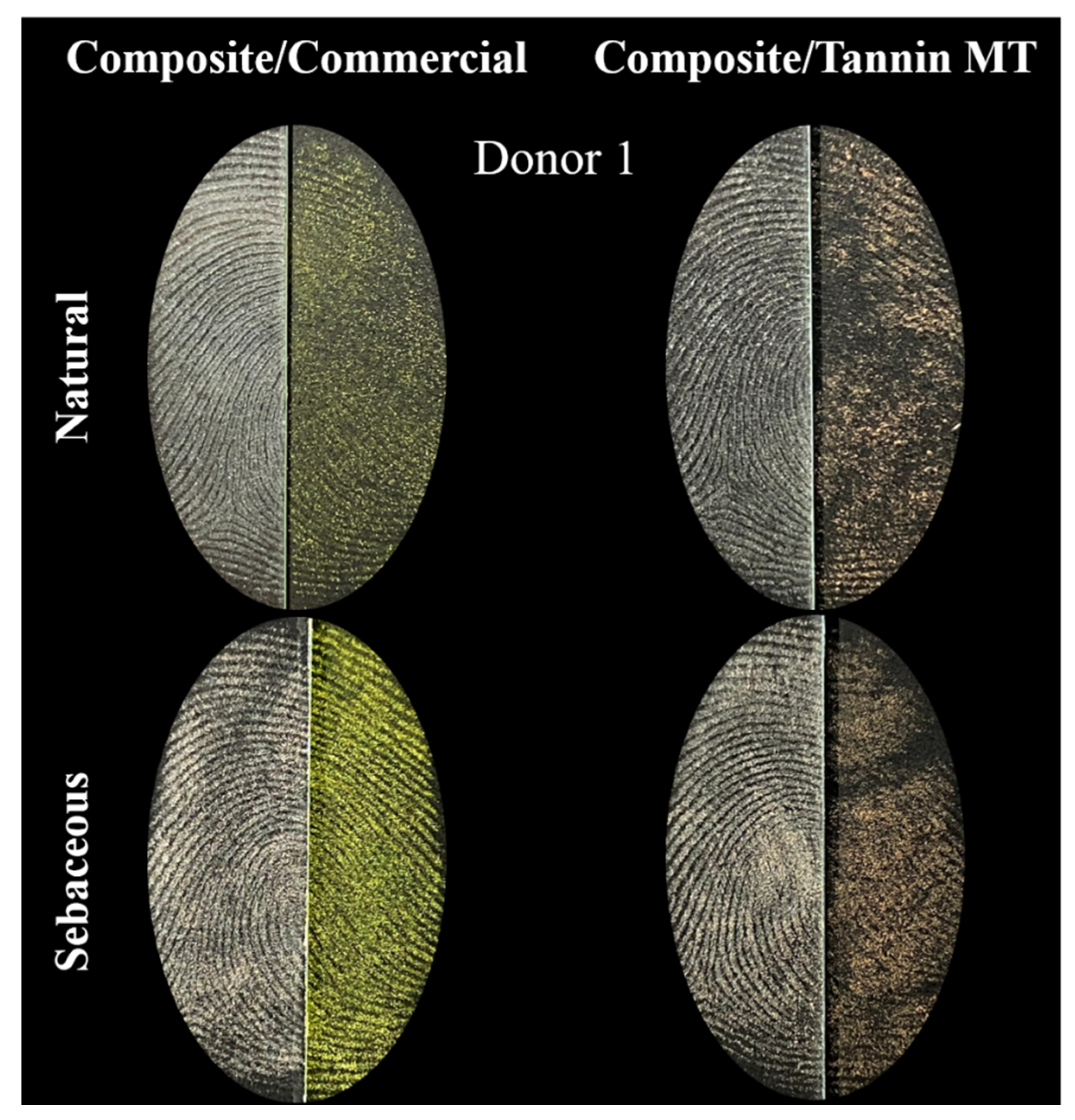
3.2. Papilloscopic Evaluation

Numerous studies emphasize the importance of including multiple donors in latent fingerprint development tests to account for biological and chemical variability. This approach captures differences influenced by factors such as age, gender, health status, and other individual conditions that affect the physical and chemical makeup of fingerprints [2,17,40,41]. In alignment with the recommendation by Sears et al. [29]—who proposed using at least five donors—this practice has become standard in the evaluation of fingerprint development powders [4,5,13].
In the initial phase of this study, four types of tannins were assessed for their effectiveness in developing both natural and sebaceous fingerprints from five donors on glass surfaces. The scoring system developed by Sears et al. [29], shown in Table 1, was employed to convert the visual quality of developed fingerprints into quantitative scores. The developed fingerprints and their corresponding scores are illustrated in Figure 4.
As shown in Figure 4, development quality varied significantly depending on the tannin used. Importantly, all tannins facilitated some degree of fingerprint visualization, as none of the samples received a score of zero. The MTH tannin exhibited the weakest performance, with scores of 1 and 2. A score of 1—indicating very faint development without identifiable ridge detail—was assigned to the natural fingerprints of Donors 2 and 4 and the sebaceous print of Donor 4. A score of 2, reflecting limited development with about one-third ridge visibility and insufficient detail for identification, was given to the natural fingerprints of Donors 1, 3, and 5 and the sebaceous prints of Donors 2 and 5. Only the sebaceous print of Donor 3 scored higher, receiving a 3 for partial ridge visibility (1/3 to 2/3), suggesting potential for identification.
The SH tannin demonstrated the highest performance for sebaceous fingerprints, with Donors 1, 2, and 3 receiving scores of 4, indicating complete ridge detail and full identifiability. Donors 4 and 5 received scores of 3. For natural fingerprints, the SH tannin yielded scores of 2 (Donors 1 and 4), 3 (Donors 3 and 5), and 4 (Donor 2), indicating moderate to high development quality. The SG tannin was less effective in developing natural fingerprints, with all samples scoring 2. However, it showed better results on sebaceous fingerprints, with scores of 3 for Donors 1 and 5, 4 for Donors 2 and 3, and 2 for Donor 4.
Among the tannins tested, the MT tannin showed the most consistent performance for natural fingerprints. All but one sample (Donor 4, score = 2) scored 3 or higher. Donors 1, 3, and 5 received scores of 3, and Donor 2 achieved a score of 4. For sebaceous fingerprints, the MT tannin yielded scores of 3 for Donors 1, 2, and 5, while Donors 3 and 4 scored 2. As seen throughout these results, the variability in fingerprint development is closely tied to the individual characteristics of the donors, which influence the chemical composition of latent residues. This is evident in the differing scores among donors even when the same tannin was used, reinforcing the need for versatile development materials capable of performing across diverse biological conditions.
In addition to the previously discussed aspects of tannin–fingerprint interaction, certain chemical characteristics of tannins play a critical role in the fingerprint development process. For fresh latent fingerprints, water constitutes the primary component facilitating powder adhesion [42]. In this context, the infrared spectral region between 2800 and 3600 cm−1, associated with strong intermolecular interactions such as tannin–tannin, tannin–water, and water–water bonding [30], is particularly relevant. These interactions, primarily involving hydrogen bonds and Van der Waals forces, enhance the attachment of tannin-based powders to fingerprint residues, thereby improving development quality [42]. The axial O–H bond deformations observed in tannins are indicative of their capacity to form such interactions, which aids in adhering effectively to the hydrophilic constituents of natural fingerprints.
Conversely, sebaceous fingerprints are predominantly composed of lipophilic substances such as fatty acids, waxes, glycerides, and sterols, with lower levels of water, amino acids, and inorganic ions [43]. Tannins adhere to these residues through hydrogen bonding between their functional groups, such as hydroxyl and carbonyl moieties (C=O, C–O, C–OH, and C=C), and the fatty components in sebum [44]. This chemical affinity supports the effective visualization of sebaceous fingerprints using tannin-based powders. Additionally, latent fingerprints often contain inorganic salts like sodium (Na), potassium (K), calcium (Ca), and chloride (Cl), which further enhance adhesion through ionic and hydrogen bonding [45]. The chlorine found in tannins may also interact with these metal ions, including iron (Fe), strengthening powder’s adherence [46].
Figure 5 illustrates the development results of both natural and sebaceous fingerprints from the same five donors previously assessed, but now using the composite material. The scoring results demonstrate a marked improvement in fingerprint development upon incorporating spent black wattle bark ashes into MT tannin. Natural fingerprints from Donors 1 and 2, and the sebaceous fingerprint from Donor 1, received a score of 3. Natural fingerprints from Donors 3, 4, and 5, along with sebaceous fingerprints from Donors 2 through 5, achieved the maximum score of 4. All fingerprints developed using the composite were classified as identifiable, underscoring the composite’s effectiveness.
The enhancement is attributed to the synergistic combination of organic and inorganic components in the composite. The organic material, with its deep coloration, improves contrast and visual clarity, while the inorganic ashes promote stronger adhesion to fingerprint residues. This dual mechanism significantly increases development efficiency [47]. The ashes’ metal content further explains this improvement: sodium enhances the powder’s hygroscopic nature, aiding moisture retention and adherence [36]; potassium, an alkali metal, supports even powder distribution [48]; and zinc (Zn) and copper (Cu)—the most abundant metals in the ashes—contribute to enhanced adhesion and visibility. Notably, copper-based powders are known to interact favorably with lipids and amino acids in fingerprint residues, yielding sharper and clearer ridge details [49].
In papilloscopy studies, the depletion series is a widely used methodology to determine the relative sensitivity of a fingerprint development technique. This approach consists of the sequential deposition of the same fingerprint on a preselected surface [29]. From the first deposition to the last, there is a progressive decrease in the residues present in the fingerprint due to the transfer of residual components to the surface [50]. In this context, the lower a powder can effectively develop a fingerprint in the depletion series, the higher its sensitivity. However, it is important to consider potential sources of human error, especially related to pressure application, which must be kept constant in all depositions to ensure the reproducibility and reliability of the results [29]. For depletion studies, sebaceous fingerprints are used, and the number of deposited marks varies depending on the type of surface analyzed. According to Sears et al. [29], on non-porous surfaces such as glass, a depletion series with 20 or more marks is often required for sensitivity differences to be observed.
To assess the composite’s sensitivity, a depletion study involving 50 sequential fingerprint depositions was performed. Fingerprints from Depositions 1, 10, 20, 30, 40, and 50 were developed using the composite and analyzed. This procedure was conducted across five donors. Figure 6 displays the depletion series for Donor 1, while data for Donors 2 through 5 are provided in Supplementary Figure S3. In Figure 6, Fingerprints 1 and 10 exhibited strong contrast and clear ridge detail. However, as deposition progressed, contrast gradually declined, though ridge minutiae remained identifiable. The results varied across donors, as shown in Figure S3, likely due to individual differences in fingerprint composition, particularly the types and quantities of fatty acids influenced by diet, cosmetics, and personal habits [51].
For Donor 2, all fingerprints showed ridge details with minimal visual change across the series. Donor 3′s fingerprints remained well-developed through to Deposition 30, but clarity was lost by Depositions 40 and 50. Donor 5 exhibited lower contrast as early as Deposition 10, and ridge details were no longer visible by Depositions 40 and 50. Donor 4 maintained visible ridge patterns up to Deposition 20, although Fingerprints 10 and 20 had lower contrast than Deposition 1; later fingerprints lacked identifiable features. These findings indicate that the composite’s performance varies depending on the donor, reflecting individual chemical variability in fingerprint residues. While the composite reliably developed early depositions with high contrast and clarity, a consistent decline was observed in later stages. This trend underscores the composite’s potential for sensitive fingerprint detection, particularly in early depositions, but also highlights the need for further refinement to enhance its effectiveness in more challenging depletion scenarios.
In addition to the previously discussed inter-donor differences in fingerprint composition, it is important to note that intra-donor variability also plays a significant role. The quantity and composition of fingerprint residue—particularly sweat—can fluctuate within the same individual due to factors such as emotional state, environmental conditions, and psychological influences [29]. To reduce the impact of such variability and enhance consistency, this study employed a method where each fingerprint was bisected along the central axis. This approach allowed side-by-side comparisons of the proposed composite, a standard commercial powder, and pure MT tannin.
Figure 7 illustrates natural and sebaceous fingerprints from Donor 1. When comparing development quality, the composite outperformed the commercial powder in rendering the natural fingerprint, offering sharper and more defined ridge details. For the sebaceous fingerprint, the composite’s performance was comparable with the commercial powder. Pure MT tannin—comprising 50% of the composite—was also effective in developing both fingerprint types.
Notably, minor differences were observed between two natural fingerprints from Donor 1 developed using the composite and the commercial powder, with the latter exhibiting slightly greater contrast in one instance. This reinforces the concept of compositional variability even within the same individual. Additional fingerprints developed using both the composite and the commercial powder are shown in Supplementary Figure S4. These results demonstrate that the composite effectively developed natural fingerprints for Donors 2 and 4, while for Donors 3 and 5, its performance aligned closely with that of the commercial powder. For sebaceous fingerprints, the composite performed similarly to the commercial powder for Donors 2 and 5, and showed superior results for Donors 3 and 4.
Supplementary Figure S5 presents fingerprints developed using both the composite and pure MT tannin. In this analysis, the composite consistently outperformed pure MT tannin for all natural fingerprints, providing significantly higher contrast and ridge clarity. For sebaceous fingerprints, the composite also yielded effective results across all donors. However, ridge detail was still visible in prints developed with pure MT tannin for Donors 2, 3, and 5.
Overall, the composite demonstrated equivalent or superior performance compared with both the standard commercial powder and pure MT tannin in developing natural and sebaceous fingerprints. Its enhanced sharpness and contrast, particularly for natural prints, highlights its potential as a highly sensitive material. Furthermore, the observed intra-donor variability underscores the importance of using versatile development materials capable of adapting to complex and fluctuating fingerprint chemistries.
A detailed evaluation was also conducted, where the characteristics of the three levels of biometric identification were identified. The first level includes the fundamental types (left loop, right loop, arch, and whorl), with the right loop shown in Figure 8. The second level involves the minutiae, some of which are highlighted in the image, such as bifurcations, island, ridge ends, dots, short ridges, and deltas. Finally, the third level is related to all dimensional attributes of ridges, including width, shape, contour, sweat pores, scars, folds, and other permanent details, where the sweat pores are highlighted in the figure below [2]. On the basis of the evaluation performed, it was possible to identify characteristics from all three levels of biometric identification, demonstrating the efficiency of the method used in the detailed analysis of fingerprints. This detailing reinforces the applicability of the composite in biometric studies and forensic analysis, highlighting its potential to capture both the general characteristics and unique details of fingerprints, which are essential for identification processes.
To conclude the fingerprint study with the composite developed in this work, fingerprints deposited on different surfaces were analyzed (Figure 9). The versatility of a latent fingerprint powder is essential for its applicability in operational scenarios, where surfaces can vary greatly [17]. Thus, the analyses were performed on glass surfaces, such as a smartphone screen and a bottle; plastic surfaces, such as a mouse and a computer keyboard; and metal surfaces, such as a knife and door handle. The results showed that the composite was efficient in revealing latent fingerprints on all tested surfaces, although performance varied. On the smartphone, the fingerprint showed excellent sharpness and contrast. On the bottle, despite this also being made of glass, the contrast was lower, possibly due to the specific coloration of the material. On plastic surfaces, both the keyboard and the mouse showed good sharpness and contrast in the revealed fingerprints. On the other hand, on metal surfaces, the performance was inferior: although the ridges of the fingerprint were relatively clear on the knife, the sharpness on the door handle was lower, compromising the identification of fingerprint details. Thus, it was observed that the developed composite demonstrated versatility and efficiency in revealing latent fingerprints on various surfaces, with superior performance on glass and plastic.
This study reinforces the significance of selecting diverse donors and underscores the impact of tannin type on the quality of latent fingerprint development. The observed variation in scores for both natural and sebaceous fingerprints highlights the influence of specific chemical interactions—particularly hydrogen bonding and other intermolecular forces—on the adhesion and visualization of latent prints. Notably, incorporating ashes from exhausted black wattle (Acacia mearnsii) bark into MT tannin emerged as a highly effective strategy, markedly enhancing the performance of the fingerprint-developing composite. This improvement can be attributed to the synergistic combination of organic and inorganic elements, which bolstered both visual contrast and powder adhesion. The presence of key metals—copper, zinc, sodium, and potassium—in the ashes played a pivotal role by promoting increased ridge sharpness and more uniform fingerprint development.
The depletion series further confirmed that donor-specific factors significantly affect the chemical composition of sebaceous fingerprints. Impressively, the composite maintained good sensitivity up to the 30th deposition cycle, indicating its durability and effectiveness across repeated uses. Evaluations using divided fingerprints revealed that the composite performed on par with, or even surpassed, both the standard commercial powder and pure MT tannin. This was especially evident in the development of natural fingerprints, where the composite provided superior ridge clarity and visual contrast.
Moreover, the composite successfully captured features across all three levels of biometric fingerprint analysis, ranging from general ridge flow (Level 1) to minutiae and pore detail (Levels 2 and 3). This indicates its potential for high-resolution forensic applications requiring accurate and detailed analyses. Surface tests confirmed the composite’s versatility, yielding excellent results on glass and plastic substrates. While its performance was somewhat reduced on metal surfaces, this outcome underscores the need for further optimization to extend its effectiveness across a broader range of operational environments.

4. Conclusions

This study represents a pioneering contribution to the exploration of tannins as latent fingerprint (LFP) development agents, while simultaneously addressing a critical environmental challenge associated with tannin extraction industries. By investigating the reuse of exhausted black wattle (Acacia mearnsii) bark—a byproduct often regarded as waste with substantial environmental implications—this work opens a promising avenue for sustainable material valorization. In particular, the composite developed from MT tannin combined with the ashes of exhausted bark demonstrated both environmental and forensic relevance. It proved to be a highly effective material for LFP development, showing consistent performance across different donors and substrates. Notably, on glass surfaces, the composite enabled visualization of third-level biometric details such as sweat pores, emphasizing its high sensitivity and resolution.
The composite stood out for its excellent balance of sensitivity, efficiency, and adaptability to various operational conditions. Its formulation, based on accessible, low-cost, scalable, and easy-to-handle components, underscores its practicality and applicability in real-world forensic contexts. These characteristics not only highlight the material’s potential for biometric and forensic applications but also reinforce its role as an innovative, sustainable alternative within the field of latent fingerprint development. Ultimately, this study aligns environmental stewardship with scientific advancement, offering a viable path toward more eco-conscious forensic technologies.

Supplementary Materials

The following supporting information can be downloaded at https://www.mdpi.com/article/10.3390/org6020027/s1. Figure S1: FTIR-ATR transmission (4000–500 cm−1). Figure S2: FTIR-ATR transmission (2000–500 cm−1). Figure S3: Depletion series of Donors 2 to 5. Figure S4: Half-and-half fingerprints (natural and sebaceous), with the left half developed using the composite and the right half developed using commercial Orange® powder purchased from Sirchie®. Figure S5: Half-and-half fingerprints (natural and sebaceous), with the left half developed using the composite and the right half developed using MT tannin.

Author Contributions

Conceptualization, D.T.B.; methodology, D.T.B.; validation, A.F.L.; formal analysis, D.H.B. and L.M.G.; investigation, D.T.B., A.F.L., D.H.B., R.B.M., J.P.d.S. and G.Q.S.; data curation, D.T.B. and A.F.L.; writing—original draft preparation, D.T.B.; writing—review and editing, A.F.L. and E.G.B.; supervision, C.M.P.d.P. and N.L.V.C.; funding acquisition, C.M.P.d.P.; project administration, C.M.P.d.P. All authors have read and agreed to the published version of the manuscript.

Funding

Financial support for this research by FAPERGS (Research Support Foundation of the Rio Grande do Sul State 22/2551-0000840-2), Coordination for Improvement of Higher-Level Personnel (CAPES 001), and the Forensic National Institute of Science and Technology (Grant number 465450/2014-8).

Data Availability Statement

The data can be made available on request.

Acknowledgments

The authors are thankful to FAPERGS, CAPES, the Forensic National Institute of Science and Technology, and the Brazilian Federal Police for their assistance.

Conflicts of Interest

The authors declare no conflicts of interest.

Abbreviations

The following abbreviations are used in this manuscript.
LFPsLatent Fingerprints
IBGEBrazilian Institute of Geography and Statistics
UV–VisUltraviolet–Visible Spectroscopy
FTIRFourier Transform Infrared Spectroscopy
EDSEnergy-Dispersive X-ray Spectroscopy
FAASFlame Atomic Absorption Spectrometer
FAESFlame Atomic Emission Spectrometer

References

  1. Silveira, C.I.; Pereira, S.A.; de Leon, V.B.; Camacho, J.T.; Leitzke, A.F.; Freitag, R.A.; Pereira, C.M.P.; Rosa, B.N.D. Reutilização Do Ácido Acetilsalicílico Preparado Nas Aulas De Química Orgânica Experimental Como Revelador De Impressões Digitais: Uma Aplicação No Ensino De Química. Química Nova 2024, 48, e-20250062. [Google Scholar] [CrossRef]
  2. Bueno, D.T.; Leitzke, A.F.; Crizel, R.L.; Jansen-Alves, C.; Bertizzolo, E.G.; da Silva, J.P.; Sejanes, G.Q.; Mariotti, K.C.; de Pereira, C.M.P. Characterization of Bixin by UV-Visible Spectroscopy and HPLC, and Its Application as Latent Fingermark Developer. Analytica 2024, 5, 107–118. [Google Scholar] [CrossRef]
  3. Christofdis, G.; Morrissey, J.; Birkett, J.W. Detection of fngermarks—Applicability to metallic surfaces: A literature review. J. Forensic Sci. 2018, 63, 1616–1627. [Google Scholar] [CrossRef] [PubMed]
  4. Leitzke, A.F.; Bueno, D.T.; Poletti, T.; Maron, G.K.; Lopes, B.V.; Morais, E.V.; Inacio, A.P.O.L.; Silveira, C.I.; Silva, J.P.; Dias, D.; et al. The effectiveness of natural indigo/kaolinite composite powder in the development of latent fingermarks. Egypt. J. Forensic Sci. 2024, 14, 19. [Google Scholar] [CrossRef]
  5. da Rosa, B.N.; da Rosa, M.P.; Poletti, T.; de Lima, N.P.K.; Maron, G.K.; Lopes, B.V.; Mariotti, K.C.; Beck, P.H.; Carreño, N.L.V.; Pereira, C.M.P. Green composites from thiophene chalcones and rice husk lignin: An alternative of powder for latent fingermark. Surfaces 2022, 5, 481–488. [Google Scholar] [CrossRef]
  6. Leitzke, A.F.; Berneira, L.M.; Rosa, B.N.D.; Moreira, B.C.; Mariotti, K.D.C.; Venzke, D.; Pereira, C.M. A Química de Produtos Naturais aplicados a reveladores de impressões digitais latentes. Química Nova 2022, 45, 424–434. [Google Scholar] [CrossRef]
  7. Vadivel, R.; Nirmala, M.; Anbukumaran, K. Commonly available, everyday materials as non-conventional powders for the visualization of latent fingerprints. Forensic Chem. 2021, 24, 100339. [Google Scholar] [CrossRef]
  8. da Rosa, B.N.; Maron, G.K.; Lopes, B.V.; Rocha, A.C.S.; de Moura Gatti, F.; Machado, J.O.A.; Barichello, J.M.; Mariotti, K.C.; Trossini, G.H.G.; Carreño, N.L.V.; et al. Dimethylaminochalcones with silicon dioxide and zinc oxide as latent fingermark developer powder. Mater. Chem. Phys. 2023, 295, 127033. [Google Scholar] [CrossRef]
  9. Nicolodi, C.; Da Rosa, B.N.; Da Silva, C.C.; Berneira, L.M.; Pacheco, B.S.; Poletti, T.; Venzke, D.; Mariotti, K.C.; Pereira, C.M.P. Aplicação de condimentos na revelação de impressões digitais latentes: Um experimento no ensino de química. Quim. Nova 2019, 42, 962–970. [Google Scholar] [CrossRef]
  10. da Rosa, B.N.; Venzke, D.; Poletti, T.; de Lima, N.P.K.; Camacho, J.T.; Mariotti, K.C.; dos Santos, M.A.Z.; Pizzuti, L.; Carreño, N.L.V.; Pereira, C.M.P. Microwave assisted synthesis of thiocarbamoylpyrazoles and application as an alternative latent fingermark developer. J. Braz. Chem. Soc. 2020, 31, 1327–1331. [Google Scholar] [CrossRef]
  11. da Rosa, B.N.; Mariotti, K.C.; Pacheco, B.S.; Silva, C.C.; Carreño, N.L.V.; Nicolodi, C.; Poletti, T.; Silva, A.C.A.; Silva, A.F.; Giongo, J.L.; et al. Fluorescent phenylthiazoles: Application as latent fingermark and their cytotoxicity against NOK-SI cell line. Chem. Data Collect. 2021, 33, 100700. [Google Scholar] [CrossRef]
  12. Venzke, D.; Poletti, T.; Rosa, B.N.; Berneira, L.M.; Lima, N.P.K.; Oliveira, T.F.; Carreño, N.L.V.; Mariotti, K.C.; Duarte, L.S.; Nobre, S.M.; et al. Preparation of fluorescent bisamides: A new class of fingermarks developers. Chem. Data Collect. 2021, 33, 100680. [Google Scholar] [CrossRef]
  13. Pacheco, B.S.; Da Silva, C.C.; Da Rosa, B.N.; Mariotti, K.C.; Nicolodi, C.; Poletti, T.; Segatto, N.V.; Collares, T.; Seixas, F.K.; Paniz, O.; et al. Monofunctional curcumin analogues: Evaluation of green and safe developers of latent fingerprints. Chem. Pap. 2021, 75, 3119–3129. [Google Scholar] [CrossRef]
  14. Passos, L.F.; Berneira, L.M.; Poletti, T.; Mariotti, K.C.; Carreño, N.L.V.; Hartwig, C.A.; Pereira, C.M.P. Evaluation and characterization of algal biomass applied to the development of fingermarks on glass surfaces. Aust. J. Forensic Sci. 2021, 53, 337–346. [Google Scholar] [CrossRef]
  15. Lima, N.P.; Rosa, B.N.; Poletti, T.; Moreira, B.C.; Leitzke, A.F.; Mariotti, K.C.; Carreño, N.L.V.; Pereira, C.M. As clássicas hidrazonas como reveladores de impressões digitais: Uma proposta de química orgânica experimental. Química Nova 2023, 46, 215–221. [Google Scholar] [CrossRef]
  16. Poletti, T.; Berneira, L.M.; Bueno, D.T.; da Silva, C.C.; da Silva, R.; Pereira, C.M. Chemical evaluation and application of cinnamaldehyde-derived curcumins as potential fingerprint development agents. Talanta Open 2022, 6, 100133. [Google Scholar] [CrossRef]
  17. Bueno, D.T.; Leitzke, A.F.; da Silva, J.P.; Bonemann, D.H.; Sejanes, G.Q.; da Rosa, B.N.; Poletti, T.; Maron, G.K.; Lopes, B.V.; Goularte, M.P.; et al. Application of Κ-Carrageenan for One-Pot Synthesis of Hybrids of Natural Curcumin with Iron and Copper: Stability Analysis and Application in Papiloscopy. Colorants 2025, 4, 3. [Google Scholar] [CrossRef]
  18. Ferrazzano, G.F.; Amato, I.; Ingenito, A.; Zarrelli, A.; Pinto, G.; Pollio, A. Plant polyphenols and their anti-cariogenic properties: A review. Molecules 2011, 16, 1486–1507. [Google Scholar] [CrossRef]
  19. Pietta, P.; Minoggio, M.; Bramati, L. Plant polyphenols: Structure, occurrence and bioactivity. Stud. Nat. Prod. Chem. 2003, 28, 257–312. [Google Scholar]
  20. Sun, X.; Zhang, J.; You, Y. Enhancement of Cu (II) removal by carbon disulfide modified black wattle tannin gel. Colloids Surf. A Physicochem. Eng. Asp. 2021, 608, 125594. [Google Scholar] [CrossRef]
  21. Hameed, Y.T.; Idris, A.; Hussain, S.A.; Abdullah, N. A tannin-based agent for coagulation and flocculation of municipal wastewater: Chemical composition, performance assessment compared to Polyaluminum chloride, and application in a pilot plant. J. Environ. Manag. 2016, 184, 494–503. [Google Scholar] [CrossRef] [PubMed]
  22. Lee, C.S.; Robinson, J.; Chong, M.F. A review on application of flocculants in wastewater treatment. Process Saf. Environ. Prot. 2014, 92, 489–508. [Google Scholar] [CrossRef]
  23. Oliveira, A.L.; de Freitas, A.D.S.; Rodrigues, J.S.; do Carmo, K.P.; Ferreira, M.; Botaro, V.R. Enhancement of UV-C irradiation protection of polystyrene matrix by tannin (Acacia mearnsii De Wild) additivation: Accelerated aging test. J. Mater. Res. 2023, 38, 1593–1608. [Google Scholar] [CrossRef]
  24. Prietsch, A.V.G.; Weimer, P.; Spies, L.M.; Haubert, R.; Nunes, A.J.S.; Kreutz, O.C.; Morisso, F.D.P.; Maluf, R.W.; Moura, A.B.D.; Ziulkoski, A.L.; et al. Isolation of Valuable Bioactive Compounds from Aerial Parts of Black Wattle, a Forest Solid Waste. Waste Biomass Valorization 2021, 12, 4953–4964. [Google Scholar] [CrossRef]
  25. Venter, P.B.; Senekal, N.D.; Kemp, G.; Amra-Jordaan, M.; Khan, P.; Bonnet, S.L.; Van Der Westhuizen, J.H. Analysis of commercial proanthocyanidins. Part 3: The chemical composition of wattle (Acacia mearnsii) bark extract. Phytochemistry 2012, 83, 153–167. [Google Scholar] [CrossRef]
  26. Cesprini, E.; De Iseppi, A.; Giovando, S.; Tarabra, E.; Zanetti, M.; Šket, P.; Marangon, M.; Tondi, G. Chemical characterization of cherry (Prunus avium) extract in comparison with commercial mimosa and chestnut tannins. Wood Sci. Technol. 2022, 56, 1455–1473. [Google Scholar] [CrossRef]
  27. Rodrigues, T.L.; Pedroso, P.D.C.; de Freitas, J.H.C.; Carvalho, A.C.P.; Flores, W.H.; Morais, M.M.; da Rosa, G.S.; de Almeida, A.R.F. Obtaining of a rich-cellulose material from black wattle (Acacia mearnsii De Wild.) bark residues. Environ. Sci. Pollut. Res. 2023, 30, 113055–113067. [Google Scholar] [CrossRef] [PubMed]
  28. IAL—Instituto Adolfo Lutz. Métodos Físico-Químicos Para Análise de Alimentos, 4th ed.; IAL: São Paulo, Brazil, 2008; 1020p. [Google Scholar]
  29. Sears, V.G.; Bleay, S.M.; Bandey, H.L.; Bowman, V.J. A methodology for finger mark research. Sci. Justice 2012, 52, 145–160. [Google Scholar] [CrossRef]
  30. da Cruz, J.A.; da Silva, A.B.; Ramin, B.B.S.; Souza, P.R.; Popat, K.C.; Zola, R.S.; Kipper, M.J.; Martins, A.F. Poly (vinyl alcohol)/cationic tannin blend films with antioxidant and antimicrobial activities. Mater. Sci. Eng. C 2020, 107, 110357. [Google Scholar] [CrossRef]
  31. Facchi, D.P.; Lima, A.C.; de Oliveira, J.H.; Lazarin-Bidóia, D.; Nakamura, C.V.; Canesin, E.A.; Bonafé, E.G.; Monteiro, J.P.; Visentainer, J.V.; Muniz, E.C.; et al. Polyelectrolyte complexes based on alginate/tanfloc: Optimization, characterization and medical application. Int. J. Biol. Macromol. 2017, 103, 129–138. [Google Scholar] [CrossRef]
  32. Lütke, S.F.; Igansi, A.V.; Pegoraro, L.; Dotto, G.L.; Pinto, L.A.; Cadaval Jr, T.R. Preparation of activated carbon from black wattle bark waste and its application for phenol adsorption. J. Environ. Chem. Eng. 2019, 7, 103396. [Google Scholar] [CrossRef]
  33. Cataldo, F.; Ursini, O.; Angelini, G. A Green Synthesis of Colloidal Silver Nanoparticles and Their Reaction with Ozone. Chem. Bull. 2013, 2, 700–705. [Google Scholar]
  34. Li, G.W.; Hong, L.; Tong, M.S.; Deng, H.H.; Xia, X.H.; Chen, W. Determination of tannic acid based on luminol chemiluminescence catalyzed by cupric oxide nanoparticles. Anal. Methods 2015, 7, 1924–1928. [Google Scholar] [CrossRef]
  35. Costadinnova, L.; Hristova, M.; Kolusheva, T.; Stoilova, N. Conductometric Study of The Acidity Properties of Tannic Acid (Chinese Tannin). J. Univ. Chem. Technol. Metall. 2012, 47, 289–296. [Google Scholar]
  36. Rodrigues, L.A.; Parmentier, J.; Parra, J.B.; Thim, G.P. Preparation of nodular carbon cryogel from simple and inexpensive polycondensation reaction of commercial modified black wattle tannin. J. Sol-Gel Sci. Technol. 2013, 6, 519–526. [Google Scholar] [CrossRef]
  37. Szczurek, A.; Amaral-Labat, G.; Fierro, V.; Pizzi, A.; Masson, E.; Celzard, A. The use of tannin to prepare carbon gels. Part I: Carbon aerogels. Carbon 2011, 49, 2773–2784. [Google Scholar] [CrossRef]
  38. Caldeira, M.V.W.; Neto, R.M.R.; Schumacher, M.V. Content and exportation of micronutrients in black wattle (Acacia mearnsii de wild.) of Australian batemans bay provenance. Rev. Árvore 2003, 27, 9–14. [Google Scholar] [CrossRef]
  39. Krug, F.J.; Rocha, F.R.P. Métodos de Preparo de Amostras para Análise Elementar; EditSBQ: São Paulo, Brazil, 2016; 572p. [Google Scholar]
  40. De Alcaraz-Fossoul, J.; Mestres Patris, C.; Balaciart Muntaner, A.; Barrot Feixat, C.; Gené Badia, M. Determination of latent fingerprint degradation patterns—A real fieldwork study. Int. J. Leg. Med. 2013, 127, 857–870. [Google Scholar] [CrossRef]
  41. Girod, A.; Ramotowski, R.; Weyermann, C. Composition of fingermark residue: A qualitative and quantitative review. Forensic Sci. Int. 2012, 223, 10–24. [Google Scholar] [CrossRef]
  42. Gomes, F.M.; de Pereira, C.M.P.; Mariotti, K.C.; Pereira, T.M.; dos Santos, N.A.; Romão, W. Study of latent fingerprints. Forensic Chem. 2023, 35, 100525. [Google Scholar] [CrossRef]
  43. Poletti, T.; Berneira, L.M.; Passos, L.F.; da Rosa, B.N.; de Pereira, C.M.; Mariotti, K.D.C. Preliminary efficiency evaluation of development methods applied to aged sebaceous latent fingermarks. Sci. Justice 2021, 61, 378–383. [Google Scholar] [CrossRef] [PubMed]
  44. Low, W.Z.; Khoo, B.E.; Aziz, Z.B.A.; Low, L.W.; Teng, T.T.; Bin Abdullah, A.F.L. Application of acid-modified Imperata cylindrica powder for latent fingerprint development. Sci. Justice 2015, 55, 347–354. [Google Scholar] [CrossRef] [PubMed]
  45. Cadd, S.; Islam, M.; Manson, P.; Bleay, S. Fingerprint composition and aging: A literature review. Sci. Justice 2015, 55, 219–238. [Google Scholar] [CrossRef] [PubMed]
  46. Sebastiany, A.P.; Pizzato, M.C.; Del Pino, J.C.; Salgado, T.D.M. A utilização da ciência forense e da investigação criminal como estratégia didática na compreensão de conceitos científicos. Educ. Química 2013, 24, 49–56. [Google Scholar] [CrossRef]
  47. Sodhi, G.S.; Kaur, J. Powder method for detecting latent fingerprints: A review. Forensic Sci. Int. 2001, 120, 172–176. [Google Scholar] [CrossRef]
  48. Singh, H.; Kour, S.; Selvaraj, M. Magnetically separable template assisted iron nanoparticle for the enhancement of latent fingerprints. J. Indian. Chem. Soc. 2022, 99, 100661. [Google Scholar] [CrossRef]
  49. Atri, S.; Bumbrah, G.S.; Verma, G.; Joshi, B. Nanostructured assessment of copper oxide for latent fingerprint recognition. J. Radiat. Res. Appl. Sci. 2024, 17, 101018. [Google Scholar] [CrossRef]
  50. Fieldhouse, S. Consistency and reproducibility in fingermark deposition. Forensic Sci. Int. 2011, 207, 96–100. [Google Scholar] [CrossRef]
  51. Croxton, R.S.; Baron, M.G.; Butler, D.; Kent, T.; Sears, V.G. Variation in amino acid and lipid composition of latent fingerprints. Forensic Sci. Int. 2010, 199, 93–102. [Google Scholar] [CrossRef]
Figure 1. General structure of (a) hydrolyzable tannins and (b) condensed tannins.
Organics 06 00027 g001
Figure 2. Tannin samples (MT, MTH, SG, and SH, respectively), provided by Tanac SA.
Organics 06 00027 g002
Figure 3. (a) FTIR-ATR transmission (4000–500 cm1), (b) FTIR-ATR transmission (2000–500 cm1) and (c) UV–vis absorption spectra of tannins.
Organics 06 00027 g003
Figure 4. Natural and sebaceous fingerprints developed with the tannins MT, MTH, SH, and SG, along with their respective scores according to Sears et al., shown in Table 1.
Organics 06 00027 g004
Figure 5. Natural and sebaceous fingerprints developed with the composite along with their respective scores according to Sears et al., shown in Table 1.
Organics 06 00027 g005
Figure 6. Depletion series of fingerprints from donor 1.
Organics 06 00027 g006
Figure 7. Half-and-half fingerprints (natural and sebaceous) of Donor 1, with the left half revealed using the composite and the right half revealed with the commercial powder Orange® from Sirchie® or MT tannin.
Organics 06 00027 g007
Figure 8. Sebaceous fingerprint developed with the composite and its detailed identification through the three levels.
Organics 06 00027 g008
Figure 9. Development of latent fingerprints using the composite on different objects representing porous and non-porous surfaces.
Organics 06 00027 g009
Table 1. Classification scheme used for the evaluation of developed fingerprints according to Sears et al. [29].
ScoreLevel of Detail
0No evidence of a mark
1Weak development, evidence of contact but no ridge details
2Limited development, about 1/3 of ridge details are presented but probably cannot be used for identification purposes
3Strong development, between 1/3 and 2/3 of ridge details, identifiable fingermark
4Very strong development, full ridge details, identifiable fingermark
Table 2. Percentage of elements detected in the different tannin samples using EDS analysis (software PCEDX Pro Version Ver.2.01).
Elements (%)MTMTHSHSG
Cl87.39180.33981.41986.129
K8.7948.3764.3885.586
Ca2.5962.4401.3531.254
Na--5.1516.380
Al-2.2492.198-
S0.8456.0455.2130.404
Fe0.1620.1680.0640.084
P-0.167--
Ag0.0940.0980.1120.070
Mn0.0690.0720.039-
Cu0.0490.0450.0420.025
Br--0.020-
Cr---0.069
Table 3. Metal concentrations in the ashes of spent bark used as latent fingerprint developers. Results are expressed as the mean ± standard deviation (n = 3).
ElementsConcentration (mg kg−1)
Cr32.5 ± 0.9
Cu46.3 ± 0.3
Fe2329 ± 207
K2957 ± 127
Mn10.8 ± 0.2
Na10,636 ± 1010
Ni22.8 ± 1.16
Zn97.4 ± 8.4
Disclaimer/Publisher’s Note: The statements, opinions and data contained in all publications are solely those of the individual author(s) and contributor(s) and not of MDPI and/or the editor(s). MDPI and/or the editor(s) disclaim responsibility for any injury to people or property resulting from any ideas, methods, instructions or products referred to in the content.

