Article

Macrocyclic Azopyrrole: Synthesis, Structure and Fluoride Recognition

Tianjin Key Laboratory of Structure and Performance for Functional Molecule, College of Chemistry, Tianjin Normal University, Tianjin 300387, China
*
Author to whom correspondence should be addressed.
Organics 2025, 6(2), 25; https://doi.org/10.3390/org6020025
Submission received: 5 March 2025 / Revised: 27 May 2025 / Accepted: 30 May 2025 / Published: 5 June 2025

Abstract

:
A macrocyclic receptor based on azopyrrole and polyether was synthesized, and its structure was characterized by NMR (1H and 13C), HRMS and X-ray crystallography. In the solid state, the macrocyclic molecules could bind methanol through a pair of N-H…O hydrogen bonds and further self-assembled into tubular structures through C-H…N hydrogen bonds. This revealed that the crystal could still keep its porous properties after the included molecules were removed. The UV–Vis titration indicates that the macrocylic receptor can chromogenically and selectively sense fluoride ion in DMSO solution, and the sensing mechanism was rationalized by 1H NMR.

1. Introduction

Azo dyes, compounds containing N=N double bond, are widely used in many fields for their reversible tans-cis isomerization ability and ease of synthesis [1]. Compared to azobenzene-analogous, heterocyclic azo dyes, in which the N=N group connects at least one heterocyclic substituent, have higher color brilliance and strength as well as better color fastness, and have been increasingly studied recently [2,3]. Among the various types of heterocyclic azo dyes, azopyrroles have received much attention for the design of advanced materials and devices because of their promising absorption properties. For examples, most azopyrroles can reversibly convert from the thermodynamically stable E-isomer to a Z-isomer, which can be used as photoswitches [4,5,6,7]. Lee et al. found that azopyrrole has multiple Brønsted basic sites and can be used in three-stage binary switching [8]. 2,5-Bisazopyrroles and their BF2 complexes are near-infrared absorbing azo dyes [9] and can be applied to fabricate bulk heterojunction organic solar cells and photovoltaic devices [10,11]. Previously, we found that 5,5’-bisdiazo-dipyrromethane compounds commonly formed hydrogen-bonded interlocked dimers through quadruple N-H…N hydrogen bonds [12].
On the other hand, anions are ubiquitous in nature and play important roles in the fields of chemistry, biology, and the environment. As result, studies on anion recognition have attracted great attention in the recent years [13,14,15,16]. Colorimetric and fluorescent receptors, which have a high sensitivity, fast response and technical simplicity, are especially desirable [17,18]. Artificial colorimetric and fluorescent anion sensor are always made up of two parts, binding sites and covalently linked signaling units. The pyrrole group is a commonly used binding site in the design of anion receptors because it can form a strong hydrogen bonding interaction with anions [19]. Calixpyrrole, dipyrrolequinoxaline, dipyrromethane, and pyrrolic amide are all well-known receptors [20,21,22,23,24,25,26,27,28]. The azo N=N group, as a signal unit, has already been used in the design of colorimetric anion sensors [29,30,31,32,33]. Azopyrrole, which contains the binding site pyrrole group and signal part azo group, can be used as a colorimetric anion sensor. Chauhan et al. have proved that 1-arylazo-5,5-dimethyl dipyrromethanes are versatile chromogenic probes for anions [34]. Recently, we also found that some azopyrroles have anion-sensing ability in DMSO solution [35,36]. At present, the anion sensors based on azopyrroles are acyclic and their anion binding ability is moderate. Macrocyclic hosts always have enhanced affinity and selectivity for target guests due to their highly pre-organized binding cavities [37,38,39,40,41]. With these facts in mind, herein, a macrocyclic compound (2), which consists of azopyrrole and polyether groups, was synthesized and its structure was characterized by X-ray crystallography. The anions sensing property of compound 2 was studied in a UV–Vis titration experiment and rationalized by 1H NMR.

2. Materials and Methods

2.1. Materials and Apparatuses

1H NMR and 13C NMR spectra were recorded in DMSO-d6, with TMS as an internal standard, on a BRUKER AV-400 spectrometer at 400 MHz and 100 MHz, respectively. HR-MS analyses were performed on a Thermo Scientific LTQ Orbitrap XL spectrometer equipped with an ESI source. TGA were carried out using TGA Q500 instrument. The Powder X-ray diffraction patterns were recorded on a D8 ADVANCEX-Ray diffractometer. UV–Vis spectra were recorded on the SHIMADZU UV-2550 spectrometer. Melting points (mp) were recorded on an electro-thermal digital melting point apparatus and uncorrected. 5,5′-bis(3-hydroxyphenyldiazo)-dipyrromethane (1) was prepared according to the procedure in [42]. (Ethane-1,2-diylbis(oxy))bis(ethane-2,1-diyl) bis(4-methylbenzenesulfonate) and other reagents are commercially available and were used without further purification.

2.2. Preparation of Compound 2

5,5′-bis(3-hydroxyphenyldiazo)-dipyrromethane (1) 0.422 g (1 mmol), 0.275 g (2 mmol) K2CO3 and 30 mL acetonitrile were mixed and refluxed. A solution of (ethane-1,2-diylbis(oxy))bis(ethane-2,1-diyl) bis(4-methylbenzenesulfonate) 0.458 g (1 mmol) in 10 mL acetonitrile was added 30 min later, and the mixture was refluxed. The reaction progress was monitored by TLC. After 12 h, the reaction was complete, and the solution was evaporated under reduced pressure. The residue was purified by column chromatography on silica gel with ethyl acetate/petroleum ether (v:v = 1:3), affording the compound 2, orange powder, 43%, m.p. = 268 °C. 1H NMR (400 MHz, DMSO-d6): δ 0.85 (t, 6H, J = 7.2 Hz, -CH3), 2.29 (q, 4H, J = 7.2 Hz, -CH2-), 3.65 (s, 4H, -CH2-), 3.79 (t, 4H, J = 5.2 Hz, -CH2-), 4.18 (t, 4H, J = 5.2 Hz, -CH2-), 6.28 (d, 2H, J = 3.6 Hz, PyCH), 6.80 (d, 2H, J = 4.0 Hz, PyCH), 6.97–6.99 (m, 2H, ArCH), 7.26–7.28 (m, 2H, ArCH), 7.32 (t, 2H, J = 1.6 Hz, ArCH), 7.39 (t, 2H, J = 8.0 Hz, ArCH), 11.65 (s, 2H, PyNH); 13C NMR (100 MHz, DMSO-d6): δ 8.0, 25.0, 42.9, 67.1, 68.5, 69.9, 109.4, 114.9, 130.1, 142.7, 145.8, 154.1, 159.1; HRESI-MS: Calcd[M+1]+ 557.2798, Found [M+1]+ 557.2877.

2.3. X-Ray Crystallography

The single crystal of compound 2 suitable for X-ray crystallographic study was grown by slowly evaporating its methanol solution. Diffraction data were measured on a BRUKER SMART APEX II CCD diffractometer with Cu Kα radiation (λ = 1.54184 Å) by scan mode at 150(2) K. The SADABS program [43] was implemented for semi-empirical multi-scan attenuation correction. The structure was confirmed by a direct routine and refined with the full-matrix least-squares technique using the SHELXT [44] and SHELXL [45] programs through the OLEX-2 [46] interface. The final refinement was performed by full matrix least-squares methods with anisotropic thermal parameters for all non-hydrogen atoms on F2. Crystallographic data and refinement parameters of the crystal are summarized in Table 1.

2.4. Titration Studies

The Job’s plot method [47] was applied to determine the stoichiometry of the complex formed between the macrocycle 2 and the fluoride. Two stock solutions of 2 (1 × 10⁻⁵ mol/L) and the fluoride (1 × 10⁻⁵ mol/L) in DMSO were mixed in different proportions from 1:0 to 0:1, keeping the total concentration of [2 + Fˉ] constant at 1 × 10⁻⁵ mol/L. The UV–Vis spectra of the solutions were measured. The Job’s plot was generated by Origin as a function of χΔA Vs χ, where χ is the molar fraction of 2 and ΔA = A − A0.
To study the fluoride (as tetrabutylammonium salts) binding ability of the macrocycle 2, an increasing fluoride were added to a constant concentration (2.5 × 10⁻⁵mol/L) DMSO solution of the 2 and the UV–Vis spectra were recorded. According to the spectra change, the association constants (Ka) of 2 with fluoride was calculated by non-linear least-square analysis using the equation for the 1:1 type host–guest complex [48],
Δ A = 1 2 Δ ε [ H ] 0 + [ G ] + 1 / K a Δ ε 2 ( [ H ] 0 + [ G ] + 1 / K a ) 2 4 Δ ε [ H ] 0 [ G ]
where [H]0 denotes the concentration of host 2 and [G] refers to the concentration of fluoride, and ΔA is the change in absorbance.
1H NMR titration experiments were measured on 400 MHz NMR spectrometers. Small aliquots of Bu4NF in DMSO-d6 (1 mol/L) were introduced to 0.4 mL DMSO-d6 solution of 2 (5 × 10⁻³ mol/L) in an incremental fashion (corresponding to 0, 0.25, 0.5, 1.0 and 4.0 equiv of fluoride), and their corresponding spectra were recorded.

3. Results

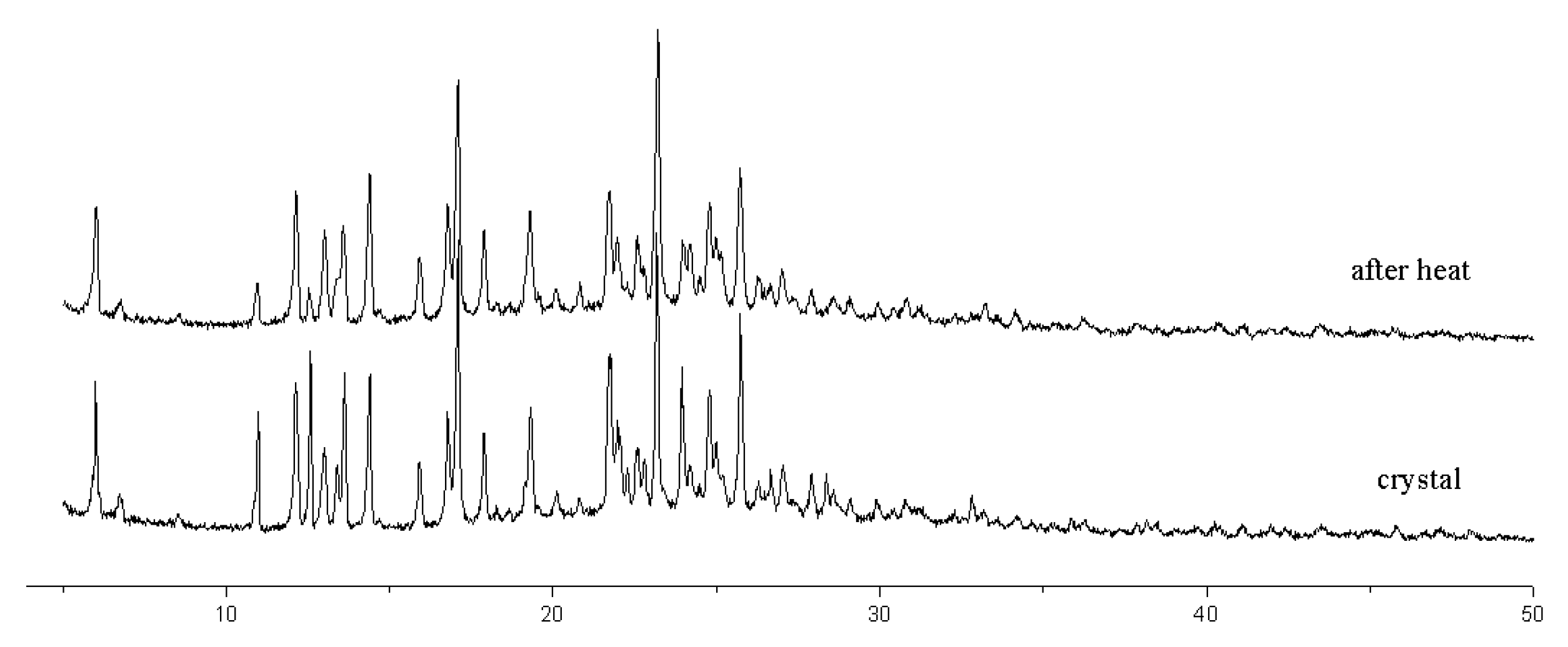
3.1. The Synthesis of Compound 2

The macrocyclic compound 2 was synthesized by the condensation reaction between 5,5′-bis(3-hydroxyphenyldiazo)-dipyrromethane and (ethane-1,2-diylbis(oxy))bis(ethane-2,1-diyl) bis(4-methylbenzenesulfonate) in a diluted solution of acetonitrile with moderate yield (Scheme 1). The structure of compound 2 was characterized by 1H, 13C NMR, HR-MS (S1-S3) and single-crystal X-ray diffraction analysis. Notably, no photochromic property or E/Z isomerization of compound 2 in DMSO solution was observed. This is potentially because of azo-hydrazone tautomerism [49,50].

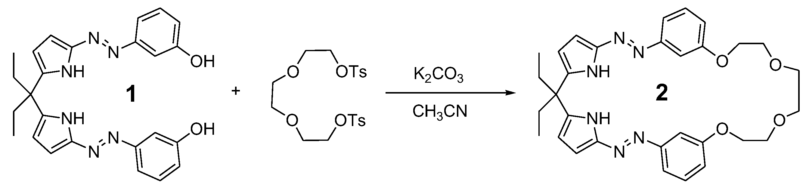
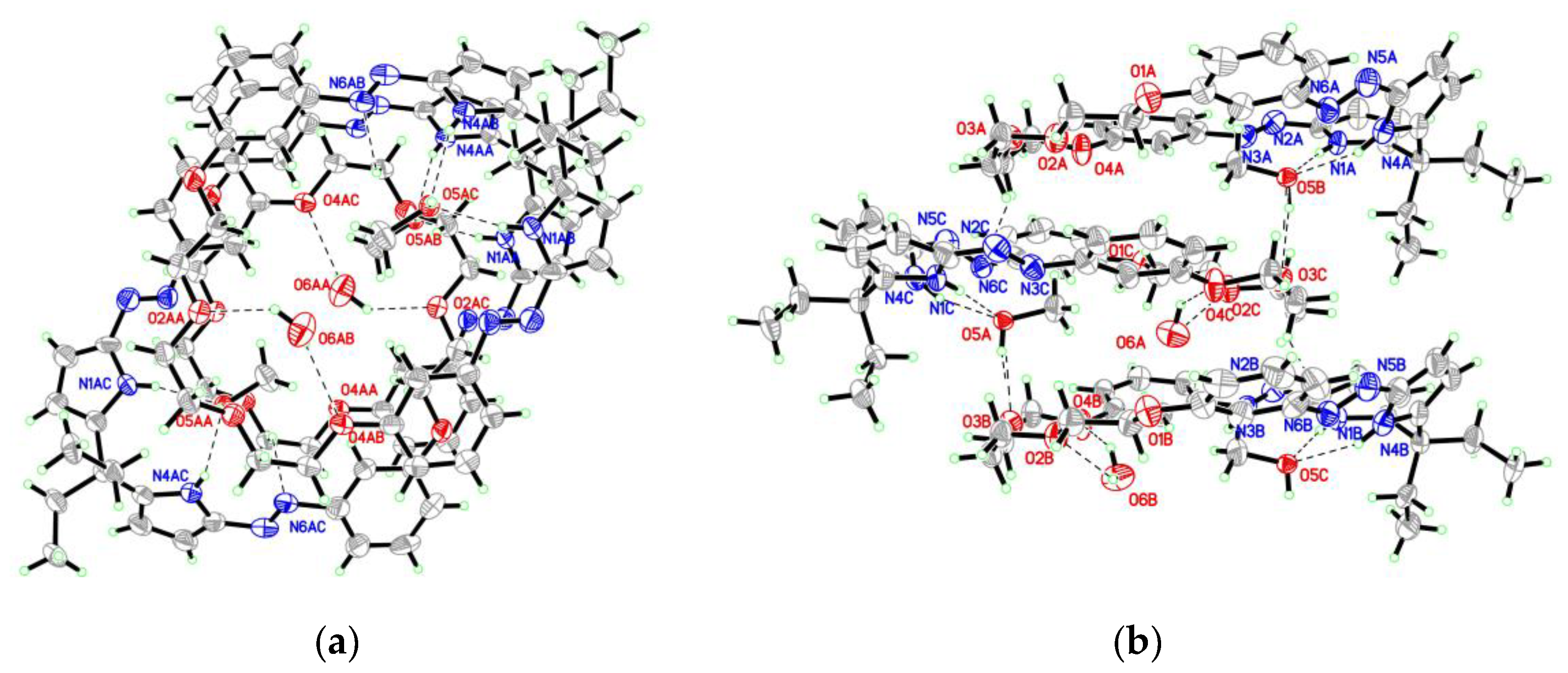
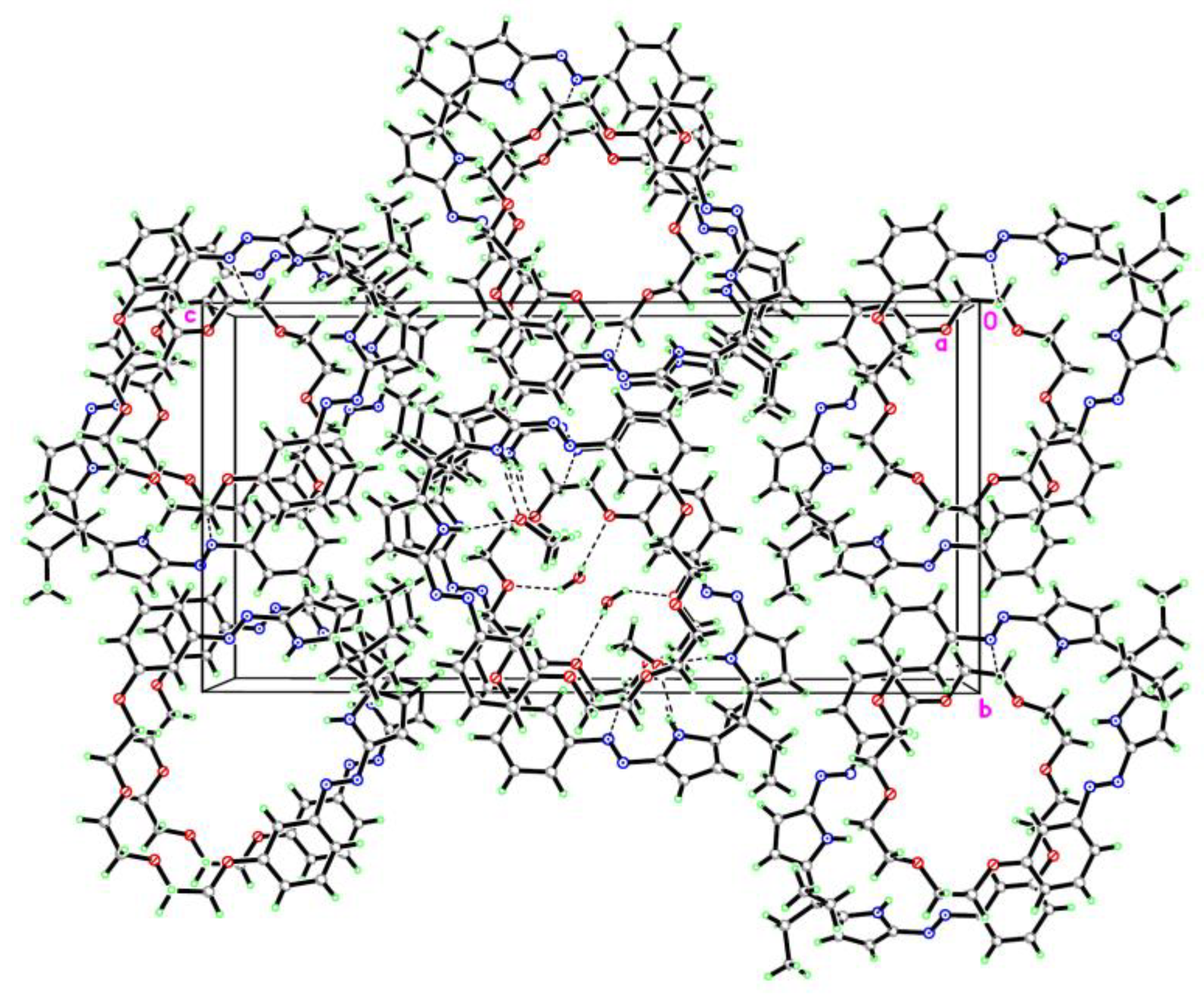
3.2. The Crystal Structure of Compound 2

Compound 2 crystallizes in the orthorhombic space group, P212121, with one molecule of 2, as well as one methanol and half water molecule in the asymmetric unit. The structure of compound 2 is shown in Figure 1, in which both N=N bonds adopt an E configuration identical to the previous azopyrrole compounds [42,51,52]. The selected bond distances and torsion angles are listed in Table 2. The N(2)-N(3) and N(5)-N(6) bond distances are 1.240 (8) and 1.208 (8) Å, respectively, which are slight shorter than previous results (1.239–1.292 Å) [42,51,52]. The dihedral angles of O3-C21-C20-O4, O2-C23-C22-O3, and O2-C24-C25-O1 are 66.0°, −67.4°, and −175.5°, respectively. In the crystal of compound 2, the two pyrrole NH groups point to the same side of the molecule, and the angle between them is 63.6°. It is quietly different with bis-diarylazo- dipyrromethanes, in which the two pyrrole rings point in the opposite direction [42,51,52]. The reason should be the methanol binding through a pair of N-H…O (N1-H1…O5: H…O = 2.15 Å, N…O = 3.001 Å, ∠N-H…O = 169° and N4-H4…O5: H…O = 2.14 Å, N…O = 2.917 Å, ∠N-H…O = 149°) hydrogen bonds (Figure 2). In the crystal, bridged by methanol through aforesaid N-H…O and O5-H5…O3 (O5-H5…O3: H…O = 1.95 Å, O…O = 2.758 Å, ∠O-H…O = 166°) hydrogen bonds, molecules of compound 2 assemble into tubular structure along an axis (Figure 2). The tubular is also stabilized by C21-H21B…N6 (C21-H21B…N6: H…N = 2.61 Å, C…N = 3.314 Å, ∠C-H…N = 129°) hydrogen bonds between two neighboring 2 molecules. The included water molecule donates its two hydrogen atoms and connects with crown ether part of 2 molecule through pair of O-H…O (O6-H6A…O2: H…O = 2.31 Å, O…O = 2.957 Å, ∠O-H…O = 133° and O6-H6A…O4: H…O = 2.36 Å, O…O = 2.917 Å, ∠O-H…O = 127°) hydrogen bonds (Figure 2). The parameters of the hydrogen bonds were also listed in Table 3. After remove the solvents, the cavity is about 10.0% of the total volume of the crystal, as calculated using the program PLATON [53]. The tubes are packed with each other through Van der Waals interactions (Figure 3).
To study the stability of the crystal, thermogravimetric analysis (TGA) was carried out. The TGA curve of the crystal of 2 is shown in Figure 4. It clearly shows a gradual weight loss of 9.0% before 70 °C, corresponding to the release of methanol and water molecules (calc. 8.3% for the crystal structure). The TGA curve also demonstrates that compound 2 starts to decompose after 250 °C, which indicates that compound 2 is stable to heat. It is consistent with other azopyrrole compounds [54]. The powder X-ray diffraction plots of crystal of 2 before and after heat at 100 °C for 6 h were shown in Figure 5. The two plots are almost the same. It means that the tubular structure of the crystal is unaffected after removing of the solvents.

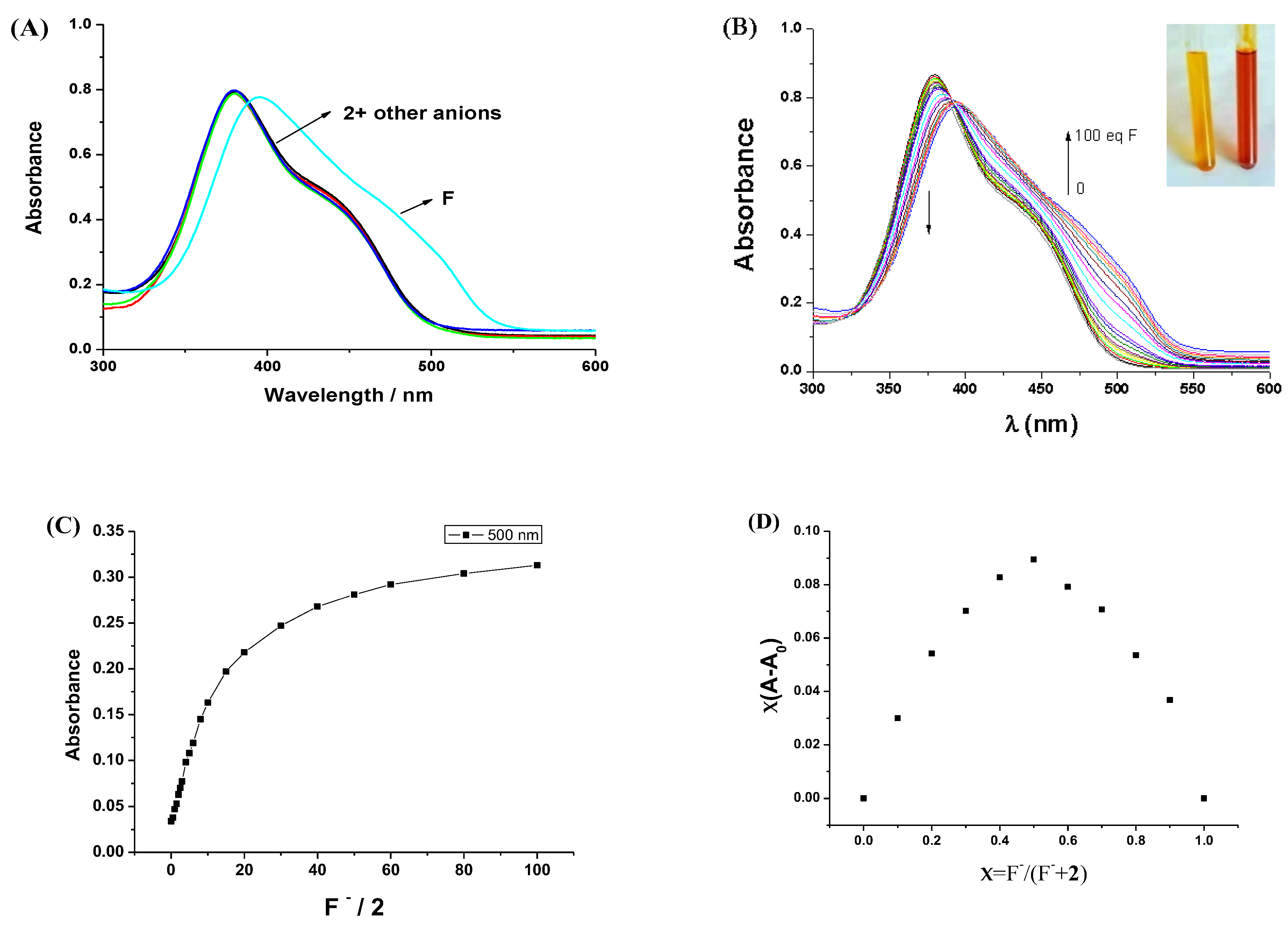
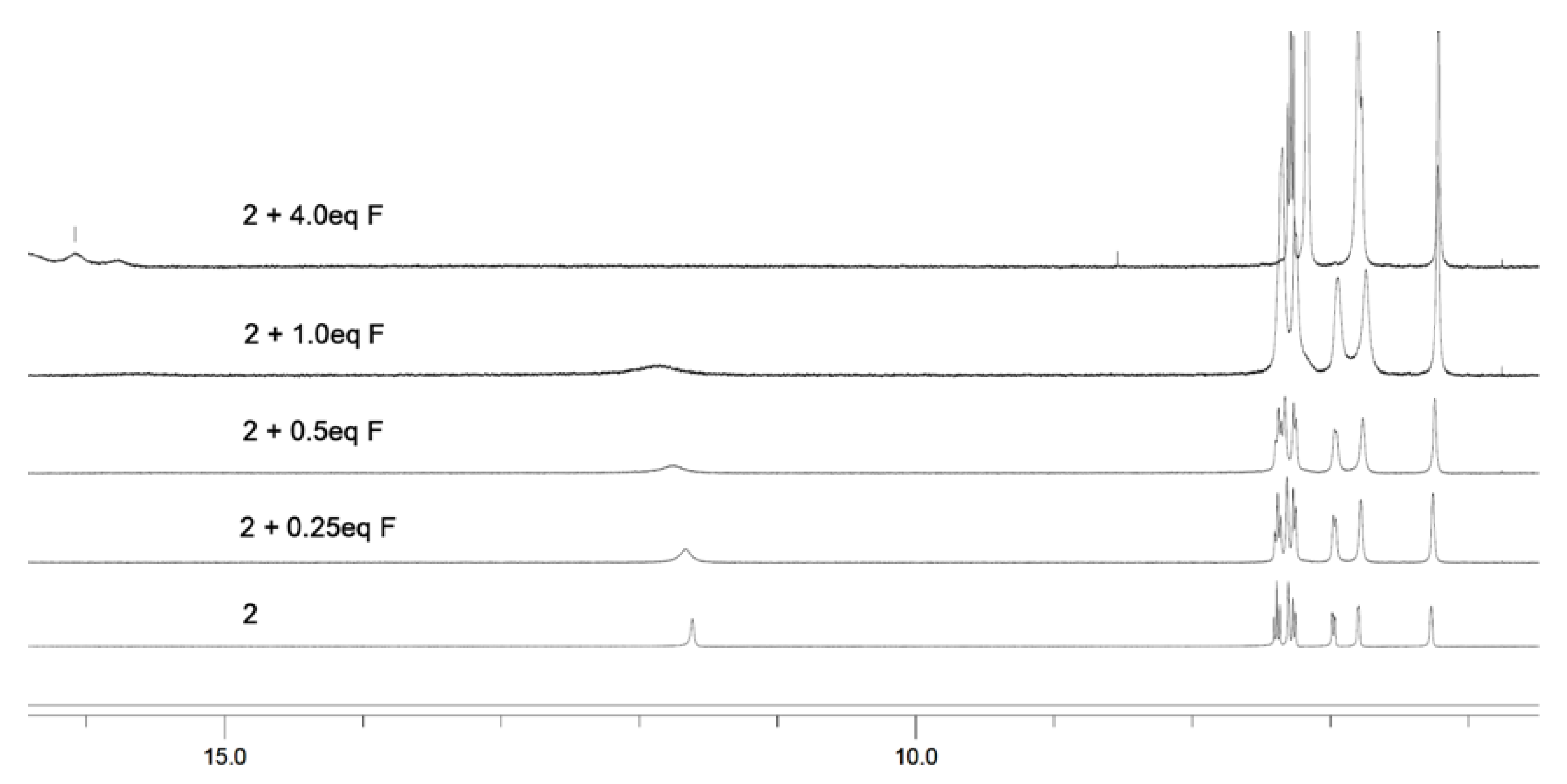
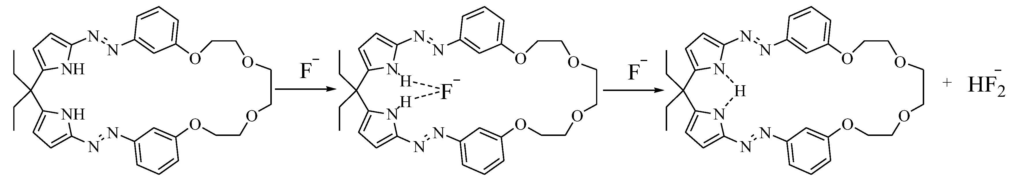
3.3. The Anions Recognition of Compound 2

The anions binding of macrocycle 2 with common anions such as Fˉ, Clˉ, Brˉ, Iˉ, AcOˉ, H2PO4ˉ, HSO4ˉ, and NO3ˉ were studied in DMSO solution using a UV–Vis titration method. As shown in Figure 6A, only fluoride caused the UV–Vis spectrum change in compound 2 after the addition of 100 equivalent anions. Fluoride also caused the solution color to change from orange to red, whereas other anions did not. This indicates that compound 2 can selectively sense fluoride in DMSO solution. The UV–Vis spectra changes of compound 2 with the addition of fluoride are shown in Figure 6B. In DMSO solution, the λmax of compound 2 is at 380 nm, which can be assigned to the absorption of the azopyrrole group and consistent with other azopyrroles [35]. Upon the gradual addition of fluoride, the intensity of the peak at 380 nm decreases, and the intensity of a new peak at 500 nm increases (Figure 6C). This is why the solution color changes. A linear response for fluoride was observed in the 1.25 × 10⁻⁵–2.5 × 10⁻⁴ M range. The limit of detection was 3.5 × 10⁻⁶ M in DMSO solution. The Job’s plot analysis at 500 nm suggests that compound 2 formed a 1:1 complex with fluoride (Figure 6D). Calculated by non-linear least-square analysis [48], the association constant Ka = 1846 M−1, which is slightly higher than previous acyclic azopyrrole receptor [36].
To interpret F¯ sensing mechanism of probe 2, 1H NMR titration experiments were performed in DMSO-d6. The 1H NMR spectra changes in probe 2 upon gradual addition of fluoride ion are shown in Figure 7. The signals of pyrrole NH moved downfield and became broader upon the addition of only 0.25–1 equivalent of F¯ ion. No solution color change was observed. This implies that probe 2 first interacted with fluoride ion through the hydrogen bonds. When four equivalents of F¯ were added, the resonance signal of the N-H proton disappeared and peaks at 16.1 ppm for HF2¯ appeared, and the solution color changed from orange to red (Figure 6B inset). It indicates that more fluoride ions caused the deprotonation of the pyrrole NH. The deprotonation of pyrrole NH proton was also evident in the upfield shifts of aromatic protons of azopyrrole. According to the 1H NMR spectra changes, the possible binding mode of probe 2 with fluoride is shown in Scheme 2.

4. Conclusions

The synthesis and structural characterization of a macrocyclic compound 2, which consist of bis-azopyrroles and polyether parts, was described. The structure of macrocyle 2 was confirmed by X-ray crystallography. The macrocycle 2 can bind methanol via pyrrole NH groups through a pair of N-H…O hydrogen bonds, and further self-assembled into tubular structures through C-H…N hydrogen bonds. It revealed that the crystal of macrocyle 2 can still maintain its porous properties after removing the included methanol molecules by heating. UV–Vis titration experiments suggested that macrocyle 2 can selectively sense fluoride in DMSO solution. The 1H NMR titration experiments indicate that compound 2 firstly bound fluoride through hydrogen bonds and then was deprotonated with more fluoride. The polyether may has the ability to interact with cations, such as Li+ and Na+. The ion-pair recognition of compound 2 to inorganic fluoride is under study.

Supplementary Materials

The following supporting information can be downloaded at: https://www.mdpi.com/article/10.3390/org6020025/s1, Figure S1: 1H NMR spectrum of compound 2 in DMSO-d6 solution; Figure S2: 13C NMR spectrum of compound 2 in DMSO-d6 solution; Figure S3: The HR-MS spectrum of compound 2. Crystallographic data for compound 2 have been deposited with the Cambridge Crystallographic Data Centre as supplementary publication No. CCDC 2117984. Copies of the data can be obtained free of charge on application to CCDC, 12 Union Road, Cambridge CB2 1EZ, UK [fax: (+44)1233-336-033; e-mail: deposit@ccdc.cam.ac.uk].

Author Contributions

Y.A. and Z.Y. wrote the main manuscript text; Y.A. and Y.S. prepared the compound, and did other experiments. All authors have read and agreed to the published version of the manuscript.

Funding

This research did not receive any specific grant from funding agencies in the public, commercial, or not-for-profit sectors.

Data Availability Statement

All data generated or analyzed during this study are included in this published article and its supplementary information file.

Conflicts of Interest

The authors declare no conflicts of interest.

References

  1. Ali, Y.; Hamid, S.A.; Rashid, U. Biomedical Applications of Aromatic Azo Compounds. Mini-Rev. Med. Chem. 2018, 18, 1548–1558. [Google Scholar] [CrossRef] [PubMed]
  2. Towns, A.D. Developments in azo disperse dyes derived from heterocyclic diazo components. Dyes Pigments 1999, 42, 3–28. [Google Scholar] [CrossRef]
  3. Emanuele, L.; D’Auria, M. The Use of Heterocyclic Azo Dyes on Different Textile Materials: A Review. Organics 2024, 5, 277–289. [Google Scholar] [CrossRef]
  4. Weston, C.E.; Richardson, R.D.; Haycock, P.R.; White, A.J.P.; Fuchter, M.J. Arylazopyrazoles: Azoheteroarene photoswitces offering quantitative isomerization and long thermal half-lives. J. Am. Chem. Soc. 2014, 136, 11878–11881. [Google Scholar] [CrossRef]
  5. Calbo, J.; Weston, C.E.; White, A.J.P.; Rzepa, H.S.; Contreras-Garcia, J.; Fuchter, M.J. Tuning Azoheteroarene photoswitch performance through heteroaryl design. J. Am. Chem. Soc. 2017, 139, 1261–1274. [Google Scholar] [CrossRef]
  6. Gallardo-Rosas, D.; Guevara-Vela, J.M.; Rocha-Rinza, T.; Toscano, R.A.; Lopez-Cortes, J.G.; Ortega-Alfaro, M.C. Structure and isomerization behavior relationships of new push-pull azo-pyrrole photoswitches. Org. Biomol. Chem. 2024, 22, 4123–4134. [Google Scholar] [CrossRef]
  7. Coelho, P.J.; Castro, M.C.R.; Fonseca, A.M.C.; Raposo, M.M.M. Photoswitching in azo dyes bearing thienylpyrrole and benzothiazole heterocyclic systems. Dyes Pigments 2011, 91, 745–748. [Google Scholar] [CrossRef]
  8. Lee, H.Y.; Olasz, A.; Chen, C.-H.; Lee, D. Three-stage binary switching of azoaromatic polybase. Org. Lett. 2012, 14, 6286–6289. [Google Scholar] [CrossRef]
  9. Li, Y.; Patrick, B.O.; Dolphin, D. Near-infrared absorbing azo dyes: Synthesis and X-ray crystallographic and spectral characterization of monoazopyrroles, bisazopyrroles, and a boron-azopyrrole complex. J. Org. Chem. 2009, 74, 5237–5243. [Google Scholar] [CrossRef]
  10. Mikroyannidis, J.A.; Tsagkournos, D.V.; Sharma, S.S.; Kumar, A.; Vijay, A.D.; Sharma, G.D. Efficient bulk heterojunction solar cells based on low band gap bisazo dyes containing anthracene and/or pyrrole units. Sol. Energy Mater. Sol. Cells 2010, 94, 2318–2327. [Google Scholar] [CrossRef]
  11. Mikroyannidis, J.A.; Roy, M.S.; Sharma, G.D. Synthesis of new low band gap dyes with BF2-azopyrrole complex and their use for dye-sensitized solar cells. J. Power Sources 2010, 195, 5391–5398. [Google Scholar] [CrossRef]
  12. Yin, Z.; Wang, W.; Guo, J.; Wang, J.; He, J.; Cheng, J.-P. Quadruple hydrogen bonded self-assemblies of 5,5′-bisdiazo-dipyrromethane. CrystEngComm 2008, 10, 957–959. [Google Scholar] [CrossRef]
  13. Beer, P.D.; Gale, P.A. Anion recognition and sensing: The state of the art and future perspectives. Angew. Chem. Int. Ed. 2001, 40, 486–516. [Google Scholar] [CrossRef]
  14. Busschaert, N.; Caltagirone, C.; Rossom, W.V.; Gale, P.A. Applications of supramolecular anion recognition. Chem. Rev. 2015, 105, 8038–8155. [Google Scholar] [CrossRef]
  15. Gale, P.A.; Howe, E.N.W.; Wu, X. Anion receptor chemistry. Chem 2016, 1, 351–422. [Google Scholar] [CrossRef]
  16. Zhao, J.; Yang, D.; Yang, X.-J.; Wu, B. Anion coordination chemistry: From recognition to supramolecular assembly. Coord. Chem. Rev. 2019, 378, 415–444. [Google Scholar] [CrossRef]
  17. Goshisht, M.K.; Tripathi, N. Fluorescence-based sensors as an emerging tool for anion detection: Mechanism, sensory materials and applications. J. Mater. Chem. C 2021, 9, 9820–9850. [Google Scholar] [CrossRef]
  18. Martinez-Manez, R.; Sancenon, F. Fluorogenic and chromogenic chemosensors and reagents for anions. Chem. Rev. 2003, 103, 4419–4476. [Google Scholar] [CrossRef]
  19. Dydio, P.; Lichosyt, D.; Jurezakm, J. Amide- and urea-functionalized pyrroles and benzopyrroles as synthetic, neutral anion receptors. Chem. Soc. Rev. 2011, 40, 2971–2978. [Google Scholar] [CrossRef]
  20. Gale, P.A.; Sessler, J.L.; Kral, V.; Lynch, V. Calix[4]pyrroles: Old yet new anion-binding agents. J. Am. Chem. Soc. 1996, 118, 5140–5141. [Google Scholar] [CrossRef]
  21. Lv, Y.; Xu, J.; Guo, Y.; Shao, S. A novel colorimetric and fluorometric anion sensor based on BODIPY-calix[4]pyrrole conjugate. J. Incl. Phenom. Macrocycl. Chem. 2012, 72, 95–101. [Google Scholar] [CrossRef]
  22. Gale, P.A.; Anzenbacher, P., Jr.; Sessler, J.L. Calixpyrroles II. Coord. Chem. Rev. 2001, 222, 57–102. [Google Scholar] [CrossRef]
  23. Aldakov, D.; Anzenbacher, P., Jr. Sensing of aqueous phosphates by polymers with dual modes of signal transduction. J. Am. Chem. Soc. 2004, 126, 4752–4753. [Google Scholar] [CrossRef] [PubMed]
  24. Pohl, R.; Aldakov, D.; Kubat, P.; Jursikova, K.; Marquez, M.; Anzenbacher, P., Jr. Strategies toward improving the performance of fluorescence-based sensors for inorganic anions. Chem. Commun. 2004, 1282–1283. [Google Scholar] [CrossRef]
  25. Aleskovic, M.; Basaric, N.; Halasz, I.; Liang, X.; Qin, W.; Mlinaric-Majerski, K. Aryl substituted adamantine-dipyrromethanes: Chromogenic and fluorescent anion sensors. Tetrahedron 2013, 69, 1725–1734. [Google Scholar] [CrossRef]
  26. Renic, M.; Basaric, N.; Mlinaric-Majerski, K. Adamantane-dipyrromethanes: Novel anion receptors. Tetrahedron Lett. 2007, 48, 7873–7877. [Google Scholar] [CrossRef]
  27. Gale, P.A.; Camiolo, S.; Tizzard, G.J.; Chapman, C.P.; Light, M.E.; Coles, S.J.; Hursthouse, M.B. 2-Amidopyrroles and 2,5-diamidopyrroles as simple anion binding agents. J. Org. Chem. 2001, 66, 7849–7853. [Google Scholar] [CrossRef]
  28. Yin, Z.; Li, Z.; Yu, A.; He, J.; Cheng, J.P. o-Di-(pyrrole-2-carboxamides)-phenylene: Pseudopolymorphs and anions recognition. Tetrahedron Lett. 2004, 45, 6803–6806. [Google Scholar] [CrossRef]
  29. Damade, K.R.; More, D.H. Azo based chromogenic sensor: An approach for naked eye detection of biologically relevant anions and metal cations. Mini-Rev Org. Chem. 2024, 21, 514–525. [Google Scholar] [CrossRef]
  30. Kaur, N.; Dhaka, G.; Singh, J. Simple naked-eye ratiometric and colorimetric receptor for anions based on azo dye featuring with benzimidazole unit. Tetrahedron Lett. 2015, 56, 1162–1165. [Google Scholar] [CrossRef]
  31. Morsavi, H.Z.; Mohammadi, A.; Yaghoubi, S.; Khereshki, N.Z. Design and synthesis of a novel optical chemoreceptor based on naphthalene azo dye for detection cyanide ion in aqueous medium an real samples. Chem. Pap. 2024, 78, 397–407. [Google Scholar]
  32. Kaur, M.; Singh, H.; Kaur, N.; Singh, N. A biginelli-azophenol based robust sensor for rapid diagnosis of cyanide in real samples. Dyes Pigments 2021, 195, 109702. [Google Scholar] [CrossRef]
  33. Park, S.M.; Saini, S.; Park, J.E.; Singh, N.; Jang, D.O. A benzothiazole-based receptor for colorimetric detection of Cu2+ and S2- ions in aqueous media. Tetrahedron Lett. 2021, 73, 153115. [Google Scholar] [CrossRef]
  34. Chauhan, S.M.S.; Bisht, T.; Garg, B. 1-Arylazo-5,5-dimethyl dipyrromethanes: Versatile chromogenic probes for anions. Sensors Actuators B 2009, 141, 116–123. [Google Scholar] [CrossRef]
  35. Li, T.; Liu, L.-J.; Yin, Z. 2-(2-hydroxylphenyl)dizao-dipyrromethane: Synthesis, structure and fluoride ion detection. Chinese J. Struct. Chem. 2017, 36, 47–52. [Google Scholar]
  36. Sun, Y.; An, Y.; Huang, J.; Yin, Z. Bis-arylazo-naphthobipyrrole: Synthesis, structure, photophysical properties, DFT calculations and anions sensing. Dyes Pigments 2025, 236, 112661. [Google Scholar] [CrossRef]
  37. Wang, M.-X. Nitrogen and oxygen bridged calixaromatics: Synthesis, structure, fictionalization, and molecular recognition. Acc. Chem. Res. 2012, 45, 182–195. [Google Scholar] [CrossRef]
  38. Ogoshi, T.; Yamagishi, T.; Nakamoto, Y. Pillar-shaped macrocyclic hosts pillar[n]arenes: New key players for supramolecular chemistry. Chem. Rev. 2016, 116, 7937–8002. [Google Scholar] [CrossRef] [PubMed]
  39. Liu, Z.; Nalluri, S.K.M.; Stoddart, J.F. Surveying macrocyclic chemistry: From flexible crown ethers to rigid cyclophanes. Chem. Soc. Rev. 2017, 46, 2459–2478. [Google Scholar] [CrossRef]
  40. Fan, L.; Du, M.; Kong, L.; Cai, Y.; Hu, X. Recognition site modifiable macrocycle: Synthesis, functional group variation and structural inspection. Molecules 2023, 28, 1338. [Google Scholar] [CrossRef]
  41. Mohammed, F.A.; Xiao, T.; Wang, L.; Elmes, R.B.P. Macrocyclic receptors for anion recognition. Chem. Commun. 2024, 60, 11812–11836. [Google Scholar] [CrossRef] [PubMed]
  42. Yin, Z.; Wang, M.; Wang, W.; Wang, X. Solvents effects on the self-assemblies of 55-bis(3-hydroxyphenyldizao)-dipyrromethane. J. Mol. Struct. 2013, 1003, 85–90. [Google Scholar] [CrossRef]
  43. Sheldrick, G.M. SADABS; University of Gottingen: Gottingen, Germany, 1996. [Google Scholar]
  44. Sheldrick, G.M. SHELXT-Integrated space-group and crystal-structure determination. Acta Crystallogr. A 2015, 71, 3–8. [Google Scholar] [CrossRef] [PubMed]
  45. Sheldrick, G.M. Crystal structure refinement with SHELXL. Acta Crystallogr. C 2015, 71, 3–8. [Google Scholar] [CrossRef]
  46. Dolomanov, O.V.; Bourhis, L.J.; Gildea, R.J.; Howard, J.A.K.; Puschmann, H. OLEX2: A complete structure solution, refinement and analysis program. J. Appl. Crystallogr. 2009, 42, 339–341. [Google Scholar] [CrossRef]
  47. Renny, J.S.; Tomasevich, L.L.; Tallmadge, E.H.; Collum, D.B. Method of continuous variations: Applications of Job Plots to the study of molecular associations in organometallic chemistry. Angew. Chem. Int. Ed. 2013, 52, 11998–12013. [Google Scholar] [CrossRef]
  48. Connors, K.A. Binding Constants: The Measurement of Molecular Complex Stability; Wiley: New York, NY, USA, 1987. [Google Scholar]
  49. Sun, Y.; Sun, J.; Yin, Z. Synthesis, crystal structrues and solvent dependent cis-trans conversion of 2-(4-hydroxylphenyl)-azopyrrole. J. Mol. Struct. 2023, 1294, 136415. [Google Scholar] [CrossRef]
  50. Chen, J.; Yin, Z. Cooperative intramolecular hydrogen bonding induced azo-hydrazone tautomerism of azopyrrole: Crystallographic and spectroscopic studies. Dyes Pigments 2014, 102, 94–99. [Google Scholar] [CrossRef]
  51. Yin, Z.; Wang, W.; Du, M.; Wang, X.; Guo, J. Crystal engineering of 5,5-bisdiazo-dipyrromethane with halogen...π synthons. CrystEngComm 2009, 11, 2441–2446. [Google Scholar] [CrossRef]
  52. Shi, H.-M.; Yin, Z. Synthesis and crystal structure of 55-bis(4-hydroxylphenyl)diazo-dipyrromethane. Chin. J. Struct. Chem. 2013, 32, 1093–1099. [Google Scholar]
  53. Spek, A.L. Single-crystal structure validation with the program PLATON. J. Appl. Crystallogr. 2003, 36, 7–13. [Google Scholar] [CrossRef]
  54. Zhang, H.; Yin, Z. Discrete cage form of water hexamer in the hydrophilic channels assembled by heterocyclic azopyrrole. J. Mol. Struct. 2015, 1092, 9–13. [Google Scholar] [CrossRef]
Scheme 1. Synthesis of compound 2.
Organics 06 00025 sch001
Figure 1. ORTEP diagram of compound 2 showing 30% probability of displaced ellipsoids.
Organics 06 00025 g001
Figure 2. (a) Top view and (b) side view of tube structure self-assembled by hydrogen bonds in the crystal.
Organics 06 00025 g002
Figure 3. Crystal packing diagram of crystal of 2 (only one channel was filled with solvents for clarity).
Organics 06 00025 g003
Figure 4. The TGA curve of the crystal of 2.
Organics 06 00025 g004
Figure 5. Powder X-ray diffraction plots of crystal of 2 before (below) and after heat (top).
Organics 06 00025 g005
Figure 6. (A) UV–Vis spectral changes in 2 in DMSO (2.5 × 10⁻⁵ M) after the addition of 100 equivalent anions; (B) UV–Vis spectra change in compound 2 in DMSO solution (2.5 × 10⁻⁵ M) with the addition of fluoride (inset: color change of the solution); (C) changes observed at 500 nm with the addition of fluoride ions; and (D) the stoichiometry analysis of complex formation by Job’s plot analysis at 500 nm ([2] + [F¯] = 1 × 10⁻⁵ M).
Organics 06 00025 g006
Figure 7. Partial 1H NMR spectrum of 2 in DMSO-d6 with the addition of different equivalents of fluoride.
Organics 06 00025 g007
Scheme 2. Proposed binding mode of receptor 2 with fluoride.
Organics 06 00025 sch002
Table 1. Crystal data of compound 2.
Crystal2
CCDC No.2117984
Empirical formulaC64H82N12O11
Formula weight1195.41
Crystal systemOrthorhombic
Space groupP212121
a(Å)7.6407(17)
b(Å)14.487(3)
c(Å)28.857(6)
α(°)90
β(°)90
γ(°)90
V(Å3)3194.2(12)
Z2
Dcalc.(g/cm3)1.243
μ(mm−1)0.086
F(000)1276.0
Reflections collected16356
Independent reflections5539 [Rint = 0.0676, Rsigma = 0.0865]
R1/wR2 [I > 2σ(I)]0.0752/0.1930
R1/wR2 (all data)0.01550/0.2431
GoF on F20.946
Table 2. Selected bond lengths (Å) and torsion angles (°) of compound 2.
BondsLength (Å)Torsion AnglesDegree (°)
N2-N31.240(8)N1-C4-N2-N30.2(11)
N5-N61.208(8)C3-C4-N2-N3179.4(8)
N1-C11.366(8)C4-N2-N3-C14179.5(6)
N1-C41.364(8)C19-C14-N3-N26.1(11)
C1-C21.391(10)C15-C14-N3-N2172.5(7)
C2-C31.383(11)N4-C8-N5-N63.5(11)
C3-C41.385(11)C7-C8-N5-N6174.3(8)
N2-C41.403(9)C8-N5-N6-C28174.4(6)
N3-C141.473(9)C29-C28-N6-N521.5(10)
N4-C51.353(8)C27-C28-N6-N5155.9(7)
N4-C81.399(10)C20-O4-C16-C1712.3(11)
C5-C61.381(11)O4-C20-C21-O366.0(8)
C6-C71.424(13)O3-C22-C23-O267.4(7)
C7-C81.294(12)O2-C24-C25-O1175.5(6)
N5-C81.426(11)C25-O1-C26-C278.0(11)
N6-C281.535(10)N1-C1-C9-C1051.6(9)
C16-O41.359(8)N1-C1-C9-C571.3(9)
C26-O11.362(8)N4-C5-C9-C1034.7(9)
O4-C201.439(8)N4-C5-C9-C187.7(8)
O1-C251.409(8)
Table 3. Hydrogen bond parameters in the crystal of 2.
D-H…AD…A (Å)H…A (Å)∠D-H…A (°)
N1-H1…O5 i3.001 (8)2.15169
N4-H4…O5 i2.917 (8)2.14149
O5-H5…O32.758 (8)1.95166
O6-H6A…O2 ii2.957 (13)2.31133
O6-H6A…O4 ii2.951 (13)2.36127
C21-H21B….N6 i3.314 (11)2.61129
Symmetry code i: 1/2 + x, 3/2 − y, 1 − z; ii: −1/2 + x, 3/2 − y, 1 − z.
Disclaimer/Publisher’s Note: The statements, opinions and data contained in all publications are solely those of the individual author(s) and contributor(s) and not of MDPI and/or the editor(s). MDPI and/or the editor(s) disclaim responsibility for any injury to people or property resulting from any ideas, methods, instructions or products referred to in the content.

