

Article

Real-World Outcomes of Cabergoline Treatment in Non-Functioning Pituitary Adenomas: An Insight into Dose Responsiveness and Radiological Follow-Up at a UK Tertiary Centre


1
Norfolk and Norwich University Hospital, Norwich NR4 7UY, UK
2
Norwich Medical School, University of East Anglia, Norwich NR4 7TJ, UK
3
Midland Regional Hospital Mullingar, N91 Y330 Mullingar, Ireland
4
National Hospital for Neurology and Neurosurgery, London WC1N 3BG, UK
5
Faculty of Medical Sciences, University College London, London WC1E 6BT, UK
*
Author to whom correspondence should be addressed.
Endocrines 2026, 7(2), 15; https://doi.org/10.3390/endocrines7020015
Submission received: 30 January 2026 / Revised: 19 March 2026 / Accepted: 31 March 2026 / Published: 8 April 2026
(This article belongs to the Section Neuroendocrinology and Pituitary Disorders)

Abstract

Introduction: Evidence on the use of dopamine agonists (DAs) for managing residual or recurrent non-functioning pituitary adenomas (NFPAs) is limited. We aim to evaluate the use of cabergoline (CAB) for NFPAs. Methods: A retrospective cohort study was conducted at a single UK centre, between November 2011 and December 2025. Twenty-six patients were identified. Ten patients were excluded due to CAB intolerance or discontinuation (n = 5), insufficient data (n = 4), or invalid scan due to patient movement (n = 1). The remaining 16 patients (mean age 68.9 ± 4 years (range 42–89 years old), 7/16 females) were included. CAB was initiated in cases where surgery or radiotherapy were not appropriate (e.g., due to age and/or comorbidities, or patient choice). Radiological response was assessed using at least two scans separated by a minimum interval of six months. Tumour shrinkage was defined as a reduction in volume of 20% or more, growth as an increase of 20% or more, and stabilisation as interval change of less than 20%. Results: Overall, tumour shrinkage was observed in 7/16 (43.8%) patients, stabilisation in the remaining 9/16 (56.3%) patients, over 503 ± 51 days (range of 117–934 days) (from the date of CAB initiation to latest MRI scan). There was a statistically significant reduction in tumour volume (p = 0.0335). In five patients with documented tumour growth prior to CAB initiation, growth rates retarded or reversed post-CAB initiation. Conclusions: Our findings in this small cohort potentially suggests that cabergoline can retard, arrest, or even reverse tumour growth in selected patients with NFPAs. Our review also highlights ongoing uncertainty regarding optimal dosing, approaches to dose up-titration, follow-up imaging intervals, and objective criteria for defining radiological response. Our results may provide a proof of concept for future, larger-scale prospective studies and controlled trials to validate the conclusions drawn.

1. Introduction

Non-functioning pituitary adenomas (NFPAs) are benign tumours characterised by the absence of clinical or biochemical evidence of pituitary hormonal hypersecretion, aside from associated hyperprolactinaemia due to pituitary stalk deviation [1]. Due to their non-secretory nature, NFPAs are often discovered incidentally on imaging or remain clinically silent until significant tumour growth leads to compressive symptoms, particularly visual loss and hypopituitarism [2]. NFPAs have an annual reported incidence of 20.3 cases per 1 million [3], and they are further classified according to their pituitary hormone and transcription factor profile, as defined by the 2017 World Health Organization (WHO) classification for endocrine tumours [4].
The mainstay of treatment for symptomatic NFPAs is transsphenoidal surgery (TSS). However, complete resection is often not feasible due to factors such as large tumour size, local invasion of adjacent structures, or unfavourable tumour consistency [5]. Long-term follow-up is therefore essential, and further intervention is often required in cases with significant residual tumour, particularly where the visual apparatus is at risk or where post-operative tumour growth occurs.
For those with progressive residual tumours, multimodal treatment strategies, including repeat surgery and adjuvant fractionated stereotactic radiotherapy or radiosurgery, may be employed to achieve prolonged disease control [6]. This is especially important given that, over a mean of 6.1 years, untreated residual tumour tissue is associated with a reported progression rate of 34.8% [7].
In cases where surgery or radiotherapy is not possible—due to frailty, comorbidities, or tumour location, or when patients decline these options—dopamine agonists (DAs) may be considered. DAs have been used in such cases to provide a non-invasive treatment alternative, particularly when tumour progression threatens to compromise visual function [8,9,10,11,12,13,14,15,16,17,18,19,20,21,22,23,24].
This retrospective cohort study examines the efficacy of cabergoline (CAB), a dopamine agonist, in the management of NFPAs, aiming to better understand its role in this challenging clinical context.

2. Materials and Methods

2.1. Study Design

A retrospective study was conducted from a single centre, which looked at the management of NFPAs using dopamine agonists. Our methods and results were reported according to the STROBE guidelines [25].

2.2. Setting

A search on our electronic outpatient letters database (electronic document template) was carried out using the keywords “non-functioning”, “incidentaloma”, “cabergoline”, “quinagolide”, “bromocriptine” and “dopamine agonist”.

2.3. Outcomes

Radiological response was based on one or two follow-up (FU) scans with an interval of at least six months. Tumour shrinkage was defined as a reduction in volume of 20% or more, growth as an increase in volume of 20% or more, and stabilisation as a variability in volume of less than 20%. Tumour volume was calculated based on the maximum x, y, and z diameters measured on sagittal and coronal images. Assuming a spherical volume, the formula 4/3 πr3 would then be used for volume calculation, with r being the mean of the x, y, and z radii.

2.4. Data Measurement

Data collection included tumour size before initial surgery (in patients who underwent TSS), tumour size on diagnosis when intervention with CAB was considered, and on post-treatment with CAB, along with the dosing schedule of CAB for individual patients. All MRI scans were interpreted independently by two radiologists. Demographic data including patient age at diagnosis, gender, baseline visual fields, prolactin levels and cabergoline dosage were also collected. All eligible patients were systematically included from the database search. The pre-CAB scan was defined as the last pituitary MRI scan prior to CAB initiation. The 1st FU scan was the first scan post-CAB initiation. The 2nd FU scan was the second scan post-CAB initiation. The latest MRI scan was the latest scan in patients who had two FU scans. The treatment interval was calculated in days, from the date of recommendation of cabergoline (based on the outpatient clinic or advice letter) to the date of the scan, excluding the end date from the calculation. The interval between scans was calculated in days, between the actual imaging, excluding the end date from the calculation.

2.5. Statistical Analysis

Statistics were performed using the IBM SPSS Statistics Software Version 29.0.2.0. The normality of tumour volume changes between pre- and post-treatment with CAB were assessed with Kolmogorov–Smirnov and Shapiro–Wilk tests (p ≤ 0.05). Descriptive statistics such as means and standard deviations were used to summarise the normally distributed data, whilst medians and interquartile ranges were used to summarise the data that were not normally distributed.
The Wilcoxon signed-rank test was utilised to evaluate the significance of tumour volume changes between the pre-CAB and the latest MRI scans. This paired non-parametric test was utilised due to the non-parametric distribution of tumour volumes. For patients with more than one FU scan, the latest scan was used for analysis. A within-patient paired comparison was used, where each patient’s tumour volume pre-CAB initiation was compared with their own latest FU scan post-CAB initiation.
Growth rate changes between pre- and post-treatment with cabergoline were assessed using a paired t-test. Spearman’s rank correlation was performed to assess the relationship between CAB dose and tumour volume change between the pre-CAB and the latest MRI scans, as well as between treatment interval and tumour volume change between the pre-CAB and the latest MRI scans.
Using our keywords, a total of 26 patients were identified under the Endocrine Department from November 2011–December 2025. Our study included 16 patients in total after applying the exclusion criterion (Figure 1).

3. Results

3.1. Patient and Sample Characteristics

The 16 analysed subjects had a mean age of 68.9 ± 4 years (range 42–89 years old) and 7/16 (43.8%) were females. All 16 patients were offered CAB as the dopamine agonist of choice. Out of the 16, six patients were offered CAB post-surgery (Table 1).
The median tumour volume pre-CAB initiation was 3073.3 mm3 (IQR 1477.5–4387.3 mm3) (Table 1).
Mean tumour diameter pre-CAB initiation (measured as the longest diameter in the axial plane) [26] was 20.6 ± 1.3 mm (range of 12.2–31.9 mm) (Table 1).
At the time of CAB initiation, in the non-operated group (ten patients), 9/10 had normal visual fields on direct confrontation, and one had bitemporal superior quadrantanopia on Goldmann perimetry. In the operated group (six patients), while there was evidence of visual field compromise pre-surgery (bitemporal hemianopia on Goldmann perimetry) in all patients, there was complete resolution post-surgery and at the time of CAB initiation. In the operated group, 5/6 (83.3%) patients (IDs 6, 10, 13, 15, 16) of the operated group had normal visual fields on confrontation at the time CAB initiation, whilst 1/6 (16.7%) patients (ID 7) had a pre-existing blind right eye due to glaucoma at the time of CAB initiation (Table 1).

3.2. Histopathology of Post-Surgical Patients

Three patients had gonadotroph adenomas, one had a corticotroph adenoma, one had a plurihormonal adenoma with mixed corticotroph and gonadotrophin lineage, and histopathology was not available for the remaining one patient.

3.3. Prolactin Concentrations

The median prolactin concentrations prior CAB initiation were 241 mIU/L (IQR 151.8–535.3 mIU/L) (normal range of 40–530 mIU/L).

3.4. Other Pituitary Hormonal Profile Concentrations

Full pituitary hormonal profiles were also performed prior to CAB initiation and during follow-up, including thyroid, adrenal, growth hormone and gonadotroph axes. Three patients had pre-existing hypothyroidism and remained on levothyroxine pre and post-CAB initiation. There were no significant changes to their levothyroxine dose following CAB initiation. In the remaining patients, all were eupituitary prior to treatment, and no new endocrine dysfunction was observed following CAB treatment.

3.5. Cabergoline Dosing and Monitoring:

The median CAB dosage was 1250 mcg weekly (IQR 625–2375 mcg weekly), with variable titrating schedules to achieve maximal dosing (Table 1).
No incidence of CAB-related hormonal disturbance or visual disturbance occurred during follow-up for all of our patients.

3.6. Radiotherapy

In total, 2/16 (12.5%) patients (ID 6, 7) received radiotherapy treatment prior to CAB initiation.

4. Radiological Response Following Cabergoline Initiation

4.1. First FU Scan

After CAB initiation, there was a variable interval to the first FU scan (from the date of CAB initiation) (90–612 days) with a mean interval of 275 ± 36 days (Table 2).
Out of the 16 patients, tumour shrinkage was demonstrated in 4/16 (25%) patients (IDs 1, 2, 11, 15) (including one post-surgery patient, ID 15) while the remaining 12/16 (75%) patients (IDs 3, 4, 5, 6, 7, 8, 9, 10, 12, 13, 14, 16) demonstrated tumour stability (including five post-surgery patients; IDs 6, 7, 10, 13, 16) (Table 2). Whilst there was still absolute tumour growth in 8/16 (50%) patients (IDs 4, 5, 7, 9, 10, 13, 14, 16), they were classified as stabilisation, as the volume increase remained below the >20% threshold for growth.

4.2. Second FU Scan

Out of the 16 patients, a second FU scan was performed in 11/16 (68.8%) patients at the time of undertaking the data collection.
Post-CAB initiation, there was a variable interval to the second FU scan (from the date of CAB initiation) (247–934 days) with a mean interval of 561 ± 56 days (Table 3).
Out of the 11, five (45.5%) patients (IDs 2, 5, 6 (post-surgery patient), 8, 15 (post-surgery patient)) demonstrated shrinkage while the remaining (54.5%) patients (IDs 3, 4, 7 (post-surgery patient), 9, 10 (post-surgery patient), 13 (post-surgery patient)) demonstrated tumour stability (Table 3). Whilst there was still absolute tumour growth in 3/11 (27.3%) patients (IDs 3, 9, 10), they were classified as stabilisation, as the volume increase remained below the >20% threshold for growth.

4.3. Combining Both First and Second FU Scans

Overall, combining the first and second FU scans (using the latest scan in patients who had two scans), 7/16 (43.8%) patients demonstrated tumour shrinkage (IDs 1, 2, 5, 6 (post-surgery and radiotherapy patient), 8, 11, 15 (post-surgery patient)) and the remaining (56.3%) patients demonstrated stabilisation (IDs 3, 4, 7 (post-surgery and radiotherapy patient), 9, 10 (post-surgery patient), 12, 13 (post-surgery patient), 14, 16 (post-surgery patient)). Whilst there was still absolute tumour growth in 6/16 (37.5%) patients (IDs 3, 9, 10, 13, 14, 16), they were classified as stable, given that the overall volume increase remained less than 20%.
Using the 20% threshold for defining growth, none of the patients demonstrated interval growth post-CAB initiation, at a mean interval (from the date of CAB initiation) of 503 ± 51 days (117–934 days) (Table 4).

4.4. Tumour Volume

The median tumour volume pre-CAB initiation was 3073.3 mm3 (IQR 1477.5–4387.3 mm3).
The median tumour volume post-CAB initiation at the first FU scan was 2483.8 mm3 (IQR 1120.6–3767.0 mm3).
The median tumour volume post-CAB initiation at the second FU scan was 1887.6 mm3 (IQR 943.0–4134.6 mm3).
Overall (using the latest scan for those that received two scans), the median tumour volume post-CAB initiation at the latest MRI scan was 2022.9 mm3 (IQR 958.7–3083.3 mm3).
Overall, there was a statistically significant reduction in tumour volume between the pre-CAB scan and the latest MRI scan (p = 0.0335) (Wilcoxon signed-rank test). For patients with more than one FU scan, the latest scan was used for analysis here.

4.5. Maximal Tumour Diameter

The mean tumour diameter pre-CAB initiation (measured as the longest diameter in the axial plane) [26] was 20.6 ± 1.3 mm (range of 12.2–31.9 mm).
The median tumour diameter on the first FU scan was 19.8 mm (IQR 16.3–23 mm).
The median tumour diameter on the second FU scan (for 11 patients) was 19.2 mm (IQR 14.5–22.6 mm).

4.6. Correlation Between CAB Dose and Radiological Response

Overall, there was a moderate positive correlation (rs = 0.53, p = 0.036) (Spearman’s rank correlation) between CAB dosage and tumour volume change from the pre-CAB MRI scan to the latest MRI scan, as well as a moderate positive correlation (rs = 0.50, p = 0.061) (Spearman’s rank correlation) between the CAB treatment interval and tumour volume change from the pre-CAB MRI scan to the latest MRI scan. However, this was not statistically significant (p = 0.061).

4.7. Five Patients with Documented Absolute Tumour Growth Prior to CAB Initiation

In five patients (IDs 1, 3, 6, 8, 9), additional MRI scans prior to CAB initiation were available. All five patients had positive growth rates before CAB treatment, of which 3/5 (60%) (IDs 3, 6, 9) experienced tumour growth whilst the remaining two (IDs 1, 8) experienced tumour stabilisation between the additional MRI prior to CAB initiation and the pre-CAB scan (Table 5). Patient ID 6 had a large tumour remnant post-surgery with evidence of post-surgical interval growth. Absolute tumour growth occurred in two patients (IDs 1, 8) but they were classified as stabilisation as the volume increase remained below the >20% threshold for growth.

4.8. Growth Rate

The data for growth rates pre- and post-CAB initiation were only available for five patients (IDs 1, 3, 6, 8, 9). Due to a variable time period between the additional MRI scan performed prior to CAB initiation and the pre-CAB scan between patients, the growth rate was calculated from the absolute tumour volume change (pre-CAB scan volume–additional MRI prior to CAB-initiation volume) divided by the number of days elapsed between the scans, excluding the end date from the calculation.
Growth rates prior to CAB initiation ranged from +0.04 mm3/day (ID 1) to +2.01 mm3/day (ID 8), with a mean growth rate of +1.20 ± 0.41 mm3/day (Table 5).
Post-CAB initiation, tumour growth rates either retarded or reversed in all five patients at the first FU scan, over 245–918 days (mean time of 471 ± 121 days). By the first FU scan, the mean growth rate dropped to −1.87 ± 0.93 mm3/day (range of −0.17–−5.09 mm3/day). Patient 8 had the largest reduction, transitioning from +2.01 mm3/day pre-treatment to −5.09 mm3/day after initiation of CAB (Table 6).
By the second FU scan, patients 6 and 8 demonstrated sustained reductions in tumour volume whilst patients 3 and 9 demonstrated stability in 419–918 days (mean time of 644 ± 141 days). The mean growth rate was −1.52 ± 1.11 mm3/day (range of −4.50–+0.28 mm3/day) (Table 6). A second scan for patient 1 was not available.
The analysis of the tumour growth rates before and after CAB initiation (from pre-CAB to first FU scan) demonstrated a mean reduction of 3.070 ± 1.281 mm3/day in 245–918 days (mean time of 471 ± 121 days) (one-tailed p-value = 0.037, paired t-test); 95% CI: −0.486 to +6.626 mm3/day. Cohen’s d was 1.072 (>0.8).

5. Discussion

5.1. Principal Findings

Our study has shown that the use of cabergoline in patients with NFPAs is associated with a statistically significant shrinkage or stabilisation of tumour volume.
When comparing responses in all 16 patients by their latest scan (mean interval of 503 ± 51 days), 7/16 (43.8%) demonstrated tumour shrinkage and the remaining 9/16 (56.3%) patients demonstrated stabilisation. An overall statistically significant reduction in tumour volume was also observed (p = 0.0335).
Out of 16 patients in our cohort, 4/16 (25%) demonstrated shrinkage on the first FU scan at a mean time of 275 ± 36 days. The second FU scan, which was undertaken at an extended interval in comparison on 11/16 (68.8%) patients, demonstrated tumour reduction in 5/11 (45.5%) of patients at a mean time of 561 ± 56 days. Three patients (IDs 5, 6, 8) had initially shown stability on the first FU scan and subsequently demonstrated shrinkage on the second FU scan.
Out of five patients (IDs 1, 3, 6, 8, 9) who had data on pre- and post-treatment growth rates, a mean reduction of 3.070 ± 1.281 mm3/day was observed at a mean time of 471 ± 121 days (one-tailed p-value = 0.037), demonstrating evidence of retarded growth post-cabergoline initiation.

5.2. Comparison with Related Literature

The current main treatment option for vision threatening or enlarging NFPAs is surgery. In untreated, surgery-naïve NFPAs, the risk of tumour growth has been reported to be 27% by 4 years [27] and 51% by 5 years [28].
Following TSS, some patients may still have residual tumours. The rates of achieving complete resection (leaving no residual tumour) [29] have been reported to range between 43 and 90% for endoscopic and 40 and 65% for microscopic TSS [30]. In residual disease, where there is an obvious tumour remnant [29], regrowth has been reported at 34.8% [7] and 36.2% at five years after partial excision [31]. In particular, patients with extrasellar remnants had lower recurrence-free survival rates than those with intrasellar remnants (23.1% and 58.3% respectively) at ten years [32]. Complete resection is associated with higher progression-free survival rates compared with residual disease (92.4% vs. 72.1% at five years) [33], supporting the notion that tumour remnants carry a risk of regrowth if left unchecked.
If regrowth of the tumour remnant occurs, the options at present are further surgery or radiotherapy. In patients that were offered post-operative radiotherapy, 85–95% achieved tumour control at five to ten years post-operatively [34]. Its routine use, however, is limited due to the association with a high rate of complications, including a 13.62% risk of developing hypopituitarism and 2.55% risk of panhypopituitarism [35]. Repeated surgery is necessary in cases of impending or definite visual compromise but carries an additional risk of post-operative hypopituitarism, cerebrospinal fluid leak, meningitis, cranial nerve damage, and visual compromise, along with a mortality rate of 0.3–0.5% [36]. Subsequent surgery and radiotherapy also carries an increased risk of corticotropic and thyrotropic deficiency rates (38% and 59% after second or third surgical operation, and 40% and 73% after radiotherapy, respectively) [37].
Treatment with DAs, in particular cabergoline, have been evaluated for their potential clinical use in managing NFPAs in the past [8,9,10,11,12,13,14,15,16,17,18,21,22,24,38]. In addition to this, NFPAs express dopamine receptors (DR) [23], primarily dopamine receptor 2 (DR2) [39]. Based on this, DAs have been the most studied, providing a potential therapeutic benefit in managing NFPAs. Overall, dopamine agonists like bromocriptine, quinagolide, and notably cabergoline (1–3 mg/week over 6–12 months) in the above studies demonstrate significant potential in reducing residual tumour growth post-surgery.

Role of Dopamine Agonists in Management of NFPAs

Most NFPAs have dopamine receptors and predominantly comprise DR2 [39]. DAs can reduce gonadotropin secretion [24] and inhibit thymidine incorporation in vitro [38], providing a potential therapeutic benefit in NFPAs. However, the variable DR expression in NFPAs suggests that some patients are more likely to respond to DA therapy than others, warranting further investigation into the selection criteria for treatment [23]. Emerging evidence also suggests that NFPA behaviour is based on a complex interplay of multiple lineages, further warranting future research to focus on lineage-specific mapping, which may further refine patient selection for DA therapy in the future [40].
Most of the literature surrounding the use of cabergoline have been retrospective studies [3,7,10,11,12,14,15,16,18,19,21,22,27,28,29,31,32,33,36,37]. More recently, a meta-analysis reported that cabergoline treatment resulted in tumour shrinkage in 19% of patients and prevented tumour progression in around half of the treated patients, supporting a potential therapeutic role of dopamine agonists in select NFPA patients [41]. A historical cohort analysis across two centres spanning 8.8 ± 6.5 years, with different standard practices for post-TSS NFPA management (bromocriptine and cabergoline) therapy vs. conservative follow-up, demonstrated that 21/55 (38.2%) patients who received DAs had tumour shrinkage (defined as at least a 2 mm change in diameter), whereas none in the conservative follow-up group exhibited tumour shrinkage [12]. Another retrospective study with the same definitions for tumour size change showed that 5/25 (20%) had tumour reduction, 12/25 (48%) demonstrated stabilisation, whilst 8/25 (32%) demonstrated growth over a follow-up period of 4.6 ± 3.4 years whilst treated with cabergoline, with at least 1 mg/week [19]. Another retrospective study with the same definitions for tumour shrinkage as our study (20% or more) found that tumour shrinkage was documented in 29 patients (66%), whereas in 11 (25%) the tumour increased in size and in four (9%), it remained stable [42], which was comparable to our findings. A longer-term study spanning 4.6 ± 3.4 years, however, found less-promising results, where five tumours (20%) decreased in size (mean decrease of 5.0 ± 3.0 mm), 12 tumours (48%) remained stable, and eight (32%) increased in size (mean growth of 5.0 ± 3.3 mm) with cabergoline treatment [19].
There was, however, a randomised clinical trial spanning over two years, demonstrating that in patients with remnant NFPAs post-TSS, out of those who received cabergoline, tumour shrinkage was exhibited in 28.8%, stabilisation in 66.1%, and growth in 5.1% of patients, as opposed to the observation group, which had corresponding figures of 10.5% for shrinkage, 73.7% for stabilisation, and 15.8% for growth [22].
The effectiveness of short-term treatment and the optimal dosing strategy with cabergoline has also been investigated. In one study spanning six months, cabergoline of 2 mg/week showed >25% tumour shrinkage in 6/19 (31.6%) patients, >10% shrinkage in 9/19 (47.4%) patients, and growth in 4/19 (21.1%) patients [15]. Further supporting this, in another trial, 5/9 (55.6%) patients experienced a >25% tumour shrinkage after one year, at a dose of 1–3 mg/week of cabergoline post-TSS [10]. The potential efficacy of cabergoline is further reinforced by minor shrinkage (>10%) in 7/13 (53.8%) patients after treatment for one year, with an initial dosage of 0.25 mg/week, increased by 0.25 mg increments to a maximum of 1 mg/week [11]. Furthermore, another study in 2022 showed that 20/22 (90.9%) patients with residual tumours post-TSS who were treated with cabergoline for a mean time of 13 months (range of 10.5–17 months) exhibited no tumour progression, compared with the higher progression rates in the observation group, reinforcing the potential role of cabergoline in managing NFPAs [21]. A recent review also suggested that cabergoline doses of 1.5–3 mg/week may be effective in actively growing remnants and high-risk pituitary adenomas [16].
Other dopamine agonists were also studied in the management of NFPAs. Placebo-controlled trials, using a variation in drug protocols, such as bromocriptine (2.5–60 mg/day), quinagolide (300–600 mg/day) and cabergoline (1–3 µg/week), demonstrated that the cumulative evidence for tumour shrinkage after DA therapy in 199 patients was 27.6% [8].
Bromocriptine also shows potential therapeutic benefit. In 32 post-TSS patients with residual NFPAs, after six months of treatment, 11/32 (34.3%) showed tumour reduction, 13/32 (40.6%) showed stabilisation, whilst 8/32 (25%) showed growth. After two years, 10/32 (31.3%) demonstrated further tumour shrinkage, showing that bromocriptine could play a role in managing NFPAs post-TSS [18]. Tumour shrinkage was also demonstrated in a patient treated by another DA (CV 205–502), with two others reporting an improvement in visual field defects [24].
In another trial, when patients were treated with either cabergoline or quinagolide, 11/18 (61.1%) patients demonstrated tumour shrinkage over 89.7 months [9]. This is similar to our findings, where 6/12 (50%) demonstrated shrinkage but over a shorter mean interval of around 14 months.
Overall, DAs such as bromocriptine, quinagolide and cabergoline show promising results, as they have also been found to reduce residual tumour growth post-operatively, when they were given even before any tumour growth was detected [14]. Out of all the various dopamine agonists used, cabergoline at 1–3 mg/week, spanning 6–12 months, was associated with the highest likelihood of tumour shrinkage and was superior to expectant follow-up protocols [9,10,11,15,16].

6. Strengths and Limitations

Our study has several strengths. The use of pre-defined radiological criteria ensures a consistent and objective evaluation of treatment response. Additionally, the inclusion of an MRI-based volumetric analysis performed by experienced radiologists enhances the reliability of tumour measurements. Our retrospective design reflects real-world practice, capturing the heterogeneity of patient characteristics, treatment protocols, and outcomes, enhancing the clinical relevance of the findings. Beyond binary outcomes like shrinkage or stabilisation, tumour growth rates before and after treatment were calculated, which shows promising results and supports the potential clinical value of cabergoline.
However, there are limitations to our study. The small sample size (n = 16) limits statistical power. This is compounded by the retrospective nature of the study, which introduces selection bias. The variability in follow-up intervals and heterogeneity in cabergoline dosing, as well as a lack of a control group with randomisation, limits the ability to contextualise the efficacy of cabergoline relative to standard care. There is also a timing discrepancy between the pre-CAB scan and the date of cabergoline initiation, and additionally, the outpatient letter date may not reflect the actual cabergoline start date, introducing potential over or underestimation of cabergoline’s effect. Other potential confounders such as dopamine receptor expression levels, baseline health and comorbidities of the patients were also not accounted for, as well as the inclusion of patients who had received radiotherapy previously.

7. Conclusions

In this single-centre retrospective study on our limited cohort of patients with NFPAs, the use of cabergoline was associated with either tumour shrinkage or stabilisation in all patients with NFPAs, as well as the retardation or reversal of growth rates in five patients. A total of 43.8% (7/16) of the cohort demonstrated tumour shrinkage at a mean follow-up of 503 ± 51 days, with a statistically significant reduction in tumour volume (p = 0.0335). The growth rate analysis of five patients demonstrated a deceleration in tumour growth (reduction of 3.070 ± 1.281 mm3/day, p = 0.037) post-treatment, over 471 ± 121 days.
Importantly, our review does not seek to redefine the role of cabergoline in the management of NFPAs but highlights a lack of clarity on optimal dosing and approaches to dose up-titration, along with no recommended interval for FU imaging for assessing dose responsiveness. In addition, the objective criteria for defining the radiological response to treatment is also lacking, with variation across published evidence so far. Using real-world data from our centre, we demonstrated a moderate dose–response association (rs = 0.53, p = 0.036) (Spearman’s rank correlation) between cabergoline exposure and tumour volume reduction, alongside highlighting the limited value in early imaging (<12 months) for assessing response, post-initiation of cabergoline in NFPAs.
While encouraging, these findings should be interpreted with caution given the small sample size, retrospective design, lack of a control group and heterogeneity in treatment dosing and duration. Our study provides a possible proof of concept that cabergoline may have a role in selected patients where surgery or radiotherapy is not feasible.

8. Recommendations and Going Forward

Surgical resection remains the standard treatment for symptomatic NFPAs, particularly in the presence of visual compromise or mass effect. However, cabergoline may be considered in selected patients, such as those unsuitable or declining surgical intervention, and those with slowly progressive disease or residual tumour following surgery. Larger prospective studies are required to better define its role and establish clearer treatment pathways.

Author Contributions

Conceptualisation, R.A.; data curation, T.T. and E.S.; formal analysis, T.T., investigation, T.T., E.S. and L.S.; methodology, R.A.; supervision, R.A.; validation, A.B., F.S., K.D., N.D. and H.J.M.; visualisation, T.T.; writing—original draft, T.T.; writing—review and editing, A.B., F.S., K.D., N.D. and H.J.M. All authors have read and agreed to the published version of the manuscript.

Funding

This research did not receive any specific grant from any funding agency in the public, commercial or not-for-profit sector.

Institutional Review Board Statement

As the results in this work refer to observational, fully anonymised data, NHS Research Ethics Committee (REC) approval was not required.

Informed Consent Statement

For retrospective, observational research and secondary analysis of medical records, a project that analyzes something that has already happened or uses previously collected data (e.g., most surveys; or examining the outcomes of surgery at a hospital, where the researchers did not have any role in deciding who was operated on), informed consent is usually not required.

Data Availability Statement

The authors confirm that the data supporting the findings of this study are available within the article, Further inquiries can be directed to the corresponding author.

Acknowledgments

The authors acknowledge Ian Nunney (University of East Anglia) for assisting with the data analysis.

Conflicts of Interest

There are no conflicts of interest that could be perceived as prejudicing the impartiality of the research reported.

References

  1. Drummond, J.; Roncaroli, F.; Grossman, A.B.; Korbonits, M. Clinical and Pathological Aspects of Silent Pituitary Adenomas. J. Clin. Endocrinol. Metab. 2019, 104, 2473–2489. [Google Scholar] [CrossRef]
  2. Molitch, M.E. Nonfunctioning Pituitary Tumors and Pituitary Incidentalomas. Endocrinol. Metab. Clin. N. Am. 2008, 37, 151–171. [Google Scholar] [CrossRef]
  3. Olsson, D.S.; Nilsson, A.G.; Bryngelsson, I.-L.; Trimpou, P.; Johannsson, G.; Andersson, E. Excess Mortality in Women and Young Adults With Nonfunctioning Pituitary Adenoma: A Swedish Nationwide Study. J. Clin. Endocrinol. Metab. 2015, 100, 2651–2658. [Google Scholar] [CrossRef]
  4. World Health Organization. WHO Classification of Tumours of Endocrine Organs; WHO: Geneva, Switzerland, 2017. [Google Scholar]
  5. Solari, D.; Cavallo, L.M.; Graziadio, C.; Corvino, S.; Bove, I.; Esposito, F.; Cappabianca, P. Giant Non-Functioning Pituitary Adenomas: Treatment Considerations. Brain Sci. 2022, 12, 1256. [Google Scholar] [CrossRef]
  6. Chibbaro, S.; Signorelli, F.; Milani, D.; Cebula, H.; Scibilia, A.; Bozzi, M.; Messina, R.; Zaed, I.; Todeschi, J.; Ollivier, I.; et al. Primary Endoscopic Endonasal Management of Giant Pituitary Adenomas: Outcome and Pitfalls from a Large Prospective Multicenter Experience. Cancers 2021, 13, 3603. [Google Scholar] [CrossRef] [PubMed]
  7. Reddy, R.; Cudlip, S.; Byrne, J.V.; Karavitaki, N.; Wass, J.A.H. Can we ever stop imaging in surgically treated and radiotherapy-naive patients with non-functioning pituitary adenoma? Eur. J. Endocrinol. 2011, 165, 739–744. [Google Scholar] [CrossRef]
  8. Colao, A.; Di Somma, C.; Pivonello, R.; Faggiano, A.; Lombardi, G.; Savastano, S. Medical therapy for clinically non-functioning pituitary adenomas. Endocr. Relat. Cancer 2008, 15, 905–915. [Google Scholar] [CrossRef] [PubMed]
  9. de Herder, W.W.; Reijs, A.E.M.; Feelders, R.A.; van Aken, M.O.; Krenning, E.P.; Tanghe, H.L.J.; van der Lely, A.-J.; Kewkkeboom, D.J. Dopamine agonist therapy of clinically non-functioning pituitary macroadenomas. Is there a role for 123I-epidepride dopamine D2 receptor imaging? Eur. J. Endocrinol. 2006, 155, 717–723. [Google Scholar] [CrossRef][Green Version]
  10. Pivonello, R.; Matrone, C.; Filippella, M.; Cavallo, L.M.; Di Somma, C.; Cappabianca, P.; Coalo, A.; Annunziato, L.; Lombardi, G. Dopamine Receptor Expression and Function in Clinically Nonfunctioning Pituitary Tumors: Comparison with the Effectiveness of Cabergoline Treatment. J. Clin. Endocrinol. Metab. 2004, 89, 1674–1683. [Google Scholar] [CrossRef] [PubMed]
  11. Lohmann, T.; Trantakis, C.; Biesold, M.; Prothmann, S.; Guenzel, S.; Schober, R.; Paschke, R. Minor Tumour Shrinkage in Nonfunctioning Pituitary Adenomas by Long-Term Treatment with the Dopamine Agonist Cabergoline. Pituitary 2001, 4, 173–178. [Google Scholar] [CrossRef]
  12. Greenman, Y.; Cooper, O.; Yaish, I.; Robenshtok, E.; Sagiv, N.; Jonas-Kimchi, T.; Yuan, X.; Gertych, A.; Shinom, I.; Ram, Z.; et al. Treatment of clinically nonfunctioning pituitary adenomas with dopamine agonists. Eur. J. Endocrinol. 2016, 175, 63–72. [Google Scholar] [CrossRef]
  13. Even-Zohar, N.; Greenman, Y. Management of NFAs: Medical treatment. Pituitary 2018, 21, 168–175. [Google Scholar] [CrossRef]
  14. Greenman, Y.; Tordjman, K.; Osher, E.; Veshchev, I.; Shenkerman, G.; Reider-Groswasser, I.I.; Segev, Y.; Ouaknine, G.; Stern, N. Postoperative treatment of clinically nonfunctioning pituitary adenomas with dopamine agonists decreases tumour remnant growth. Clin. Endocrinol. 2005, 63, 39–44. [Google Scholar] [CrossRef]
  15. Garcia, E.C.; Naves, L.A.; Silva, A.O.; de Castro, L.F.; Casulari, L.A.; Azevedo, M.F. Short-term treatment with cabergoline can lead to tumor shrinkage in patients with nonfunctioning pituitary adenomas. Pituitary 2013, 16, 189–194. [Google Scholar] [CrossRef]
  16. Vargas-Ortega, G.; Gonzalez-Virla, B.; Romero-Gameros, C.A. Pharmacological Treatment of Non-Functioning Pituitary Adenomas. Arch. Med. Res. 2023, 54, 102917. [Google Scholar] [CrossRef] [PubMed]
  17. Greenman, Y.; Bronstein, M.D. Cabergoline should be attempted in progressing non-functioning pituitary macroadenoma. Eur. J. Endocrinol. 2021, 185, D11–D20. [Google Scholar] [CrossRef] [PubMed]
  18. Mahmoud, M.; Moussa, A.A.; Abokresha, A.E.; Yousef, H.A. Postoperative Use of Dopamine Agonist in Controlling the Residual Mass in Non Functioning Pituitary Adenoma. Open J. Mod. Neurosurg. 2020, 10, 267–274. [Google Scholar] [CrossRef][Green Version]
  19. Ayalon-Dangur, I.; Turjeman, A.; Hirsch, D.; Robenshtok, E.; Tsvetov, G.; Gorshtein, A.; Masri, H.; Shraga-Slutzky, I.; Manisterski, Y.; Akirov, A.; et al. Cabergoline treatment for surgery-naïve non-functioning pituitary macroadenomas. Pituitary 2024, 27, 52–60. [Google Scholar] [CrossRef]
  20. Fachi, M.M.; Deus Bueno, L.; Oliveira, D.C.; Silva, L.L.; Bonetti, A.F. Efficacy and safety in the treatment of hyperprolactinemia: A systematic review and network meta-analysis. J. Clin. Pharm. Ther. 2021, 46, 1549–1556. [Google Scholar] [CrossRef]
  21. Iglesias, P.; Biagetti, B.; Araujo-Castro, M.; Alcázar, V.; Guerrero-Pérez, F.; Rivero, N.; Casteràs, A.; Gómez, C.G.; Izquierdo, B.G.; Torres, V.V.; et al. Effect of cabergoline on tumor remnant after surgery in nonfunctioning pituitary adenoma. J. Neurooncol. 2022, 160, 351–359. [Google Scholar] [CrossRef]
  22. Batista, R.L.; Musolino, N.R.C.; Cescato, V.A.S.; da Silva, G.O.; Medeiros, R.S.S.; Herkenhoff, C.G.B.; Trarbach, E.; Cunha-Neto, M. Cabergoline in the Management of Residual Nonfunctioning Pituitary Adenoma. Am. J. Clin. Oncol. 2019, 42, 221–227. [Google Scholar] [CrossRef] [PubMed]
  23. Colao, A.; Ferone, D.; Lastoria, S.; Cerbone, G.; Di Sarno, A.; Di Somma, C.; Lucci, R.; Lombardi, G. Hormone levels and tumour size response to quinagolide and cabergoline in patients with prolactin-secreting and clinically non-functioning pituitary adenomas: Predictive value of pituitary scintigraphy with 123 I-methoxybenzamide. Clin. Endocrinol. 2000, 52, 437–445. [Google Scholar] [CrossRef]
  24. Kwekkeboom, D.J.; Lamberts, S.W.J. Long-term treatment with the dopamine agonist CV 205–502 of patients with a clinically non-functioning, gonadotroph, or α -subunit secreting pituitary adenoma. Clin. Endocrinol. 1992, 36, 171–176. [Google Scholar] [CrossRef] [PubMed]
  25. Vandenbroucke, J.P.; von Elm, E.; Altman, D.G.; Gøtzsche, P.C.; Mulrow, C.D.; Pocock, S.J. Strengthening the Reporting of Observational Studies in Epidemiology (STROBE). Epidemiology 2007, 18, 805–835. [Google Scholar] [CrossRef]
  26. Eisenhauer, E.A.; Therasse, P.; Bogaerts, J.; Schwartz, L.H.; Sargent, D.; Ford, R.; Dancey, J.; Arbuck, S.; Gwyther, S.; Mooney, M.; et al. New response evaluation criteria in solid tumours: Revised RECIST guideline (version 1.1). Eur. J. Cancer 2009, 45, 228–247. [Google Scholar] [CrossRef]
  27. Hwang, K.; Kwon, T.; Park, J.; Joo, J.-D.; Han, J.H.; Oh, C.W.; Kim, C.Y. Growth Pattern and Prognostic Factors of Untreated Nonfunctioning Pituitary Adenomas. J. Korean Neurosurg. Soc. 2019, 62, 256–262. [Google Scholar] [CrossRef]
  28. Constantinescu, S.M.; Duprez, T.; Fomekong, E.; Raftopoulos, C.; Alexopoulou, O.; Maiter, D. Natural history and surgical outcome of incidentally discovered clinically nonfunctioning pituitary macroadenomas. Endocr. Connect. 2023, 12, e230224. [Google Scholar] [CrossRef]
  29. Hussein, Z.; Grieve, J.; Dorward, N.; Miszkiel, K.; Kosmin, M.; Fersht, N.; Bouloux, P.M.; Jaunmuktane, Z.; Baldeweg, S.E.; Marcus, H.J. Non-functioning pituitary macroadenoma following surgery: Long-term outcomes and development of an optimal follow-up strategy. Front. Surg. 2023, 10, 1129387. [Google Scholar] [CrossRef]
  30. Yu, S.; Du, Q.; Yao, S.; Zhang, K.; Wang, J.; Zhu, Z.; Jiang, X.B. Outcomes of endoscopic and microscopic transsphenoidal surgery on non-functioning pituitary adenomas: A systematic review and meta-analysis. J. Cell. Mol. Med. 2018, 22, 2023–2027. [Google Scholar] [CrossRef]
  31. Tampourlou, M.; Ntali, G.; Ahmed, S.; Arlt, W.; Ayuk, J.; Byrne, J.V.; Chavda, S.; Cudlip, S.; Gittoes, N.; Grossman, A.; et al. Outcome of Nonfunctioning Pituitary Adenomas That Regrow After Primary Treatment: A Study From Two Large UK Centers. J. Clin. Endocrinol. Metab. 2017, 102, 1889–1897. [Google Scholar] [CrossRef] [PubMed]
  32. O’Sullivan, E.P.; Woods, C.; Glynn, N.; Behan, L.A.; Crowley, R.; O’Kelly, P.; Smith, D.; Thompson, C.J.; Agha, A. The natural history of surgically treated but radiotherapy-naïve nonfunctioning pituitary adenomas. Clin. Endocrinol. 2009, 71, 709–714. [Google Scholar] [CrossRef]
  33. Fong, K.Y.; Lim, M.J.R.; Fu, S.; Low, C.E.; Chan, Y.H.; Deepak, D.S.; Xu, X.; Thong, M.; Jain, S.; Teo, K.; et al. Postsurgical outcomes of nonfunctioning pituitary adenomas: A patient-level meta-analysis. Pituitary 2023, 26, 461–473. [Google Scholar] [CrossRef]
  34. Minniti, G.; Flickinger, J.; Tolu, B.; Paolini, S. Management of nonfunctioning pituitary tumors: Radiotherapy. Pituitary 2018, 21, 154–161. [Google Scholar] [CrossRef]
  35. De Nigris Vasconcellos, F.; Vilela, M.A.D.; Garcia Torrico, F.; Scalise, M.A.; Vargas, V.P.S.; Mendieta, C.D.; Pichardo-Rojas, P.; Rosi, M.E.A.; Fleury, L.T.; de Brito Rebelo, N.D.; et al. Stereotactic radiosurgery for recurrent/residual nonfunctioning pituitary adenoma: A single-arm systematic review and meta-analysis. Acta Neurochir. 2024, 166, 392. [Google Scholar] [CrossRef] [PubMed]
  36. Barker, F.G.; Klibanski, A.; Swearingen, B. Transsphenoidal Surgery for Pituitary Tumors in the United States, 1996–2000: Mortality, Morbidity, and the Effects of Hospital and Surgeon Volume. J. Clin. Endocrinol. Metab. 2003, 88, 4709–4719. [Google Scholar] [CrossRef] [PubMed]
  37. Charleux, T.; Vendrely, V.; Huchet, A.; Trouette, R.; Ferrière, A.; Tabarin, A.; Jecko, V.; Loiseau, H.; Dupin, C. Management after initial surgery of nonfunctioning pituitary adenoma: Surveillance, radiotherapy or surgery? Radiat. Oncol. 2022, 17, 165. [Google Scholar] [CrossRef]
  38. Florio, T.; Barbieri, F.; Spaziante, R.; Zona, G.; Hofland, L.J.; van Koetsveld, P.M.; Feelders, R.A.; Stalla, G.K.; Theodoropoulou, M.; Culler, M.D.; et al. Efficacy of a dopamine-somatostatin chimeric molecule, BIM-23A760, in the control of cell growth from primary cultures of human non-functioning pituitary adenomas: A multi-center study. Endocr. Relat. Cancer 2008, 15, 583–596. [Google Scholar] [CrossRef] [PubMed]
  39. Vieira Neto, L.; Wildemberg, L.E.; Moraes, A.B.; Colli, L.M.; Kasuki, L.; Marques, N.V.; Gasparetto, E.L.; de Castro, M.; Takiya, C.M.; Gadelha, M.R. Dopamine receptor subtype 2 expression profile in nonfunctioning pituitary adenomas and in vivo response to cabergoline therapy. Clin. Endocrinol. 2015, 82, 739–746. [Google Scholar] [CrossRef]
  40. Haydar Ali Tajuddin, A.; Isa, N.F.; Mohd Nasir, M.H. Biomarkers Driving Precision Medicine in Nonfunctioning Pituitary Neuroendocrine Tumors: A Systematic Review of Recent Literature. J. Clin. Endocrinol. Metab. 2025, 111, e1195–e1204. [Google Scholar] [CrossRef]
  41. Botelho, M.S.; Franzini, Í.A.; Nunes-Nogueira Vdos, S.; Boguszewski, C.L. Treatment of non-functioning pituitary adenoma with cabergoline: A systematic review and meta-analysis. Pituitary 2022, 25, 810–818. [Google Scholar] [CrossRef]
  42. Vargas-Ortega, G.; González-Virla, B.; Balcázar-Hernández, L.; Arreola-Rosales, R.; Benitez-Rodríguez, F.J.; Félix, B.L.; Mercado, M. Efficacy of cabergoline therapy in patients with non-functioning pituitary adenomas: A single center clinical experience. Arch. Endocrinol. Metab. 2022, 66, 506–511. [Google Scholar] [CrossRef] [PubMed]
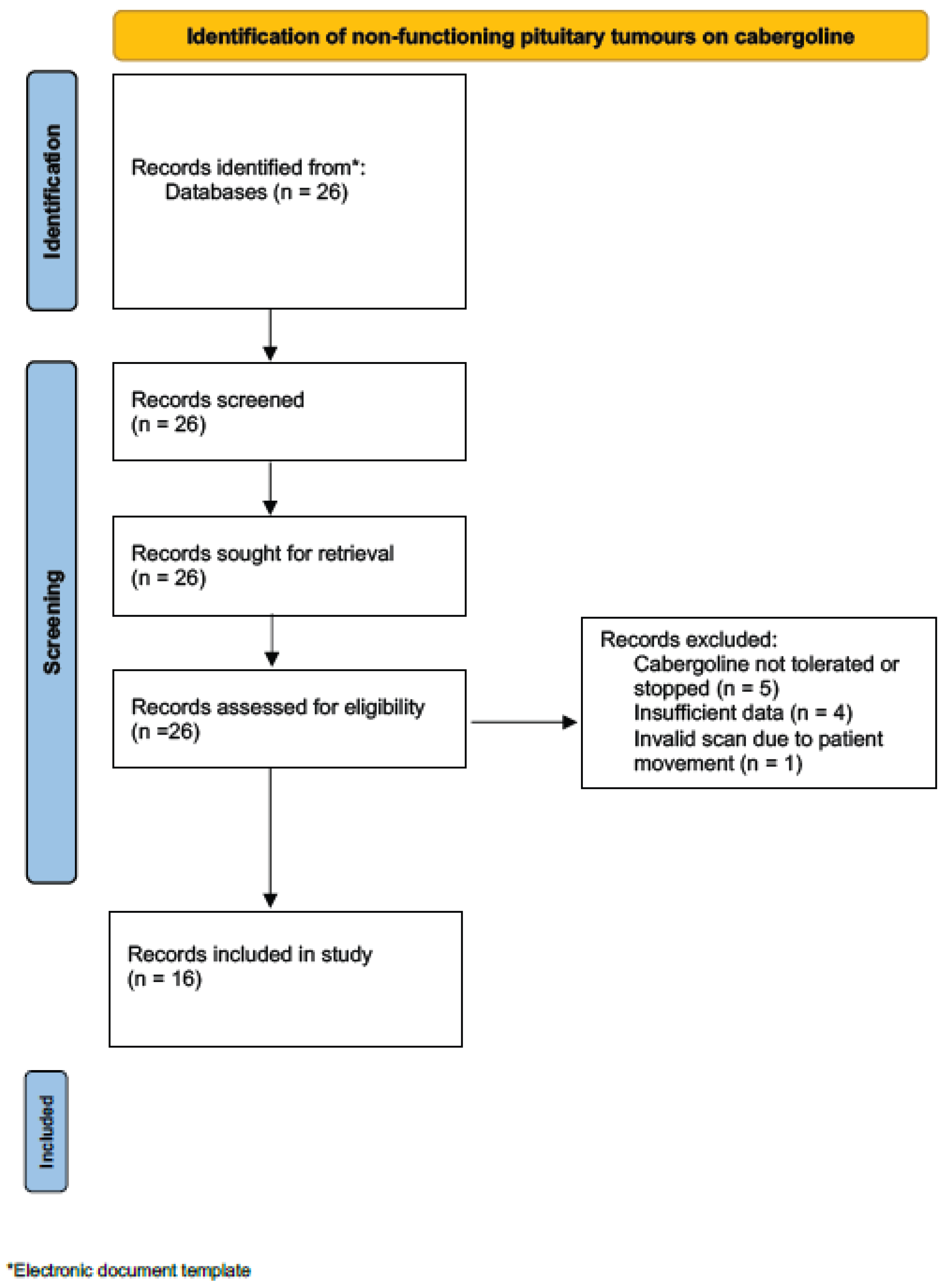
Figure 1. PRISMA flow diagram illustrating the selection process of patients.
Endocrines 07 00015 g001
Table 1. Patient characteristics.
IDGenderAgePre or Post- SurgeryPre-CAB Maximal Diameter of Tumour (mm)Pre-CAB Tumour Volume (mm3)Baseline Visual Fields (on Initiation of Cabergoline)Indication for Starting Cabergoline: (1) Surgery not Possible Due to Patient Age/Comorbidities, or (2) Surgery Declined by the Patient, (3) Enlarging Tumour, (4) Substantial Tumour Remnant Post-Surgery Cabergoline Titrating ScheduleFinal Weekly Cabergoline Dosage (mcg)
1Female44Pre17.91618.4Normal2 + 3250 mcg once weekly250
2Male49Pre23.95012.5Normal2500 mcg twice weekly1000
3Male76Pre14.6703.3Normal1 + 3500 mcg thrice weekly1500
4Female72Pre14.81014.5Normal1500 mcg once weekly500
5Male86Pre19.93648.4Normal1250 mcg once weekly250
6Male55Post20.33535.0Normal (bitemporal hemianopia pre-surgery which resolved afterwards)2 + 3 + 4250 mcg thrice weekly750
7Female42Post18.72239.9Blind right eye post-surgery (due to glaucoma)2 + 4250 mcg twice weekly500
8Male89Pre28.17513.1Normal1 + 3250 mcg once daily1750
9Male85Pre23.63902.4Normal1 + 3250 mcg once weekly for 9 months, then increased to 1000 mcg once weekly for 6 months, then increased to 1000 mcg twice weekly for 2 months, then increased to 1000 mcg thrice weekly3000
10Female49Post31.911,453.0Normal (bitemporal hemianopia pre-surgery which resolved afterwards)2 + 4500 mcg once weekly for 7 weeks, then increased to 1000 mcg once weekly for 3 weeks, then increased to 1500 mcg once weekly for 3 weeks, then increased to 2000 mcg once weekly for 3 weeks, then increased to 2500 mcg once weekly for 3 weeks, then increased to 3000 mcg once weekly3000
11Male83Pre21.54872.2Normal1500 mcg twice weekly1000
12Male86Pre20.52648.8Normal1500 mcg twice weekly1000
13Female63Post26.03497.7Normal1 + 2 + 41000 mcg thrice weekly3000
14Female80Pre21.02424.1Bitemporal superior quadrantanopia1 + 2500 mcg once daily3500
15Female75Post15.01336.6Normal1 + 3500 mcg once weekly for 6 weeks, then increased to 500 mcg twice weekly for 6 weeks, then increased to 500 mcg thrice weekly1500
16Male69Post12.2653.5Normal1 + 3500 mcg thrice weekly1500
Table 2. Tumour volumes in the pre-CAB scan to 1st FU scan.
IDPre-CAB Volume (mm3)1st FU Volume (mm3)Pre-CAB vs. 1stTime Between Pre-CAB Scan to 1st FU Scan (Days)Duration of CAB Treatment (CAB Initiation—1st FU Scan) (Days)Result Pre-CAB vs. 1st FU
11618.41177.10.73918358Shrinkage
25012.51674.60.33266214Shrinkage
3703.3690.60.98436315Stable
41014.51064.11.05335326Stable
53648.43942.51.08181153Stable
63535.02919.90.83245139Stable
72239.92395.11.0735090Stable
87513.16124.60.82273200Stable
93902.44599.51.18482187Stable
1011,453.013,215.51.15445225Stable
114872.23279.50.67378117Shrinkage
122648.82158.10.81398357Stable
133497.73591.41.03464346Stable
142424.12572.41.06611501Stable
151336.6943.00.71315181Shrinkage
16653.5722.61.11698612Stable
Table 3. Tumour volumes in the pre-CAB scan to 2nd FU scan.
IDPre-CAB Volume (mm3)2nd FU Volume (mm3)Pre-CAB vs. 2nd FUTime Between Pre-CAB Scan to 2nd FU Scan (Days)Duration of CAB Treatment (CAB Initiation- 2nd FU Scan) (Days)Result Pre-CAB vs. 2nd FU
25012.5518.40.10510458Shrinkage
3703.3796.31.131055934Stable
41014.5974.30.96701692Stable
53648.41814.70.50538510Shrinkage
63535.02726.70.77419313Shrinkage
72239.91887.60.84507247Stable
87513.14711.80.63623550Shrinkage
93902.44134.61.06817551Stable
1011,453.013,215.51.15899679Stable
133497.72887.11.03774656Stable
151336.6943.00.71704570Shrinkage
Table 4. Tumour volumes in pre-CAB scan, and tumour volumes when combining the 1st and 2nd FU scans (using the latest scan in patients who had two scans).
IDPre-CAB Volume (mm3)Latest FU Volume (mm3)Pre-CAB vs. Latest FUTime Between Pre-CAB Scan to Latest FU Scan (Days)Duration of CAB Treatment (CAB Initiation—Latest FU Scan) (Days)Result Pre-CAB vs. Latest FU
11618.41177.10.73918358Shrinkage
25012.5518.40.10510458Shrinkage
3703.3796.31.131055934Stable
41014.5974.30.96701692Stable
53648.41814.70.50538510Shrinkage
63535.02726.70.77419313Shrinkage
72239.91887.60.84507247Stable
87513.14711.80.63623550Shrinkage
93902.44134.61.06817551Stable
1011,453.013,215.51.15899679Stable
114872.23279.50.67378117Shrinkage
122648.82158.10.81398357Stable
133497.72887.11.03774656Stable
142424.12572.41.06611501Stable
151336.6943.00.71704570Shrinkage
16653.5722.61.11698612Stable
Table 5. Positive growth rates of patients with enlarging tumours.
IDAdditional MRI Prior to CAB Initiation Volume (mm3)Pre-CAB Volume (mm3)Additional MRI vs. Pre-CABGrowth Rate from Additional MRI to Pre-CAB (mm3/days)
11574.31618.41.03+0.04
3523.6703.31.34+0.38
61838.83535.01.92+1.62
85937.67513.11.27+2.01
93671.33902.41.06+1.96
Table 6. Growth rate of tumours, between the additional MRI prior to CAB initiation to pre-CAB scan, between pre-CAB scan to 1st FU scan, and between the pre-CAB scan to 2nd FU scan.
IDAdditional MRI Prior to CAB Initiation Volume (mm3)Pre-CAB Volume (mm3)1st FU Volume (mm3)2nd FU Volume (mm3)Additional MRI vs. Pre-CABGrowth Rate from Additional MRI to Pre-CAB (mm3/days)Pre-CAB vs. 1st FU ScanGrowth Rate from Pre-CAB to 1st FU (mm3/days)Pre-CAB vs. 2nd FU ScanGrowth Rate from Pre-CAB to 2nd FU (mm3/days)Result Additional MRI vs. Pre-CABResult Pre-CAB vs. 1st FUResult Pre-CAB vs. 2nd FU
11574.31618.41177.1N/A1.03+0.040.73−0.17N/AN/AStableShrinkageN/A
3523.6703.3690.6796.31.34+0.380.98−0.031.13+0.09IncreaseStableStable
61838.83535.02919.92726.71.92+1.620.83−2.510.77−1.93IncreaseStableShrinkage
85937.67513.16124.64711.81.27+2.010.82−5.090.63−4.50IncreaseStableShrinkage
93671.33902.44599.54134.61.06+1.961.18−1.541.06+0.28StableStableStable
Disclaimer/Publisher’s Note: The statements, opinions and data contained in all publications are solely those of the individual author(s) and contributor(s) and not of MDPI and/or the editor(s). MDPI and/or the editor(s) disclaim responsibility for any injury to people or property resulting from any ideas, methods, instructions or products referred to in the content.


