Next Issue
Volume 2, September
Previous Issue
Volume 2, March


Endocrines, Volume 2, Issue 2 (June 2021) – 10 articles

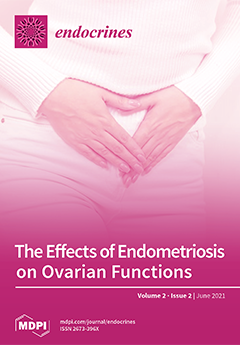
Cover Story (view full-size image): Endometriosis is an estrogen-dependent chronic inflammatory disease. Fibrotic changes in affected anatomical site, such as uterus, fallopian tubes, and ovaries, may cause functional disturbances in these reproductive organs. The ovarian function can be classified into three basic elements: (1) production of ovarian hormones; (2) maintenance of follicular development until post-ovulatory luteal stage; (3) reservoir of dormant oocytes (ovarian reserve). Endometriosis may affect these functions, but the exact mechanism is still yet to be determined. Diminished ovarian reserve may be a main deterioration factor of endometriosis-derived ovarian dysfunction. Early medical and surgical interventions may be beneficial to maintain ovarian function, but evidence is still lacking. View this paper
  • Issues are regarded as officially published after their release is announced to the table of contents alert mailing list.
  • You may sign up for e-mail alerts to receive table of contents of newly released issues.
  • PDF is the official format for papers published in both, html and pdf forms. To view the papers in pdf format, click on the "PDF Full-text" link, and use the free Adobe Reader to open them.
Order results
Result details
Section
Select all
Export citation of selected articles as:
7 pages, 1518 KiB  
Case Report
Autoimmune Hypophysitis with Late Renal Involvement: A Case Report
by Stefano Iuliano, Maria Carmela Zagari, Margherita Vergine, Alessandro Comi, Michele Andreucci, Gemma Patella, Stefania Giuliano, Sandro La Vignera, Antonio Brunetti, Antonio Aversa and Emanuela A. Greco
Endocrines 2021, 2(2), 160-166; https://doi.org/10.3390/endocrines2020016 - 17 Jun 2021
Viewed by 2046
Abstract
We report a case of a 50-year-old male admitted to the Endocrinology Unit because of persistent headaches, nausea, feeling tired, sudden weight loss, cold intolerance, decreased appetite, and lack of sex interest. Diagnostic workup showed a 6-millimeter pituitary tumor without signs of compression, [...] Read more.
We report a case of a 50-year-old male admitted to the Endocrinology Unit because of persistent headaches, nausea, feeling tired, sudden weight loss, cold intolerance, decreased appetite, and lack of sex interest. Diagnostic workup showed a 6-millimeter pituitary tumor without signs of compression, and a condition of progressive panhypopituitarism. After 12 months of hormone replacement therapy, the patient was hospitalized because of sudden weight gain, periorbital-peripheral edema, severe dyslipidemia, hypertension, and proteinuria. Corticosteroid therapy was shifted from oral to continuous intravenous infusion, and once the diagnosis of “immune complex-mediated glomerulonephritis with mesangial deposits suggestive for membranoproliferative glomerulonephritis type IIIIgG4-positive” was made, the immunosuppressant mycophenolate (1500 mg/day) was started. After a 6-month follow-up, the complete resolution of renal symptoms was accompanied by the disappearance of a pituitary lesion and the patient was back to prior hormone replacement therapy. Autoimmune hypophysitis (AH) is a rare autoimmune inflammatory disease of the pituitary gland that can impair hormone secretion and function. IgG4-hypophysitis is rare and is usually associated with other IgG4-related diseases. Herein, we describe a rare case of AH associated with late renal disease, and without any other organ involvement. Full article
Show Figures

Figure 1

10 pages, 599 KiB  
Communication
Association between Physical Activity and Seasonal Variations in Metabolic and Vascular Function in Adults
by Hiroto Honda, Makoto Igaki, Motoaki Komatsu and Shin-ichiro Tanaka
Endocrines 2021, 2(2), 150-159; https://doi.org/10.3390/endocrines2020015 - 11 Jun 2021
Viewed by 2424
Abstract
This article highlights the association between physical activity (PA) and seasonal variations in metabolic and vascular function in adults. Increasing PA is an important method for preventing cardiovascular disease (CVD) and all-cause mortality by improving blood glucose, blood pressure, blood lipid profiles, body [...] Read more.
This article highlights the association between physical activity (PA) and seasonal variations in metabolic and vascular function in adults. Increasing PA is an important method for preventing cardiovascular disease (CVD) and all-cause mortality by improving blood glucose, blood pressure, blood lipid profiles, body fat, insulin resistance, and vascular function, such as endothelial function. Conversely, various factors, such as seasonal climate conditions, may affect the amount of PA that individuals undertake. Changes in PA often induce seasonal variations in metabolic and vascular function; the deterioration of such functions in winter is the most prominent, and there is clear evidence of an increased risk of CVD in this season. Understanding the influence of PA on seasonal variations observed in metabolic and vascular function is necessary for the management of these physiological functions. In this article summary, few studies have proven that maintaining PA can suppress the variations, and it remains unclear what types, intensities, and durations of regular PA are effective for circumventing seasonal impact. In addition to further studies, there is a need to educate individuals about the strategies to manage PA and other aspects of their lifestyles throughout the year, particularly in winter. Full article
Show Figures

Figure 1

8 pages, 1429 KiB  
Communication
The Effects of Endometriosis on Ovarian Functions
by Michio Kitajima, Kanako Matsumoto, Itsuki Kajimura, Ayumi Harada, Noriko Miyashita, Asako Matsumura, Yuriko Kitajima and Kiyonori Miura
Endocrines 2021, 2(2), 142-149; https://doi.org/10.3390/endocrines2020014 - 3 Jun 2021
Cited by 5 | Viewed by 3923
Abstract
Infertility is a main manifestation of endometriosis, though the exact pathogenesis of endometriosis-associated infertility remains unclear. Compromised ovarian functions may be one of the causes of endometriosis related infertility. The ovarian function can be classified into three basic elements, (1) production of ovarian [...] Read more.
Infertility is a main manifestation of endometriosis, though the exact pathogenesis of endometriosis-associated infertility remains unclear. Compromised ovarian functions may be one of the causes of endometriosis related infertility. The ovarian function can be classified into three basic elements, (1) production of ovarian hormones, (2) maintenance of follicular development until ovulation, and (3) reservoir of dormant oocytes (ovarian reserve). The effects of endometriosis on ovarian hormone production and follicular development are inconclusive. Ovarian endometrioma is common phonotype of endometriosis. Development of endometrioma per se may affect ovarian reserve. Surgery for endometriomas further diminish ovarian reserve, especially women with bilateral involvement. Early intervention with surgery and/or medical treatment may be beneficial, though firm evidence is lacking. When surgery is chosen in women at reproductive age, specific techniques that spare ovarian function should be considered. Full article
(This article belongs to the Special Issue Impact of Endometriosis on Women’s Health)
Show Figures

Figure 1

9 pages, 260 KiB  
Article
Vitamin D and Subclinical Cardiovascular Damage in Essential Hypertension
by Marcello Magurno, Daniele Crescibene, Martina Spinali, Velia Cassano, Giuseppe Armentaro, Keti Barbara, Sofia Miceli, Marta L. Hribal, Maria Perticone and Angela Sciacqua
Endocrines 2021, 2(2), 133-141; https://doi.org/10.3390/endocrines2020013 - 31 May 2021
Cited by 1 | Viewed by 2769
Abstract
Vitamin D deficiency is linked to cardiac dysfunction, vascular remodeling, metabolic syndrome and insulin resistance (IR). The aim of the present study was to evaluate the association between vitamin D levels and cardiovascular (CV) organ damage in a large cohort of newly diagnosed [...] Read more.
Vitamin D deficiency is linked to cardiac dysfunction, vascular remodeling, metabolic syndrome and insulin resistance (IR). The aim of the present study was to evaluate the association between vitamin D levels and cardiovascular (CV) organ damage in a large cohort of newly diagnosed treatment-naïve hypertensive patients, and the role of IR in this context. We enrolled 500 Caucasian individuals, without CV or renal complications. Subjects underwent a complete evaluation and measurements of vitamin D, standard laboratory determinations and instrumental examination, including echocardiography and applanation tonometry. Linear regression analyses were performed to assess the correlation between pulse wave velocity (PWV) and left ventricular mass index (LVMI) with different covariates. PWV was significantly correlated with age (p < 0.0001), LDL cholesterol (p < 0.0001), BMI (p = 0.001), pulse pressure (PP) (p = 0.005) and high sensitivity C-reactive protein (hs-CRP) (p = 0.006), while an inverse correlation was observed with vitamin D levels (p < 0.0001), Matsuda index (p < 0.0001) and estimated glomerular filtration ratio (e-GFR) (p = 0.006). LVMI significantly correlated with PP (p < 0.0001), hs-CRP (p < 0, 0001) and age (p = 0.001), while an inverse relationship was observed with vitamin D levels (p < 0.0001), Matsuda’s insulin sensitivity index (ISI) (p < 0.0001) and e-GFR (p < 0.0001). Vitamin D was the strongest predictor of PWV and LVMI, explaining, respectively, 28.3% and 19.1% of their variation. Our study suggests that low vitamin D might be a biomarker of end-organ damage. Full article
12 pages, 2317 KiB  
Review
Natural Killer Cell Mobilization in Breast and Prostate Cancer Survivors: The Implications of Altered Stress Hormones Following Acute Exercise
by Erik D. Hanson, Lauren C. Bates, Kaileigh Moertl and Elizabeth S. Evans
Endocrines 2021, 2(2), 121-132; https://doi.org/10.3390/endocrines2020012 - 19 May 2021
Cited by 1 | Viewed by 2763
Abstract
Natural killer (NK) cells from the innate immune system are integral to overall immunity and also in managing the tumor burden during cancer. Breast (BCa) and prostate cancer (PCa) are the most common tumors in U.S. adults. Both BCa and PCa are frequently [...] Read more.
Natural killer (NK) cells from the innate immune system are integral to overall immunity and also in managing the tumor burden during cancer. Breast (BCa) and prostate cancer (PCa) are the most common tumors in U.S. adults. Both BCa and PCa are frequently treated with hormone suppression therapies that are associated with numerous adverse effects including direct effects on the immune system. Regular exercise is recommended for cancer survivors to reduce side effects and improve quality of life. Acute exercise is a potent stimulus for NK cells in healthy individuals with current evidence indicating that NK mobilization in individuals with BCa and PCa is comparable. NK cell mobilization results from elevations in shear stress and catecholamine levels. Despite a normal NK cell response to exercise, increases in epinephrine are attenuated in BCa and PCa. The significance of this potential discrepancy still needs to be determined. However, alterations in adrenal hormone signaling are hypothesized to be due to chronic stress during cancer treatment. Additional compensatory factors induced by exercise are reviewed along with recommendations on standardized approaches to be used in exercise immunology studies involving oncology populations. Full article
(This article belongs to the Special Issue Exercise Endocrinology)
Show Figures

Figure 1

12 pages, 585 KiB  
Review
Testosterone Boosters Intake in Athletes: Current Evidence and Further Directions
by Artemii Lazarev and Eduard Bezuglov
Endocrines 2021, 2(2), 109-120; https://doi.org/10.3390/endocrines2020011 - 17 May 2021
Cited by 5 | Viewed by 37013
Abstract
“Testosterone boosters” (TB)—are supplements that are claimed to increase testosterone levels in the human body. While the consumption of TB may be popular among athletes, there is insufficient evidence both about the safety and the real efficacy of TB. In our review, we [...] Read more.
“Testosterone boosters” (TB)—are supplements that are claimed to increase testosterone levels in the human body. While the consumption of TB may be popular among athletes, there is insufficient evidence both about the safety and the real efficacy of TB. In our review, we searched MEDLINE/PubMed and Cochrane Library for studies on the effects of 15 substances that are claimed to increase testosterone levels Anacyclus pyrethrum; Bulbine natalensis; Epimedium (horny goat weed); L-arginine; L-carnitine; magnesium; Mucuna pruriens; pantothenic acid; selenium; shilajit Eurycoma longifolia (Tongkat Ali); Serenoa repens (saw palmetto); boron; Withania somnifera (ashwagandha); and Trigonella foenum-graecum (fenugreek) in athletes and healthy adults under 55 years of age. We found such studies regarding 10 out of 15 substances: L-arginine (3 studies); L-carnitine (2); magnesium (1); selenium (2); shilajit (1); Tongkat Ali (2); Serenoa repens (1); boron (3); ashwagandha root (2); and fenugreek (7). Many of them fail to prove the efficacy of these substances to increase testosterone levels. Tongkat Ali, ashwagandha, and fenugreek were the substances with the strongest evidence. The positive effect of magnesium and shilajit on testosterone concentration was shown in single studies. Conflicting data found that L-arginine, L-carnitine, Serenoa repens, selenium and boron do not appear to increase testosterone levels. There are almost no data on the safety profile of various TB components; however, certain TB components may be linked to coagulation, and pancreatic and hepatic disorders. Based on the review, the authors conclude that at present TB cannot be recommended for use by athletes due to insufficient data on their efficacy and safety. Full article
(This article belongs to the Special Issue Exercise Endocrinology)
Show Figures

Figure 1

10 pages, 285 KiB  
Review
Endocrine Responses to Sport-Related Brain Injury in Female Athletes: A Narrative Review and a Call for Action
by Donna Duffy, Kirsty J. Elliott-Sale, Hanna Gardner, Samantha Goldenstein and Laurie Wideman
Endocrines 2021, 2(2), 99-108; https://doi.org/10.3390/endocrines2020010 - 23 Apr 2021
Cited by 5 | Viewed by 3328
Abstract
Sport-related brain injury (SRBI) occurs when a blow to the head causes the brain to move back and forth in the skull, and can lead to neuroendocrine dysfunction. Research has shown that males and females experience and recover from SRBI differently, yet most [...] Read more.
Sport-related brain injury (SRBI) occurs when a blow to the head causes the brain to move back and forth in the skull, and can lead to neuroendocrine dysfunction. Research has shown that males and females experience and recover from SRBI differently, yet most of what is known regarding diagnosis, treatment, and recovery of SRBI is based on male normative data even though females meet or exceed incidence numbers of SRBIs compared to those of males. Females also have been known to have worse outcomes and a greater number of symptoms following SRBI than males. Research is limited as to why females have worse outcomes, but sex hormones have been suggested as a potential reason. SRBI may cause a dysregulation of the hypothalamic–pituitary–gonadal (HPG) axis, which is responsible for regulating the sex hormones estrogen and progesterone. Initial research has shown that SRBI may suppress estrogen and progesterone, and the concentration of these sex hormones could be indicative of injury severity and recovery trajectory. This review discusses the sex-specific differences in SRBI and also the future direction of research that is needed in order to identify the repercussions of SRBIs for female athletes, which will eventually lead to better clinical treatment, sideline care, and recovery profiles. Full article
(This article belongs to the Special Issue Exercise Endocrinology)
8 pages, 247 KiB  
Article
Inter-Reader Agreement of ATA Sonographic Risk in Thyroid Nodules with Bethesda Category III Indeterminate Cytology
by Maria I. Linares Valderrama, Marilyn A. Arosemena, Anu Thekkumkattil, Russ A. Kuker, Rosa P. Castillo, Charif Sidani, Manuel Lora Gonzalez, Sabina Casula and Atil Y. Kargi
Endocrines 2021, 2(2), 91-98; https://doi.org/10.3390/endocrines2020009 - 22 Apr 2021
Cited by 1 | Viewed by 1972
Abstract
Background: Substantial inter-observer variation has been documented in the recognition and description of specific sonographic features as well as for ATA sonographic risk (ASR). This raises the question if the risk stratification proposed by the ATA guidelines is reproducible and applicable for nodules [...] Read more.
Background: Substantial inter-observer variation has been documented in the recognition and description of specific sonographic features as well as for ATA sonographic risk (ASR). This raises the question if the risk stratification proposed by the ATA guidelines is reproducible and applicable for nodules with indeterminate cytology. The aim of the study was to determine the inter-reader agreement (IRR) among radiologists using the 2015 ASR stratification in indeterminate thyroid nodules. Methods: Three board certified radiologists who were blinded to clinical data and to each other, interpreted the ultrasound findings of 179 nodules that had Bethesda III cytology. The nodules were classified into high suspicion (HS), intermediate (IS), low (LS), very low (VLS). Echogenicity, composition, shape taller than wide, vascularity, type of margins, presence and type of calcifications were also described. Results: The majority consensus revealed that 28%, 27%, 39% and 5% were described as high, intermediate, low and very low ASR, respectively. The inter-reader agreement was near perfect (k 0.82 CI 95% (0.77–0.87)). Nodules were paired into a higher risk (HS + IS) and lower risk (LS + VLS) categories with substantial agreement (k 0.7) in both categories. Conclusion: A near perfect agreement among readers was observed when stratifying indeterminate cytology nodules for ASR. Full article
12 pages, 779 KiB  
Article
Influence of Menstrual Cycle or Hormonal Contraceptive Phase on Energy Intake and Metabolic Hormones—A Pilot Study
by Johanna K. Ihalainen, Ida Löfberg, Anna Kotkajuuri, Heikki Kyröläinen, Anthony C. Hackney and Ritva S. Taipale-Mikkonen
Endocrines 2021, 2(2), 79-90; https://doi.org/10.3390/endocrines2020008 - 16 Apr 2021
Cited by 11 | Viewed by 5724
Abstract
Sex hormones are suggested to influence energy intake (EI) and metabolic hormones. This study investigated the influence of menstrual cycle (MC) and hormonal contraceptive (HC) cycle phases on EI, energy availability (EA), and metabolic hormones in recreational athletes (eumenorrheic, NHC = 15 and [...] Read more.
Sex hormones are suggested to influence energy intake (EI) and metabolic hormones. This study investigated the influence of menstrual cycle (MC) and hormonal contraceptive (HC) cycle phases on EI, energy availability (EA), and metabolic hormones in recreational athletes (eumenorrheic, NHC = 15 and monophasic HC-users, CHC = 9). In addition, 72-h dietary and training logs were collected in addition to blood samples, which were analyzed for 17β-estradiol (E2), progesterone (P4), leptin, total ghrelin, insulin, and tri-iodothyronine (T3). Measurements were completed at four time-points (phases): Bleeding, mid-follicular (FP)/active 1, ovulation (OVU)/active 2, mid-luteal (LP)/inactive in NHC/CHC, respectively. As expected, E2 and P4 fluctuated significantly in NHC (p < 0.05) and remained stable in CHC. In NHC, leptin increased significantly between bleeding and ovulation (p = 0.030) as well as between FP and OVU (p = 0.022). No group differences in other measured hormones were observed across the MC and HC cycle. The mean EI and EA were similar between phases, with no significant differences observed in macronutrient intake over either the MC or HC. While the MC phase might have a small, but statistically significant effect on leptin, the findings of the present study suggest that the MC or HC phase does not significantly alter ad libitum EI or EA in recreational athletes. Full article
(This article belongs to the Special Issue Exercise Endocrinology)
Show Figures

Figure 1

14 pages, 830 KiB  
Review
Exercise as a Therapeutic Intervention in Gestational Diabetes Mellitus
by Konstantina Dipla, Andreas Zafeiridis, Gesthimani Mintziori, Afroditi K. Boutou, Dimitrios G. Goulis and Anthony C. Hackney
Endocrines 2021, 2(2), 65-78; https://doi.org/10.3390/endocrines2020007 - 26 Mar 2021
Cited by 15 | Viewed by 6744
Abstract
Gestational Diabetes Mellitus (GDM) is defined as any degree of glucose intolerance with onset or first recognition during pregnancy. Regular exercise is important for a healthy pregnancy and can lower the risk of developing GDM. For women with GDM, exercise is safe and [...] Read more.
Gestational Diabetes Mellitus (GDM) is defined as any degree of glucose intolerance with onset or first recognition during pregnancy. Regular exercise is important for a healthy pregnancy and can lower the risk of developing GDM. For women with GDM, exercise is safe and can affect the pregnancy outcomes beneficially. A single exercise bout increases skeletal muscle glucose uptake, minimizing hyperglycemia. Regular exercise training promotes mitochondrial biogenesis, improves oxidative capacity, enhances insulin sensitivity and vascular function, and reduces systemic inflammation. Exercise may also aid in lowering the insulin dose in insulin-treated pregnant women. Despite these benefits, women with GDM are usually inactive or have poor participation in exercise training. Attractive individualized exercise programs that will increase adherence and result in optimal maternal and offspring benefits are needed. However, as women with GDM have a unique physiology, more attention is required during exercise prescription. This review (i) summarizes the cardiovascular and metabolic adaptations due to pregnancy and outlines the mechanisms through which exercise can improve glycemic control and overall health in insulin resistance states, (ii) presents the pathophysiological alterations induced by GDM that affect exercise responses, and (iii) highlights cardinal points of an exercise program for women with GDM. Full article
(This article belongs to the Special Issue Exercise Endocrinology)
Show Figures

Figure 1

Previous Issue
Next Issue
Back to TopTop