Case Report

Recurrent Borderline Ovarian Tumors in the Adolescent Population: Case Report

1
Bowman Gray Center for Medical Education, Wake Forest University School of Medicine, Central Campus, Winston-Salem, NC 27101, USA
2
Department of Obstetrics and Gynecology, Atrium Health Wake Forest Baptist Medical Center, Winston-Salem, NC 27157, USA
*
Author to whom correspondence should be addressed.
Reprod. Med. 2025, 6(1), 4; https://doi.org/10.3390/reprodmed6010004
Submission received: 2 December 2024 / Revised: 9 January 2025 / Accepted: 13 January 2025 / Published: 5 February 2025

Abstract

:
Background and Clinical Significance: Borderline ovarian tumors (BOTs) are a rare diagnosis, especially in the adolescent population. This can make initial management and surveillance strategies difficult, given the limited guidelines and experience in this young age group. Case Presentation: We present two cases of recurrent serous BOTs diagnosed in adolescent patients. Both patients were initially treated with fertility-sparing surgery and followed with transabdominal pelvic ultrasounds. Secondary surgical debulking of recurrent disease with uterine preservation was successful in both patients with a long-term disease-free status. Conclusions: Although rare, BOTs can occur in adolescent patients and should be on the differential for ovarian masses in this age group. Fertility-sparing surgical techniques, reproductive endocrinology consultation, surveillance strategies, and hormone replacement therapy should all be taken into consideration when treating adolescent patients with BOTs.

1. Introduction

Borderline ovarian tumors (BOTs) account for 15–20% of all epithelial ovarian malignancies, and approximately 3000 people in the United States are diagnosed each year [1,2]. BOTs are divided by histologic features—most commonly serous, followed by mucinous, endometrioid, clear-cell, and transitional-cell, respectively [3]. Most patients are diagnosed at an early stage with a 10-year survival rate of >90% [3]. The average age for the diagnosis of BOTs is 45 years, and many patients undergo fertility-sparing treatment [3]. Relapse rates of patients with BOTs range from 7 to 30% and are influenced by the presence of peritoneal implants, micropapillary histologic pattern, and microinvasion [2,4].
Within children and adolescents, BOTs are rare and account for only 10–30% of ovarian epithelial tumors and less than 1% of all ovarian tumors [3,4]. Although there are established guidelines for adult patients with these tumors, there is little data in the adolescent population regarding diagnosis, treatment, and follow-up. Here, we present two cases of BOTs diagnosed in adolescent patients with subsequent recurrence on the contralateral ovary and highlight the surgical and survivorship considerations of young patients with BOTs.

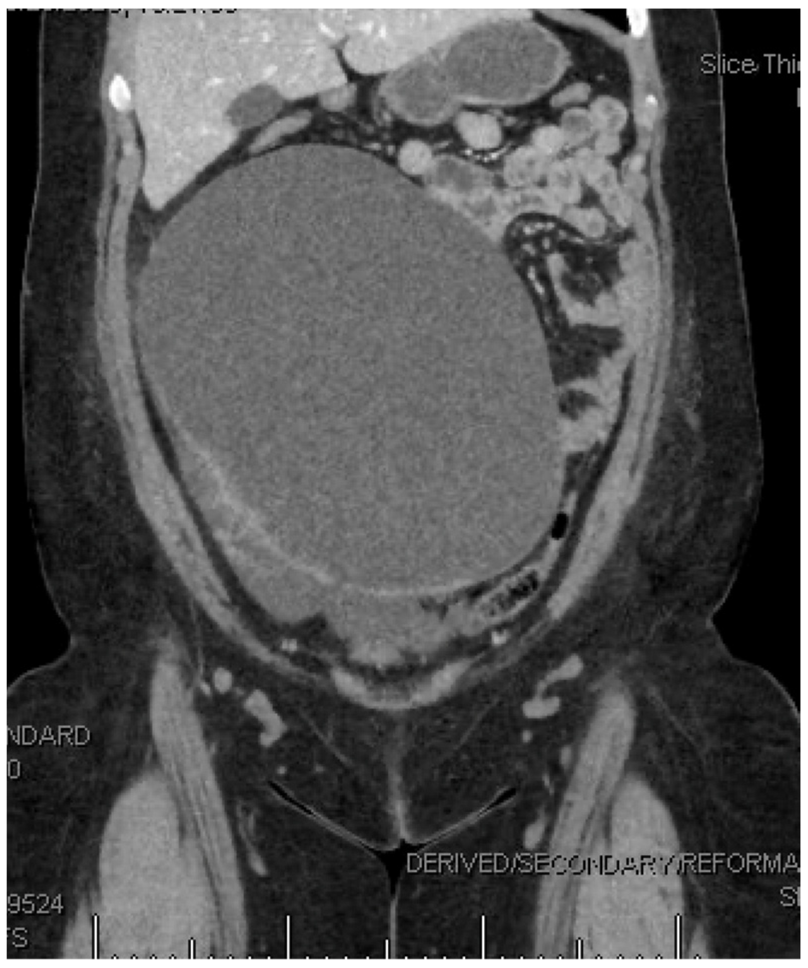
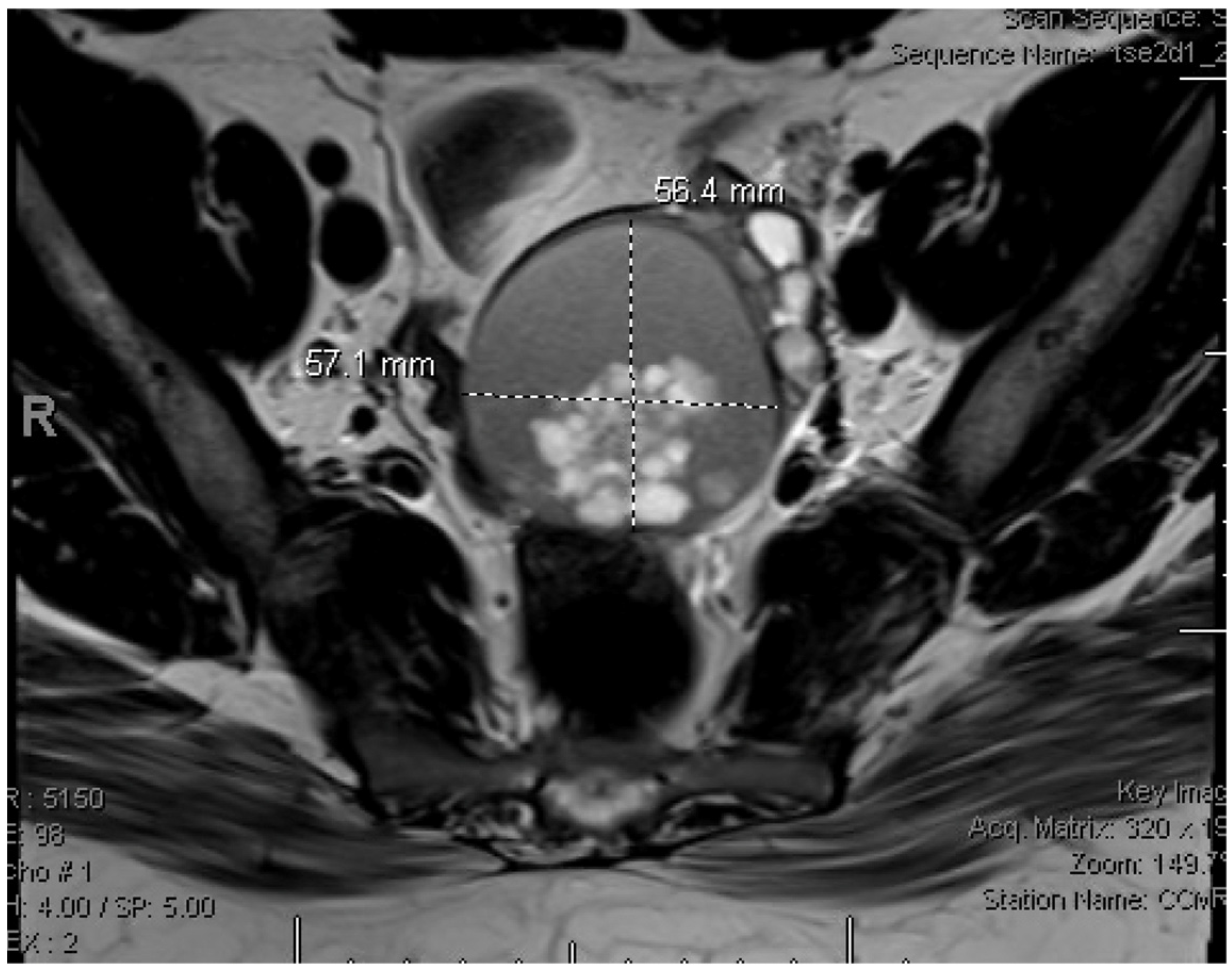
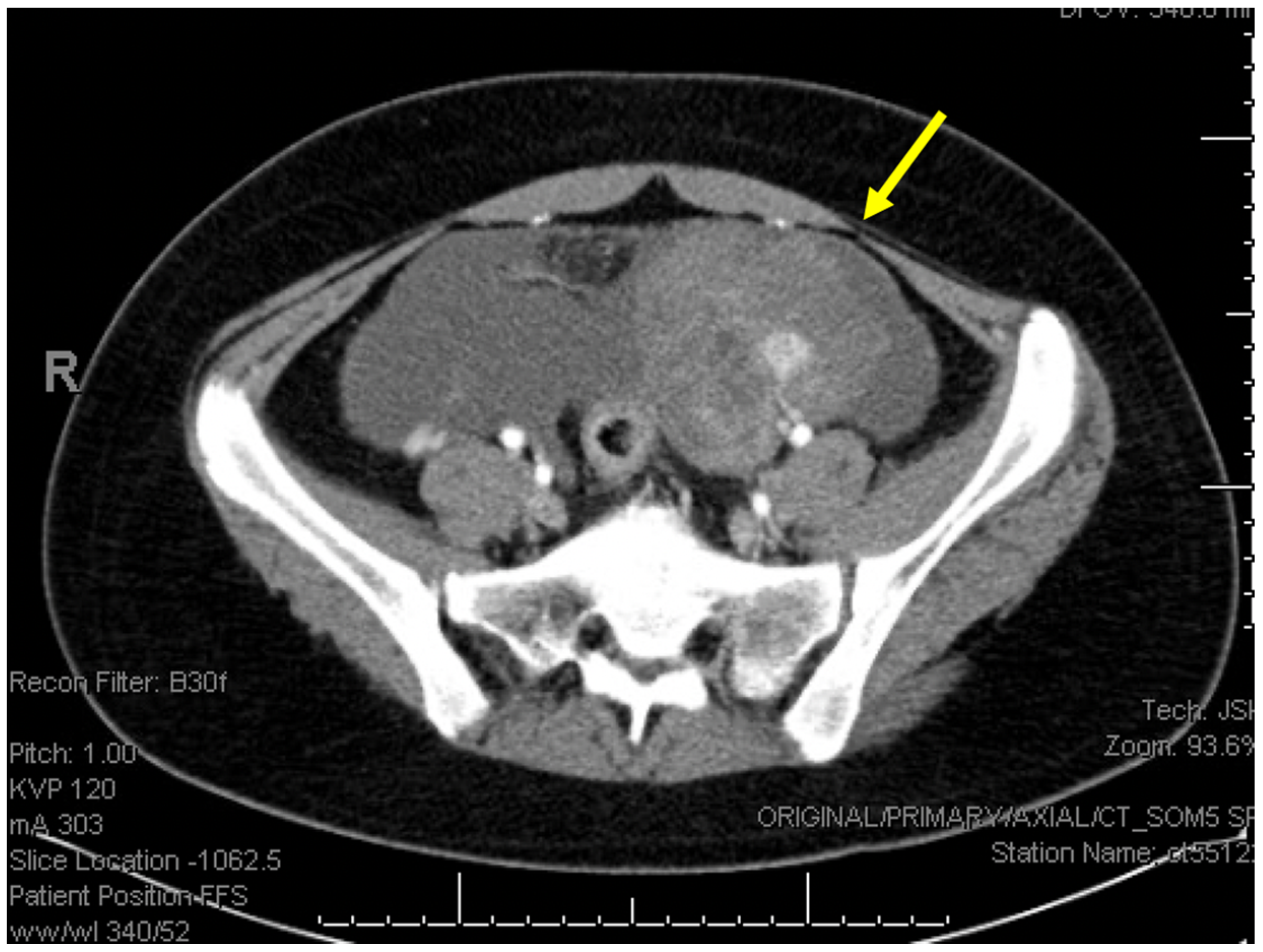
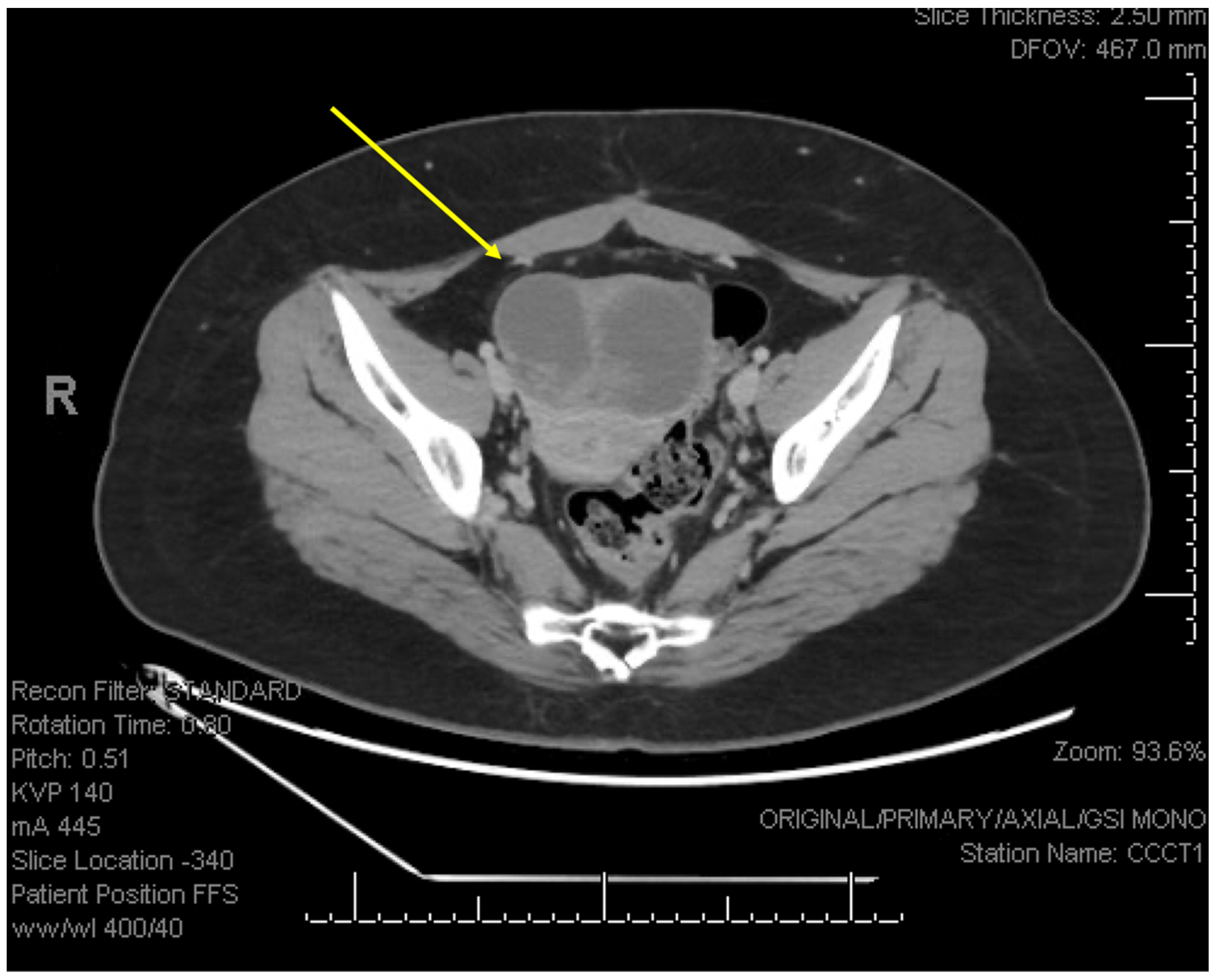
2. Detailed Case Description

The first patient presented at age 19 with several months of intermittent abdominal pain and a palpable abdominal mass on exam. Computed tomography (CT) scan with intravenous (IV) contrast showed a 21.2 × 13.9 × 24.1 cm complex, cystic pelvic mass (Figure 1) and an elevated CA125 of 345 IU/mL. She then underwent surgical management with intraoperative findings notable for a 20 cm right ovarian mass, a 5 cm mass adherent to posterior cul-de-sac and right pelvic sidewall, and normal-appearing left tube and ovary. Intraoperative frozen section performed on the right adnexal mass noted a serous BOT. Her procedure included an abdominal right salpingo-oophorectomy via vertical midline incision, removal of pelvic mass, infracolic omentectomy, and peritoneal biopsies. There was no intraoperative spillage of the mass and no residual disease at the end of the case. Final diagnosis showed stage IIB serous BOT.
She was followed closely by gynecologic oncology with surveillance visits, transabdominal pelvic ultrasounds, and CA125 levels every 6 months. She was diagnosed with recurrent disease 26 months after her initial surgery when an enlarging left adnexal cyst was found on pelvic ultrasound. Additional magnetic resonance imaging (MRI) showed a 5 cm mass in the left ovary with an internal solid component (Figure 2), CA125 was normal at 16 IU/mL. Discussion of cystectomy versus salpingo-oophorectomy was had with the patient, and, given the risk of recurrence with cystectomy, she opted to undergo laparoscopic left salpingo-oophorectomy, uterine preservation, and peritoneal nodule biopsy, with final pathology confirming recurrent serous BOT with a non-invasive peritoneal implant. She has continued on surveillance and has been without evidence of disease recurrence for 24 months.
The second patient, at age 12, presented to an outside emergency department (ED) with acute onset abdominal pain. Transabdominal pelvic ultrasound showed a 10 × 12 cm left adnexal mass with clinical concern for ovarian torsion. She underwent laparoscopic removal of a left paratubal cyst with intraoperative spillage of cyst contents and left ovarian detorsion, and final pathology demonstrated a serous BOT. CA125 was not collected at that time. Eleven months later, at age 13, she was referred to gynecologic oncology for ongoing surveillance. Transabdominal pelvic ultrasound imaging was obtained, showing a 9 cm solid mass in her left ovary and an additional complex posterior uterine mass with elevated CA125 to 374 IU/mL. CT scan demonstrated a 7 cm left ovarian mass with moderate to large volume ascites (Figure 3).
The decision was made to proceed with surgical management. Intraoperative findings were notable for ascites, a 9 cm friable mass of the left fallopian tube and ovary, a 3 cm area of indurated tissue along the right pelvic sidewall, a 4 cm plaque overlying the bladder peritoneum, and a 2 cm cyst posterior to the uterus. The right tube and ovary were normal in appearance, and there was no intraoperative spillage of cystic contents. Her procedure included an exploratory laparotomy via vertical midline incision with left salpingo-oophorectomy, omentectomy, and tumor debulking of the involved implants, with sparing of her uterus and right tube and ovary. Pathology confirmed recurrence of left serous BOT with non-invasive peritoneal and omental implants. She was followed with surveillance visits, transabdominal pelvic ultrasound, and CA125 levels every 3 months for 12 months and then every 6 months thereafter.
She presented to the ED again at the age of 16, 21 months from her most recent surgery, with acute onset abdominal pain. CT scan demonstrated a 11.0 × 6.6 × 6.3 cm right adnexal mass (Figure 4), and the decision was made to proceed with surgical management. Intraoperative findings were notable for an 8 cm right multilobulated cystic ovarian mass and normal appear ovarian stroma, cystic right fallopian tube, normal uterus, and surgically absent left adnexa. Her procedure included a laparoscopic right salpingo-oophorectomy. There was no spillage of cystic contents or residual disease at the end of the case. Final pathology confirmed recurrent serous BOT. She has been in follow-up for 24 months without any evidence of recurrent disease.
Given their young age, both patients were referred to Reproductive Endocrinology and Infertility (REI) to discuss egg retrieval prior to the removal of their remaining ovary, which they ultimately declined. In addition, both patients were able to preserve their uteruses and are being treated with hormone replacement therapy in the setting of surgical menopause.

3. Discussion

BOTs are rare in the adolescent population, and most data come from case reports, limited case series, and expert opinion. Based on review of the available literature, there are fewer than ten reported cases of BOTs diagnosed at an age under 13, as in one of our patients [1,5,6]. In two case series of a total of twenty-eight adolescent patients, two patients were aged 12, as in one of our patients, with the majority post-menarchal, with an average age at diagnosis of 15.5 in one study and 17.5 in the other [5,7]. Here, we include a review of the literature and outline four important considerations in the treatment of adolescent patients with BOTs (surgical management, surveillance, fertility considerations, and hormone replacement therapy).
  • Surgical management:
In case studies of BOTs in adolescents, data support conservative management with staging and fertility-sparing surgery when feasible [5]. However, all series note the importance of close follow-up and surveillance, as recurrence rates with fertility-sparing surgery are 7–30% [8,9].
One study found the risk of progression to invasive carcinoma in BOTs to be between 2 and 3% [10]. For early-stage serous BOTs, the only prognostic factor of invasive disease recurrence is the initial stage [11]. Mucinous BOTs are more likely to recur in the form of invasive adenocarcinoma [11,12,13]. Certain pathologic features (invasive peritoneal implants, micropapillary pattern, stromal microinvasion, or mucinous tumor with intraepithelial carcinoma) are associated with a higher risk of invasive recurrence, and these pathologic findings can be used to guide management [11].
Given that mucinous BOTs are more frequently unilateral and more likely to recur as invasive adenocarcinoma, the recommendation is for salpingo-oophorectomy rather than cystectomy for these tumors [3,13]. For serous BOTs without any high-risk pathologic findings, it is reasonable to perform cystectomy if feasible, and uterine conservation should be performed. Studies assessing the impact of uterine-sparing surgery on the survival outcomes of women with BOTs have shown no significant difference in the risk of death due to BOTs or due to any cause, with mixed results about the risk of recurrence [14,15]. It is important to await final pathological diagnosis rather than using frozen section procedures to guide management, as frozen sections can be inaccurate, particularly in mucinous ovarian neoplasms [16]. Given the young age and implications of surgical menopause, final pathologic diagnosis prior to proceeding with a more extensive surgery is preferred.
  • Fertility Considerations:
For adolescents with this diagnosis, it is critical that we discuss referral to REI to discuss assisted reproductive technologies and egg retrieval, given the risk of recurrence. Case series report an approximately 54% rate of spontaneous pregnancies after conservatively treated BOTs [12]. Palomba et al. demonstrated that cystectomy improves fertility results, so this should be performed if feasible for serous BOTs [17]. Fertility-sparing surgery includes the preservation of at least one ovary and the uterus, as was performed in the index surgery in the presented cases. At diagnosis, or when there is concern about the need for removal of the remaining ovary, referral to REI should be issued to discuss options including oocyte and ovarian tissue cryopreservation. Uterine preservation can also allow for donor egg pregnancy and should be performed when feasible.
  • Surveillance:
In both cases presented, the patients developed recurrent disease in the contralateral ovary 26–42 months post-operatively, stressing the importance of active and ongoing surveillance. The consensus report from the Society of Gynecologic Oncology (SGO) for post-treatment surveillance of patients with BOTs recommends surveillance, including physical (pelvic) examination every 3–6 months, pelvic ultrasounds for women with fertility-sparing surgery, and CA125 level if initially elevated [9]. It is recommended that patients undergo complete hysterectomy with bilateral salpingo-oophorectomy at the completion of childbearing.
Special consideration must be made for young and/or non-sexually active patients if unable to perform pelvic or transvaginal examinations. The author’s recommendation for these situations is to consider using transabdominal ultrasound if feasible with patient body habitus, CA125, and physical examination and symptoms for guidance. In our cases, initial recurrence of an adnexal mass was diagnosed on transabdominal pelvic ultrasound. Thus, transabdominal pelvic ultrasound appears to be an acceptable alternative to transvaginal ultrasound for the detection of recurrent masses in adolescent patients.
  • Hormone Replacement:
For patients, especially adolescent patients, who ultimately require removal of both ovaries, as in our cases, it is important to discuss the role of hormone replacement therapy (HRT). To mitigate adverse sequelae following surgical menopause, it is recommended to immediately start HRT and continue until the age of menopause (average age 51) unless contraindicated, and longer if patients are experiencing symptoms of menopause [18]. HRT should include progesterone for endometrial protection if the uterus is left in situ. Several studies have looked at HRT after surgical menopause in patients with BOTs. Rousset-Jablonski et al. recommend HRT for serous BOTs after surgical menopause, particularly in cases without high-risk criteria (discussed below), and another study found no recurrent cases among 72 patients who used HRT after BOT diagnosis [18,19].
Certain pathologic features (invasive peritoneal implants, micropapillary patterns, stromal microinvasion, or mucinous tumor with intraepithelial carcinoma) are associated with a higher risk of invasive recurrence, and these pathologic findings can be used to guide risk benefit discussions with patients [11]. Although both of the patients discussed had disease recurrence, the benefits of HRT were felt to outweigh the risk of further recurrence given their young age, and both patients were initiated on estradiol patch and progesterone. Additionally, both patients have been without evidence of disease since initiating HRT.

4. Conclusions

BOTs can occur in an adolescent population and have a propensity to recur in the contralateral ovary. Tumor-specific risk factors such as micropapillary pattern, advanced stage, peritoneal implants, microinvasion or stromal invasion, and lymph node invasion should be used to guide management. When feasible, fertility-sparing surgery is recommended, and close surveillance should be used as discussed above, given the up-to-30% risk of recurrence. HRT and assisted reproductive technologies should also be discussed with adolescent patients.

Author Contributions

Conceptualization, J.P.D., M.F. and C.M.; methodology, J.P.D., M.F. and C.M.; writing—original draft preparation, J.P.D., M.F. and C.M.; writing—review and editing, J.P.D., M.F. and C.M.; supervision; J.P.D. and C.M. All authors have read and agreed to the published version of the manuscript.

Funding

This research received no external funding.

Institutional Review Board Statement

Not applicable.

Informed Consent Statement

Written informed consent has been obtained from the patients to publish this paper.

Data Availability Statement

No new data were created or analyzed in this study. Data sharing is not applicable to this article.

Conflicts of Interest

The authors declare no conflicts of interest.

References

  1. Zhao, J.; Wang, D.; Wang, R.; He, Y.; Jia, C.; Pan, L.; Ma, S.; Wu, M.; Wang, W.; Cheng, X.; et al. Fertility-sparing surgery in children and adolescents with borderline ovarian tumors: A retrospective study. J. Ovarian Res. 2024, 17, 96. [Google Scholar] [CrossRef] [PubMed]
  2. Veran-Taguibao, S.; Taguibao, R.A.A.; Gallegos, N.; Farzaneh, T.; Kim, R.; Lin, F.; Lu, D. A Rare Case of Ovarian Serous Borderline Tumor with Brain Metastasis. Case Rep. Pathol. 2019, 2019, 2954373. [Google Scholar] [CrossRef] [PubMed]
  3. Fischerova, D.; Zikan, M.; Dundr, P.; Cibula, D. Diagnosis, treatment, and follow-up of borderline ovarian tumors. Oncologist 2012, 17, 1515–1533. [Google Scholar] [CrossRef] [PubMed]
  4. Gershenson, D.M. Management of borderline ovarian tumours. Best Pract. Res. Clin. Obstet. Gynaecol. 2017, 41, 49–59. [Google Scholar] [CrossRef] [PubMed]
  5. Xu, M.; Wang, B.; Shi, Y. Borderline ovarian tumor in the pediatric and adolescent population: A clinopathologic analysis of fourteen cases. Int. J. Clin. Exp. Pathol. 2020, 13, 1053–1059. [Google Scholar] [PubMed]
  6. Aggarwal, A.; Lucco, K.L.; Lacy, J.; Kives, S.; Gerstle, J.T.; Allen, L. Ovarian epithelial tumors of low malignant potential: A case series of 5 adolescent patients. J. Pediatr. Surg. 2009, 44, 2023–2027. [Google Scholar] [CrossRef]
  7. Childress, K.J.; Patil, N.M.; Muscal, J.A.; Dietrich, J.E.; Venkatramani, R. Borderline Ovarian Tumor in the Pediatric and Adolescent Population: A Case Series and Literature Review. J. Pediatr. Adolesc. Gynecol. 2018, 31, 48–54. [Google Scholar] [CrossRef] [PubMed]
  8. Kasaven, L.S.; Chawla, M.; Jones, B.P.; Al-Memar, M.; Galazis, N.; Ahmed-Salim, Y.; El-Bahrawy, M.; Lavery, S.; Saso, S.; Yazbek, J. Fertility Sparing Surgery and Borderline Ovarian Tumours. Cancers 2022, 14, 1485. [Google Scholar] [CrossRef]
  9. Salani, R.; Backes, F.J.; Fung, M.F.; Holschneider, C.H.; Parker, L.P.; Bristow, R.E.; Goff, B.A. Posttreatment surveillance and diagnosis of recurrence in women with gynecologic malignancies: Society of Gynecologic Oncologists recommendations. Am. J. Obstet. Gynecol. 2011, 204, 466–478. [Google Scholar] [CrossRef] [PubMed]
  10. Zanetta, G.; Rota, S.; Chiari, S.; Bonazzi, C.; Bratina, G.; Mangioni, C. Behavior of borderline tumors with particular interest to persistence, recurrence, and progression to invasive carcinoma: A prospective study. J. Clin. Oncol. 2001, 19, 2658–2664. [Google Scholar] [CrossRef] [PubMed]
  11. Morice, P.; Uzan, C.; Fauvet, R.; Gouy, S.; Duvillard, P.; Darai, E. Borderline ovarian tumour: Pathological diagnostic dilemma and risk factors for invasive or lethal recurrence. Lancet Oncol. 2012, 13, 103–115. [Google Scholar] [CrossRef] [PubMed]
  12. Daraï, E.; Fauvet, R.; Uzan, C.; Gouy, S.; Duvillard, P.; Morice, P. Fertility and borderline ovarian tumor: A systematic review of conservative management, risk of recurrence and alternative options. Human Reprod. Update 2013, 19, 151–166. [Google Scholar] [CrossRef]
  13. Koskas, M.; Uzan, C.; Gouy, S.; Pautier, P.; Lhommé, C.; Haie-Meder, C.; Duvillard, P.; Morice, P. Prognostic factors of a large retrospective series of mucinous borderline tumors of the ovary (excluding peritoneal pseudomyxoma). Ann. Surg. Oncol. 2011, 18, 40–48. [Google Scholar] [CrossRef] [PubMed]
  14. Raimondo, D.; Raffone, A.; Zakhari, A.; Maletta, M.; Vizzielli, G.; Restaino, S.; Travaglino, A.; Krishnamurthy, S.; Mabrouk, M.; Casadio, P.; et al. The impact of hysterectomy on oncological outcomes in patients with borderline ovarian tumors: A systematic review and meta-analysis. Gynecol. Oncol. 2022, 165, 184–191. [Google Scholar] [CrossRef]
  15. Raimondo, D.; Raffone, A.; Scambia, G.; Maletta, M.; Lenzi, J.; Restaino, S.; Mascilini, F.; Trozzi, R.; Mauro, J.; Travaglino, A.; et al. The impact of hysterectomy on oncological outcomes in postmenopausal patients with borderline ovarian tumors: A multicenter retrospective study. Front. Oncol. 2022, 12, 1009341. [Google Scholar] [CrossRef] [PubMed]
  16. Shah, J.S.; Mackelvie, M.; Gershenson, D.M.; Ramalingam, P.; Kott, M.M.; Brown, J.; Gauthier, P.; Nugent, E.; Ramondetta, L.M.; Frumovitz, M. Accuracy of Intraoperative Frozen Section Diagnosis of Borderline Ovarian Tumors by Hospital Type. J. Minim. Invasive Gynecol. 2019, 26, 87–93. [Google Scholar] [CrossRef] [PubMed]
  17. Palomba, S.; Zupi, E.; Russo, T.; Falbo, A.; Del Negro, S.; Manguso, F.; Marconi, D.; Tolino, A.; Zullo, F. Comparison of two fertility-sparing approaches for bilateral borderline ovarian tumours: A randomized controlled study. Hum. Reprod. 2007, 22, 578–585. [Google Scholar] [CrossRef] [PubMed]
  18. Rousset-Jablonski, C.; Pautier, P.; Chopin, N. Tumeurs frontières de l’ovaire. Recommandations pour la pratique clinique du CNGOF–Contraception hormonale et THM/THS après tumeur frontière de l’ovaire [Borderline Ovarian Tumours: CNGOFS Guidelines for Clinical Practice-Hormonal Contraception and MHT/HRT after Borderline Ovarian Tumour]. Gynecol. Obstet. Fertil. Senol. 2020, 48, 337–340. [Google Scholar]
  19. Mascarenhas, C.; Lambe, M.; Bellocco, R.; Bergfeldt, K.; Riman, T.; Persson, I.; Weiderpass, E. Use of hormone replacement therapy before and after ovarian cancer diagnosis and ovarian cancer survival. Int. J. Cancer 2006, 119, 2907–2915. [Google Scholar] [CrossRef] [PubMed]
Figure 1. Patient 1: CT abdomen and pelvis with intravenous administration of contrast agent at initial presentation, showing a 21.2 × 13.9 × 24.1 cm complex adnexal mass.
Reprodmed 06 00004 g001
Figure 2. Patient 1: Pelvic MRI showing a 5 cm complex left adnexal mass concerning for recurrent serous borderline ovarian tumor.
Reprodmed 06 00004 g002
Figure 3. Patient 2: CT abdomen and pelvis with intravenous administration of contrast agent, at the time of first recurrence, showing a 9 cm complex left adnexal mass (arrow), with clinical concern for recurrent serous borderline ovarian tumor.
Reprodmed 06 00004 g003
Figure 4. Patient 2: CT abdomen and pelvis with intravenous administration of contrast agent showing 11.0 × 6.6 × 6.3 cm complex right adnexal mass (arrow) concerning for recurrent serous borderline ovarian tumor.
Reprodmed 06 00004 g004
Disclaimer/Publisher’s Note: The statements, opinions and data contained in all publications are solely those of the individual author(s) and contributor(s) and not of MDPI and/or the editor(s). MDPI and/or the editor(s) disclaim responsibility for any injury to people or property resulting from any ideas, methods, instructions or products referred to in the content.

