Case Report

Paradoxical Coronary Artery Embolism Through a Patent Foramen Ovale in a Young Adult

1
Department of Internal Medicine, Catholic Health System, University at Buffalo, Buffalo, NY 14203, USA
2
Department of Interventional Cardiology, Mercy Hospital of Buffalo, Buffalo, NY 14220, USA
*
Author to whom correspondence should be addressed.
Hearts 2026, 7(2), 12; https://doi.org/10.3390/hearts7020012
Submission received: 10 March 2026 / Revised: 1 April 2026 / Accepted: 2 April 2026 / Published: 7 April 2026

Abstract

We describe the case of a 26-year-old man who presented with acute chest pain and was found to have single-vessel coronary occlusion most consistent with probable paradoxical embolism. Coronary angiography demonstrated complete occlusion of the ramus intermedius artery. Aspiration thrombectomy restored flow without stent implantation. Intravascular ultrasound showed no plaque rupture, atherosclerosis, or coronary dissection, supporting but not definitively confirming an embolic etiology. Transthoracic and transesophageal echocardiography subsequently identified a large patent foramen ovale with bidirectional shunting. Lower-extremity Doppler studies and an extensive hypercoagulable evaluation were negative. The patient later underwent successful percutaneous closure of the patent foramen ovale. This case highlights probable paradoxical coronary embolism as a rare cause of acute myocardial infarction in a young patient without significant atherosclerotic disease and underscores the value of multimodality imaging in supporting the diagnosis and guiding management.

1. Introduction

Paradoxical embolism refers to the passage of thrombotic material from the venous to the arterial circulation through a right-to-left shunt. The most common anatomical substrate is a patent foramen ovale (PFO), which is present in roughly one quarter of adults [1,2]. Although PFO is usually clinically silent, transient increases in right-sided cardiac pressure may permit embolic material to cross the interatrial septum and enter the systemic circulation.
Paradoxical embolism is most commonly discussed in the setting of cryptogenic stroke, but systemic arterial embolization may also affect peripheral arteries, visceral vessels, and, rarely, the coronary circulation [3,4,5,6]. Coronary embolism accounts for a small proportion of acute myocardial infarction cases and may be under-recognized, particularly in younger patients without significant atherosclerotic risk factors or angiographic evidence of coronary artery disease [7,8]. In such cases, identification of an embolic source has important implications for secondary prevention.
Multimodality imaging is central to diagnosis. Coronary angiography identifies the culprit vessel and pattern of occlusion, while intravascular imaging such as intravascular ultrasound (IVUS) can help exclude plaque rupture, plaque erosion, and spontaneous coronary artery dissection [9,10]. Echocardiography, particularly transesophageal echocardiography (TEE), is essential for identifying a PFO or other intracardiac shunt and for excluding alternative cardiac sources of embolism [11,12].
We report a case of acute myocardial infarction in a young adult most consistent with probable paradoxical coronary embolism through a large PFO. The case illustrates how angiography, IVUS, transthoracic echocardiography, and TEE can be integrated to establish the diagnosis and guide definitive management.

2. Case Description

A 26-year-old man with a history of tobacco use presented to the emergency department with sudden onset substernal chest pain radiating to the left arm. The pain was severe, pressure-like, and associated with palpitations and nausea. He denied exertional dyspnea, orthopnea, paroxysmal nocturnal dyspnea, prior episodes of exertional chest pain, syncope, recent surgery, prolonged immobility, or known thromboembolic disease. He had no known cardiovascular history and denied illicit drug use. Family history was notable for premature myocardial infarction in his father, but there was no known family history of thrombophilia, venous thromboembolism, spontaneous coronary artery dissection, or inherited cardiac disease.
On presentation he was hemodynamically stable, with a blood pressure of 132/99 mmHg, a heart rate of 66 beats per minute, and an oxygen saturation of 92% on room air. Cardiac examination revealed normal heart sounds without murmurs, rubs, or gallops. Lungs were clear bilaterally. Peripheral pulses were intact and symmetric, and there was no lower-extremity edema or calf tenderness.
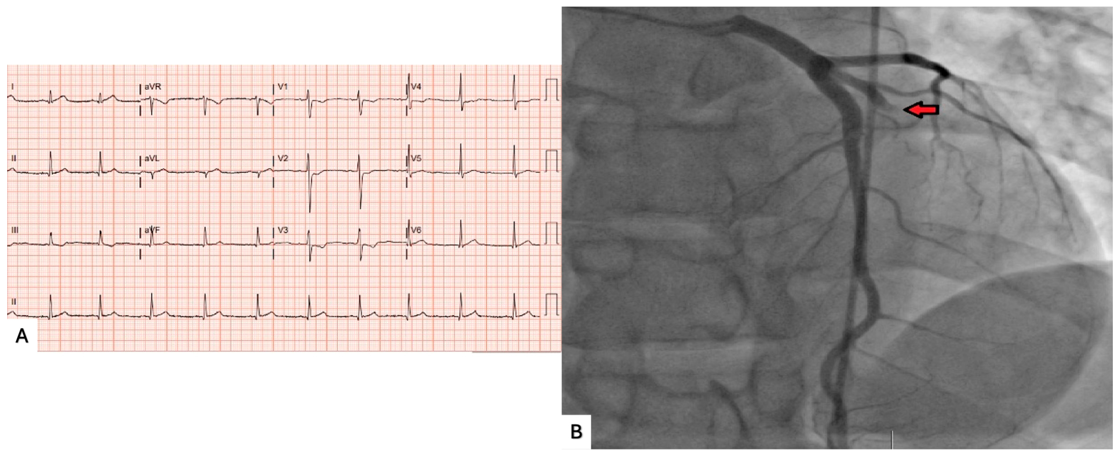
Initial laboratory testing showed elevated high-sensitivity cardiac troponin of 152 pg/mL, which later peaked at 615 pg/mL. Electrocardiography demonstrated T-wave inversions in leads V1–V4, consistent with ischemia (Figure 1). Given persistent symptoms and rising biomarkers, emergent coronary angiography was performed.
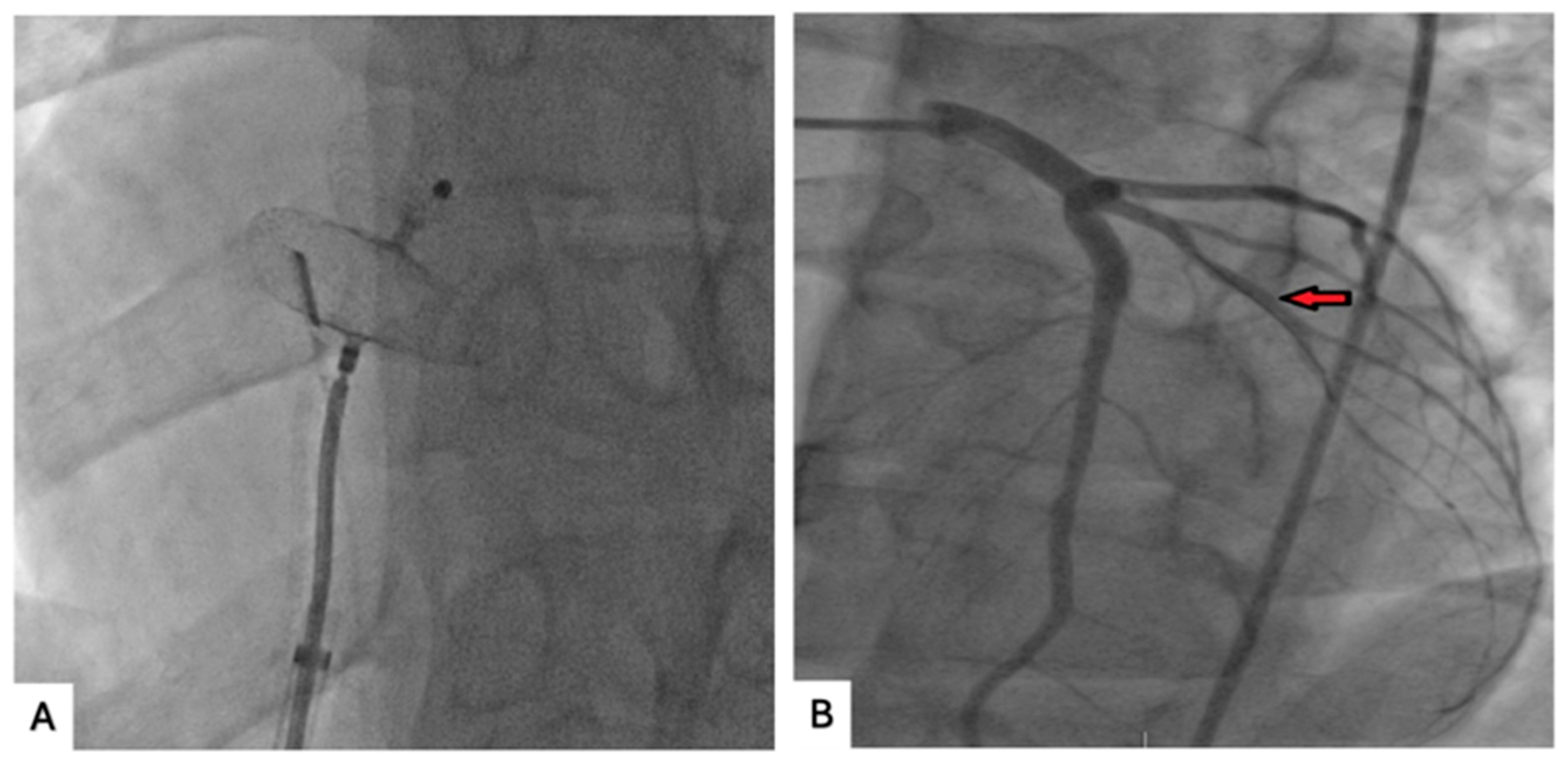
Angiography demonstrated complete occlusion of the ramus intermedius artery. Aspiration thrombectomy was performed with restoration of coronary flow and without need for stent placement. Post-thrombectomy angiography confirmed brisk restoration of flow (Figure 2A). IVUS of the culprit segment showed no plaque rupture, no evidence of underlying atherosclerotic disease, and no coronary dissection, findings that supported, but did not definitively confirm, an embolic mechanism rather than primary atherothrombotic myocardial infarction (Figure 2B).
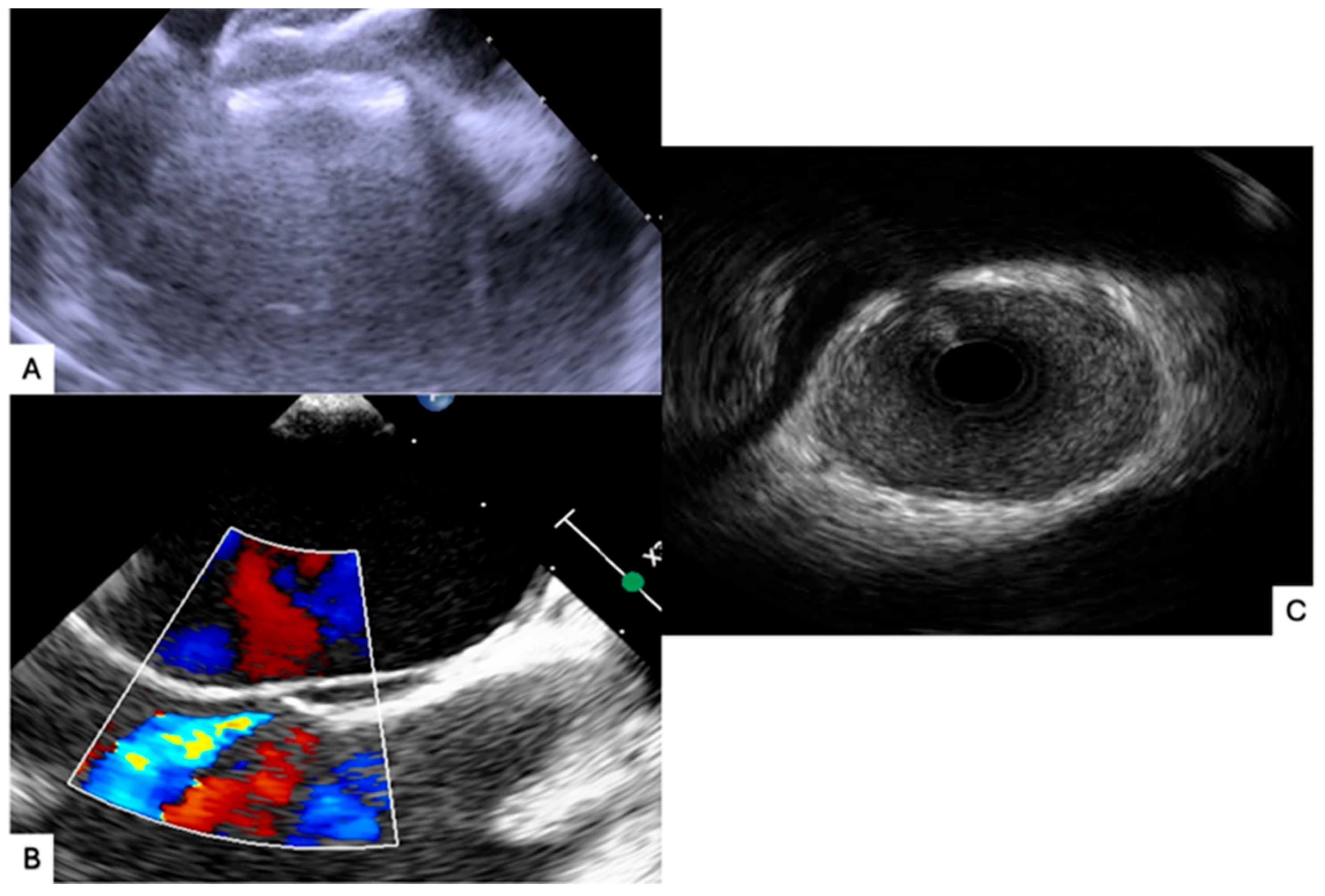
Additional evaluation was undertaken to identify the embolic source. Lower-extremity Doppler ultrasound studies were negative for deep vein thrombosis. Transthoracic echocardiography demonstrated mildly reduced left ventricular systolic function with estimated ejection fraction 45–49% and global hypokinesis. Bubble study demonstrated interatrial shunting. Transesophageal echocardiography confirmed a large patent foramen ovale with bidirectional shunting across the interatrial septum and no evidence of left atrial appendage thrombus or intracardiac mass (Figure 3).
Given the patient’s age, absence of coronary atherosclerosis on IVUS, and presence of a large PFO with bidirectional shunting, probable paradoxical coronary embolism was considered the most likely mechanism of myocardial infarction. A hematologic evaluation performed during hospitalization was unrevealing and included antiphospholipid antibodies, antinuclear antibodies, protein S antigen, and antithrombin III. It is recognized that hypercoagulable testing performed during the acute phase may have limitations and does not exclude all transient or inherited prothrombotic states. The patient remained clinically stable after thrombectomy and was discharged on antithrombotic therapy and guideline-directed medical therapy. During hospitalization, the diagnostic evaluation proceeded chronologically from coronary angiography and IVUS to transthoracic echocardiography, transesophageal echocardiography, lower-extremity Doppler studies, and hematologic testing. Four weeks later, he underwent successful percutaneous closure of the PFO using a 30 mm Amplatzer Talisman device PFO Occluder (Abbott, Abbott Park, IL, USA). He remained asymptomatic without recurrent thromboembolic events.

3. Discussion

Coronary embolism is an uncommon cause of acute myocardial infarction and is typically considered after more common etiologies, such as plaque rupture, have been excluded [7,8,13,14,15]. In the present case, several features support a non-atherothrombotic mechanism: the patient was young with minimal conventional cardiovascular risk factors, angiography demonstrated an isolated abrupt occlusion of the ramus intermedius artery, and intravascular ultrasound (IVUS) revealed no evidence of plaque rupture, underlying atherosclerosis, or coronary dissection. Taken together, these findings are most consistent with a probable paradoxical coronary embolism.
The mechanism of paradoxical embolism involves the passage of thrombotic material from the venous to the arterial circulation through a right-to-left shunt, most commonly a patent foramen ovale (PFO) [1,2,3]. Favorable hemodynamic conditions for this process include transient increases in right atrial pressure exceeding left atrial pressure, such as during Valsalva maneuver, coughing, or physical exertion, which facilitate right-to-left shunting across the interatrial septum. Once in the arterial circulation, embolic material may lodge in end organs, including the coronary arteries.
Coronary embolism is less frequently observed than cerebral embolism, likely due to differences in vascular anatomy and flow dynamics. The cerebral circulation receives a greater proportion of cardiac output, and emboli preferentially travel along direct flow paths from the aortic arch to the cerebral vessels. In contrast, the coronary arteries arise at acute angles from the aortic root and are primarily perfused during diastole, which may reduce the likelihood of embolic entry and lodging within the coronary circulation.
Multimodality imaging was central to establishing the diagnosis in this case. Coronary angiography identified the culprit lesion and pattern of occlusion, while IVUS excluded plaque rupture, erosion, and dissection, thereby reducing the likelihood of a primary atherothrombotic event [9,10,16]. Echocardiographic evaluation further supported an embolic mechanism. Transthoracic echocardiography with bubble study demonstrated interatrial shunting, and transesophageal echocardiography confirmed the presence of a large PFO with bidirectional flow, providing a plausible anatomical route for paradoxical embolization [11,12,17].
A structured differential diagnosis for myocardial infarction with non-obstructive coronary arteries (MINOCA) was considered. Coronary vasospasm was deemed less likely given the presence of a fixed occlusion requiring thrombectomy rather than transient vasoconstriction. Spontaneous coronary artery dissection was excluded based on IVUS findings demonstrating no dissection or intramural hematoma. In situ coronary thrombosis without plaque rupture remains a possible alternative; however, the absence of underlying atherosclerosis and the presence of a large right-to-left shunt favor an embolic mechanism. Other potential embolic sources were not identified despite evaluation.
Although the findings strongly support an embolic etiology, definitive proof of paradoxical coronary embolism remains challenging. No venous thrombus was identified on lower-extremity Doppler imaging, and the hypercoagulable evaluation was unrevealing. Additionally, extended venous imaging and prolonged rhythm monitoring to exclude paroxysmal atrial fibrillation were not performed. Therefore, the diagnosis should be considered presumptive but highly likely, based on the integration of clinical presentation, imaging findings, and exclusion of alternative etiologies as described in prior case reports and observational studies [18,19,20,21,22].
Management of paradoxical coronary embolism requires both treatment of the acute coronary event and strategies to reduce the risk of recurrence [23,24]. In this case, aspiration thrombectomy successfully restored coronary flow without the need for stent implantation, which was appropriate given the absence of underlying coronary artery disease. The role of PFO closure in non-neurologic paradoxical embolism is less well defined than in cryptogenic stroke because the condition is rare and no randomized trial has specifically addressed coronary events. However, available data and guideline statements support closure in carefully selected patients when a causal relationship is strongly suspected [25,26,27,28,29,30,31,32]. In our patient, the presence of a large PFO, absence of competing mechanisms, and young age supported percutaneous closure as a reasonable strategy to reduce the risk of recurrent embolic events.
Failure to recognize paradoxical coronary embolism may lead to unnecessary stent placement and missed opportunities to address the underlying embolic source. This case highlights the importance of maintaining a high index of suspicion for embolic mechanisms in young patients presenting with acute coronary syndromes without clear evidence of atherosclerotic disease and demonstrates the value of integrating coronary and structural imaging to guide diagnosis and management.

4. Conclusions

Probable paradoxical coronary embolism is an uncommon but important cause of acute myocardial infarction, particularly in younger patients without significant atherosclerotic disease. This case underscores the need to consider embolic etiologies when coronary imaging does not support plaque rupture and demonstrates the value of combining angiography, IVUS, transthoracic echocardiography, and TEE to support the diagnosis. Recognition of the likely underlying mechanism allowed for targeted treatment with aspiration thrombectomy followed by percutaneous PFO closure to reduce the risk of recurrent embolic events.

5. Case Report Limitations

This report is subject to the inherent limitations of a single case. A formal CARE checklist timeline figure was not included, although the clinical course is described chronologically. In addition, patient perspective was not available. Despite these limitations, the case provides a clear diagnostic pathway supported by multimodality imaging and highlights important considerations in the evaluation of suspected embolic myocardial infarction.

Author Contributions

Conceptualization: W.M.; Investigation: S.S. and M.T.; Data curation: M.S.; Writing—original draft preparation: M.S.; Writing—review and editing: W.M. and T.S.; Supervision: W.M. All authors have read and agreed to the published version of the manuscript.

Funding

This research received no external funding.

Institutional Review Board Statement

Ethical review and approval were waived for this study because it represents a retrospective single-patient case report without identifiable patient information. According to U.S. Department of Health and Human Services regulations governing human subjects research (45 CFR 46), case reports describing one or a small number of patients that are not designed to produce generalizable knowledge are not considered human subjects research and therefore do not require formal Institutional Review Board (IRB) review or approval. Oversight of research activities at Catholic Health is conducted through the Catholic Health Institutional Review Board. Institutional guidance regarding IRB procedures can be found at: https://www.chsbuffalo.org/physicians/physician-resources/boards-committees/institutional-review-board/ (accessed on 7 June 2025).

Informed Consent Statement

Written informed consent was obtained from the patient for publication of this case report and accompanying images.

Data Availability Statement

All relevant clinical data supporting the findings of this case report are included within the article. Further inquiries can be directed to the corresponding author.

Acknowledgments

The authors thank colleagues and mentors for their support in manuscript preparation.

Conflicts of Interest

The authors declare no conflicts of interest.

References

  1. Koutroulou, I.; Tsivgoulis, G.; Karacostas, D.; Grigoriadis, N.; Karapanayiotides, T. Epidemiology of patent foramen ovale in general population and in stroke patients: A narrative review. Front. Neurol. 2020, 11, 281. [Google Scholar] [CrossRef] [PubMed]
  2. Kent, D.M.; Wang, A.Y. Patent Foramen Ovale and Stroke: A Review. JAMA 2025, 334, 1463–1473. [Google Scholar] [CrossRef] [PubMed]
  3. Windecker, S.; Stortecky, S.; Meier, B. Paradoxical embolism. J. Am. Coll. Cardiol. 2014, 64, 403–415. [Google Scholar] [CrossRef] [PubMed]
  4. Barrios, D.D.; Chinitz, J.S. Paradoxical embolism due to patent foramen ovale. Rev. Esp. Cardiol. 2017, 70, 207–209. [Google Scholar]
  5. Hakim, F.A.; Kransdorf, E.P.; Abudiab, M.M.; Janardhanan, R.; Ammash, N.M.; Khandheria, B.K. Paradoxical coronary artery embolism: A rare cause of myocardial infarction. J. Community Hosp. Intern. Med. Perspect. 2014, 4, 24290. [Google Scholar] [CrossRef]
  6. Boumaaz, M.; Moutaouakil, F.; Rachdi, S.; Benyass, A.; Ounissi, M.; Ait Houssa, M.; Ismaili, N. Myocardial infarction caused by a proven paradoxical coronary embolism through a patent foramen ovale. Pan Afr. Med. J. 2020, 37, 198. [Google Scholar]
  7. Shibata, T.; Kawakami, S.; Noguchi, T.; Tanaka, T.; Asaumi, Y.; Kanaya, T.; Nagai, T.; Nakao, K.; Fujino, M.; Nagatsuka, K.; et al. Prevalence, clinical features, and prognosis of acute myocardial infarction attributable to coronary artery embolism. Circulation 2015, 132, 241–250. [Google Scholar] [CrossRef]
  8. Raphael, C.E.; Heit, J.A.; Reeder, G.S.; Bois, M.C.; Maleszewski, J.J.; Tilbury, R.T.; Holmes, D.R. Coronary embolus: An underappreciated cause of acute coronary syndromes. JACC Cardiovasc. Interv. 2018, 11, 172–180. [Google Scholar] [CrossRef]
  9. Hong, Y.J.; Ahn, Y.; Jeong, M.H. Role of intravascular ultrasound in patients with acute myocardial infarction. Korean Circ. J. 2015, 45, 259–265. [Google Scholar] [CrossRef]
  10. Vazquez-Fuster, J.I.; Gomez-Lara, J.; Brugaletta, S.; Roura, G.; Romaguera, R.; Ferreiro, J.L.; Sabaté, M. Intravascular ultrasound in acute myocardial infarction. Diagnostics 2023, 13, 3400. [Google Scholar]
  11. Mojadidi, M.K.; Bogush, N.; Caceres, J.D.; Msaouel, P.; Tobis, J.M. Diagnostic accuracy of transesophageal echocardiography for the detection of patent foramen ovale: A meta-analysis. J. Am. Soc. Echocardiogr. 2014, 27, 1053–1060. [Google Scholar]
  12. Calvert, P.A.; Rana, B.S.; Kydd, A.C.; Shapiro, L.M. Patent foramen ovale: Anatomy, outcomes, and closure. Heart 2011, 97, 812–818. [Google Scholar] [CrossRef]
  13. Popovic, B.; Agrinier, N.; Bouchahda, N.; Pinelli, S.; Maigrat, C.H.; Metzdorf, P.A.; Suty, C.S.; Juillière, Y.; Camenzind, E. Coronary embolism among ST-segment-elevation myocardial infarction patients: Mechanisms and management. Circ. Cardiovasc. Interv. 2018, 11, e005587. [Google Scholar] [CrossRef]
  14. Cheng, T.O. Paradoxical embolism: Historical perspective and clinical implications. Int. J. Cardiol. 2016, 221, 948–953. [Google Scholar]
  15. Wong, R.; Alenezi, F.; Altisent, O.A.J.; Puri, R.; Rodés-Cabau, J. Paradoxical embolism leading to myocardial infarction: Revisiting the significance of patent foramen ovale. JACC Case Rep. 2025, 30, 105330. [Google Scholar] [CrossRef] [PubMed]
  16. Johnson, T.W.; Räber, L.; Di Mario, C.; Bourantas, C.; Jia, H.; Mattesini, A.; Gonzalo, N.; de la Torre Hernandez, J.M.; Prati, F.; Koskinas, K.; et al. Clinical use of intracoronary imaging. Part 2: Acute coronary syndrome, ambiguous lesions, and plaque characterization. Eur. Heart J. 2019, 40, 2566–2584. [Google Scholar] [CrossRef]
  17. Messe, S.R.; Kasner, S.E. Patent foramen ovale and secondary stroke prevention. Circulation 2018, 138, 1613–1621. [Google Scholar]
  18. Tukaye, D.N.; Chandra, A.; Mowery, A.; Bikkina, M. Paradoxical thromboembolism and ST-elevation myocardial infarction. Case Rep. Cardiol. 2017, 2017, 7058578. [Google Scholar]
  19. Chen, J.; Li, L.; Liu, Y.; Wang, Y.; Zhang, H.; Zhao, X. Acute cerebral infarction with acute myocardial infarction due to paradoxical embolism through a patent foramen ovale. BMC Neurol. 2020, 20, 119. [Google Scholar]
  20. Mojaddedi, S.; Farahani, A.V.; Abbasi, S.H.; Naderi, N.; Darvish-Kazem, S. Patent foramen ovale-related paradoxical embolism after noncardiac surgery. J. Med. Cases 2022, 13, 105–109. [Google Scholar] [CrossRef]
  21. Ibanez, B.; James, S.; Agewall, S.; Antunes, M.J.; Bucciarelli-Ducci, C.; Bueno, H.; Caforio, A.L.P.; Crea, F.; Goudevenos, J.A.; Halvorsen, S.; et al. 2017 ESC Guidelines for the management of ST-segment elevation myocardial infarction. Eur. Heart J. 2018, 39, 119–177. [Google Scholar] [CrossRef] [PubMed]
  22. Capodanno, D.; Milazzo, G.; Vitale, L.; Di Stefano, D.; Di Salvo, M.; Grasso, C.; Tamburino, C. Updating the evidence on patent foramen ovale closure. Int. J. Cardiol. 2014, 175, 166–173. [Google Scholar]
  23. Onwordi, E.N.; Gamal, A.; Zaman, A. Anticoagulant therapy for acute coronary syndromes. Interv. Cardiol. Rev. 2018, 13, 87–92. [Google Scholar] [CrossRef] [PubMed]
  24. Collet, J.P.; Thiele, H.; Barbato, E.; Barthélémy, O.; Bauersachs, J.; Bhatt, D.L.; Dendale, P.; Dorobantu, M.; Edvardsen, T.; Folliguet, T.; et al. 2020 ESC Guidelines for the management of acute coronary syndromes. Eur. Heart J. 2021, 42, 1289–1367. [Google Scholar] [CrossRef]
  25. Saver, J.L.; Carroll, J.D.; Thaler, D.E.; Smalling, R.W.; MacDonald, L.A.; Marks, D.S.; Tirschwell, D.L. Long-term outcomes of patent foramen ovale closure or medical therapy after stroke. N. Engl. J. Med. 2017, 377, 1022–1032. [Google Scholar] [CrossRef]
  26. Søndergaard, L.; Kasner, S.E.; Rhodes, J.F.; Andersen, G.; Iversen, H.K.; Nielsen-Kudsk, J.E.; Settergren, M.; Sjöstrand, C.; Roine, R.O.; Hildick-Smith, D.; et al. Patent foramen ovale closure or antiplatelet therapy for cryptogenic stroke. N. Engl. J. Med. 2017, 377, 1033–1042. [Google Scholar] [CrossRef]
  27. Mas, J.L.; Derumeaux, G.; Guillon, B.; Massardier, E.; Hosseini, H.; Mechtouff, L.; Arquizan, C.; Béjot, Y.; Vuillier, F.; Detante, O.; et al. Patent foramen ovale closure or anticoagulation versus antiplatelets after stroke. N. Engl. J. Med. 2017, 377, 1011–1021. [Google Scholar] [CrossRef]
  28. Pristipino, C.; Sievert, H.; D’Ascenzo, F.; Louis Mas, J.; Meier, B.; Scacciatella, P.; Hildick-Smith, D.; Gaita, F.; Toni, D.; Kyrle, P.; et al. European position paper on the management of patients with patent foramen ovale. Eur. Heart J. 2019, 40, 3182–3195. [Google Scholar] [CrossRef]
  29. Elzanaty, A.M.; Patel, N.; Mojadidi, M.K. Patent foramen ovale closure in the management of cryptogenic stroke: State of the art. J. Clin. Med. 2021, 10, 103. [Google Scholar]
  30. Kheiri, B.; Abdalla, A.; Osman, M.; Ahmed, S.; Hassan, M.; Bachuwa, G. Patent foramen ovale closure versus medical therapy after cryptogenic stroke: An updated meta-analysis. Cardiol. J. 2019, 26, 47–55. [Google Scholar] [CrossRef]
  31. Handke, M.; Harloff, A.; Olschewski, M.; Hetzel, A.; Geibel, A. Patent foramen ovale and cryptogenic stroke. N. Engl. J. Med. 2007, 357, 2262–2268. [Google Scholar] [CrossRef]
  32. Alkhouli, M.; Busu, T.; Shah, K.; Osman, M.; Alqahtani, F.; Raybuck, B. Incidence and outcomes of patent foramen ovale closure in contemporary practice. JACC Cardiovasc. Interv. 2018, 11, 727–737. [Google Scholar]
Figure 1. Initial electrocardiogram and coronary angiography. (A) Presenting electrocardiogram demonstrating T-wave inversions in leads V1–V4 consistent with anteroseptal ischemia. (B) Coronary angiography in the AP cranial projection demonstrating complete occlusion of the ramus intermedius artery (red arrow).
Hearts 07 00012 g001
Figure 2. Intravascular imaging and restoration of coronary flow. (A) Coronary angiography following aspiration thrombectomy demonstrating restoration of flow in the ramus intermedius artery. (B) Intravascular ultrasound demonstrating absence of plaque rupture, atherosclerosis, or coronary dissection (red arrow).
Hearts 07 00012 g002
Figure 3. Echocardiographic identification of patent foramen ovale. (A) Transthoracic echocardiogram with agitated saline demonstrating interatrial shunting. (B) Transesophageal echocardiogram demonstrating a patent foramen ovale. (C) Transesophageal echocardiogram with color Doppler demonstrating bidirectional flow across the interatrial septum. All figures represent original clinical images obtained during the patient’s diagnostic evaluation. Written informed consent for publication of these images was obtained from the patient.
Hearts 07 00012 g003
Disclaimer/Publisher’s Note: The statements, opinions and data contained in all publications are solely those of the individual author(s) and contributor(s) and not of MDPI and/or the editor(s). MDPI and/or the editor(s) disclaim responsibility for any injury to people or property resulting from any ideas, methods, instructions or products referred to in the content.