Case Report

Pott’s Puffy Tumor: Two-Case Series and Contemporary Management Approach

ENT&HNS Department, Faculty of Medicine, Alanya Alaaddin Keykubat University, 07400 Alanya, Antalya, Türkiye
*
Author to whom correspondence should be addressed.
Sinusitis 2025, 9(2), 22; https://doi.org/10.3390/sinusitis9020022
Submission received: 11 August 2025 / Revised: 29 September 2025 / Accepted: 29 October 2025 / Published: 3 November 2025

Abstract

Pott’s Puffy Tumor (PPT) is a rare but potentially life-threatening complication of frontal sinusitis, characterized by subperiosteal abscess formation and frontal bone osteomyelitis. Although predominantly seen in adolescents, adult cases are increasingly recognized. Early diagnosis is essential to prevent severe orbital and intracranial sequelae. We present two patients with distinct clinical features: a 31-year-old female with chronic frontal sinusitis complicated by sequestrated bone extrusion through a cutaneous fistula, and a 16-year-old male with an acute presentation of subperiosteal abscess, nasal polyp-related obstruction of the osteomeatal complex (OMC), and orbital cellulitis. Both patients underwent combined surgical and medical management, including broad-spectrum intravenous antibiotics, functional endoscopic sinus surgery, and external drainage. In the adult, necrotic bone was excised, and the anterior frontal wall was reconstructed with titanium mesh to restore sinus anatomy and drainage, while in the adolescent, early abscess drainage and polyp removal ensured frontal recess patency and prevented osteomyelitis. Postoperative follow-up demonstrated complete resolution without recurrence. These cases highlight that PPT can occur in both acute and chronic settings of chronic rhinosinusitis with nasal polyps, emphasizing the importance of prompt imaging, multidisciplinary evaluation, and individualized surgical strategies to optimize outcomes and minimize life-threatening complications.

1. Introduction

Pott’s Puffy Tumor (PPT) is a serious complication of acute frontal sinusitis, primarily affecting children and adolescents, and is characterized by frontal bone osteomyelitis and a subperiosteal abscess [1,2].
Although it was first described in the 18th century, its true prevalence remains unclear due to its rarity. Recent literature suggests that, in addition to adolescents who are classically affected, adult cases are increasingly recognized, likely due to improved imaging and heightened clinical awareness [3,4]. With the widespread use of antibiotics, PPT is now mostly reported in isolated case reports or small series [5]. The most common etiological factor is acute or chronic rhinosinusitis, while other contributing factors include frontal trauma, odontogenic infections, diabetes mellitus, and immunosuppressive conditions [2,3]. The infection may spread intracranially via bony erosion, preexisting anatomical channels, or septic thrombophlebitis through Haversian canals [4,6], which can lead to serious complications such as acute meningitis, epidural or subdural abscess, cavernous sinus thrombosis, cerebritis, and frontal lobe abscesses [7,8]. Although PPT can occur across a wide age range, adolescents remain the most frequently affected group [6]. The frontal sinuses typically complete their development by late adolescence, and diploic veins—thin-walled and valveless—facilitate hematogenous spread of infection from the sinuses to the brain [4].
Frontal bone involvement may result in demineralization and necrosis. Infection of the inferior frontal sinus wall can extend to the orbit, causing orbital complications [9]. Neurological symptoms may be masked or absent, especially in patients receiving antibiotics, and intracranial complications may remain asymptomatic until advanced stages, when mental status changes, neck stiffness, papilledema, or seizures may appear [9].
Common presenting symptoms include forehead swelling, frontal headache, fever, periorbital edema, and purulent or non-purulent rhinorrhea [3]. Intracranial involvement may manifest with nausea, vomiting, photophobia, cranial nerve palsies, seizures, or altered mental status, whereas extracranial complications may include periorbital edema, cutaneous fistulas, diplopia, or proptosis [2,7,9].
Computed tomography (CT) and magnetic resonance imaging (MRI) are essential for diagnosis. CT delineates bony anatomy and air–bone/soft tissue interfaces, whereas MRI provides superior soft tissue resolution and is considered the gold standard for detecting intracranial or orbital complications [6,10]. Polymicrobial infections, often including anaerobic bacteria, are frequently identified in cultures from PPT patients [6,11].
Optimal management involves broad-spectrum intravenous antibiotics, surgical drainage of the abscess, debridement of necrotic tissue, and endoscopic sinus surgery to restore frontal sinus ventilation [12]. Suspected cases should be hospitalized promptly and treated with IV antibiotics, hydration, and analgesia without delay [8].
Differences in management exist between the acute and chronic stages of the disease. In one of our cases, frontal bone erosion was observed in the chronic stage, whereas in the other case, a subperiosteal abscess was present in the acute stage. Treatment approaches were adjusted accordingly.
Accordingly, the aim of this study is to present two cases of Pott’s Puffy Tumor—one juvenile and one adult—and to compare their clinical presentations and management strategies with current literature.

2. Case Reports

2.1. Case 1

A 31-year-old female patient presented to our clinic with complaints of a fistulized defect on the skin of the left frontal region and purulent discharge from the defect for the past 3 months. The patient reported intermittent exacerbations of pain and discharge that partially resolved with antibiotic treatment. She described the initial formation of an abscess in the frontal region approximately 3 months prior, which subsequently fistulized to the skin following medical treatment, exposing a sequestrated bony fragment through the defect. She had no prior sinonasal surgery, trauma, or systemic disease, but reported allergic rhinitis and active smoking.
On physical examination, a 2 × 2 cm sequestrated bone fragment fistulized to the skin was observed in the left frontal region, surrounded by purulent discharge. (Figure 1a,b). Bilateral ocular movements were normal, and visual acuity was intact. Ocular movements and visual acuity were normal. Endoscopic examination revealed purulent discharge around the left middle turbinate and nasopharynx. Neurological examination was normal. Consultations with ophthalmology and neurosurgery revealed no orbital or intracranial complications on cranial CT and orbital MRI.
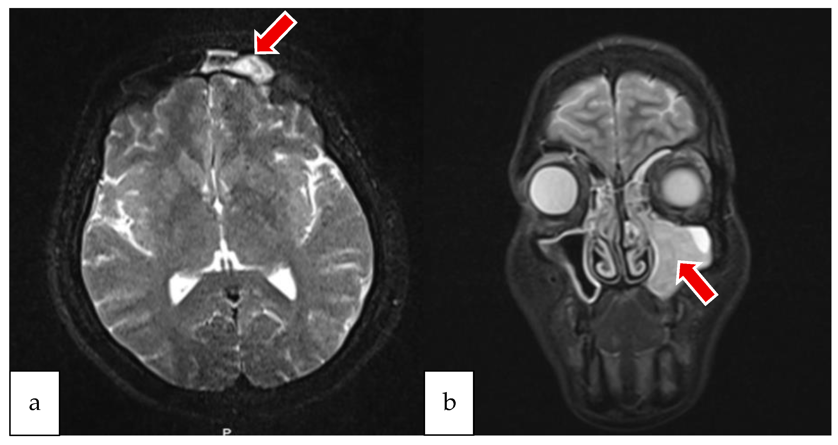
Preoperative CT-PNS imaging revealed a depressed fracture in the left FS, mucosal thickening in the FS, bilateral ethmoidal cells, and the right maxillary sinus. Complete opacification of the left maxillary sinus extending into the left nasal cavity was noted due to mucosal thickening (Figure 2). The patient’s preoperative MRI is presented in Figure 3.
Laboratory findings included WBC 10.64 × 10⁹/L (59.4% neutrophils, 27.1% lymphocytes, 5.9% monocytes, 0.8% basophils), Hb 13 g/dL, Hct 38.8%, MCV 86.6 fL, Plt 323,000/μL, CRP 0.1 mg/dL (normal: 0–0.5 mg/dL), and ESR 22 mm/hour. Routine biochemistry was unremarkable.
Empiric intravenous broad-spectrum antibiotic therapy was initiated after culture sampling from the fistulous defect; in the preoperative period, intravenous ampicillin–sulbactam was administered, cefazolin was given intraoperatively, and during the postoperative course, intravenous ceftriaxone therapy was initiated following consultation with the infectious diseases department. The patient underwent functional endoscopic sinus surgery (FESS). Purulent secretions were observed in the left maxillary sinus and OMC, and a mass compatible with a nasal polyp (NP) was seen obliterating the frontal recess (FR) lateral to the middle turbinate. Simultaneously, the sequestrated bone fragment in the skin was excised through an external approach. The posterior wall of the FS was intact, but the anterior wall was necrotic, and the FR was obstructed by polypoid tissue. Following resection of the polyp, the FR was opened, and the abscess within the FS was drained. The defect in the anterior table was repaired using a titanium mesh plate. The skin defect was closed with an advancement flap (Figure 4a,b).
Histopathological evaluation of the surgical specimen obtained from the nasal cavity revealed features of an NP, including acute inflammatory cell infiltration, areas of necrotic tissue, the presence of a few degenerated bony spicules, and submucosal glandular structures. Postoperatively, the patient was discharged with a treatment regimen including broad-spectrum oral antibiotics, intranasal corticosteroids, nasal saline irrigation, oral antihistamines, and nasal moisturizers. She was also counseled regarding lifestyle modifications and the detrimental effects of tobacco use.
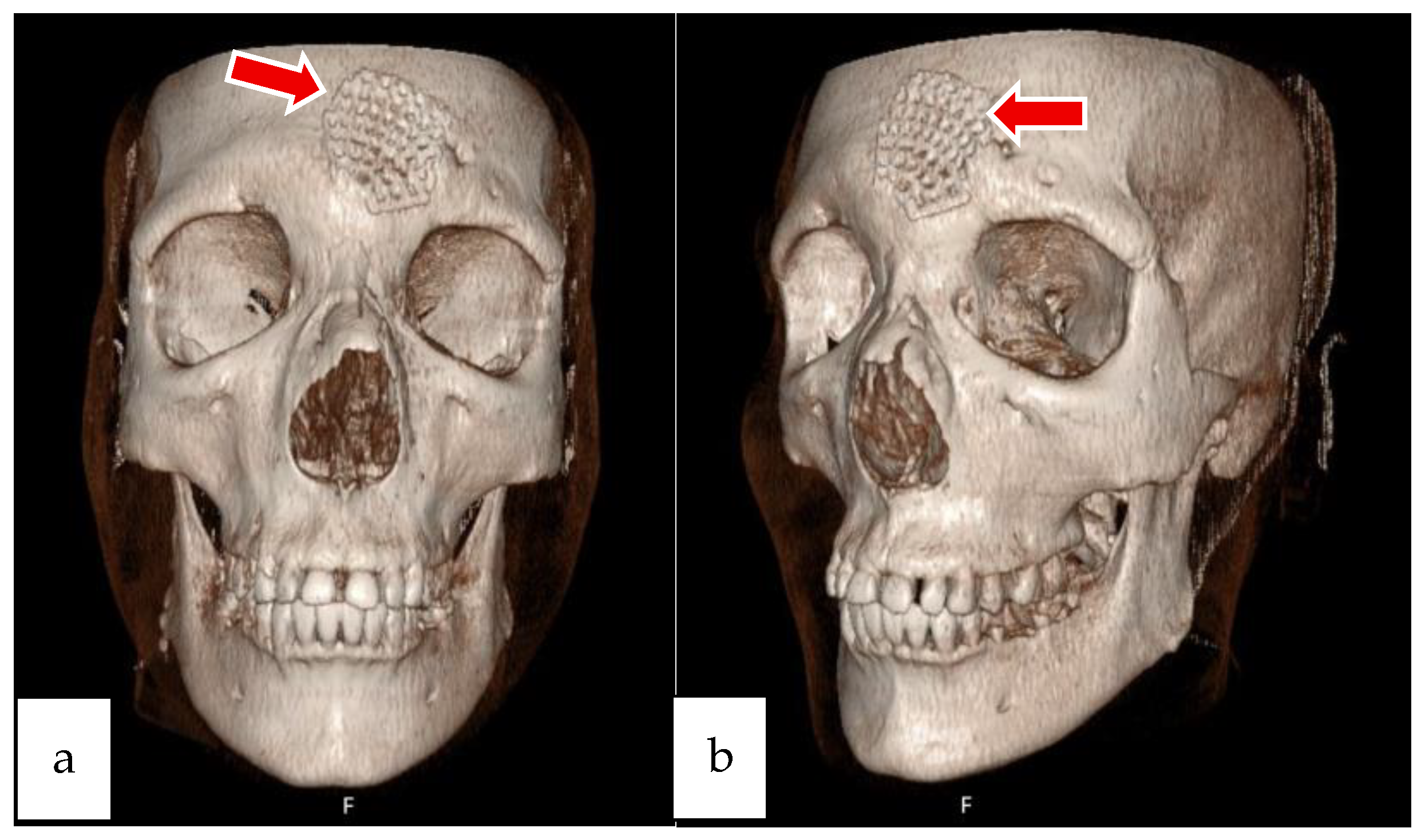
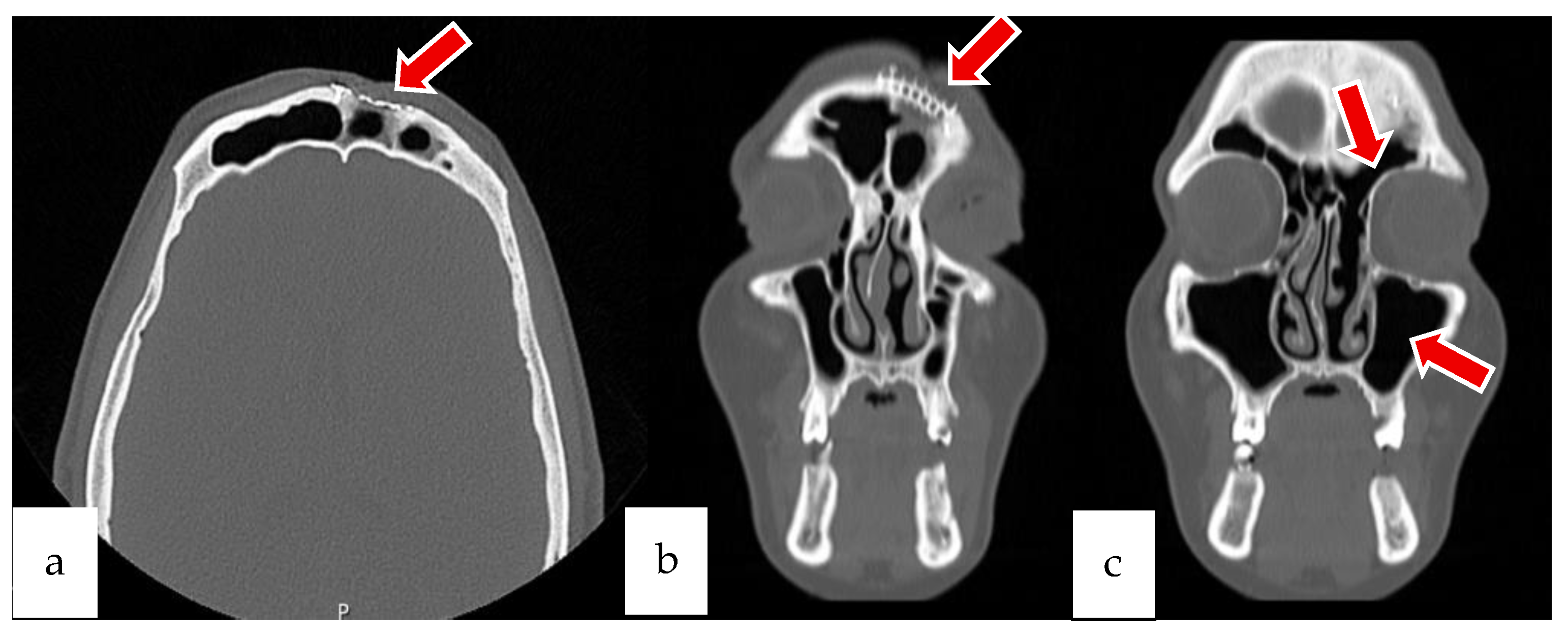
Follow-up at 3 months revealed resolution of drainage, patent FR, and healthy flap, although the patient continued smoking. Radiological and clinical follow-up confirmed the stability of the reconstruction: postoperative images demonstrated complete healing of the skin incision (Figure 5), correct placement of the titanium mesh (Figure 6), and restoration of FS integrity (Figure 7 and Figure 8).
In conclusion, in the chronic phase of frontal sinusitis, bone erosion of the anterior table of the frontal bone was observed. Sequestrated bone fragments were surgically removed, and the FS was cleared endoscopically. The defect resulting from the bone erosion was reconstructed using a plate and screws. The primary objectives of this approach were to ensure patency of the FR, frontoethmoid, and maxillary sinus pathways, thereby facilitating adequate sinus ventilation and minimizing mucosal inflammation.

2.2. Case 2

A 16-year-old male patient was referred to our clinic from the emergency department with complaints of swelling and redness in the right frontal region and right upper eyelid, which had developed over the course of five days following the onset of headache. The patient had a known diagnosis of asthma and a family history of allergic rhinitis. He also reported the use of tobacco and related products. There was no known history of prior sinonasal surgery or trauma.
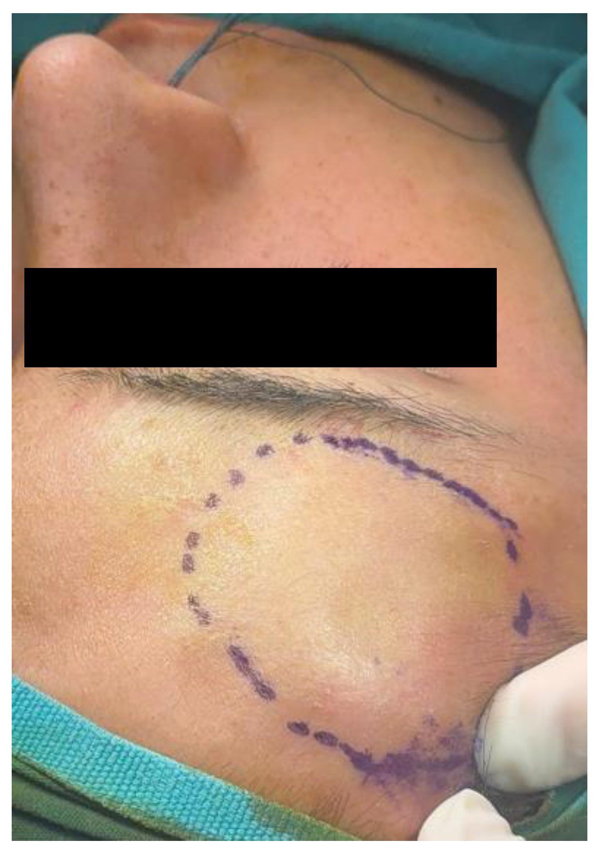
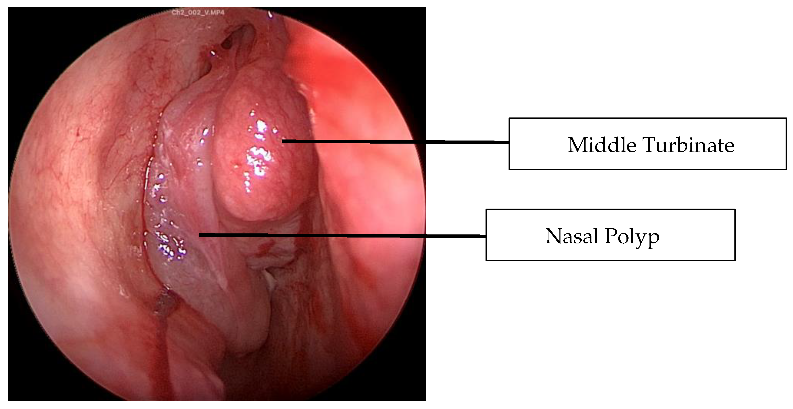
On physical examination, a fluctuant and hyperemic swelling measuring approximately 5 × 4 cm was observed in the right frontal region (Figure 9). Edema, chemosis, and hyperemia were present in the right upper eyelid. Ocular movements were full in the left eye; however, lateral gaze was restricted in the right eye. There was no proptosis. Edema of the right eyelid was also noted. Endoscopic nasal examination revealed purulent discharge from the right maxillary ostium and FR, along with an NP located at the level of the middle turbinate (Figure 10). Neurological examination was normal.
Laboratory findings included WBC 22.54 × 10⁹/L (89.9% neutrophils, 4.1% lymphocytes, 5.9% monocytes, 0.1% basophils), hemoglobin 14.6 g/dL, hematocrit 44.4%, MCV 89.9 fL, platelet count 273,000/μL, and C-reactive protein (CRP) level of 223 mg/dL (reference range: 0–0.5 mg/dL).
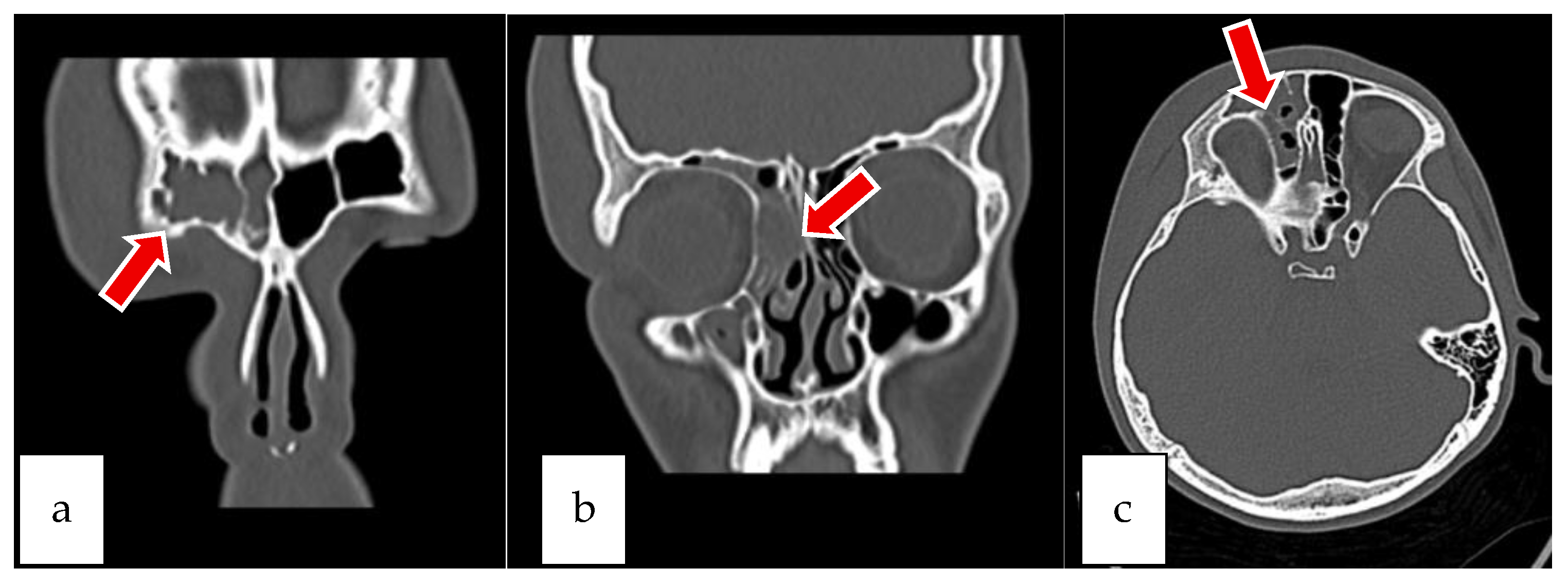
Preoperative CT-PNS showed no bony defect in the right FS. However, near-total opacification of the right FS and ethmoidal air cells was observed, along with mucosal thickening in the right maxillary sinus and signs of inflammation in the cells adjacent to the right orbit (Figure 11a–c).
MRI revealed soft tissue intensities in the right half of the FS and a subcutaneous collection measuring approximately 39 × 9 mm in the scalp at the same level, with peripheral contrast enhancement after intravenous contrast administration (suggestive of abscess). In the right periorbital region, soft tissue edema consistent with periorbital cellulitis was noted. The soft tissue intensities extended into the anterior ethmoid air cells on the right side. Additionally, contrast enhancement was detected in the adjacent dura mater near the right FS, suggestive of possible meningitis (Figure 12a–e).
The patient was consulted with ophthalmology and neurosurgery departments. The ophthalmology team confirmed that lateral gaze was intact and visual acuity was preserved, with no evidence of paresis or other ocular abnormalities. However, due to the absence of meningeal irritation signs, intracranial surgical intervention was not considered.
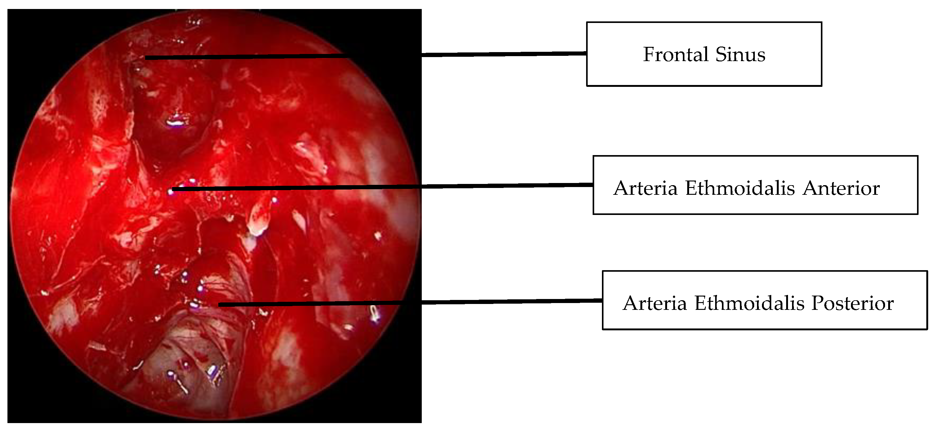
While the patient was under intravenous broad-spectrum antibiotic therapy, surgical intervention was performed; the patient had been receiving cefotaxime and metronidazole under pediatric care, and following consultation with the pediatric infectious diseases team, the regimen was switched to vancomycin. No growth was observed in the culture due to ongoing antibiotic treatment. An external surgical approach was applied via a 2–3 cm vertical incision at the right frontotemporal junction, allowing for subperiosteal abscess drainage. Areas of dehiscence were observed on the posterior table; however, no significant bony defect was noted. Simultaneously, FESS was performed. Polypoid tissues obstructing the right OMC and FR were excised. A total ethmoidectomy was carried out, and the abscess within the FS was drained (Figure 13 and Figure 14).
Histopathological examination of the surgical specimen revealed an inflammatory NP with markedly inflamed respiratory-type mucosa showing features of chronic active inflammation, accompanied by a fibrinoleukocytic exudate containing sparse bacterial colonization.
Postoperatively, the patient was discharged with the same treatment regimen. He was evaluated at postoperative months 1, 2, 3, and 5. No complaints were reported, and the purulent drainage had resolved (Figure 15). Despite continued tobacco use, no signs of obstruction were observed at the surgical site. The postoperative limitation in right lateral gaze had resolved, extraocular movements were full in all directions, and visual acuity remained normal.
In conclusion, this case presented during the acute phase. The primary goal of treatment was drainage of the abscess and early intervention on the subperiosteal abscess to prevent the development of osteomyelitis. FESS was performed to access the FR, remove polyps in the area, and ensure adequate drainage. This intervention restored normal sinus ventilation, prevented osteomyelitis in the surrounding bone, and reduced mucosal inflammation. Follow-up evaluations demonstrated good nasal drainage, absence of mucopurulent discharge, and resolution of inflammatory findings, reflecting the success of both the surgical procedure and the treatment protocol.

3. Discussion

Chronic rhinosinusitis (CRS) is an inflammatory disorder of the sinonasal mucosa, which may occur with or without NP. Inflammatory mediators play a critical role in disease pathogenesis [13,14]. FS drainage occurs via the FR, and obstruction of this pathway predisposes to frontal sinusitis. In patients with NPs, particularly when extensive polypoid tissue obstructs the FR, impaired drainage can facilitate infection, which may extend to the anterior wall of the FS, causing osteitis and ultimately PPT [15].
PPT is more frequently seen in males, with forehead swelling (74.7%), frontal headache (67%), fever (59.3%), and signs of acute or chronic sinusitis (39.6%) being the most common manifestations. Sinusitis is the leading predisposing factor (49.5%), while intracranial involvement is observed in 38.1% of cases [16]. Our cases included a 16-year-old male and a 31-year-old female, both with NP-related FR obstruction (Figure 10). Neither had a history of sinonasal surgery, suggesting that CRS with NPs was the primary etiological factor. The pediatric case demonstrated dural enhancement and a frontal abscess on MRI but no meningeal signs, whereas the adult patient presented with chronic erosion and cutaneous fistulization.
The most commonly reported pathogens in PPT are Streptococci (26.1%), Fusobacterium (19.0%), and Staphylococcus spp. (16.6%) [17]. In our cases, cultures were negative, likely due to prior oral antibiotic use. This underlines the importance of early sampling before empirical therapy. Optimal treatment of PPT requires combined medical and surgical management. Broad-spectrum intravenous antibiotics, FESS for drainage, and resection of necrotic bone are central to management. Inadequate treatment risks anterior table erosion with cutaneous fistula formation [18] or orbital extension with permanent visual impairment [9,19]. PPT should therefore be suspected in any patient presenting with erythematous scalp swelling [18].
In our pediatric case, orbital cellulitis was present. Early external drainage combined with FESS successfully restored ocular motility and preserved vision, preventing osteomyelitis. In the adult case, chronic erosion led to necrotic bone extrusion and a cutaneous fistula, necessitating excision of sequestrated bone and anterior wall reconstruction. These outcomes highlight differences between acute and chronic disease: in acute settings, early abscess drainage and frontal recess clearance prevent osteomyelitis, whereas in chronic cases, reconstruction may be required to restore sinus integrity and ventilation.
The anterior FS wall is thinner than the posterior wall, making it more prone to abscess formation. While the primary surgical goals include eradication of infection, drainage of collections, and removal of necrotic bone [15,19], the choice of approach remains debated. Endoscopic surgery provides improved visualization, less morbidity, and better cosmetic outcomes, but may be insufficient in extensive abscesses or intracranial complications [20,21]. External approaches, including frontal trephination, allow rapid drainage but carry aesthetic drawbacks. Advances in technology have expanded the role of endoscopic frontal sinusotomy, which is now preferred when feasible [20]. In line with the meta-analysis, the majority of reported patients required surgical intervention, most frequently through a combined approach or external drainage in conjunction with FESS [16]. Our pediatric case, treated with early external drainage and FESS, closely reflects this strategy, which has been shown to reduce the duration of antibiotic therapy and improve outcomes by preventing osteomyelitis. Similarly, our adult case required a more extensive surgical approach, including excision of necrotic bone and anterior wall reconstruction, which goes beyond the standard techniques most frequently described. This difference underscores that while combined surgical strategies are effective in both acute and chronic settings, the extent of intervention must be tailored to the disease stage: early, limited drainage and sinus clearance suffice in acute disease, whereas chronic cases with bone erosion demand more radical surgical management to restore both function and anatomy.
Our cases illustrate that PPT can develop in both acute and chronic CRS with NPs. While the pediatric patient benefited from early drainage and polyp removal to prevent osteomyelitis, the adult case required extensive reconstruction due to chronic bone erosion. These differences underline the need for individualized treatment strategies based on disease stage and severity. Rapid imaging, multidisciplinary collaboration, and timely intervention are crucial to achieve favorable outcomes and prevent life-threatening complications.

4. Conclusions

In both cases, PPT developed as a complication of CRSwNP, yet the clinical presentations reflected different disease stages. The adult patient, representing the chronic phase, required removal of sequestrated bone and anterior table reconstruction, while the pediatric patient, presenting acutely, benefited from early drainage and polyp removal to restore sinus ventilation and prevent osteomyelitis.
These outcomes clearly demonstrate that timely diagnosis and individualized management are essential to avoid severe sequelae. Forehead or eyelid swelling should raise suspicion for PPT, and prompt imaging with CT and/or MRI is indispensable to assess the extent of disease. Optimal treatment requires a multidisciplinary approach, combining endoscopic and, when necessary, external surgical techniques with broad-spectrum antibiotics.
Ultimately, these cases highlight the importance of recognizing PPT in both acute and chronic CRS with NPs, underlining that early intervention and tailored surgical strategies lead to favorable functional and aesthetic results while preventing life-threatening complications.

Author Contributions

Conceptualization, M.B.K. and G.O.K.; methodology, G.O.K.; software, O.B. and G.O.K.; validation, M.B.K., O.B. and G.O.K.; formal analysis, G.O.K.; investigation, M.B.K., O.B. and G.O.K.; resources, M.B.K. and G.O.K.; data curation, M.B.K. and G.O.K.; writing—original draft preparation, M.B.K. and O.B.; writing—review and editing, M.B.K. and G.O.K.; visualization, M.B.K., O.B. and G.O.K.; supervision, G.O.K.; project administration, G.O.K. All authors have read and agreed to the published version of the manuscript.

Funding

This research received no external funding.

Institutional Review Board Statement

Ethical review and approval were not applicable for this study, as the two cases presented were based on routine clinical care, without any experimental procedures or identifiable patient data.

Informed Consent Statement

Written informed consent has been obtained from the patients or the patient’s legal guardian to publish this paper.

Data Availability Statement

The datasets generated and/or analyzed during the current study are not publicly available due to patient confidentiality.

Conflicts of Interest

The authors declare no conflicts of interest.

Abbreviations

The following abbreviations are used in this manuscript:
CRSChronic Rhinosinusitis
PPTPott’s Puffy Tumor
CRSwNPChronic Rhinosinusitis with Nasal Polyp
IVIntravenous
CTComputed Tomography
CT-PNSComputed tomography of paranasal sinus
MRIMagnetic Resonance Imaging
FESCFunctional Endoscopic Sinus Surgery
FRFrontal Recess
NPNasal Polyp
WBCWhite Blood Cell count
HbHemoglobin
HctHematocrit
MCVMean Corpuscular Volume
PLTPlatelet Count
CRPC-Reactive Protein
ESRErythrocyte Sedimentation Rate
IVKMIntravenous Contrast Material
OMKOstiomeatal Complex
FSFrontal Sinus

References

  1. Tibesar, R.J.; Azhdam, A.M.; Borrelli, M. Pott’s Puffy Tumor. Ear Nose Throat J. 2021, 100 (Suppl. S6), 870s–872s. [Google Scholar] [CrossRef] [PubMed]
  2. Badr, S.A.; Myriam, L.; Youssef, O.; Sami, R.; Reda, A.; Mohamed, M. Management of a Pott puffy tumor: Case report and literature review. Int. J. Surg. Case Rep. 2024, 123, 110241. [Google Scholar] [CrossRef] [PubMed]
  3. Rohde, R.L.; North, L.M.; Murray, M.; Khalili, S.; Poetker, D.M. Pott’s puffy tumor: A comprehensive review of the literature. Am. J. Otolaryngol. 2022, 43, 103529. [Google Scholar] [CrossRef]
  4. Blumfield, E.; Misra, M. Pott’s puffy tumor, intracranial, and orbital complications as the initial presentation of sinusitis in healthy adolescents, a case series. Emerg. Radiol. 2011, 18, 203–210. [Google Scholar] [CrossRef]
  5. Williams, S.P.; Wilkie, M.D. Pott’s puffy tumour: An unforgettable complication of frontal sinusitis. BMJ Case Rep. 2014, 2014, bcr2014204061. [Google Scholar] [CrossRef]
  6. Ketenci, I.; Unlü, Y.; Tucer, B.; Vural, A. The Pott’s puffy tumor: A dangerous sign for intracranial complications. Eur. Arch. Otorhinolaryngol. 2011, 268, 1755–1763. [Google Scholar] [CrossRef]
  7. Hasan, I.; Smith, S.F.; Hammond-Kenny, A. Potts puffy tumour: A rare but important diagnosis. J. Surg. Case Rep. 2019, 2019, rjz099. [Google Scholar] [CrossRef]
  8. Perić, A.; Milojević, M.; Ivetić, D. A Pott’s Puffy Tumor Associated with Epidural—Cutaneous Fistula and Epidural Abscess: Case Report. Balkan Med. J. 2017, 34, 284–287. [Google Scholar] [CrossRef]
  9. Nisa, L.; Landis, B.N.; Giger, R. Orbital involvement in Pott’s puffy tumor: A systematic review of published cases. Am. J. Rhinol. Allergy 2012, 26, e63–e70. [Google Scholar] [CrossRef]
  10. Klivitsky, A.; Erps, A.; Regev, A.; Ashkenazi-Hoffnung, L.; Pratt, L.T.; Grisaru-Soen, G. Pott’s Puffy Tumor in Pediatric Patients: Case Series and Literature Review. Pediatr. Infect. Dis. J. 2023, 42, 851–856. [Google Scholar] [CrossRef] [PubMed]
  11. Giannoni, C.M.; Stewart, M.G.; Alford, E.L. Intracranial complications of sinusitis. Laryngoscope 1997, 107, 863–867. [Google Scholar] [CrossRef]
  12. Daloiso, A.; Mondello, T.; Boaria, F.; Savietto, E.; Spinato, G.; Cazzador, D.; Emanuelli, E. Pott’s Puffy Tumor in Young Age: A Systematic Review and Our Experience. J. Clin. Med. 2024, 13, 6428. [Google Scholar] [CrossRef]
  13. De Corso, E.; Corbò, M.; Montuori, C.; Furno, D.; Seccia, V.; Di Cesare, T.; Pipolo, C.; Baroni, S.; Mastrapasqua, R.; Rizzuti, A.; et al. Blood and local nasal eosinophilia in chronic rhinosinusitis with nasal polyps: Prevalence and correlation with severity of disease. Acta Otorhinolaryngol. Ital. 2025, 45, 39–46. [Google Scholar] [CrossRef]
  14. Tatsuki, S.; Tsuda, T.; Takeda, K.; Obata, S.; Inohara, H. A Case of Pott’s Puffy Tumor in a Patient With Eosinophilic Chronic Rhinosinusitis. Cureus 2024, 16, e60893. [Google Scholar] [CrossRef] [PubMed]
  15. Mutlu, V.; Yörük, Ö.; Özkan, Ö. Isolated complicated unilateral acute frontal sinusitis of polyp origin obstructing the mouth of the frontonasal duct. ENTCase 2015, 1, 21–29. [Google Scholar]
  16. Kokot, K.; Fercho, J.M.; Duszyński, K.; Jagieło, W.; Miller, J.; Chasles, O.G.; Yuser, R.; Klecha, M.; Matuszczak, R.; Nowiński, E.; et al. Pott’s Puffy Tumor in the Adult Population: Systematic Review and Meta-Analysis of Case Reports. J. Clin. Med. 2025, 14, 4062. [Google Scholar] [CrossRef] [PubMed]
  17. Bem, L.S., Jr.; Medeiros, M.N.C.; Gadelha, L.d.S.P.N.; Neri, W.J.R.; Cavalcanti, M.A.G. Pott’s Puffy Tumor—Overview of Case Series. J. Meml. Med. 2021, 3, 10–17. [Google Scholar]
  18. Terui, H.; Numata, I.; Takata, Y.; Ogura, M.; Aiba, S. Pott’s Puffy Tumor Caused by Chronic Sinusitis Resulting in Sinocutaneous Fistula. JAMA Dermatol. 2015, 151, 1261–1263. [Google Scholar] [CrossRef]
  19. Emejulu, J.-K. Complicated Pott’s Puffy Tumour Involving both Frontal and Parietal Bones: A Case Report. J. Infect. Dis. Ther. 2014, 2, 5. [Google Scholar] [CrossRef]
  20. Jung, J.; Lee, H.C.; Park, I.H.; Lee, H.M. Endoscopic Endonasal Treatment of a Pott’s Puffy Tumor. Clin. Exp. Otorhinolaryngol. 2012, 5, 112–115. [Google Scholar] [CrossRef]
  21. Parida, P.K.; Surianarayanan, G.; Ganeshan, S.; Saxena, S.K. Pott’s puffy tumor in pediatric age group: A retrospective study. Int. J. Pediatr. Otorhinolaryngol. 2012, 76, 1274–1277. [Google Scholar] [CrossRef]
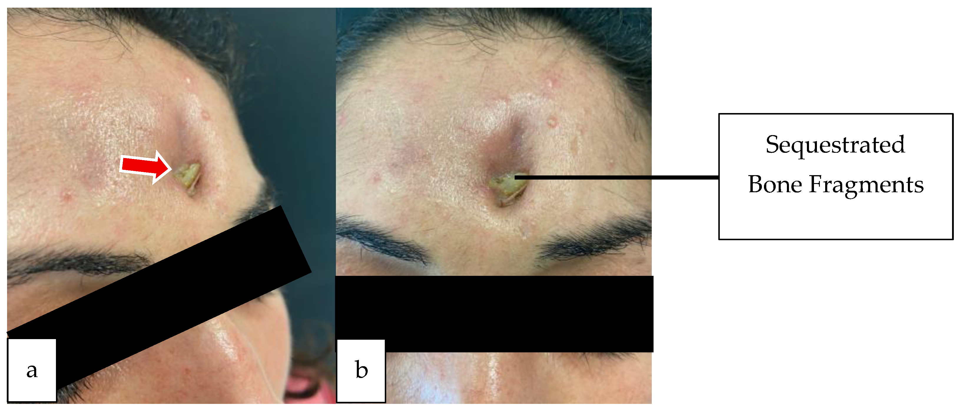
Figure 1. Pre-operative patient images of the first case. (a) Oblique view showing sequestrated frontal bone fragments extruding through a cutaneous fistula. (b) Frontal view showing purulent discharge and bone exposure.
Sinusitis 09 00022 g001
Figure 2. Pre-operative PNS CT of the first case. (a) Coronal section; arrow showing soft tissue density filling the left maxillary sinus and extending into the osteomeatal complex. (b) Axial section; arrow demonstrating a defect in the anterior table of the left frontal sinus associated with sequestrated bone fragments and surrounding inflammatory tissue. (c) Coronal section; arrow showing mucosal thickening within the left frontal sinus, consistent with obstructive sinusitis.
Sinusitis 09 00022 g002
Figure 3. Pre-operative facial MRI of the first case. (a) Axial T2-weighted contrast-enhanced MRI; arrow showing soft tissue density in the left frontal sinus. (b) Coronal section; arrow demonstrating soft tissue density in the left maxillary and ethmoid sinuses.
Sinusitis 09 00022 g003
Figure 4. Intraoperative images of the first case. (a) Elevation of the U-shaped flap and removal of sequestrated bone fragments from the defective site (arrows). (b) Reconstruction of the anterior frontal wall with a titanium mesh (arrows).
Sinusitis 09 00022 g004
Figure 5. Post-operative clinical image of the first case showing healed skin incision and resolution of discharge. (a) Left oblique view. (b) Frontal view. (c) Right oblique view.
Sinusitis 09 00022 g005
Figure 6. Post-operative X-Ray of first case. Red arrows indicate the titanium mesh. (a) Lateral graphy. (b) Antero-posterior graphy.
Sinusitis 09 00022 g006
Figure 7. Post-operative maxillofacial CT (3D) of first case. Red arrows indicate the titanium mesh. (a) Antero-posterior view (b) Left oblique view.
Sinusitis 09 00022 g007
Figure 8. Post-operative maxillofacial CT of first case. (a) Coronal section of the non-contrast frontal sinus CT. (b) Frontal sinus repaired with titanium mesh. (c) Resolution of soft tissue density in the maxillary and frontal sinuses.
Sinusitis 09 00022 g008
Figure 9. Per-operative image of second case (a fluctuant and hyperemic swelling measuring approximately ~5 × 4 cm was observed in the right frontal region).
Sinusitis 09 00022 g009
Figure 10. Pre-operative endoscopic image showing an NP obstructing the right middle meatus at the level of the middle turbinate.
Sinusitis 09 00022 g010
Figure 11. Pre-operative CT-PNS of the second case. (a,b) Coronal sections showing near-total opacification of the right frontal sinus and ethmoidal cells. (c) Axial section; arrow indicating mucosal thickening in the right maxillary sinus and orbital-adjacent inflammation.
Sinusitis 09 00022 g011
Figure 12. Pre-operative MRI of the second case. (a,b) Coronal MR images showing soft tissue density in the right FS and a subcutaneous collection in the overlying scalp with peripheral rim enhancement (abscess). (c) Axial MR image showing FS soft tissue density. (d,e) Axial and coronal MR images; arrows showing subcutaneous collection in the overlying scalp with peripheral rim enhancement (abscess).
Sinusitis 09 00022 g012
Figure 13. Intraoperative endoscopic image (right nasal passage): arrows indicating FS, anterior and posterior ethmoidal arteries.
Sinusitis 09 00022 g013
Figure 14. Per-operative endoscopy (Right Nasal Passage). (a) Aspiration of mucopurulent material from the right OMC and FR during surgery. (b) Excision of an NP from the right nasal passage.
Sinusitis 09 00022 g014
Figure 15. Post-operative endoscopic image of the second case showing patent (a) FS and (b) maxillary sinus drainage pathways.
Sinusitis 09 00022 g015
Disclaimer/Publisher’s Note: The statements, opinions and data contained in all publications are solely those of the individual author(s) and contributor(s) and not of MDPI and/or the editor(s). MDPI and/or the editor(s) disclaim responsibility for any injury to people or property resulting from any ideas, methods, instructions or products referred to in the content.