Communication

Efficacy of the PlasmaShield®, a Non-Thermal, Plasma-Based Air Purification Device, in Removing Airborne Microorganisms

1
College of Science and Engineering, Flinders University, Adelaide, SA 5042, Australia
2
Flinders Microscopy and Microanalysis (FMMA), Flinders University, Adelaide, SA 5042, Australia
*
Author to whom correspondence should be addressed.
Electrochem 2022, 3(2), 276-284; https://doi.org/10.3390/electrochem3020019
Submission received: 25 April 2022 / Revised: 14 May 2022 / Accepted: 19 May 2022 / Published: 1 June 2022
(This article belongs to the Collection Feature Papers in Electrochemistry)

Abstract

:
Airborne microorganisms play a significant role in the transmission of infectious diseases. As such, improving indoor microbial air quality can enhance infection control in numerous settings. This study examined the efficacy of the PlasmaShield® air purification device to remove airborne microorganisms under laboratory conditions. Pure cultures of model microorganisms at varying concentrations were aerosolized using a 1-jet Collison nebulizer through stainless-steel removable piping prior to reaching the PlasmaShield® device. The surviving microorganisms were captured using the Staplex® MBS-6 Six Stage Microbial Air Sampler and enumerated via culture on agar plates. The positive-hole-corrected colony/plaque-forming units were compared with the negative control (microorganisms aerosolized through an empty PlasmaShield® casing). The PlasmaShield® statistically significantly (p < 0.05) reduced airborne Escherichia coli, Staphylococcus epidermidis, Bacteriophage MS2 and Cladosporium sp. compared with the negative control. The maximum removal achieved was estimated to be 4 × log10 E. coli (99.99% removal), 4 × log10 S. epidermidis (99.97% removal), 7 × log10 MS2 (99.99998% removal) and 5 × log10 Cladosporium sp. (99.999% removal). Scanning electron microscope images of the surviving microorganisms showed that the PlasmaShield® damaged the cell membrane of these model microorganisms. This study provides proof-of-concept evidence to support the use of this technology to improve indoor microbial air quality.

1. Introduction

Indoor air quality is recognized as a significant factor affecting human health [1]. Within clinical and non-clinical settings, it is recognized that inadequate air ventilation is associated with increased risk of transmission of infectious diseases [2,3,4]. Recent studies during the current COVID-19 pandemic have also shown that overcrowding and poor ventilation promote the airborne transmission of SARS-CoV-2 [5,6,7].
There are a range of engineering solutions available to improve microbial air quality through air purification technologies. This includes devices utilizing air filtration, ultraviolet irradiation, electrostatic precipitation, negative air ionization, photocatalytic oxidation, air ozonation, passive solar exposure, pulsed light and plasma-based disinfection [8]. This study examines the efficacy of the PlasmaShield®, a novel air purification device utilizing electron beam irradiation and non-thermal plasma-based disinfection, to remove four airborne microorganisms.
Escherichia coli, Staphylococcus epidermidis, bacteriophage MS2 and Cladosporium sp. were chosen as the model microorganisms. E. coli is a Gram-negative bacterium found in the intestines of humans and animals and is recognized as an indicator of fecal contamination. It is the most commonly used model bacteria [9,10,11,12]. S. epidermidis is a coagulase-negative, Gram-positive bacteria that is part of the skin’s natural flora. It does not cause disease in healthy individuals, but is increasingly associated with hospital acquired infections and indwelling medical device-associated infections [13]. This is further complicated by the emergence of antibiotic resistant strains [14]. Its significance as an opportunistic pathogen is linked to its biofilm forming capabilities [15], which protects it from disinfection methods enabling long term persistence in the environment [16]. MS2 is a single stranded RNA virus that infects E. coli and other members of the Enterobacteriaceae. It is a recognized indicator organism for enteric viruses and fecal contamination [17]. It is a suitable surrogate for viral pathogens and has been used as a model organism in virus inactivation studies [18,19,20] and to investigate virus survival during aerosol dispersion [21,22]. Cladosporium sp. is one of the most common fungi found in air samples [23]. It has been isolated from a range of indoor environments including hospitals [24], aged care facilities [25], schools [26] and office buildings [27].

2. Materials and Methods

2.1. PlasmaShield Design

The PlasmaShield® is a non-thermal plasma-based air purification device (U.S. Patent No. 10,744,515). Briefly, it contains a discharge electrode located centrally within a cylindrical ground electrode, and a power supply electrically connected to the discharge and the ground electrode. This produces a uniform electric field which destroys microorganisms in the air by electroporation [28] and a low-energy electron beam irradiation which inactivates microorganisms by damaging their nucleic acid [29]. In addition, a corona discharge from the discharge electrode generates ions (excited N and NO molecules) and free electrons, which potentially provide a secondary disinfection mechanism. These ions are produced within a narrow region (100 µm) around the discharge electrode tip inside the plasma reactor. The nature of these excited species suggests that they react with other molecules and are therefore unable to travel a noticeable distance from their generation point [28]. Independent testing has shown that the ozone emissions generated by the device do not exceed 2 ppb [30]. It is also certified by the California Air Resources Board (CARB) as an air cleaning device, meeting the ozone regulation of less than 50 ppb [31].

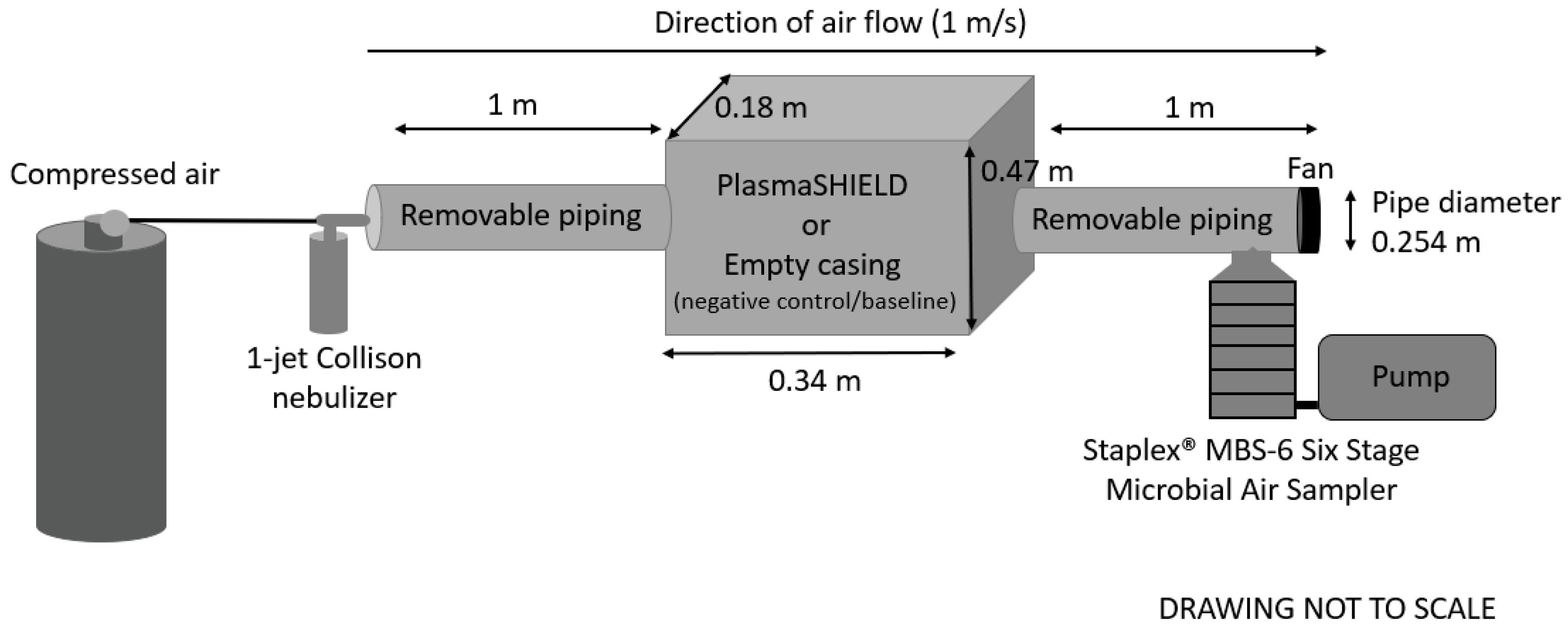
2.2. Experimental Setup

The PlasmaShield® device in its commercial configuration was provided by PlasmaShield LTD (Keswick, SA, Australia). The efficacy of the device to remove model microorganisms was tested using the experimental setup shown in Figure 1. Briefly, each microorganism was aerosolized (at a range of concentrations) using a 1-jet Collison nebulizer, (CH Technologies: Westwood, NJ, USA) through stainless steel removable piping (0.254 m (10 inch) diameter × 1 m in length) prior to reaching the PlasmaShield® device. The remaining microorganisms were captured 1 m after the device using the Staplex® MBS-6 Six Stage Microbial Air Sampler (The Staplex Company: Brooklyn, NY, USA) for 60 m at a sampling flow rate of 28.3 L/min (1 CFM). Each stage of the air sampler contained a different filter size with 400 holes. Stage 6 contained the finest filter and collected particles of 0.65 to 1.1 µm in size. Stages 5, 4, 3, 2 and 1 contained 1.1 µm to 2.1 µm, 2.1 µm to 3.3 µm, 3.3 to 4.7 µm, 4.7 to 7.0 µm and 7.0 µm and above, respectively. Surviving microorganisms were enumerated via culture on agar plates. After incubation, the colonies/plaques present on each agar plate were counted and recorded as positive hole corrected using the correction described by Macher [32]. The positive hole corrected counts for each of the six stages were added together and the total from the three trials averaged. For each microorganism/concentration tested a negative control, or baseline, was determined by replacing the PlasmaShield® with an empty casing. The air flow rate was controlled at 1 m/s, which was confirmed using an anemometer (Pocket ProTM + Multi 2, HACH Company: Melbourne, Australia), this gave a volume flow rate of approximately 51 L/s (the PlasmaShield® rated capacity is 86 L/s). The air pressure released from the air cylinder was set to 10 psi. The pressure drop at 1 m/s flow rate was 0.007 psi. All experiments were conducted in triplicate.

2.3. Escherichia coli

E. coli (American Type Culture Collection (ATCC) 700891) culture was incubated at 37 °C for 12 h in nutrient broth (Oxoid®: Adelaide, Australia). The broth culture was then centrifuged at 3000 rpm for 15 min and the pellet was washed with phosphate-buffered saline (PBS) (Fisher BioReagents®: Adelaide, Australia). The washed pellet was resuspended in PBS and the optical density of the culture adjusted to 1 (109 colony forming units (CFU)/mL) at 600 nm (spectrophotometer OD600) using PBS. The culture was then serially diluted with PBS to obtain the desired concentrations (107 CFU/mL, 108 CFU/mL or 109 CFU/mL). After each experiment E. coli were captured using the Staplex® MBS-6 Six Stage Microbial Air Sampler on plate count agar (PCA) (Oxoid®) and incubated overnight at 37 °C.

2.4. Staphylococcus epidermidis

S. epidermidis (ATCC 14990) culture was incubated at 37 °C for 12 h in nutrient broth. The broth culture was then centrifuged at 3000 rpm for 15 min and the pellet was washed with PBS. The washed pellet was resuspended in PBS and the optical density of the culture was adjusted to 1 (109 CFU/mL) at 600 nm (spectrophotometer OD600) using PBS. The culture was then serially diluted with PBS to obtain the desired concentration (107 CFU/mL, 108 CFU/mL, 109 CFU/mL or 109 CFU/mL). For each replicate trial and concentration, 60 mL of the culture was added to a 1-jet Collison nebulizer to produce aerosols (Figure 1). After each experiment, surviving S. epidermidis were captured using the Staplex® MBS-6 Six Stage Microbial Air Sampler on PCA and incubated overnight at 37 °C.

2.5. Bacteriophage MS2

MS2 (ATCC 15597-B1) was propagated using the double agar layer method. The bottom layer (of the tryptone soya agar (TSA)-E. coli agar plates) consisted of TSA (Oxoid®) and the top layer consisted of 4.5 mL of soft TSA mixed with 500 µL of overnight E. coli (ATCC 700891) culture (which had been incubated overnight at 37 °C in typtone soya broth (Oxoid®)) and 200 µL of MS2 culture. The plates were then incubated overnight at 37 °C. The plaques were harvested in peptone water (Oxoid®) and purified by centrifugation at 3000 rpm for 15 min to separate the host cell debris and the MS2. The supernatant was filtered through a 0.22 μm Millex-GP Syringe Filter Unit (Millipore, catalog number SLGP033RS, Ireland) and used as a stock solution. This stock was serially diluted in sterile water to achieve desired concentrations (107 plaque forming units (PFU)/mL, 1010 PFU/mL, 1012 PFU/mL and 1015 PFU/mL) which were confirmed by plating and counting plaques using the double agar layer method described above. After each experiment, surviving MS2 were captured using the Staplex® MBS-6 Six Stage Microbial Air Sampler on TSA-E. coli agar plates, which were incubated overnight at 37 °C.

2.6. Cladosporium sp.

An environmental culture of Cladosporium sp. previously isolated from indoor air [33] was incubated for three days at 25 °C on to malt extract agar (MEA) (Oxoid®). After incubation, a small piece of agar at the edge of the fungal colony (where the mycelium is highly active) was cut using a sterile scalpel and placed on to a fresh MEA plate. The agar was placed upside down to allow the growth to touch the fresh agar plate. The inoculated plates were then incubated for 7 days at 25 °C until the lawn cultures were visible, and spores were produced. The fungal plates were flooded with 10 mL of 0.05% Tween 80 solution (Labchem, AJAX Laboratory Chemicals, Adelaide, Australia) and the spores were separated from the hyphae using a sterile glass slide. The solution was then filtered through sterile absorbent cotton wool pads to remove the hyphal debris. This step was repeated three times. The spore solution was mixed well and serially diluted in 0.05% Tween 80 solution to obtain the desired concentration (107 spores/mL, 109 spores/mL and 1010 spores/mL). The concentration of the spore suspension was determined using a hemocytometer (spores/mL). After each experiment, surviving Cladosporium sp. were captured using the Staplex® MBS-6 Six Stage Microbial Air Sampler on MEA and incubated at 25 °C for three days.

2.7. Scanning Electron Micrscopy (SEM)

Experiments were conducted using the setup described in 2.1 (see Figure 1); however, the Staplex® MBS-6 Six Stage Microbial Air Sampler was replaced with a liquid impinger sampler (BioSampler® SKC Inc: Eighty Four, PA, USA). Captured microorganisms were then deposited onto cleaned glass slides (oxygen plasma treated for 5 min). Samples were then fixed by adding to 3% glutaraldehyde solution in PBS (pH 7.2) at 21 °C for one hr. The glutaraldehyde solution was then discarded and samples were washed twice for 5 min with PBS. Samples were then dehydrated using three × 15 min washes of increasing ethanol concentrations (70%, 90% and 100% ethanol). The samples were then immediately dried using a Leica CPD300 Critical Point Dryer. Glass slides were then mounted onto SEM stubs using carbon adhesive tape and coated with 5nm of Platinum using an Emitech K57X sputter coater. SEM analysis was performed using a FEI Inspect F50 SEM, with a beam voltage of (10–30 kV).

3. Results

Efficacy of PlasmaShield® to Remove Airborne Escherichia coli, Staphylococcus epidermidis, Bacteriophage MS2 and Cladosporium sp.

The PlasmaShield® statistically significantly (p < 0.05) reduced airborne E. coli, S. epidermidis, MS2, Cladosporium sp. compared with the negative control (Table 1). The negative control for each model microorganism could only be calculated at the lowest concentration tested. As the test concentration increased the number of CFU or PFU per plate was too many to count and as such the negative control for these higher concentrations were estimated by extrapolating from the highest countable concentration (e.g., the negative control from the lowest test concentration). The maximum removal achieved was estimated to be 4 × log10 E. coli (99.99% removal), 4 × log10 S. epidermidis (99.97% removal), 7 × log10 MS2 (99.99998% removal) and 5 × log10 Cladosporium sp. (99.999% removal) (Table 1). The SEM images (Figure 2) demonstrate that the PlasmaShield® damages the cell membrane of these model microorganisms. Figure 2A,C,E,G show intact E. coli, S. epidermidis, MS2 and Cladosporium, respectively. These cells were collected during the baseline trials with the PlasmaShield® turned off. Figure 2B,D,F,H show cell debris of E. coli, S. epidermidis, MS2 and Cladosporium, respectively, collected after treatment with the PlasmaShield®.

4. Discussion

Currently, HEPA (“high efficiency particulate air”) filtration is most commonly used method for improving indoor air quality [34,35]. However, there are several limitations with this approach. This includes the need to regularly change and dispose of contaminated filters as well as the energy requirements needed to overcome the high pressure drop that occurs across the filters [35,36]. New air purification technologies provide an alternative to HEPA filtration and can potentially overcome these limitations. Previous studies have shown that plasma-based devices have microbial inactivation/sterilization capabilities [37,38,39]. This includes a study by Tanaka et al. [38] that demonstrated that a chemical reactor with plasma-assisted catalytic technology (PACT) achieved a 99.99% disinfection efficacy against airborne feline calicivirus (FCV), which is considered an appropriate surrogate for norovirus [38]. Similarly, this current study demonstrated that the PlasmaShield® had disinfection capabilities against airborne model microorganisms E. coli, S. epidermidis, MS2 and Cladosporium sp.
A limitation of this study was that the number of CFU or PFU per plate for the baseline/negative control measurements were often too many to count (Table 1). For these trials, the negative control was estimated by extrapolating from a trial conducted with a lower test concentration. This does not consider the potential for additional loss of microbial cells through physical inactivation during the aerosolization process. This may have resulted in a slight overestimation of the baseline and therefore the disinfection removal efficacy. However, other approaches to either reduce the aerosolization time or to dilute the baseline to a countable level would have required additional handling steps that would have also added additional uncertainty. The manufacturer of the PlasmaShield® states that the device utilizes two modes of action. This includes electron beam irradiation (through multilayer electron beam emitters) and irreversible electroporation (an intense electric field). Previous studies describing the antimicrobial mechanism of action of non-thermal plasma-based devices describe the breaking of bacterial peptidoglycan bonds. As these bonds are structurally important, their breaking leads to cell wall damage, enabling reactive oxygen species to enter through the site of injury [40,41,42]. However, there is limited research investigating the mechanism of action responsible for the breaking of these bonds. It is also thought to be dependent on the impinging plasma species [42]. The destruction of the cell wall by the PlasmaShield® was observed in this study through the SEM images and supports these mechanisms of action.
The antimicrobial efficacy of the PlasmaShield® demonstrated in this study was achieved under controlled laboratory conditions at a constant flow rate and air pressure. Future research is needed to determine the influence of environmental factors, such as airflow, humidity, temperature, and particulate matter, on the efficacy of the PlasmaShield® disinfection capabilities.

5. Conclusions

The airborne transport of pathogens represents a significant source of infection in many settings, such as medical facilities, crowded locations, facilities that handle such pathogens, and, recently, quarantine facilities used to control the spread of COVID-19. This study demonstrated that the PlasmaShield® air purification device effectively removed model airborne microorganisms under controlled conditions. This provides proof-of-concept evidence to support the use of this technology to improve indoor microbial air quality.

Author Contributions

Conceptualization, H.W., K.E.R. and P.S.; methodology, H.W., K.E.R., P.S., M.A.N., T.P.K., E.J.K. and A.S.; data curation, T.P.K., E.J.K., M.A.N. and A.S.; writing—original draft preparation, H.W.; writing—review and editing, all authors. All authors have read and agreed to the published version of the manuscript.

Funding

This research was funded by PlasmaShield LTD (Keswick, South Australia, Australia) through contract research.

Institutional Review Board Statement

Not applicable.

Informed Consent Statement

Not applicable.

Acknowledgments

The authors also would like to acknowledge the facilities, and the scientific and technical assistance, of Microscopy Australia and the Australian National Fabrication Facility (ANFF) under the National Collaborative Research Infrastructure Strategy, at the South Australian Regional Facility, Flinders Microscopy and Microanalysis, Flinders University.

Conflicts of Interest

The funders had no role in the experimental design; in the collection, analyses, interpretation of data or writing of manuscript.

References

  1. Tran, V.V.; Park, D.; Lee, Y.-C. Indoor air pollution, related human diseases, and recent trends in the control and improvement of indoor air quality. Int. J. Environ. Res. Public Health 2020, 17, 2927. [Google Scholar] [CrossRef] [PubMed] [Green Version]
  2. Mouchtouri, V.A.; Rudge, J.W. Legionnaires’ disease in hotels and passenger ships: A systematic review of evidence, sources, and contributing factors. J. Travel Med. 2015, 22, 325–337. [Google Scholar] [CrossRef] [PubMed] [Green Version]
  3. Tomlinson, B.; Cockram, C. Sars: Experience at Prince of Wales hospital, Hong Kong. Lancet 2003, 361, 1486–1487. [Google Scholar] [CrossRef]
  4. Hoge, C.W.; Reichler, M.R.; Dominguez, E.A.; Bremer, J.C.; Mastro, T.D.; Hendricks, K.A.; Musher, D.M.; Elliott, J.A.; Facklam, R.R.; Breiman, R.F. An epidemic of pneumococcal disease in an overcrowded, inadequately ventilated jail. N. Engl. J. Med. 1994, 331, 643–648. [Google Scholar] [CrossRef]
  5. Liu, Y.; Ning, Z.; Chen, Y.; Guo, M.; Liu, Y.; Gali, N.K.; Sun, L.; Duan, Y.; Cai, J.; Westerdahl, D. Aerodynamic analysis of SARS-CoV-2 in two Wuhan hospitals. Nature 2020, 582, 557–560. [Google Scholar] [CrossRef]
  6. Li, Y.; Qian, H.; Hang, J.; Chen, X.; Cheng, P.; Ling, H.; Wang, S.; Liang, P.; Li, J.; Xiao, S. Probable airborne transmission of SARS-CoV-2 in a poorly ventilated restaurant. Build. Environ. 2021, 196, 107788. [Google Scholar] [CrossRef]
  7. Noorimotlagh, Z.; Jaafarzadeh, N.; Martínez, S.S.; Mirzaee, S.A. A systematic review of possible airborne transmission of the COVID-19 virus (SARS-CoV-2) in the indoor air environment. Environ. Res. 2021, 193, 110612. [Google Scholar] [CrossRef]
  8. Griffiths, W.D.; Bennett, A.; Speight, S.; Parks, S. Determining the performance of a commercial air purification system for reducing airborne contamination using model micro-organisms: A new test methodology. J. Hosp. Infect. 2005, 61, 242–247. [Google Scholar] [CrossRef]
  9. enHealth Council. Guidance on Use of Rainwater Tanks; National Public Health Partnership; Australian Government Department of Health and Ageing: Canberra, Australia, 2004; Volume 3432-JN8304.
  10. United States Environmental Protection Agency. Recreational Water Quality Criteria; Office of Water, Ed.; United Stated Environmental Protection Agency: Washington, DC, USA, 2012.
  11. Food Standards Australia New Zealand. Australian and New Zealand Food Standards Code; Australian Department of Health: Canberra, Australia, 2015.
  12. Blount, Z.D. The natural history of model organisms: The unexhausted potential of E. coli. Elife 2015, 4, e05826. [Google Scholar] [CrossRef]
  13. Gomes, F.; Teixeira, P.; Oliveira, R. Mini-review: Staphylococcus epidermidis as the most frequent cause of nosocomial infections: Old and new fighting strategies. Biofouling 2014, 30, 131–141. [Google Scholar] [CrossRef]
  14. Weßels, C.; Strommenger, B.; Klare, I.; Bender, J.; Messler, S.; Mattner, F.; Krakau, M.; Werner, G.; Layer, F. Emergence and control of linezolid-resistant Staphylococcus epidermidis in an ICU of a German hospital. J. Antimicrob. Chemother. 2018, 73, 1185–1193. [Google Scholar] [CrossRef]
  15. Büttner, H.; Mack, D.; Rohde, H. Structural basis of Staphylococcus epidermidis biofilm formation: Mechanisms and molecular interactions. Front. Cell. Infect. Microbiol. 2015, 5, 14. [Google Scholar]
  16. Wojtyczka, R.D.; Orlewska, K.; Kępa, M.; Idzik, D.; Dziedzic, A.; Mularz, T.; Krawczyk, M.; Miklasińska, M.; Wąsik, T.J. Biofilm formation and antimicrobial susceptibility of Staphylococcus epidermidis strains from a hospital environment. Int. J. Environ. Res. Public Health 2014, 11, 4619–4633. [Google Scholar] [CrossRef] [Green Version]
  17. McMinn, B.R.; Ashbolt, N.J.; Korajkic, A. Bacteriophages as indicators of fecal pollution and enteric virus removal. Lett. Appl. Microbiol. 2017, 65, 11–26. [Google Scholar] [CrossRef] [Green Version]
  18. Kohn, T.; Mattle, M.J.; Minella, M.; Vione, D. A modeling approach to estimate the solar disinfection of viral indicator organisms in waste stabilization ponds and surface waters. Water Res. 2016, 88, 912–922. [Google Scholar] [CrossRef]
  19. Silverman, A.I.; Nguyen, M.T.; Schilling, I.E.; Wenk, J.; Nelson, K.L. Sunlight inactivation of viruses in open-water unit process treatment wetlands: Modeling endogenous and exogenous inactivation rates. Environ. Sci. Technol. 2015, 49, 2757–2766. [Google Scholar] [CrossRef] [Green Version]
  20. Hosseini, S.R.S.; Dovom, M.R.E.; Yavarmanesh, M.; Abbaszadegan, M. Thermal inactivation of ms2 bacteriophage as a surrogate of enteric viruses in cow milk. J. Consum. Prot. Food Saf. 2017, 12, 341–347. [Google Scholar] [CrossRef]
  21. Turgeon, N.; Toulouse, M.-J.; Martel, B.; Moineau, S.; Duchaine, C. Comparison of five bacteriophages as models for viral aerosol studies. Appl. Environ. Microbiol. 2014, 80, 4242–4250. [Google Scholar] [CrossRef] [Green Version]
  22. Zuo, Z.; Kuehn, T.H.; Bekele, A.Z.; Mor, S.K.; Verma, H.; Goyal, S.M.; Raynor, P.C.; Pui, D.Y. Survival of airborne ms2 bacteriophage generated from human saliva, artificial saliva, and cell culture medium. Appl. Environ. Microbiol. 2014, 80, 2796–2803. [Google Scholar] [CrossRef] [Green Version]
  23. Flannigan, B.; Samson, R.A.; Miller, J.D. Microorganisms in Home and Indoor Work Environments: Diversity, Health Impacts, Investigation and Control; CRC Press: Boca Raton, FL, USA, 2016. [Google Scholar]
  24. Verde, S.C.; Almeida, S.M.; Matos, J.; Guerreiro, D.; Meneses, M.; Faria, T.; Botelho, D.; Santos, M.; Viegas, C. Microbiological assessment of indoor air quality at different hospital sites. Res. Microbiol. 2015, 166, 557–563. [Google Scholar] [CrossRef]
  25. Yilmaz, O.; Asan, A.; Aydogdu, H.; Sen, B. Airborne fungal diversity inside a nursing home in Edirne, Turkey. Fresenius Environ. Bull. 2017, 26, 7025–7033. [Google Scholar]
  26. Reddy, M.K.; Srinivas, T. Mold allergens in indoor play school environment. Energy Procedia 2017, 109, 27–33. [Google Scholar] [CrossRef]
  27. Liu, Z.; Zhu, Z.; Zhu, Y.; Xu, W.; Li, H. Investigation of dust loading and culturable microorganisms of HVAC systems in 24 office buildings in Beijing. Energy Build. 2015, 103, 166–174. [Google Scholar] [CrossRef]
  28. Khazaei, S.V. Gas Purifying Apparatus. U.S. Patent 10,744,515, 2020. Washington, DC: U.S. Patent and Trademark Office. Available online: https://patents.google.com/patent/US10744515B2/en (accessed on 21 April 2022).
  29. Zhao, W.; Alwahabi, Z.T. Diagnostics of air purification plasma device by spatially resolved emission spectroscopy. Plasma 2022, 5, 206–220. [Google Scholar] [CrossRef]
  30. Huang, Y.; Zhou, S.; Langenbacher, J. Ozone Test Report (Report Number: 210304019gzu-001); Intertek: Guangzhou, China, 2021. [Google Scholar]
  31. The California Air Resources Board. List of Carb-Certified Air Cleaning Devices. Available online: https://ww2.arb.ca.gov/list-carb-certified-air-cleaning-devices (accessed on 21 April 2022).
  32. Macher, J.M. Positive-hole correction of multiple-jet impactors for collecting viable microorganisms. Am. Ind. Hyg. Assoc. J. 1989, 50, 561–568. [Google Scholar] [CrossRef]
  33. Schroder, T.; Gaskin, S.; Ross, K.; Whiley, H. Antifungal activity of essential oils against fungi isolated from air. Int. J. Occup. Environ. Health 2018, 23, 181–186. [Google Scholar] [CrossRef]
  34. Zhang, Y.; Mo, J.; Li, Y.; Sundell, J.; Wargocki, P.; Zhang, J.; Little, J.C.; Corsi, R.; Deng, Q.; Leung, M.H. Can commonly-used fan-driven air cleaning technologies improve indoor air quality? A literature review. Atmos. Environ. 2011, 45, 4329–4343. [Google Scholar] [CrossRef]
  35. Earnest, G.S.; Gressel, M.G.; Mickelsen, R.L.; Moyer, E.S.; Reed, L.D.; Karwacki, C.J.; Morrison, R.W.; Tevault, D.E. Guidance for Filtration and Air-Cleaning Systems to Protect Building Environments from Airborne Chemical, Biological, or Radiological Attacks; Department of Health and Human Services, National Institute for Occupational Safety and Health, Eds.; Department of Health and Human Services: Cincinatti, OH, USA, 2003; p. 78.
  36. Fisk, W.J.; Faulkner, D.; Palonen, J.; Seppanen, O. Performance and costs of particle air filtration technologies. Indoor Air 2002, 12, 223–234. [Google Scholar] [CrossRef] [PubMed] [Green Version]
  37. Kelly-Wintenberg, K.; Sherman, D.M.; Tsai, P.-Y.; Gadri, R.B.; Karakaya, F.; Chen, Z.; Roth, J.R.; Montie, T.C. Air filter sterilization using a one atmosphere uniform glow discharge plasma (the volfilter). IEEE Trans. Plasma Sci. 2000, 28, 64–71. [Google Scholar] [CrossRef]
  38. Tanaka, Y.; Fujino, K.; Larkins, G.A.; Osawa, A.; Hayashi, Y.; Taharaguchi, S. Preventing the spread of norovirus-like infections by the airborne route using plasma assisted catalytic technology (pact). J. Vet. Med. Sci. 2018, 80, 1459–1462. [Google Scholar] [CrossRef] [PubMed] [Green Version]
  39. Kelly-Wintenberg, K.; Hodge, A.; Montie, T.; Deleanu, L.; Sherman, D.; Reece Roth, J.; Tsai, P.; Wadsworth, L. Use of a one atmosphere uniform glow discharge plasma to kill a broad spectrum of microorganisms. J. Vac. Sci. Technol. A Vac. Surf. Film. 1999, 17, 1539–1544. [Google Scholar] [CrossRef]
  40. Yu, Q.; Huang, C.; Hsieh, F.H.; Huff, H.; Duan, Y. Bacterial inactivation using a low-temperature atmospheric plasma brush sustained with argon gas. J. Biomed. Mater. Res. Part B Appl. Biomater. 2007, 80, 211–219. [Google Scholar] [CrossRef]
  41. Murray, B.K.; Ohmine, S.; Tomer, D.P.; Jensen, K.J.; Johnson, F.B.; Kirsi, J.J.; Robison, R.A.; O’Neill, K.L. Virion disruption by ozone-mediated reactive oxygen species. J. Virol. Methods 2008, 153, 74–77. [Google Scholar] [CrossRef]
  42. Yusupov, M.; Bogaerts, A.; Huygh, S.; Snoeckx, R.; Van Duin, A.C.; Neyts, E.C. Plasma-induced destruction of bacterial cell wall components: A reactive molecular dynamics simulation. J. Phys. Chem. 2013, 117, 5993–5998. [Google Scholar] [CrossRef]
Figure 1. Schematic of experimental setup.
Electrochem 03 00019 g001
Figure 2. Scanning electron microscope images of baseline/negative control (A) E. coli (C) S. epidermidis (E) MS2 and (G) Cladosporium spp. and PlasmaShield® destroyed microorganisms (B) E. coli (D) S. epidermidis (F) MS2 and (H) Cladosporium sp.
Electrochem 03 00019 g002
Table 1. Removal efficacy of the PlasmaShield® compared with the negative control for each microorganism and concentration tested.
MicroorganismTest Concentration (CFU/mL or PFU/mL)Average Positive Hole Corrected CFU or PFU (±2SD)Log10 Removal Percentage Removal (%) +
Negative ControlPlasmaShield®
Escherichia coli1072875 (±22)37 (±22)298
10828,750 *16 (±4)399.9
109287,500 *15 (±2) 499.99
Staphylococcus epidermidis1062026 (±92)44 (±25)298
10720,260 *34 (±11)399.8
108202,600 *165 (±20)399.9
1092,026,000 *625 (±153)499.97
Bacteriophage MS2107117 (±30)24 (±9)180
1010117,000 *34 (±14)499.97
101211,700,000 *91 (±30)599.999
101511,700,000,000 *2757 (±886)799.99998
Cladosporium sp.1074957 (±1809)10 (±1)399.8
109495,700 *21 (±5)499.99
10104,957,000 *54 (±27)599.999
* The negative control could not be calculated at this concentration as there were too many CFU or PFU per plate. As such, this is an estimate extrapolated from the highest countable concentration for each microorganism (e.g., the negative control from the lowest test concentration). + The number of decimal points included reflects the log10 removal achieved.
Publisher’s Note: MDPI stays neutral with regard to jurisdictional claims in published maps and institutional affiliations.

