Article

Impact of Portable Radiometers on Irradiance Measurements of LED Photocuring Units

by
Matías Mederos
1,†,
Guillermo Grazioli
1,†,
Elisa de León Cáceres
1,
Andrés García
1,
José Alejandro Rivera-Gonzaga
2,
Rim Bourgi
3,4,5 and
Carlos Enrique Cuevas-Suárez
2,*
1
Dental Biomaterials Unit, Department of Preventive and Restorative Dentistry, School of Dentistry, University of the Republic, Montevideo 11600, Uruguay
2
Dental Materials Laboratory, Academic Area of Dentistry, Autonomous University of Hidalgo State, San Agustín Tlaxiaca 42160, Mexico
3
Department of Biomaterials and Bioengineering, INSERM UMR_S 1121, University of Strasbourg, 67000 Strasbourg, France
4
Department of Restorative Sciences, Faculty of Dentistry, Beirut Arab University, Beirut 115020, Lebanon
5
Department of Restorative and Esthetic Dentistry, Faculty of Dental Medicine, Saint-Joseph University of Beirut, Beirut 1107 2180, Lebanon
*
Author to whom correspondence should be addressed.
These authors contributed equally to this work.
Optics 2025, 6(3), 28; https://doi.org/10.3390/opt6030028
Submission received: 30 April 2025 / Revised: 13 June 2025 / Accepted: 26 June 2025 / Published: 30 June 2025
(This article belongs to the Special Issue Advanced Optical Imaging for Biomedicine)

Abstract

Purpose: The aim of this in vitro study was to evaluate the influence of different models of commercially available portable dental radiometers on the measurement of light irradiance emitted by light-emitting diode (LED) photocuring units. Materials and Methods: Eight LED photocuring units, all emitting light in a single-wavelength spectrum, were tested. Light irradiance (mW/cm2) was measured using six portable dental radiometers: four digital models (D1–D4) and two analog models (A1, A2). Digital model D1 was used as the reference (control). All measurements were conducted under standardized conditions, and each LED–radiometer combination was tested in triplicate. Data were analyzed using Sigma Plot 12.0 (Palo Alto, CA, USA) to verify the assumptions of normality and homogeneity of variances. A one-way analysis of variance (ANOVA) was used to assess the effect of the radiometer model on irradiance values, followed by Tukey’s post hoc test for multiple comparisons. The significance level was set at α < 0.05. Results: No statistically significant difference in irradiance was found between D1 (control) and D2. However, significantly lower values were recorded with A2, while D3, D4, and A1 produced significantly higher irradiance values compared to the control (p < 0.05). Conclusion: Irradiance measurements can vary significantly depending on the radiometer model used. Clinicians should be aware of this variability and are encouraged to regularly check the irradiance of the light-curing units used in daily practice, ensure their proper maintenance, and implement periodic monitoring to maintain effective clinical performance.

1. Introduction

Light-curing units (LCUs) are essential tools in modern dental practice due to the widespread use of resin-based materials that require light-induced polymerization to harden and achieve functional stability [1]. Among these LCUs, light-emitting diode (LED) devices have become the most common, owing to their high energy efficiency, spectral precision, and reduced heat generation compared to older technologies such as quartz–tungsten–halogen units [2,3,4]. LED LCUs typically emit light in the range of 380–550 nm, targeting photoinitiators like camphorquinone, which is widely used in dental composites [5].
The effectiveness of light curing is a critical determinant of the degree of conversion (DC) of resin monomers into a stable polymer network. A high DC not only improves mechanical performance and clinical durability but also minimizes the release of unreacted monomers, which are associated with cytotoxicity and inflammatory responses in the surrounding tissues [6,7]. Thus, photocuring quality directly impacts both the physical integrity and biocompatibility of dental restorations, making irradiance a key parameter for clinicians and researchers alike [8].
To ensure optimal polymerization, it is important to regularly verify the output of LCUs using dental radiometers. These devices measure the spectral irradiance (power per unit area) emitted by a curing light and help detect device degradation over time [9]. Portable radiometers—available in various analog and digital models—are commonly used for this purpose, typically incorporating a photodiode sensor that converts light energy into electrical signals [5].
However, recent studies have shown that discrepancies exist between the irradiance values claimed by LCU manufacturers and those measured by radiometers in clinical settings [4]. Furthermore, the accuracy and consistency of different radiometer models vary significantly, which may lead to misleading results and affect clinical decision-making, such as the selection of a light-curing unit or the evaluation of whether its irradiance is sufficient to ensure the proper polymerization of resin-based materials. For example, a recent study found that even commercially available radiometers from reputable brands yielded variable irradiance measurements under identical conditions [10].
Given this variability, the aim of the present study was to evaluate and compare the irradiance measurements of different commercially available portable dental radiometers using the same LED LCU. The null hypothesis tested was that no significant differences in irradiance measurements would be observed across the tested radiometers. The alternative hypothesis is that there will be significant differences in the light irradiance measurements when the same PU is analyzed with different radiometers.

2. Materials and Methods

2.1. Study Design

An in vitro study was carried out. A total of 8 LEDs with monowave PUs were evaluated (Table 1 and Figure 1). All devices were used with 100% charged batteries and it was verified that the fiber tip of the unit was clean, without scratches or fractures. The units were set for 20 s, using the standard cure intensity mode. Except for D1, the radiometers used in this study were not brand new; however, they had been handled with care and stored under appropriate conditions. Prior to the study, all devices were verified for consistent readings through repeated measurements under controlled conditions. As no relevant discrepancies or fluctuations were observed, we considered them reliable for comparative evaluation. Among the devices tested, the Bluephase Meter II (Radiometer D1) was used as the reference or control device due to its reported accuracy and reliability in previous studies. Notably, this radiometer was brand new and had not been used in any prior experiments or clinical settings, which further supports its role as a reference standard for the comparison.
For all irradiance measurements, the light-curing unit was fixed in a custom-made acrylic support to ensure a stable position and a perpendicular orientation between the curing light tip and the radiometer sensor surface were maintained. Each radiometer was placed on a flat, non-reflective surface, and measurements were taken without any external light interference. The distance between the curing light and each radiometer was standardized by direct contact with the sensor area, as recommended by the manufacturers. This setup minimized variability and allowed for reproducible measurement conditions.
All measurements were conducted in a light-controlled room to avoid interference from ambient illumination. The room temperature was maintained between 22 and 24 °C throughout the experiment.
Light irradiance (mW/cm2) was evaluated by using different portable dental radiometers (Figure 2 and Table 2). The evaluation was carried out at the same time by a single operator. Before measurements, the PU was fixed, allowing the tip to maintain a direct contact with the radiometer sensor. Three measurements per PU were taken and recorded. For the Bluephase Meter II radiometer, PU tip diameters were measured, and the value was recorded on the radiometer, as indicated by the manufacturer. It should be noted that new batteries were used in all radiometers for the measurements.

2.2. Statistical Analysis

The data were analyzed using Sigma Plot 12.0 (Palo Alto, CA, USA) to verify the assumptions of normal distribution and variance homogeneity. The radiometer factor on the light irradiance was assessed through a one-way analysis of variance (ANOVA). Tukey’s post hoc test was applied for multiple comparisons. In all analyses, the significance level was established at α < 0.05.

3. Results

The irradiance values measured for each light-curing unit varied significantly depending on the radiometer used (one-way ANOVA, p < 0.05).
For instance, in the case of the W1 curing unit, irradiance values ranged from 410 ± 20 mW/cm2 (A2) to 1361 ± 20 mW/cm2 (D4). The reference device D1 recorded 500 ± 10 mW/cm2, showing statistically significant differences compared to D3, D4, A1, and A2 (p < 0.05).
Similar variations were observed for other curing units. For W3, values ranged from 640 ± 20 mW/cm2 (A2) to 1790 ± 20 mW/cm2 (A1). Although D1 and D2 provided similar values (860 ± 10 mW/cm2 and 870 ± 10 mW/cm2, respectively), significant differences were found when compared to D3, D4, A1, and A2.
The highest irradiance value recorded in the study was 1990 ± 20 mW/cm2 (D3, for unit C1), while the lowest was 410 ± 20 mW/cm2 (A2, for unit W1).
These results confirm that the same light-curing unit can yield significantly different irradiance values depending on the radiometer used. This highlights the variability among commercially available radiometers. The lowercase letters in Table 3 indicate statistically significant differences between devices (Tukey’s post hoc test, p < 0.05).

4. Discussion

In the present work, the light irradiance of different light-emitting diode (LED) photocuring units was measured using portable digital and analog dental radiometers available on the market, comparing the variation between them. The results obtained suggest that the intensity values depend on the type and model of radiometer used. Therefore, we can say that the null hypothesis has been rejected.
The radiometers included in this study were selected based on their widespread use in dental practice and their representation of different measurement technologies (analog and digital). This choice aimed to provide a realistic comparison reflective of commonly available options for clinicians. Although the devices were not new, their consistent performance during preliminary checks supports the validity of the recorded measurements.
The international standardization of organization (ISO) 10650 standard [11] suggests the use of a laboratory meter to quantify the radiant energy output to measure PU irradiance [11]. This type of equipment is not usually available in dental offices. Instead, some researchers use portable dental radiometers to monitor PU irradiance [9,12]. Bluephase Meter II has been suggested as a commercial control because the irradiance values measured are similar to those reported by laboratory radiometers, and because it shows less variation in the readings than other hand-held radiometers available in the market [12,13], as confirmed by the results obtained in this study. One explanation for its consistent readings could be due to the fact that Bluephase Meter II allows for the measurement of the UP tip diameter for the final irradiance calculation.
In the present study, no differences were observed between the irradiance of the different PUs when evaluated by Bluephase Meter II: D1 (control) and D2. On the other hand, significantly lower values were reported in some units when evaluated using the analog radiometer A2, and significantly higher values when using the radiometers D3, D4, and A1 in comparison with the control. Various studies have reported a similar behavior [12,13].
Irradiance measurements of portable dental radiometers are affected by the tip diameter, light-beam profile, and emission spectrum [14]. The lack of precision in radiometers lies in two factors; first, some dental radiometers only test a small portion of the UP light emission surface, which subsequently is used as a sample to predict the total irradiance [15]. On the other hand, the light-beam profile of most PUs is not always the same, and the irradiance can significantly vary in different zones of the light-emitting tip. For this reason, the radiometer reading can vary greatly in the same PU, depending on what portion of the light emission tip is being tested [14,16]. Another reason why radiometers tend to record different readings is that many of them are not very sensitive to certain wavelengths; this makes it difficult to obtain an exact measurement, since PUs can emit light in a wide spectrum [17].
Also, commercial radiometers differ in the way they convert light into electrical current [16,18]; for this reason, there are challenges when measuring the irradiance supplied by different PUs [19]. In addition to this, the sensor diameter of the radiometers can vary, and often it is different to the diameter of the PU tip [20]. The size and shape of the sensor could modify the amount of light that actually reaches the detector. In fact, if the tip of the PU is moved above the sensor, a wide fluctuation in irradiance values can be observed [21].
When measuring the irradiance of a PU unit, the distance between the light beam and the sensor is relevant and must be 0 mm [22]. In addition, older halogen PUs needed light filtering, and this could reduce the irradiance at certain wavelengths. Generally, LED units manage to limit the wavelength spectrum in which they emit light, so this filtering is usually minimal. It should be noted that some photoinitiators used in the most current composite resins do not have the same excitation peak in terms of the required wavelength compared to the most used commercial photoinitiator, which is camphorquinone, and this wavelength may not be included in the irradiance value reported by the radiometer [23].
According to the results obtained, irradiance values vary between different radiometers. Since photoactivation time is irradiance dependent, these variations could have clinical repercussions [22]. An example of this is the W1 unit; when measured with the control radiometer (D1), this PU emits an irradiance of 500 mW/cm2, so its photoactivation time should be 40 s; on the other hand, when evaluated with the D4 radiometer, it emits an irradiance of 1361 mW/cm2, so its photoactivation time should be 20 s.
The display, especially of those digital radiometers, provides an accurate reading—for example, an intensity value of 927 mW/cm2. However, this number can often differ by up to ±20% from the actual correct value of the unit even when measured by a radiometer with the highest precision. The irradiance from 12 LED light-curing units was measured using five brands of dental radiometers [9]. On the other hand, in analog radiometers, a needle indicates the intensity value emitted by the unit, so the reading does not always refer to a precise value but rather an approximation. This has led to the belief that digital radiometers are the most appropriate [13]. For all of the above, we can say that currently the reference instruments for measuring radiant output continue to be laboratory type [24].
Despite the limitations of dental portable radiometers, they can be useful for clinicians to monitor any changes in the irradiance that may occur over time. The differences in size and costs for their acquisition are also worth mentioning. Most researchers recommend periodically monitoring the irradiance of the PU with a radiometer, documenting the results on a spreadsheet. Even if the current number is not accurate, the clinician is able to notice when there is a significant drop in irradiance and send it for repair or replacement [25].
The results expressed in this study must be interpreted with caution, since the measurements were carried out by analyzing the radiometers available on the market, and not all existing types and models were included. The selection was based on the most commonly used devices available at the time. Moreover, despite the fact that a single-time measurement may not fully capture variations in performance over time, longitudinal assessments would provide valuable insights into device consistency and reliability.
Moreover, another limitation is related to the absence of manufacturer-reported data on spectral sensitivity and measurement accuracy for several radiometers, as noted in Table 2. Since radiometer performance is highly dependent on the similarity between the device’s spectral sensitivity and the emission spectrum of the light-curing unit, this missing information introduces a degree of uncertainty regarding the absolute validity of some measurements. In addition to this, detailed information about the sensor aperture size was not available for all radiometers used in this study. Some manufacturers do not provide such specifications, and direct measurement was not feasible without compromising the integrity of the devices. This lack of information limits the ability to quantitatively assess the extent of geometric mismatch. Finally, the relatively small number of measurements per condition should be highlighted (n = 3), which may reduce the statistical power for detecting subtle differences.
Future studies should explore a wider variety of LED units and portable radiometers, as both are essential tools to guarantee the correct functioning of photopolymerization units in clinical practice. Additional parameters should be considered, such as curing time, total energy output, and depth of cure, as all are essential for ensuring effective and predictable photopolymerization in clinical practice.

5. Conclusions

The results of this in vitro study demonstrate that irradiance values vary significantly depending on the radiometer used, even when evaluating the same light-curing unit under standardized conditions. These discrepancies may affect clinical decisions related to exposure time and material polymerization, emphasizing the need for careful device selection and consistent monitoring.
Clinicians are encouraged to periodically verify the performance of their light-curing units using reliable radiometers—preferably those with proven accuracy—and to document irradiance values over time. Although portable radiometers have limitations, they remain valuable tools for identifying performance changes and ensuring effective clinical outcomes.

Author Contributions

Conceptualization, R.B., M.M. and J.A.R.-G.; methodology, R.B., G.G. and E.d.L.C.; software, E.d.L.C., A.G., C.E.C.-S. and J.A.R.-G.; validation, R.B.; formal analysis, G.G., A.G., R.B. and J.A.R.-G.; investigation, R.B. and M.M.; resources, G.G., R.B. and J.A.R.-G.; data curation, R.B. and C.E.C.-S.; writing—original draft preparation, R.B., M.M. and G.G.; writing—review and editing, C.E.C.-S., A.G. and J.A.R.-G.; visualization, A.G., E.d.L.C. and R.B.; supervision, C.E.C.-S.; project administration, G.G. and C.E.C.-S.; funding acquisition, J.A.R.-G. All authors have read and agreed to the published version of the manuscript.

Funding

This research received no external funding.

Institutional Review Board Statement

Not applicable.

Informed Consent Statement

Not applicable.

Data Availability Statement

The raw data supporting the conclusions of this article will be made available by the authors on request. No new data were created or analyzed in this study.

Conflicts of Interest

The authors declare no conflicts of interest.

References

  1. Rueggeberg, F.A.; Giannini, M.; Arrais, C.A.G.; Price, R.B.T. Light Curing in Dentistry and Clinical Implications: A Literature Review. Braz. Oral Res. 2017, 31, e61. [Google Scholar] [CrossRef] [PubMed]
  2. Jandt, K.D.; Mills, R.W. A Brief History of LED Photopolymerization. Dent. Mater. 2013, 29, 605–617. [Google Scholar] [CrossRef] [PubMed]
  3. Leprince, J.G.; Hadis, M.; Shortall, A.C.; Ferracane, J.L.; Devaux, J.; Leloup, G.; Palin, W.M. Photoinitiator Type and Applicability of Exposure Reciprocity Law in Filled and Unfilled Photoactive Resins. Dent. Mater. 2011, 27, 157–164. [Google Scholar] [CrossRef] [PubMed]
  4. Watts, D.C. Light-Curing Dental Resin-Based Composites: How It Works and How You Can Make It Work. Front. Dent. Med. 2023, 4, 1108316. [Google Scholar] [CrossRef]
  5. Guarneri, J.A.G.; Price, R.B.; Maucoski, C.; Arrais, C.A.G. The Dark Art of Light Curing in Dentistry. J. Dent. 2024, 150, 105375. [Google Scholar] [CrossRef]
  6. Obeid, A.T.; Kojic, D.D.; Felix, C.; Velo, M.; Furuse, A.Y.; Bombonatti, J.F. Effects of Radiant Exposure and Distance on Resin-Based Composite Polymerization. Am. J. Dent. 2022, 35, 172–177. [Google Scholar]
  7. Grazioli, G.; Cuevas-Suarez, C.E.; Mederos, M.; De Leon, E.; Garcia, A.; Zamarripa-Calderón, E.; Piva, E. Evaluation of Irradiance and Radiant Exposure on the Polymerization and Mechanical Properties of a Resin Composite. Braz. Oral Res. 2022, 36, e082. [Google Scholar] [CrossRef]
  8. Oh, S.; Kim, H.J.; Kim, H.-J.; Antonson, S.A.; Kim, S.-Y. Influence of Irradiation Distance on the Mechanical Performances of Resin Composites Polymerized with High-Irradiance Light Curing Units. Biomater. Res. 2022, 26, 18. [Google Scholar] [CrossRef]
  9. Maucoski, C.; Price, R.B.; Arrais, C.A.G. Irradiance from 12 LED Light Curing Units Measured Using 5 Brands of Dental Radiometers. J. Esthet. Restor. Dent. 2023, 35, 968–979. [Google Scholar] [CrossRef]
  10. Shortall, A.C.; Hadis, M.A.; Palin, W.M. On the Inaccuracies of Dental Radiometers. PLoS ONE 2021, 16, e0245830. [Google Scholar] [CrossRef]
  11. ISO 10650:20182; Dentistry—Powered Polymerization Activators. International Organization of Standardization: Geneva, Switzerland, 2018.
  12. Giannini, M.; André, C.B.; Gobbo, V.C.; Rueggeberg, F.A. Accuracy of Irradiance and Power of Light-Curing Units Measured with Handheld or Laboratory Grade Radiometers. Braz. Dent. J. 2019, 30, 397–403. [Google Scholar] [CrossRef] [PubMed]
  13. Price, R.B.; Labrie, D.; Kazmi, S.; Fahey, J.; Felix, C.M. Intra- and Inter-Brand Accuracy of Four Dental Radiometers. Clin. Oral Investig. 2012, 16, 707–717. [Google Scholar] [CrossRef] [PubMed]
  14. Assaf, C.; Fahd, J.-C.; Sabbagh, J. Assessing Dental Light-Curing Units’ Output Using Radiometers: A Narrative Review. J. Int. Soc. Prev. Community Dent. 2020, 10, 1. [Google Scholar] [CrossRef] [PubMed]
  15. Leonard, D.L.; Charlton, D.G.; Hilton, T.J. Effect of Curing-Tip Diameter on the Accuracy of Dental Radiometers. Oper. Dent. 1999, 24, 31–37. [Google Scholar]
  16. Shimokawa, C.A.K.; Turbino, M.L.; Giannini, M.; Braga, R.R.; Price, R.B. Effect of Curing Light and Exposure Time on the Polymerization of Bulk-Fill Resin-Based Composites in Molar Teeth. Oper. Dent. 2020, 45, E141–E155. [Google Scholar] [CrossRef]
  17. Hasanain, F.A.; Nassar, H.M. Utilizing Light Cure Units: A Concise Narrative Review. Polymers 2021, 13, 1596. [Google Scholar] [CrossRef]
  18. Al-Senan, D.; Ageel, F.; Aldosari, A.; Maktabi, H. Knowledge and Attitude of Dental Clinicians towards Light-curing Units: A Cross-sectional Study. Int. J. Dent. 2021, 2021, 5578274. [Google Scholar] [CrossRef]
  19. Shortall, A.C.; Felix, C.J.; Watts, D.C. Robust Spectrometer-Based Methods for Characterizing Radiant Exitance of Dental LED Light Curing Units. Dent. Mater. 2015, 31, 339–350. [Google Scholar] [CrossRef]
  20. Al-Assadi, H.Z.; Mohammed, Z.H.; Nema, T.G.; MuhamedAli, A.M.; Khudhair, N.Y. Measurement of Output Intensity of the Light-Curing Devices in Dental Private Offices: An Analytical Cross-Sectional Study. Dent. Hypotheses 2025, 16, 10–12. [Google Scholar] [CrossRef]
  21. Kameyama, A.; Haruyama, A.; Asami, M.; Takahashi, T. Effect of Emitted Wavelength and Light Guide Type on Irradiance Discrepancies in Hand-Held Dental Curing Radiometers. Sci. World J. 2013, 2013, 647941. [Google Scholar] [CrossRef]
  22. García Terra, A.; de León Cáceres, E.; Tessore, R.; Mederos, M.; Vázquez, P.; Alonso, G.; Furtado, B.; Cruz, G.; D’Angelo, D.; Grazioli, G. Decálogo de Buenas Prácticas Para El Uso y Mantenimiento de Las Unidades de Fotocurado LEDs. Odontoestomatología 2023, 25, e326. [Google Scholar] [CrossRef]
  23. Germscheid, W.; de Gorre, L.G.; Sullivan, B.; O’Neill, C.; Price, R.B.; Labrie, D. Post-Curing in Dental Resin-Based Composites. Dent. Mater. 2018, 34, 1367–1377. [Google Scholar] [CrossRef] [PubMed]
  24. Lee, S.-Y.; Chiu, C.-H.; Boghosian, A.; Greener, E.H. Radiometric and Spectroradiometric Comparison of Power Outputs of Five Visible Light-Curing Units. J. Dent. 1993, 21, 373–377. [Google Scholar] [CrossRef] [PubMed]
  25. Omidi, B.; Gosili, A.; Jaber-Ansari, M.; Mahdkhah, A. Intensity Output and Effectiveness of Light Curing Units in Dental Offices. J. Clin. Exp. Dent. 2018, 10, e555. [Google Scholar] [CrossRef]
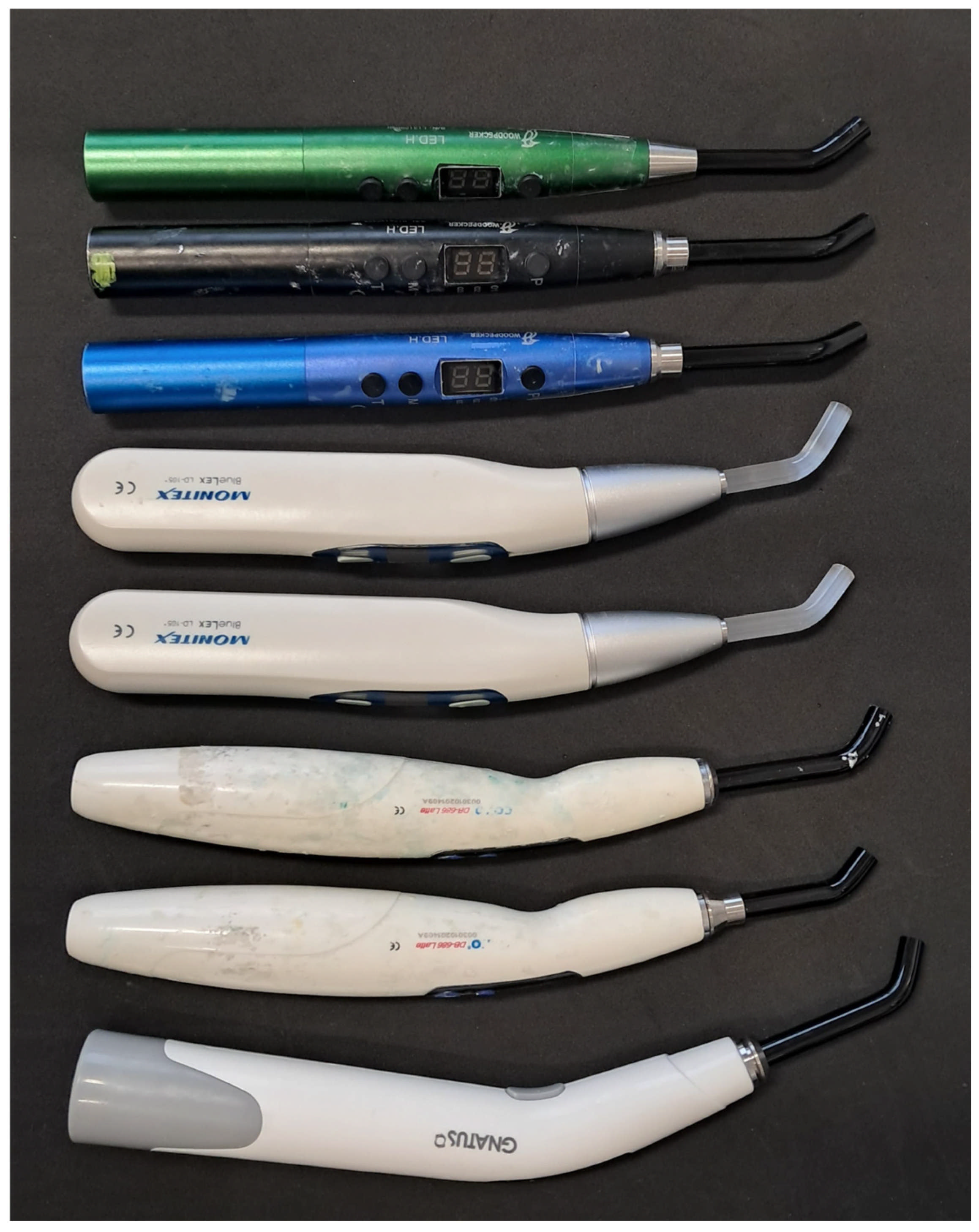
Figure 1. The light-curing units used.
Optics 06 00028 g001
Figure 2. The radiometers used in the study: D1—(control); D2—Bluephase Meter II; D3—LVCHEN LED Light Meter; D4—Radiometer LED; A1—Analogical Radiometer; and A2—Kerr Radiometer by Demetron.
Optics 06 00028 g002
Table 1. The characteristics of the polymerization units used in the present study.
Brand
(Group Code)
ModelManufacturerTip Diameter (mm)Intensity Declared by the Manufacturer
(mW/cm2)
Batch
1Woodpecker
(W1)
LED-HGuilin Woodpecker Medical Instrument; Guilin, China.8.0850–1000L1341355H
2Woodpecker
(W2)
LED-HGuilin Woodpecker Medical Instrument; Guilin, China.8.0850–1000H12050071A
3Woodpecker
(W3)
LED-HGuilin Woodpecker Medical Instrument; Guilin, China.8.0850–1000L1560597H
4Monitex
(M1)
BlueLEX LD-106MONITEX Industrial Company; New Taipei City, Taiwan.8.0100013L03778
5Monitex
(M2)
BlueLEX LD-106MONITEX Industrial Company; New Taipei City, Taiwan.8.0100013L03776
6Coxo
(C1)
DB-686-1bFoshan COXO Medical Instrument; Foshan, China.8.0120000310201409A
7Coxo
(C2)
DB-686-1bFoshan COXO Medical Instrument; Foshan, China.8.0120000310201409A
8Gnatus
(G)
Optilight MaxGnatus; San Paolo, Brazil.8.0120015070000046
Table 2. The characteristics of the radiometers used in this work.
Brand (Group Code)ManufacturerAction Mode *Spectral Range (nm)Irradiance RangeDeclared AccuracyBatch
Bluephase Meter® II **
(D1)
Ivoclar-Vivadent; Schaan, LiechtensteinDigital 380–550300–12,000±10%1300003059
Bluephase Meter® II
(D2)
Ivoclar-Vivadent; Schaan, LiechtensteinDigital 380–550300–12,000±10%1310001077
LVCHEN LED Light Meter
(D3)
Generic productDigital N/S ***N/S ***N/S ***N/S ***
Radiometer
(D4)
Generic productDigitalN/S ***N/S ***N/S ***N/S ***
Analogical Radiometer
(A1)
Generic productAnalogicalN/S ***N/S ***N/S ***N/S ***
Kerr by Demetron
(A2)
Kerr Dental; Brea, CA, EUAAnalogical400–500<2000±5%N/S ***
* The mechanism by which the intensity is recorded in mW/cm2, which can be digital or analog. ** A second Bluephase Meter® II model was used as a control group. This product is new. *** N/S: NOT SPECIFY.
Table 3. The average intensity values (in mW/cm2) and standard deviation obtained after measurement with the radiometers.
Photocuring UnitsRadiometers
D1D2D3D4A1A2
1W1500 (±10) d520 (±10) d740 (±20) c1361 (±20) a990 (±20) b410 (±20) e
2W2560 (±10) d560 (±10) d740 (±20) c1428 (±20) a990 (±20) b490 (±20) e
3W3860 (±10) d870 (±10) d1640 (±20) b 1576 (±20) c1790 (±20) a640 (±20) e
4M11020 (±10) d1020 (±10) d1490 (±20) c1514 (±20) b1590 (±20) a690 (±20) e
5M2970 (±10) d980 (±10) d1690 (±20) a1544 (±20) b1390 (±20) c690 (±20) e
6C11220 (±10) d1220 (±10) d1990 (±20) a1658 (±20) c1890 (±20) b1000 (±20) e
7C2920 (±10) d910 (±10) d1440 (±20) c1550 (±20) b1590 (±20) a780 (±20) e
8G1160 (±10) c1160 (±10) c1240 (±20) b1278 (±20) a590 (±20) e980 (±20) d
Different lowercase letters indicate the presence of statistically significant differences between radiometers for each light-curing unit (p < 0.05), as determined by one-way ANOVA followed by post hoc Tukey tests.
Disclaimer/Publisher’s Note: The statements, opinions and data contained in all publications are solely those of the individual author(s) and contributor(s) and not of MDPI and/or the editor(s). MDPI and/or the editor(s) disclaim responsibility for any injury to people or property resulting from any ideas, methods, instructions or products referred to in the content.

