Review

Advances of Machine Learning in Phased Array Ultrasonic Non-Destructive Testing: A Review

1
College of Electrical and Information Engineering, Hunan University, Changsha 410082, China
2
Shenzhen Research Institute, Hunan University, Shenzhen 518000, China
3
DEC Academy of Science and Technology Co., Ltd., Chengdu 611731, China
*
Author to whom correspondence should be addressed.
AI 2025, 6(6), 124; https://doi.org/10.3390/ai6060124
Submission received: 22 April 2025 / Revised: 6 June 2025 / Accepted: 9 June 2025 / Published: 12 June 2025
(This article belongs to the Section AI Systems: Theory and Applications)

Abstract

Recent advancements in machine learning (ML) have led to state-of-the-art performance in various domain-specific tasks, driving increasing interest in its application to non-destructive testing (NDT). Among NDT techniques, phased array ultrasonic testing (PAUT) is an advanced extension of conventional ultrasonic testing (UT). This article provides an overview of recent research advances in ML applied to PAUT, covering key applications such as phased array ultrasonic imaging, defect detection and characterization, and data generation, with a focus on multimodal data processing and multidimensional modeling. The challenges and pathways for integrating the two techniques are examined. Finally, the article discusses the limitations of current methodologies and outlines future research directions toward more accurate, interpretable, and efficient ML-powered PAUT solutions.

Graphical Abstract

1. Introduction

Phased array ultrasonic testing (PAUT), an advanced form of conventional ultrasonic testing, utilizes array transducers with digital control to steer and focus the acoustic beam, enabling the characterization of an object’s internal structure. PAUT offers broader inspection coverage, improved defect characterization, and enhanced adaptability to complex geometries [1]. These advantages have made PAUT a prominent focus of research in the field of non-destructive testing (NDT). Specifically, PAUT data are classified into several modalities based on imaging principles, including A-scan, B-scan, C-scan, S-scan, and three-dimensional (3D) volumetric data. Although PAUT data contain substantial structural characteristics, their direct interpretation is challenging due to modality coupling properties and the lack of a direct correspondence between raw signal patterns and physical geometries. Therefore, certified professionals are required to identify potential defects within PAUT data in industrial NDT scenarios. However, manual interpretation is inherently limited in scalability and consistency. This challenge has driven the development of algorithms tailored for PAUT analysis.
In early studies on the automated processing of PAUT data, heuristic optimization algorithms were primarily used to suppress noise and improve data quality [2] and sizing accuracy [3], while statistical analysis methods were applied to assess defect severity [4]. However, these methods primarily relied on manually defined model parameters and were therefore limited to the quantitative evaluation of conventional defects in geometrically regular components. More recently, machine learning (ML) is a paradigm that seeks to learn informative patterns from data [5]. For the automated processing of PAUT data, early studies primarily utilized traditional signal processing or classical ML algorithms for denoising, imaging, and feature extraction [6,7]. These algorithms are generally based on rigorous mathematical theories with compact and interpretable workflows. In the context of defect classification tasks using shallow ML models on PAUT data, a typical framework comprises three stages: signal pre-processing, feature extraction, and feature classification [8]. In the pre-processing stage, wavelet transform (WT) is used for noise suppression, Fourier transform (FT) for spectral decomposition, and Hilbert–Huang transform (HHT) for non-stationary signal analysis. Next, feature extraction quantifies time–frequency characteristics from pre-processed one-dimensional (1D) signals, including amplitude-based statistics (e.g., mean, kurtosis), FT-derived spectral energy, and HHT-based nonlinear dynamics. For two-dimensional (2D) data, morphological features are extracted using Gabor filtering, Canny edge detection, and histograms of oriented gradients. Furthermore, principal component analysis (PCA) and k-means clustering are commonly applied to compress the dimensionality of the extracted features. In the third stage, shallow ML models such as support vector machine (SVM), decision tree, artificial neural network (ANN), k-nearest neighbor (kNN), and Bayesian classifiers are employed for feature classification.
Among the studies leveraging shallow ML models, Bai et al. [9] applied PCA to extract features from ultrasonic scattering matrices, followed by quadratic discriminant analysis and a trained SVM for defect classification and size estimation. Li et al. [8] applied the lifted wavelet transform (LWT) for A-scan pre-processing and feature extraction, followed by SVM for weld defect detection. A genetic algorithm (GA) was applied to optimize feature subset selection. He et al. [10] provided a comprehensive comparison of several classical classifiers based on the A-scan classification task, including logistic regression, kNN, decision tree, naive Bayes, and SVM. Shallow ML models demonstrate reliable performance in certain cases, but their effectiveness is limited by feature extraction and selection. In complex industrial inspections, subtle feature variations and a low signal-to-noise ratio (SNR) can degrade their robustness [11].
Over the past decade, the deep learning (DL) subfield within ML has advanced rapidly, driven by specialized parallel computing hardware [12] and developments in fundamental DL theories, including novel network architectures [13], activation functions [14], and optimization algorithms [15]. These advancements have significantly expanded DL applications across various industries [16,17]. DL has been explored to meet the diverse data processing needs in PAUT imaging and defect detection. In PAUT imaging, Kumar et al. [18] used a convolutional neural network (CNN) to estimate inactive channel data in sparse arrays, mitigating grating lobe artifacts. Zhang et al. [19] introduced a Bayesian DL model tailored for super-resolution PAUT imaging. For feature extraction, Wang et al. [20] utilized contrastive learning to compute the Mahalanobis distance between normal and abnormal C-scan features, enabling anomaly detection. In defect detection and characterization, Posilovic et al. [21] employed You Only Look Once (YOLO) and Single Shot Multibox Detector (SSD) models for defect localization and identification in B-scan data. He et al. [22] proposed an enhanced Mask R-CNN model for the pixel-level segmentation of five welding defect types in S-scan data. Despite DL’s potential in PAUT, challenges remain: limited labeled data, labor-intensive annotation, and poor interpretability, often resulting in a reliance on empirical validation over theoretical advancements in practical applications.
In the field of ultrasonic-based NDT, several comprehensive reviews have discussed advancements in integrating ML techniques. Cantero-Chinchilla et al. [23] focused on theoretical foundations and progress in DL applications in conventional UT, while Yang et al. [24] provided a comprehensive review of ML advancements in the context of ultrasonic guided waves for structural health monitoring. Unlike conventional UT, PAUT generates more complex and diverse data modalities, which require more specialized ML strategies for effective data interpretation and decision-making. To bridge this gap and guide future research, this article reviews key studies and presents an overview of existing ML methods applied in PAUT-based NDT tasks, focusing on imaging, defect detection and characterization, and data generation. Notably, this article highlights ML-enhanced PAUT with focus on multimodal data processing. Figure 1 illustrates a systematic framework for the content of this article.
The structure of this article is organized as follows: Section 2 provides a brief overview of the theoretical background of PAUT, covering PAUT imaging methods and data formats. Section 3 reviews recent advancements in the application of ML to PAUT, including innovations in phased array ultrasonic imaging, defect detection and characterization, and data generation methods. Section 4 discusses the challenges of applying ML to PAUT-based NDT and explores potential solutions. Section 5 summarizes research limitations and outlines future directions. Finally, Section 6 concludes this article.

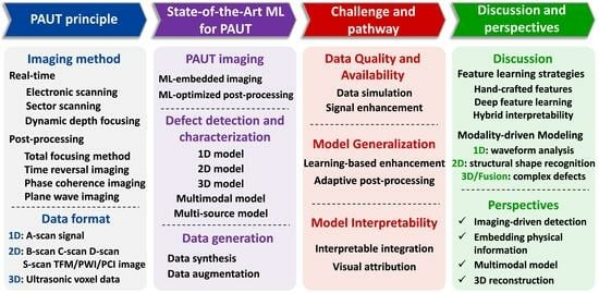
2. Overview of PAUT Fundamentals

To further analyze ML applications in PAUT-based NDT tasks, this section systematically reviews PAUT imaging techniques and data formats. The patterns of PAUT probes, imaging methods, and data formats are graphically depicted in Figure 2.

2.1. PAUT Imaging Method

Phased array ultrasonic imaging methods can be categorized into conventional real-time imaging and post-processing imaging, based on the use of phase delay control.

2.1.1. Real-Time Imaging

Real-time imaging utilizes the pulse-echo method and time delays for physical beamforming, enabling acoustic beam steering and focusing at specified angles or depths for image construction. Its rapid processing capability enables real-time NDT. This technique is classified into three types based on the scanning methods and beam control strategies.
  • Linear scanning
Linear scanning involves grouping transducer arrays along the scan direction and implementing uniform time delays for each group. This forms a sequence of focal points arranged linearly. The sequential excitation of element groups simulates the probe’s physical movement in an automated fashion. The received echo signals are processed by the delay-and-sum (DAS) algorithm to produce a 2D scan image. The DAS output I DAS at a focal point is calculated as
I DAS = n = 1 N w n s n t τ n
where N is the number of elements in the group, w n is an apodization weight for element n , s n t is the signal received by element n , and τ n is the time delay applied to elements for synthetic acoustic beam steering at the desired focal point.
  • Sector scanning
Sector scanning is a distinctive PAUT imaging method. This approach involves selecting a fixed group of adjacent elements in the transducer array and repeatedly exciting them with varying focal laws. By controlling the transmission and reception time delays of each element, the acoustic beam is steered at different angles and focused at multiple points, thereby generating a fan-shaped imaging region.
  • Dynamic depth focusing
Dynamic depth focusing [25] differs from the single-point focusing methods of linear and sector scanning. During wave transmission, a single focal point is used, but during echo reception, digital control is applied to refocus at various depths, from near to far distances. Dynamic depth focusing efficiently reduces beam divergence and enhances the depth of field, ensuring consistent image quality across the inspection direction.

2.1.2. Post-Processing Imaging

Post-processing imaging utilizes a more flexible and precise synthetic beamforming approach, leveraging multipath and multiple reflection signals for image reconstruction. This method enables virtual focusing at any point within the image, thereby improving image quality. Post-processing imaging methods can be categorized as follows:
  • Total focusing method
The total focusing method (TFM) is a post-processing imaging technique based on full matrix capture (FMC) data, first proposed by Holmes et al. [26]. In TFM, each element i is sequentially activated, and the echo signals s i j t from all N elements are received for each transmission, yielding a full matrix dataset containing N × N A-scans. During image reconstruction, TFM performs DAS processing for all possible transmit–receive pairs i , j , ensuring virtual focusing at each pixel p in the imaging region. The TFM image intensity I TFM p is given by
I TFM p = i = 1 N j = 1 N w i j s i j t = τ i p + τ j p
where τ i p is the time-of-flight from transmitter i to point p , τ j p is the time-of-flight from point p to receiver j , and w i j are weighting factors. TFM is acknowledged as the gold standard in PAUT post-processing imaging [27], but requires significant computational resources due to the O N 2 operations per pixel.
  • Time reversal imaging
To surpass the Rayleigh criterion and achieve super-resolution imaging at a fixed frequency, Lev-Ari et al. [28] devised the time reversal–multiple signal classification (TR-MUSIC) algorithm based on time reversal acoustics theory [29] and multiple signal classification. It uses singular value decomposition to decompose the N × N FMC response matrix K (at frequency ω ) into signal and noise subspaces.
K ω = U ω Σ ω V H ω
where Σ is a diagonal matrix of singular values, and U and V are unitary matrices. For defective objects, the TR-MUSIC image is formed using the noise subspace vectors U n ω , yielding high values at defect locations p where the steering vector g p , ω is orthogonal to the noise subspace.
I TR - MUSIC p 1 U n H ω g p , ω 2
This results in near-infinite inner product values at defects while remaining finite elsewhere.
  • Phase coherence imaging
Phase coherence imaging (PCI) [30] is a post-processing algorithm for PAUT based on FMC data, emphasizing phase information to minimize amplitude effects on imaging quality. PCI utilizes a phase coherence factor (PCF) or sign coherence factor (SCF) to weight aperture data. The SCF C S C F for a point p is calculated as:
C S C F p = 1 N k = 1 N sign s k t p
where s k t p is the analytic signal at the calculated time t p for aperture element k , and sign extracts the signal’s sign. The PCI image is then I PCI p = I DAS p × C SCF p p (where p is an exponent), thereby amplifying signals from point reflectors while suppressing noise. Since frequency information is independent of amplitude, PCI is less affected by signal attenuation. Moreover, in scenarios with high-frequency variance and low SNR, PCI can significantly improve image quality.
  • Plane wave imaging
Plane wave imaging (PWI) was first utilized in medical ultrasound imaging due to its ultra-high frame rate [31]. However, the unfocused nature of the acoustic beam in PWI leads to lower image resolution and contrast [32]. In the NDT field, Le Jeune et al. [33] proposed a PWI-TFM to address the long acquisition time and high computational complexity of FMC-TFM. PWI-TFM excites multiple elements simultaneously with specific time delays τ m tx to generate plane waves at specific angles θ . The beamformed signal s θ t for a plane wave transmission at angle θ received by element n is
s θ t = n = 1 N w n s n t τ n BF θ
where τ n BF θ is the beamforming delay to coherently sum echoes from direction θ across the array. A set of plane waves at different angles is transmitted, and the resulting beamformed signals are then processed using a TFM-like reconstruction, reducing the number of transmissions while increasing acoustic power and decreasing data storage demands.

2.2. PAUT Data Representation

PAUT generates diverse data formats based on different imaging and acquisition strategies, including 1D A-scan signals and 2D B-scan, C-scan, and S-scan images, as well as 3D volumetric data.
  • 1D format
The A-scan signal depicts the relationship between the ultrasonic pulse-echo amplitude and the acoustic path. A-scan data enable the preliminary assessment of internal material defect depth and dimensions.
  • 2D format
The B-scan provides a 2D cross-sectional view of the object aligned with the probe scanning and wave propagation directions, illustrating the distribution of echo amplitudes along the scanning axis.
The C-scan offers a 2D slice view of the specimen at a specific depth range, oriented perpendicular to the B-scan. Each pixel in the C-scan represents the maximum echo amplitude within a gated A-scan region. The horizontal axis of the C-scan denotes the probe’s scanning distance, while the vertical axis indicates its step size.
The S-scan is a distinct data format in PAUT. A sector-shaped cross-sectional image is generated by exciting groups of elements sequentially with varying focal laws and steering beam angles. In an S-scan, the horizontal axis represents the angular range of the sector scan, while the vertical axis shows echo amplitude distribution along the acoustic path.
  • Three-dimensional volumetric format
The 3D volumetric data can be acquired using either 2D matrix array transducers or 1D linear array transducers. The former excites and receives signals at the same position repeatedly, thereby allowing 3D data acquisition without mechanical scanning. In contrast, 1D linear arrays require mechanical scanning, using an encoder to reconstruct 3D volume from 2D slices.

3. State-of-the-Art ML for PAUT

ML in PAUT primarily focuses on imaging, defect detection, and data generation. Imaging serves as the data foundation, while data generation enhances the performance of an ML-based detection model. This section reviews recent advancements.

3.1. Phased Array Ultrasonic Imaging

Phased array ultrasonic imaging faces high beamforming computational costs and resolution constraints imposed by the Rayleigh criterion [34]. Speckle noise further degrades image quality and may obscure defect signals. Although noise reduction techniques in both transform and spatial domains mitigate noise interference [35], they often sacrifice image detail. Recent advancements in ML offer promising solutions to these challenges. ML models are applied in two ways: integrated within the imaging workflows or employed as post-processing for ultrasonic data.
Building on the integration of ML models into imaging workflows, Luiken et al. [36] proposed a simultaneous shooting method, inspired by reflection seismology, to improve data acquisition flexibility. However, this practice introduces aliasing noise and artifacts. To address this, a DL-based self-supervised denoising method using a U-net architecture is proposed, enabling data denoising without requiring clean labels for training. This enhances acquisition efficiency while preserving image quality. To further simplify the FMC-DAS imaging, Pilikos et al. [37] introduced a DL architecture that integrates data pre-processing, beamforming, and image post-processing into a single network for end-to-end optimization. Specifically, FMC data pre-processing employs a 3D-CNN, while the DAS is embedded within the network layers for imaging generation, and a 2D-CNN is employed for post-processing to output defect masks. This approach mitigates reconstruction errors from physical model inaccuracies and imaging noise. Liu et al. [38] proposed FMC-Net, a DL imaging method for directly reconstructing high-resolution ultrasonic images from FMC data, as shown in Figure 3. FMC-Net employs an encoder–decoder architecture with multi-scale residual modules and skip connections to capture complex excitation–reception features within the FMC data. It outperforms TFM and wavenumber algorithms in the visualization of sub-wavelength defects. Similarly, Molinier et al. [39] proposed a conditional generative adversarial network (GAN) to generate TFM-like images from single plane wave insonification, eliminating the need for FMC acquisition and TFM processing while enhancing contrast and drastically cutting the imaging time. In summary, ML-based methods can enhance imaging quality by improving the imaging workflow, while maintaining a comparable imaging speed during inference by transferring computational load to the training phase.
Additionally, several studies focused on post-processing to enhance the quality of ultrasonic data. The Rayleigh criterion constrains the resolution of ultrasonic phased array imaging. To surpass the diffraction limit, Gao et al. [40] proposed a label-enhanced semi-supervised Cycle GAN for TFM image super-resolution, effectively suppressing noise and artifacts. The model employs a U-net generator and a Markovian discriminator to transform TFM images into defect morphology representations, imposing stricter constraints through a reverse transformation to enhance reconstruction consistency and reduce artifacts. Zhang et al. [41] presented a two-stage DL-based network for defect super-resolution imaging. The first stage employs an enhanced residual network (ResNet) for defect localization, while the second stage utilizes DeepLab v3+ with dilated convolutions to expand the receptive field, preserving background information and refining defect features. Compared to ML modules embedded in PAUT imaging workflows, ML-based post-processing methods sacrifice workflow integration but offer greater flexibility.

3.2. Defect Detection and Characterization

This section reviews ML advancements in defect detection and characterization, focusing on the processing of diverse PAUT data modalities. The discussion is categorized into unimodal, multimodal, and multi-source models. Specifically, research on unimodal approaches is examined across 1D, 2D, and 3D models. Table 1 summarizes representative studies on PAUT unimodal models, while Table 2 presents research on multimodal and multi-source models.

3.2.1. Unimodal Models

1.
One-dimensional model for A-scan
The 1D form of PAUT data represents time-domain A-scan signals, where ML models are mainly used for classification or regression to detect and localize defects.
In binary classification tasks, the primary objective is to determine the presence of defects in A-scan signals. Shafiei et al. [42] investigated binary defect classification using A-scan signals from polyethylene joints. CNN achieved the highest F1-score on 6000 samples, outperforming classical ML models, yet errors from cold fusion/flawless signal overlap revealed challenges in resolving subcritical defects. Similarly, Choung et al. [43] detected internal discontinuity defects in wind turbine blades using an A-scan dataset from 9361 ultrasonic images, achieving nearly 99% accuracy with an 11-layer CNN. Wang et al. [44] proposed a graph convolutional network (GCN)-based approach that models acoustic–structural relationships as graphs, extracting defect-sensitive features via adjacency matrix analysis.
Besides utilizing binary classification to determine defect presence, multi-class classification has been explored to identify specific defect types within A-scan signals. Kim et al. [45] developed a seven-class classification model for weld A-scan data by integrating welding domain knowledge. The model combines two conventional features (peak width and skip distance) with two features characterizing the A-scan signal’s relationship to the welding boundary. A 1D-CNN extracts and predicts defect classes, improving accuracy from 89.77% to 98.79%. This innovative use of welding domain expertise enhances both detection accuracy and output reliability. Cheng et al. [46] tackled low-velocity impact defect depth localization in carbon fiber-reinforced polymer (CFRP) materials by comparing long short-term memory network (LSTM), CNN, and CNN-LSTM architectures. CNN-LSTM demonstrated superior depth classification accuracy, with an average relative error of 8.96%.
Regression models are used to predict defect location and size. Wang et al. [47] proposed a 1D-CNN method to reconstruct rough surface morphology from ultrasonic phased array pulse-echo signals. The model takes the normalized A-scan as the input and outputs surface morphology. SHapley Additive exPlanations (SHAP) is further applied to improve interpretability during feature extraction. Yang et al. [48] developed a quantitative analysis method for corrosion defects in engine cylinder cavities using radial basis function neural network (RBFNN) and GA. Wavelet packet energy spectrum, fractal dimension, peak features, and routine features are extracted from A-scan signals as the input to the RBFNN, which predicts defect diameter and taper angle. GA is applied to select optimal feature subsets. The optimized model reduces the defect size estimation error to within 4%.
2.
Two-dimensional model for B-scan
In the application of ML to B-scan image defect detection and characterization, Zhang et al. [49] developed an improved YOLO model for detecting cracks in high-speed railway train wheels from B-scan images. The network integrated Swin transformer modules with an enhanced feature pyramid network (FPN), and a detection head optimized for small targets. Trained on 15,000 real-world B-scan images, the model achieved an mAP@0.5 of 89% and a recall of 94%. Similarly, Chen et al. [50] developed an enhanced YOLO v8-based model to address small defect size challenges. The model integrated space-to-depth convolution to minimize information loss and employed a bi-level routing and spatial attention module for better feature extraction. Trained on 2286 B-scan images of side-drilled holes (SDH) and flat-bottom holes (FBH), the model outperformed the baseline, increasing the F1-score by 17.56% and the intersection-over-union (IoU) by 0.43%. Beyond YOLO-based models, Google’s EfficientDet [51] has been applied to B-scan image analysis. Cheng et al. [52] proposed an EfficientDet-based model for CFRP defect detection, embedding a transformer block in the backbone and using adaptive weighting in the neck to retain useful features. The trained model achieved 98.97% mAP on a dataset of artificial defect test blocks. Medak et al. [53] introduced a CNN-LSTM model for B-scan sequence detection to leverage semantic information across adjacent slices. The model employed ConvLSTM layers and 1 × 1 convolutions to process sequences, focusing on detecting defects in the middle slices. Based on the dataset from [54], it improved mAP by 2% at 512 × 512 and 3.4% at 384 × 384 resolution compared to single-image detection. The model innovatively leveraged three stacked adjacent slices as an input to enhance detection by increasing semantic density. Yang et al. [55] proposed a DL-based network for reconstructing concrete cross-section images and identifying subsurface cracks, as shown in Figure 4. The encoder–decoder architecture integrated physical information, including the serial number of investigation line and point, as well as step length, to aid registration. This approach generated global representations of crack distribution and achieved an F1-score exceeding 78% in crack detection.
Given the scarcity of defect data in industrial settings, some studies have proposed anomaly detection by training models primarily on normal B-scan data with limited defect samples. Tunukovic et al. [56] proposed an unsupervised ML model for defect detection in CFRP composites. The method employed automated gating and density-based spatial clustering of applications with noise (DBSCAN) to extract defect regions from 3D ultrasonic data, then trained an autoencoder (AE) on corresponding B-scan images. During inference, defects were identified by comparing input and reconstructed images. The model achieved area under the curve (AUC) values of 92.2% and 87.9% for simple and complex samples, respectively.
3.
Two-dimensional model for C-scan
For defect detection and characterization in C-scan images, Tunukovic et al. [57] systematically compared conventional amplitude thresholding, statistical methods, and ML-based models for defect detection in CFRP C-scan data. For ML approaches, they evaluated YOLO v5, Faster R-CNN, and RetinaNet. The study found that non-ML methods were highly sensitive to artifacts, while Faster R-CNN achieved the highest localization accuracy. Zhu et al. [58] proposed an improved YOLO v5 model for detecting delamination and adhesive defects in wind turbine blades using C-scan images. The model incorporated deformable convolution, a lightweight cross-stage partial spatial pyramid pooling fast module, and a context augmentation module. Additionally, the weighted IoU loss function was applied to accelerate convergence. The model achieved a precision of 92.6%, recall of 91.2%, and mAP@0.5 of 94.6%. To extract defect morphological information, Liu et al. [59] proposed an unsupervised C-scan semantic segmentation framework based on manifold learning. The method employed uniform manifold approximation and projection for nonlinear feature extraction, followed by a CNN and superpixel-based clustering for segmentation. Iterative optimization reduced background noise and improved defect identification. On a dataset of 1000 C-scan images, the framework outperformed PCA and k-means in IoU, demonstrating the effectiveness of unsupervised learning in PAUT defect segmentation.
4.
Two-dimensional model for S-scan
In research involving S-scan images for defect classification, Zhou et al. [60] used a 2D-CNN to classify three types of internal defects in high-voltage cable terminal lead seals. In the context of regression tasks. Jia et al. [61] innovatively applied PAUT to estimate the spatial and temporal parameters of gas–liquid two-phase flow interfaces. The ultrasonic echo signals were transformed into a 2D data matrix and subsequently converted into a ladder image. The gray wolf optimizer fine-tuned the least squares boosting model for amplitude prediction. The results demonstrated the potential of PAUT for flow pattern recognition tasks.
For S-scan defect segmentation, He et al. [22] developed an improved Mask R-CNN to detect welding defects in metro train body structures. This model incorporated context and attention blocks into the ResNet backbone and added a balanced block in the FPN to enhance feature extraction. It achieved 98.2% accuracy, outperforming baseline by 4.5%. To address the impact of multiple reflections and false signals from non-welding regions on defect identification in welds, Chen et al. [62] proposed a two-stage segmentation method based on the DeepLab v3+ model. The first stage performed a coarse extraction of the weld’s valid region, while the second stage refined the segmentation for defect detection. Wang et al. [20] developed an improved contrastive learning model for unsupervised defect detection in aircraft CFRP composites and metal L-shaped structures using S-scan and C-scan data. The model integrated the spatial transformer network and optimized squeeze-and-excitation network to extract common features from normal samples, calculated normal distribution parameters via mean and covariance, and identified defects using Mahalanobis distance.
5.
Two-dimensional model for TFM and PWI data
In the application of ML to the classification tasks of TFM or PWI images, Zhang et al. [63] proposed a defect classification method for TFM images of artillery cradle welds, employing a feature evaluation algorithm that integrated multiple criteria, including grayscale unevenness, differential moment, and mixed entropy. Quantum-behaved particle swarm optimization was used to optimize the kernel extreme learning machine, achieving a classification accuracy of 98%. For object detection in single-plane wave images, Latete et al. [64] addressed data scarcity using transfer learning. A large finite element (FE) simulation dataset was used for pre-training, followed by fine-tuning with real defect data to improve detection. This approach outperformed the 6 dB drop method in measurement accuracy, highlighting its effectiveness in data-constrained scenarios. In the context of other tasks, Zhang et al. [65] proposed a strongly generalized CNN with an end-to-end encoder–decoder architecture for segmenting single-angle plane wave images. The model removed traditional beamforming, directly generating segmented images from the raw radio frequency data. On the medical dataset, the model achieved an IoU of 96.29% and an F1-score of 98.28%, with a processing time of 0.2045 s.
6.
Three-dimensional model for volumetric data
In the classification task of ultrasonic 3D volumetric data, McKnight et al. [66] explored defect detection in ultrasonic 3D volumetric data of CFRP materials using a binary classification approach. A neural architecture search (NAS) was applied to optimize a 3D ResNet-based model. The 3D dataset was constructed using 64 stacked B-scan images from artificial defect specimens and CIVA software simulations. The NAS-optimized model surpassed VoxNet and a manually designed CustomNet in accuracy with fewer parameters, though its inference time remained a limitation. Similarly, Wang et al. [67] stacked five adjacent S-scan images as the input to a 3D CNN to predict weld defect categories.
For defect segmentation in ultrasonic 3D volumetric data, existing studies [68,69] primarily adopt the 3D U-net architecture. Specifically, Zhang et al. [69] developed a 3D U-net-based method for ship welding defect segmentation. They enhanced the network by modifying encoding stages, adding skip connections and residual blocks, and using a combined Dice and cross-entropy loss. Defect segmentation labels were generated via the 6 dB drop method, and a dataset of 196 ultrasonic 3D volumes (64 × 128 × 128) was created by stacking S-scan data. The model achieved 90.9% segmentation accuracy, demonstrating promise for welding inspection despite having high hardware demands.
Some studies have aimed to enhance the representation of 2D defect features using ultrasonic 3D volumetric data. Liu et al. [70] proposed a 3D deep convolutional AE for segmenting 2D defect features in polymer composites. The dual-layer encoder with 3D convolution and pooling enhanced feature visualization. A depth receptive field was applied to predict defect depth, reducing surface and backwall echo interference. The dataset was constructed by stacking 500 C-scan images acquired via pulsed-echo methods, forming a 3D matrix. The model achieved a mean IoU of 80% and a mean contrast-to-noise ratio (CNR) of 6.547.
Table 1. Summary of machine learning methods using unimodal PAUT data.
ApplicationReferenceML ModelInputDataset Source and SizeOutputKey Metric
ClassificationZhao 2023 [71] Multi-grained cascade forest (gcForest)A-scanArtificial defect specimen
2000 A-scan signals
Size categories of defect
(seven classes)
Acc = 97.50%
Wang 2022 [44] GCNA-scanN/APresence of defectN/A
Cheng 2023 [46] 1D-CNN and LSTMA-scanArtificial defect specimen
2694 A-scan signals
Presence of defectAcc = 96.28%,
P = 95.22%,
R = 96.49%
Siljama 2021 [72] Improved VGG16B-scanReal defect data and data augmentation
500,000 B-scan images
Presence of defectAcc = 97.5%,
P = 97.26%,
R = 96.63%
McKnight 2024 [66] 3D-CNN3D volumetric dataArtificial defect specimen and simulation data
680 3D volumetric data (64 × 1204 × 64)
Presence of defectAcc = 100.00%,
P = 100.00%,
R = 100.00%
Dimensional regressionJia 2024 [61] Least squares boosting (LSBoost)S-scanN/ACharacteristic parameters of interfacial wavesMAPE = 4.38% (Stratified flow)
MAPE = 17.26% (Plug flow)
Wang 2024 [47] 1D-CNNA-scanSimulation data
21,200 A-scan signals
Surface heightMAE = 0.0237mm (thirty-two elements),
MAE = 0.0292mm
(eight elements),
MAE = 0.0497mm
(four elements)
Yang 2016 [48]RBFNNA-scanReal defect data
320 A-scan signals
Defect size and angleRRMSE = 3.612% (Taper angle),
RRMSE = 3.453% (Diameter)
Pyle 2021 [73] 2D-CNNMultiple PWI imagesReal defect data and simulation data
26,623 PWI images
Defect size and angleMSE = ±0.29mm (Length),
MSE = ±2.9° (Angle)
Bai 2021 [74] 2D-CNNScattering matrixSimulation data
1156 scattering matrices
Defect size and angleMAE = 0.08, RMSE = 0.12, R2 = 0.92 (Size)
MAE = 4.88, RMSE = 9.54, R2 = 0.92 (Angle)
Object
detection
Yuan 2020 [75] ANNB-scanReal defect data and artificial defect specimen
35 B-scan images
Defect location and class
(three classes)
Acc = 93.00%
Chen 2024 [50] Improved YOLO v8B-scanSimulation data and public dataset
2286 B-scan images
Defect location and class
(two classes)
F1 = 75.68%, IoU = 83.79%
Medak 2022 [53] 2D-CNN and LSTMB-scan sequenceArtificial defect specimen
Over 4000 B-scan image sequences
Defect location and class
(seven classes)
mAP = 91.60% (Conv2d)
mAP =91.40% (LSTM)
Tunukovic 2024 [57] Faster R-CNNC-scanArtificial defect specimen and simulation data
Over 300 C-scan images
Defect location and class
(four classes)
P = 99.80%,
R = 96.00%,
F1 = 97.80%
Latete 2021 [64] Faster R-CNNPWI imageArtificial defect specimen and simulation data
2048 time-trace matrices
Defect location and class
(two classes)
R = 70.00%
SegmentationLiu 2022 [59] 2D-CNNC-scanArtificial defect specimen
1000 C-scan images
Defect mask and class
(three classes)
Mean IoU = 75.00%
Zhang 2022 [65] Strongly generalized CNNRadio frequency dataPublic dataset
2900 radio frequency data
Defect mask and class
(one class)
IoU = 96.29%
F1 = 98.28%
He 2023 [22] Improved Mask R-CNNS-scanReal defect data
3000 S-scan images
Defect mask and class
(five classes)
mAP = 98.20%
Zhang 2024 [69] Improved 3D U-net3D volumetric dataReal defect data
196 3D volumetric samples (64 × 128 × 128)
Defect mask and class
(one class)
Dice Acc = 90.90%
Anomaly detectionTunukovic 2024 [56] DBSCAN and AEB-scanArtificial defect specimen
11,750 B-scan images
Presence of defectAUC = 92.20% (Simple)
AUC = 87.90% (Complex)
Posilovic 2022 [76] MobileNet and Patch distribution modeling (PaDiM)B-scanArtificial defect specimen
5715 anomalous and 11,709 normal B-scan images
Presence of defectAUC = 82.00%
Wang 2023 [20] 2D-CNN and transformerS-scan and C-scanReal defect data
90 normal S-scan and C-scan images
Presence of defectIoU = 15.42%
F1 = 25.80%
Note: The abbreviations used in the table are as follows: Acc—Accuracy, P—Precision, R—Recall, F1—F1-score, IoU—Intersection-Over-Union, mAP—Mean Average Precision, MAE—Mean Absolute Error, MAPE—Mean Absolute Percentage Error, RMSE—Root Mean Square Error, RRMSE—Relative Root Mean Square Error, R2—Coefficient of Determination, AUC—Area Under the ROC Curve. N/A indicates that the dataset source and size was not specified in the referenced article.

3.2.2. Multimodal Models

In complex industrial inspections, ML models relying on a single PAUT modality often result in false positives or missed defects. For example, detecting bonding defects in wind turbine blade spars requires integrating B-scan, C-scan, and A-scan data for comprehensive analysis. Multimodal data improve fault tolerance and inspection reliability.
In the research on NDT utilizing multimodal PAUT data combined with ML, Ortiz de Zuniga et al. [77] proposed a multimodal framework that integrated S-scan and A-scan data to classify welding defects in the international thermonuclear experimental reactor (ITER) vacuum vessel. A 2D-CNN was used for S-scan classification, while an LSTM assessed whether the A-scan aligned with defect-free welding signals. The final decision was obtained by applying a logical OR operation to both outputs. The CNN achieved 100% accuracy, while the LSTM reached 83%. Similarly, Li et al. [78] employed an improved YOLO v4 to coarsely locate defects in the aircraft composite from C-scan, while a 1D-CNN classified A-scan signals from the identified regions. The results demonstrated that this cascaded approach outperformed single-modality methods. However, both studies analyzed modalities independently, without feature-level fusion. Cao et al. [79] proposed a multimodal defect classification model using S-scan and A-scan data to detect pseudo defects in welds, as shown in Figure 5. It employs ResNet and gated recurrent unit (GRU) branches to extract 324-dimensional feature vectors from S-scan and A-scan data, respectively. The tensors are then aligned, fused, and processed by a fully connected layer for three-class classification, distinguishing true and pseudo defects. The fusion model achieved 98.07% accuracy, significantly outperforming unimodal approaches.

3.2.3. Multi-Source Models

Ultrasonic phased array imaging is susceptible to artifacts and near-field blind zones [80], and struggles with materials like coarse-grained castings. Combining different NDT techniques can mitigate these limitations: radiographic testing offers high-contrast internal structures, eddy current testing detects surface and near-surface cracks, and infrared thermography identifies thermal anomalies [81]. However, multi-source data integration faces challenges in cross-modality data heterogeneity, inter-modality alignment, and fusion architecture design.
In the context of applying ML methods to integrate PAUT with other NDT techniques, Li et al. [82] developed a model integrating infrared thermography and PAUT for aircraft composite defect detection. A cascade R-CNN with fusion modules and FPN enabled the parallel processing and feature-level fusion of infrared and ultrasonic images. Evaluated on a dataset of 500 paired infrared and C-scan images, the model achieved 99.3% accuracy and 90.4% mAP. Caballero et al. [83] developed a semi-automatic method combining X-ray computed tomography (XCT) and PAUT for internal defect segmentation in composites. Specifically, a 2D-CNN was trained to segment defects using 3D ultrasonic slice data as the input and projection-registered XCT data as the labels, addressing the insufficient information of ultrasonic data in porosity defect assessment. The method achieved a 66% F1-score and 50% IoU, leveraging multi-source registration for supervised closed-loop training without manual annotations. Furthermore, Sudharsan et al. [84] proposed a fusion-based detection method combining PAUT and pulsed thermography. A coordinate transformation merged data from both modalities into volumetric datasets. A 2D-CNN integrated with a bi-planar medial axial transform algorithm extracted defect features via three orthogonal plane convolutions and quantified defect sizes. The method achieved a detection accuracy of 91.46%, outperforming single-modality methods.
Table 2. Summary of machine learning based on multimodal and multi-source models.
ApplicationsReferenceML ModelInput 1Input 2Fusion Method
ClassificationOrtiz de Zuniga 2022 [77] 2D-CNN and LSTMS-scanA-scanDecision-level fusion of two-branch classification results.
Object detectionLi 2021 [78] YOLO v4 and 1D-CNNC-scanA-scanThe C-scan is used to locate defect regions, followed by the extraction of A-scan data from these regions for defect classification.
ClassificationCao 2025 [79] ResNet and GRUS-scanA-scanThe two branches perform feature-level fusion for classification.
Object detectionLi 2021 [82] Cascade R-CNNC-scanInfrared imageParallel two-branch feature-level fusion at multiple scales.
SegmentationCaballero 2023 [83] 2D-CNNC-scanXCT slice dataThe two data sources are aligned, with the C-scan serving as the model input and XCT slices used as segmentation labels.
SegmentationSudharsan 2024 [84] Tri-planar Mask R-CNNTFM imagePulsed thermography dataThe spatial alignment of the two volumetric data enables pixel-level fusion, followed by feature extraction along the three spatial dimensions.

3.3. Generation of Phased Array Ultrasonic Data

Compared to traditional shallow ML algorithms, DL offers superior robustness and feature extraction capabilities for detection tasks. However, in industrial NDT, the rarity of defects poses a significant challenge in acquiring diverse and well-annotated defect datasets. Additionally, the resolution of phased array ultrasonic imaging is constrained by factors such as probe frequency, aperture size, and beamforming principles [85]. To enhance domain adaptability and reduce dependence on real-world defect data, researchers have explored data generation techniques to expand dataset size and enhance data quality. This section reviews two primary approaches: data synthesis and data augmentation [86]. Table 3 summarizes typical PAUT data generation studies.

3.3.1. Data Synthesis

Synthetic data are generated from scratch using physical models and simulations. Researchers employ FE methods to model acoustic fields and generate data via simulation software or custom algorithms.
Gantala et al. [87] used virtual array source aperture (VASA) and FE simulations to generate a dataset of 225 TFM images containing SDH and crack defects. MATLAB simulations modeled defects with varying sizes and shapes. Pyle et al. [73] proposed a hybrid FE and ray-based simulation method to generate crack defect PWI data. A local FE model was employed to generate the scattering matrix. Then, a ray-based model was used to efficiently create FMC data by tracing all relevant paths from the array to the defect.
In studies utilizing simulation software, Liu et al. [38] employed MATLAB’s Field II software to generate a comprehensive FMC dataset. Similarly, Pilikos et al. [37] utilized the K-wave toolbox of MATLAB to generate FMC data for end-to-end imaging network training. Zhang et al. [88] used the FE software PZFlex to simulate the propagation and reflection of ultrasonic waves in laminated composites. Wrinkle defects were introduced as sinusoidal geometric features. Furthermore, Kumbhar et al. [89] utilized COMSOL 6.0 to simulate A-scan data. A 2D angle beam ultrasonic model of a steel specimen with a rectangular defect was constructed. The ultrasonic signal propagated as a longitudinal wave through a wedge structure, generating shear waves upon refraction at the interface. Lee et al. [90] used CIVA to synthesize S-scan data for liquefied natural gas (LNG) storage tanks by adjusting parameters including waveform, incident angle, and frequency, generating 498 representative images.

3.3.2. Data Augmentation

Data augmentation focuses on transforming existing datasets to expand their size while enhancing diversity. Common techniques include conventional methods, virtual defects, and GAN-based data generation.
Conventional methods employ geometric transformations and noise injection to diversify training samples. For 1D A-scan signals, McKnight et al. [91] added noise to simulate structural and random noise. For 2D C-scan images, Zhu et al. [58] applied pixel-level augmentation, including HSV adjustments, random flips, and image-level techniques such as mosaic and cut-out. To prevent the model from overfitting to specific augmentation patterns, Virkkunen et al. [92] introduced virtual flaws to a B-scan dataset by copying and relocating defect-free regions. Similarly, Siljama et al. [72] improved data diversity by embedding extracted defect signals into different backgrounds.
GAN improves data quality and enlarges datasets by modeling real data distributions, enabling the generator to transform noise or conditional inputs into new samples. Sun et al. [93] proposed a constrained Cycle GAN to learn the nonlinear mappings between unpaired phased array and linear array images in an unsupervised manner, generating quasi-linear phased array images with a higher spatial resolution. The model incorporated ultrasonic imaging physics by introducing identical and correlation coefficient losses to ensure structural consistency and backscatter patterns. Yang et al. [55] developed a conditional GAN to augment B-scan datasets. Its five-layer Markovian discriminator enhances the local details of generated images by penalizing structures at the patch scale. To control the structural characteristics of generated data, Granados et al. [94] introduced a conditional U-net model for high-resolution multimodal TFM image generation. The model takes low-fidelity simulated multimodal TFM images and physical morphological parameters, including wave velocity, specimen geometry, reconstruction mode, and defect size and orientation, as inputs. By integrating simulation parameters with deep feature maps via the fidelity linear modulation and the parametric spatial transformer layers, the model enables precise control of generation while reducing annotation costs. Additionally, other studies have employed deep convolutional GAN [95] and SPADE GAN [96] for the augmentation of TFM and B-scan image datasets.
Table 3. Summary of PAUT data generation methods.
MethodReferenceApproachesDataset Type and Size
Data
synthesis
Zhang 2022 [88] PZFlex simulation4500 A-scan signals
Kumbhar 2023 [89] COMSOL simulationA-scan signals
N/A
Lee 2023 [90] CIVA simulation498 S-scan images
Gantala 2023 [87] FE and VASA1000 TFM images
Pyle 2021 [73] FE and ray-based simulation25,625 PWI images
Zhang 2023 [97] CIVA simulation2000 PWI images
Liu 2023 [38] MATLAB Field II simulation30,000 sets of paired FMC-TFM data
Pilikos 2020 [37] MATLAB K-wave simulation230 sets of paired FMC–mask data
Latete 2021 [64] Pogo FEA simulation2048 time-trace matrices
Data augmentationSiljama 2021 [72] Traditional data augmentation and virtual flaws500,000 B-scan images
Shi 2020 [98] Traditional data augmentation2050 B-scan images
Virkkunen 2021 [92] Virtual flaws20,000 B-scan images
Sun 2023 [93] Constrained Cycle GANB-scan images
N/A
Yang 2024 [55] PATCH GAN1159 sets of paired B-scan–mask data
McKnight 2024 [91] Cycle GAN154 C-scan images
Granados 2023 [94] Conditional U-netTFM images
N/A
Granados 2024 [99] Class-conditioned generative adversarial autoencoderTFM images
N/A
Note: N/A indicates that the dataset type and size was not specified in the referenced article.

4. Challenges in ML-PAUT Integration

ML applications in NDT are a growing trend. However, PAUT multimodal data are intricate and varied, with defect semantic features shared across different modalities, leading to potential redundancy. Key challenges in integrating ML with PAUT include defect data scarcity, model generalization, and limited ML interpretability. This section systematically explores these challenges and reviews current solutions, as depicted in Figure 6.

4.1. Data Quality and Availability

The effectiveness of ML models is primarily determined by the quality and scale of the input data, as they rely on data-driven learning. In industrial NDT, defect data are scarce and highly valuable. Unlike publicly available datasets such as ImageNet [100] and Microsoft Common Objects in Context (MS COCO) [101], industrial NDT datasets are mainly used for in-house applications.
In terms of data integrity and signal fidelity in PAUT, Song et al. [19] analyzed the impact of limited training datasets on ML model performance. They applied a variational Bayesian DL approach to quantify epistemic and aleatoric uncertainties in guided wave array imaging. This method offers probabilistic insights for defect detection, avoiding over-confidence and mitigating erroneous decision-making. Additionally, in cases of limited real or artificially manufactured defect data, the use of simulated data has gained considerable popularity. In efforts to improve the availability of simulated data, Bevan et al. [102] proposed a forward model for enhancing the usability of simulation-based TFM images. This model integrated beam spread, transmission reflection coefficients, attenuation, and scattering matrices to generate high-quality simulated datasets. For the quantitative evaluation of data availability, Schmid et al. [103] used probabilistic neural networks to analyze uncertainty in segmented TFM images, relying on synthetic datasets. Entropy-based uncertainty metrics were employed to quantify prediction confidence and variance. The study highlighted the limitations of synthetic data and the need for high-quality, real-world datasets in industrial NDT.
To mitigate low resolutions, blurred edges, and background noise in low-frequency ultrasonic images, Lei et al. [104] proposed a CNN that integrates an attention module into an AE architecture and employs cross-layer connections to fuse multi-scale features, significantly improving structural similarity and peak SNR. Yin et al. [105] proposed an artifact suppression method for A-scan signals using PCA and AE. PCA reduces data dimensionality, while AE extracts artifact components, which are then subtracted from the original signal. In terms of post-imaging image processing, Jayasudha et al. [106] developed a denoising method for C-scan images of weld defects. A 2D adaptive anisotropic diffusion filter was utilized to reduce noise, followed by an adaptive mean adjustment algorithm to optimize contrast and brightness. An improved k-means algorithm was then applied to extract the region of interest (ROI), minimizing artifact impact. Additionally, Guan et al. [107] proposed a spatially adaptive Gaussian splatting (SAGS) method for S-scan data post-processing. This approach assigns a customized Gaussian kernel to each sampling point, enabling the precise resampling of non-uniform points. SAGS dynamically adjusts the Gaussian kernel size to match the local features of the ultrasonic sound path, reducing aliasing and over-blurring.
In summary, improving data quality and availability through screening or augmentation can enhance model performance. Notably, when using purely simulated or idealized flaws, it is essential to evaluate data usability and ensure relevance to real-world industrial defects.

4.2. Model Generalization

ML generalization refers to a model’s ability to perform well on unseen data. However, the time-varying nature of ultrasonic signals and variations in probe coupling can lead to distribution mismatches between collected and test datasets. Research on improving generalization has primarily focused on data-level enhancements and model training strategies.
In efforts to enhance model generalization through data augmentation, Herve-Cote et al. [108] proposed an FMC-based data generation method. It expands limited FMC data by varying reconstruction parameters, reorganizing tensors, and applying random transformations. Incorporating prior welding geometry as contextual input into a CNN further enhances data authenticity, achieving 93% accuracy on previously unseen defects. Similarly, Koskinen et al. [109] enhanced ML model generalization for unseen defect types in B-scan images by incorporating artificial defects, simulations, and data augmentation. This research highlighted the importance of considering the minimum defect size and type in dataset design. To detect overfitting in beamforming neural networks, Zhang et al. [110] proposed a method by inputting artificial data (zeros, ones, Gaussian noise) and comparing deviations from ground truth images via qualitative and quantitative metrics.
In research aimed at improving generalization through model training strategies, Pyle et al. [111] tackled poor model generalization in PWI images from experiments and simulations. The study applied domain adaptation (DA) to compare training results across data sources, using three DA methods—MixedSet, regression with contrastive semantic alignment (RCSA), and an adversarial domain classifier—against two non-DA baselines: simulation-only and experiment-only data. RCSA minimized label-based cross-domain distances, and the adversarial domain classifier optimized domain-invariant features by confusing domain discrimination. Results showed that the adversarial domain classifier achieved the best generalization in predicting experimental notch sizes.
In summary, data-level methods improve generalization via simulation, augmentation, and artificial defects. Training-level strategies such as domain adaptation further promote invariant feature learning. These methods guide models to capture the essential semantic features of defects rather than to overfit to acquisition-specific variations.

4.3. Model Interpretability

DL models are characterized by their complex structures and numerous parameters, making it difficult to intuitively interpret their inner workings. In such safety-sensitive applications, the lack of interpretability in DL models considerably limits their broader adoption.
Several studies have focused on embedding interpretable ML models into DL frameworks to enhance the interpretability of the latter. For instance, Pyle et al. [112] introduced a dimensionality reduction method named Gaussian feature approximation (GFA) for enhancing the interpretability of regression model. By fitting 2D elliptical Gaussian functions to PWI images, GFA extracts seven geometric descriptors of defects. GFA demonstrates superior regression accuracy and interpretability compared to the 6 dB drop method and PCA. In the domain of game-theory-based feature contribution model interpretability, Shi et al. [113] developed a feature selection framework for A-scan analysis. By integrating SHAP values to quantify feature contributions and coupling sequential selection algorithms, the method identifies physically meaningful feature subsets that maximize both prediction accuracy and model transparency.
Additionally, researchers have visualized attention mechanisms in DL models to enhance interpretability by highlighting the learning process. McKnight et al. [91] applied guided gradient-weighted class activation mapping (Grad-CAM) for C-scan defect classification. This approach integrates guided backpropagation with class activation maps to reveal the model’s focus regions, and it has also been used for the qualitative analysis of model effectiveness.
In summary, model interpretability in ML-PAUT is improved through two strategies: embedding interpretable modules to guide learning, and using the visualization or feature attribution methods to reveal decision patterns. These approaches reduce model opacity and enhance trust in safety-critical NDT applications.

5. Discussion and Perspectives

5.1. Discussion

The literature review indicates that a wide range of ML approaches have been applied to PAUT-based NDT tasks, including bonding defect detection in composites, weld flaw identification in metals, and defect size estimation. Early studies primarily focused on converting traditional signal processing tasks into ML-based frameworks. With the advancement of DL, research has shifted toward more expressive models with greater capacity, enhancing both imaging and detection performance. To clarify how different modeling strategies adapt to PAUT applications, the following discussion focuses on feature extraction and modality selection.

5.1.1. Feature Extraction

Feature extraction is pivotal in integrating ML with PAUT, and the design of features is influenced by the physical properties and dimensional structure of different data modalities. Current research categorizes feature extraction strategies along two dimensions: modality-specific data structure differences, and whether features are manually designed or automatically learned.
A-scan signals are inherently time-domain waveforms. Researchers have extracted semantic information from them using both hand-crafted features—such as wavelet energy, fractal dimension, and time–frequency descriptors—and deep learning methods. For instance, ref. [48] leveraged domain-specific statistical features, whereas [43,47] demonstrated the effectiveness of 1D-CNNs in learning robust representations directly from raw waveforms, particularly under noise or weak echo conditions.
In 2D modalities such as B-scan and C-scan, CNN-based methods have been widely applied to automatically extract spatial and edge features from raw pixel data. Models like YOLO and Faster R-CNN, enhanced with multi-scale and attention modules, have shown improved performance in detecting small or low-contrast flaws [49,50,57]. For S-scan, TFM, or PWI data containing angular or beamforming information, studies have integrated convolutional structures with context modeling to better capture directional patterns. Some approaches further augment these learned features with statistical descriptors like entropy and grayscale unevenness to enhance model robustness [22,60,61,63].
For 3D volumetric data, voxel-based representations enable 3D CNNs to extract continuous spatial features from defect regions. These methods effectively model defect morphology, boundary consistency, and spatial correlations [66,67,69,70]. As data dimensionality increases, automated feature learning has gained prominence, not only for its superior representational power but also due to the limitations of handcrafted features in analyzing complex PAUT data.
However, manual and automatic methods are often complementary. Recent studies integrate explainability tools like SHAP values and embedded feature selection to interpret learned features and improve model transparency [47,113]. Such a hybrid paradigm reflects a growing interest in combining physical interpretability with high-capacity representations.
In summary, feature extraction strategies in ML-based PAUT have evolved from hand-crafted design to automatic and hybrid learning. The choice of method reflects a balance between task requirements, data modality, and deployment constraints.

5.1.2. Modality Selection

Different types of defects exhibit notable differences in structural morphology and physical scale, resulting in distinct modeling requirements and data representation preferences across PAUT modalities, even within the same defect class.
For crack-related defects, B-scan and TFM imaging methods are widely used for their ability to represent lateral crack propagation paths and edge contours. For example, ref. [49] introduced a transformer-based detection model for wheel cracks using B-scan images. In contrast, ref. [63] employed TFM images with an extreme learning machine to classify weld cracks. While both methods share similar task settings, B-scan excels in localized target representation, making it suitable for object detection. TFM provides enhanced contrast through compounding, offering superior classification performance. For polymer-based or interface-dominated materials, 3D volumetric data allow for improved spatial consistency and interlayer depth modeling. Ref. [70] applied an autoencoder network on a voxel-stacked input to segment polymer matrix cracks, achieving a favorable IoU. Although 3D data impose higher computational costs, they offer superior capability in analyzing embedded defects compared to 2D modalities.
In weld defect detection, different modalities exhibit variations in both the scale of detectable features and the modeling structure. A-scan signals excel at capturing subtle structural variations. For instance, ref. [45] improved defect classification accuracy by modeling waveform parameters and weld boundaries. However, such methods often rely on handcrafted features and suffer from limited generalization. In contrast, S-scan images retain angular information, enabling superior spatial representation. As demonstrated by [22], S-scan-based segmentation outperforms conventional models, particularly in complex weld geometries. Under conditions involving multiple defect types or ambiguous signals, multimodal fusion tends to provide more robustness. For instance, ref. [79] proposed a dual-branch fusion model using both A-scan and S-scan inputs, achieving high classification accuracy. These studies indicate that the A-scan input is well suited for local waveform anomaly analysis, S-scan for regional structural assessment, while fusion strategies are optimal for complex or morphologically ambiguous defects.
For typical composite material defects such as delamination, disbonding, and impact damage, C-scan imaging is widely adopted due to its cross-sectional view and high fidelity in contour representation. Ref. [58] applied YOLOv5 for delamination and disbonding detection in wind turbine blades, while ref. [59] developed an unsupervised superpixel segmentation method to delineate weak bonding regions in CFRP. Conversely, A-scan signals or low-dimensional time-series data are more effective for low-visibility defects (e.g., minor cracks, slight impacts). Ref. [46] demonstrated this by using a CNN-LSTM to predict impact depth in CFRP. To overcome unimodal limitations, studies like [82,83] integrated thermal imaging and XCT, respectively, improving the detection of bonding failures and porosity through multimodal alignment. In summary, C-scan is well suited for stable 2D structural anomalies, whereas A-scan and multimodal strategies are superior for capturing spatially subtle or small-scale defects.
Overall, for crack, weld, and composite defects, optimal modality selection depends on three key factors: task objectives, structural features, and data dimensions. Two-dimensional imaging (B-scan, C-scan, TFM) excels at shape-based identification, while 1D signals (A-scan) better capture physical properties and temporal patterns. For complex structures with multiple defect types, 3D and multimodal approaches prove most effective.

5.2. Perspectives

In recent years, PAUT has emerged as an efficient NDT technique, offering real-time imaging, intuitive data representation, and operational simplicity. Meanwhile, the theoretical framework of shallow ML has matured, while advances in computational hardware and algorithm optimization have enabled high-performance DL methods. ML models show significant potential in enhancing PAUT applications. Research indicates that ML integration in PAUT focuses on three areas: imaging enhancement, defect detection and characterization, and data generation. However, critical issues remain, and future directions are outlined to address them.
(1)
Imaging-driven defect characterization
Unlike visible light images, raw PAUT data lack interpretability and are not intuitively understandable [114]. Specific transformations are required to visualize the data, enhancing the contrast between defects and the background. Imaging techniques are fundamental to the effectiveness of ML applications. For the more accurate characterization of defects larger than the ultrasonic wavelength, Bevan et al. [115] generated multiview TFM images from the same ROI within an FMC dataset. These images were fused using a matched filter-based fusion approach to enhance the visualization of large defects. However, this method requires tailoring the fusion process based on prior features of the expected defect. In addition, conventional PAUT systems based on linear wave interactions fail to detect subwavelength defects. One approach to overcome this limitation is by leveraging the nonlinear response of micro-defects. Nonlinear PAUT detects these responses as higher harmonics and sub-harmonics in the received signal spectrum under single-frequency excitation [116]. The essence of the method is to improve the SNR of micro-defects. Developing more efficient and robust imaging methods is crucial for fully leveraging the potential of ML models.
Future efforts may explore the integration of linear and nonlinear imaging modes within a unified pipeline, with region-specific switching guided by lightweight ML-based signal analysis. Such integration is expected to enhance sensitivity across a wider range of defect scales without altering the hardware structure. In addition, imaging-specific augmentation techniques that simulate variations in probe angle or material structure may help improve the generalization of data-driven imaging models.
(2)
Fusion of PAUT physical information with ML
Current ML interpretability methods, such as Grad-CAM, visualize model attention but are limited to observational analysis, unable to directly intervene in model mechanisms. This poses challenges for NDT model interpretability. PAUT data integrate multiple physical processes, creating non-intuitive internal structural mappings. Existing ML models focus solely on data while neglecting underlying physical mechanisms. A feasible solution is to incorporate PAUT physics principles into ML models [117]. Sun et al. [118] integrated ultrasonic guided wave physical parameters (e.g., reflected wave intensity, angular relationships) into the neural network via hybrid model inputs and physics-constrained loss functions, achieving high-precision microcrack quantification. Gao et al. [119] incorporated the nonlinear equation of Fermat’s principle into the loss function to enforce sound wave propagation along the shortest time path, ensuring physically consistent predictions and enabling accurate interface reconstruction in dual-layer media using FMC data. The incorporation of physical information not only enhances model interpretability but also alleviates overfitting caused by the scarcity of defect data in industrial NDT.
A promising direction is to introduce physical priors into ML models in modular form, such as physics-guided loss terms or parameter constraints derived from wave propagation behavior. This strategy can enhance prediction consistency without requiring the deep reformulation of the model architecture.
(3)
Multimodal models for PAUT data
PAUT data exhibit diverse modalities and redundant features. However, most ML models for PAUT in existing studies focus on unimodal data or process multiple modalities separately before simple decision-making. Real industrial data are more complex than artificial or simulated defect data, as they contain stochastic noise from poor coupling and multiple echoes. In the field of ground-penetrating radar, researchers have proposed multimodal fusion models that integrate A-scan, B-scan, and C-scan data to detect subsurface defects in airport runways [120]. In medical image segmentation, Zhao et al. [121] developed a multimodal feature learning framework based on optical coherence tomography (OCT), leveraging both 3D volumetric data and B-scan information to enhance model performance by capturing the complementary advantages of different modalities. Thus, developing multimodal models for the effective semantic fusion of PAUT data, facilitating multi-level integration and precise decision-making, requires further investigation.
Inspired by these practices, PAUT research may benefit from the development of lightweight multimodal fusion frameworks that combine image-level and signal-level information, especially B-scan and C-scan. Preliminary work can focus on feature-level alignment using attention- or correlation-based methods, enabling mutual compensation across modalities. Such models may improve robustness in real-world noisy data scenarios.
(4)
Three-dimensional ultrasonic reconstruction for NDT
In industrial NDT, 3D ultrasonic reconstruction primarily relies on 1D linear array probes with 1D encoders, restricting the probe’s spatial pose to a 2D plane. This limitation leads to cubic voxel structures that struggle to accurately represent real-world objects, particularly large and irregular curved surfaces. In contrast, extensive research in the medical domain has explored sensor-based and sensorless freehand 3D ultrasound reconstruction [122,123]. These methods either directly capture the probe’s 6-DOF spatial pose or infer relative transformations using semantic correlations in ultrasound image sequences. These approaches offer valuable insights for improving 3D ultrasonic reconstruction in industrial NDT.
A practical path forward may involve hybrid pose tracking that combines encoder data with low-cost inertial sensors or rough surface geometry priors. Alternatively, deep-learning-based registration methods trained on simulated or controlled acquisition sequences could offer a means of estimating probe motion and enabling volumetric reconstruction.

6. Conclusions

PAUT has become a key research focus in NDT, crucial for ensuring the structural integrity of metals and composites. With industrial automation and digital transformation, ML technologies present new opportunities for efficient PAUT data processing. Due to the diverse modalities of PAUT data, existing ML models vary based on specific engineering needs. This article reviews ML applications in PAUT, analyzing current research trends and limitations from the perspective of the PAUT workflow.
The literature review indicates that ML techniques have been applied not only for defect quantification in PAUT data but also in phased array ultrasonic imaging and data generation. Depending on the complexity of the task, both shallow ML and DL methods are selectively applied. Nevertheless, most studies concentrate on data-driven supervised DL models. Some research offers valuable comparative analyses between ML models and traditional signal processing algorithms. For PAUT data acquisition, artificial defect test blocks and simulated data have been used to train ML models. While this approach alleviates data limitations, it may yield models tailored to idealized defect patterns. Furthermore, numerous datasets used in these studies have not been made publicly available, which hinders the reproducibility of the results. Therefore, greater attention should be paid to the suitability of model architectures for specific tasks. In addition, current studies rarely consider the integration of physical priors into ML frameworks, and the interpretability of deep models remains limited. The development of robust fusion strategies for multimodal PAUT data, such as A-scan, B-scan, and C-scan, is also underexplored. Moreover, volumetric reconstruction using ML under complex scanning conditions is still in its early stages. These limitations pose challenges to the practical deployment of ML-enhanced PAUT systems. Future research should focus on combining physical modeling with learning-based approaches, developing lightweight multimodal networks for on-site applications, and constructing benchmark datasets to ensure reproducibility and scalability.
Overall, this review outlines how ML techniques are applied across the PAUT workflow and summarizes key technical tasks. By analyzing challenges and current solutions, it offers a clear basis for future research in intelligent ultrasonic NDT.

Author Contributions

Y.N.: Writing—review and editing, Writing—original draft, Conceptualization. Y.H.: Project administration, Funding acquisition, Methodology. B.D.: Writing—original draft, Data curation. X.L.: Formal analysis, Visualization. H.W.: Writing—review and editing, Supervision. L.W.: Writing—review and editing. Y.C.: Investigation. All authors have read and agreed to the published version of the manuscript.

Funding

This work was supported in part by the National Natural Science Foundation of China under Grant 52377009, and in part by the Leader Talents in Science and Technology Innovation in Hunan Province under Grant 2023RC1039.

Conflicts of Interest

Author Liwen Wang and Yi Cao were employed by the company DEC Academy of Science and Technology Co., Ltd., Chengdu 611731, China. The remaining authors declare that the research was conducted in the absence of any commercial or financial relationships that could be construed as a potential conflict of interest.

References

  1. Li, W.; Zhou, Z.; Li, Y. Inspection of butt welds for complex surface parts using ultrasonic phased array. Ultrasonics 2019, 96, 75–82. [Google Scholar] [CrossRef] [PubMed]
  2. Satyanarayan, L.; Kumaran, K.B.; Krishnamurthy, C.; Balasubramaniam, K. Inverse method for detection and sizing of cracks in thin sections using a hybrid genetic algorithm based signal parametrisation. Theor. Appl. Fract. Mech. 2008, 49, 185–198. [Google Scholar] [CrossRef]
  3. Satyanarayan, L.; Rajkumar, K.; Sharma, G.; Jayakumar, T.; Krishnamurthy, C.; Balasubramaniam, K.; Raj, B. Investigations on imaging and sizing of defects using ultrasonic phased array and the synthetic aperture focusing technique. Insight-Non-Destr. Test. Cond. Monit. 2009, 51, 384–390. [Google Scholar] [CrossRef]
  4. Kleiner, D.; Bird, C.R. Signal processing for quality assurance in friction stir welds. Insight-Non-Destr. Test. Cond. Monit. 2004, 46, 85–87. [Google Scholar] [CrossRef]
  5. Janiesch, C.; Zschech, P.; Heinrich, K. Machine learning and deep learning. Electron. Mark. 2021, 31, 685–695. [Google Scholar] [CrossRef]
  6. Bai, Z.; Chen, S.; Xiao, Q.; Jia, L.; Zhao, Y.; Zeng, Z. Compressive sensing of phased array ultrasonic signal in defect detection: Simulation study and experimental verification. Struct. Health Monit. 2018, 17, 434–449. [Google Scholar] [CrossRef]
  7. Cruz, F.; Simas Filho, E.; Albuquerque, M.; Silva, I.; Farias, C.; Gouvêa, L. Efficient feature selection for neural network based detection of flaws in steel welded joints using ultrasound testing. Ultrasonics 2017, 73, 1–8. [Google Scholar] [CrossRef]
  8. Li, J.; Zhan, X.; Jin, S. An automatic flaw classification method for ultrasonic phased array inspection of pipeline girth welds. Insight Non-Destr. Test. Cond. Monit. 2013, 55, 308–315. [Google Scholar] [CrossRef]
  9. Bai, L.; Velichko, A.; Drinkwater, B.W. Characterization of defects using ultrasonic arrays: A dynamic classifier approach. IEEE Trans. Ultrason. Ferroelectr. Freq. Control 2015, 62, 2146–2160. [Google Scholar] [CrossRef]
  10. He, X.; Jiang, X.; Mo, R.; Guo, J. Quality Classification of Ultrasonically Welded Automotive Wire Harness Terminals by Ultrasonic Phased Array. Russ. J. Nondestruct. Test. 2024, 60, 415–430. [Google Scholar] [CrossRef]
  11. Chauhan, N.K.; Singh, K. A review on conventional machine learning vs. deep learning. In Proceedings of the 2018 International Conference on Computing, Power and Communication Technologies (GUCON), Greater Noida, India, 28–29 September 2018; pp. 347–352. [Google Scholar]
  12. Li, F.; Zou, F.; Rao, J. A multi-GPU and CUDA-aware MPI-based spectral element formulation for ultrasonic wave propagation in solid media. Ultrasonics 2023, 134, 107049. [Google Scholar] [CrossRef] [PubMed]
  13. He, K.; Zhang, X.; Ren, S.; Sun, J. Deep residual learning for image recognition. In Proceedings of the IEEE Conference on Computer Vision and Pattern Recognition, Las Vegas, NV, USA, 27–30 June 2016; pp. 770–778. [Google Scholar]
  14. Ramachandran, P.; Zoph, B.; Le, Q.V. Searching for activation functions. arXiv 2017, arXiv:1710.05941. [Google Scholar]
  15. Salimans, T.; Goodfellow, I.; Zaremba, W.; Cheung, V.; Radford, A.; Chen, X. Improved techniques for training gans. Adv. Neural Inf. Process. Syst. 2016, 29, 2234–2242. [Google Scholar]
  16. Latif, S.; Driss, M.; Boulila, W.; Huma, Z.E.; Jamal, S.S.; Idrees, Z.; Ahmad, J. Deep learning for the industrial internet of things (iiot): A comprehensive survey of techniques, implementation frameworks, potential applications, and future directions. Sensors 2021, 21, 7518. [Google Scholar] [CrossRef]
  17. Zhang, Y.; Zhang, T.; Wang, S.; Yu, P. An efficient perceptual video compression scheme based on deep learning-assisted video saliency and just noticeable distortion. Eng. Appl. Artif. Intell. 2025, 141, 109806. [Google Scholar] [CrossRef]
  18. Kumar, V.; Lee, P.-Y.; Kim, B.-H.; Fatemi, M.; Alizad, A. Gap-filling method for suppressing grating lobes in ultrasound imaging: Experimental study with deep-learning approach. IEEE Access 2020, 8, 76276–76286. [Google Scholar] [CrossRef]
  19. Song, H.; Yang, Y. Uncertainty quantification in super-resolution guided wave array imaging using a variational Bayesian deep learning approach. Ndt E Int. 2023, 133, 102753. [Google Scholar] [CrossRef]
  20. Wang, X.; Wang, Q.; Zhang, L.; Yu, J.; Liu, Q. Three-Dimensional Defect Characterization of Ultrasonic Detection Based on GCNet Improved Contrast Learning Optimization. Electronics 2023, 12, 3944. [Google Scholar] [CrossRef]
  21. Posilović, L.; Medak, D.; Subašić, M.; Petković, T.; Budimir, M.; Lončarić, S. Flaw detection from ultrasonic images using YOLO and SSD. In Proceedings of the 2019 11th International Symposium on Image and Signal Processing and Analysis (ISPA), Dubrovnik, Croatia, 23–25 September 2019; pp. 163–168. [Google Scholar]
  22. He, D.; Ma, R.; Jin, Z.; Ren, R.; He, S.; Xiang, Z.; Chen, Y.; Xiang, W. Welding quality detection of metro train body based on ABC mask R-CNN. Measurement 2023, 216, 112969. [Google Scholar] [CrossRef]
  23. Cantero-Chinchilla, S.; Wilcox, P.D.; Croxford, A.J. Deep learning in automated ultrasonic NDE–developments, axioms and opportunities. NDT E Int. 2022, 131, 102703. [Google Scholar] [CrossRef]
  24. Yang, Z.; Yang, H.; Tian, T.; Deng, D.; Hu, M.; Ma, J.; Gao, D.; Zhang, J.; Ma, S.; Yang, L. A review on guided-ultrasonic-wave-based structural health monitoring: From fundamental theory to machine learning techniques. Ultrasonics 2023, 133, 107014. [Google Scholar] [CrossRef] [PubMed]
  25. Freeman, S.R.; Quick, M.K.; Morin, M.A.; Anderson, R.C.; Desilets, C.S.; Linnenbrink, T.E.; O'Donnell, M. Delta-sigma oversampled ultrasound beamformer with dynamic delays. IEEE Trans. Ultrason. Ferroelectr. Freq. Control 1999, 46, 320–332. [Google Scholar] [CrossRef] [PubMed]
  26. Holmes, C.; Drinkwater, B.W.; Wilcox, P.D. Post-processing of the full matrix of ultrasonic transmit–receive array data for non-destructive evaluation. NDT E Int. 2005, 38, 701–711. [Google Scholar] [CrossRef]
  27. Fan, C.; Caleap, M.; Pan, M.; Drinkwater, B.W. A comparison between ultrasonic array beamforming and super resolution imaging algorithms for non-destructive evaluation. Ultrasonics 2014, 54, 1842–1850. [Google Scholar] [CrossRef]
  28. Lev-Ari, H.; Devancy, A. The time-reversal technique re-interpreted: Subspace-based signal processing for multi-static target location. In Proceedings of the 2000 IEEE Sensor Array and Multichannel Signal Processing Workshop. SAM 2000 (Cat. No. 00EX410), Cambridge, MA, USA, 16–17 March 2000; pp. 509–513. [Google Scholar]
  29. Fink, M.; Cassereau, D.; Derode, A.; Prada, C.; Roux, P.; Tanter, M.; Thomas, J.-L.; Wu, F. Time-reversed acoustics. Rep. Prog. Phys. 2000, 63, 1933. [Google Scholar] [CrossRef]
  30. Camacho, J.; Parrilla, M.; Fritsch, C. Phase coherence imaging. IEEE Trans. Ultrason. Ferroelectr. Freq. Control 2009, 56, 958–974. [Google Scholar] [CrossRef]
  31. Miura, K.; Shidara, H.; Ishii, T.; Ito, K.; Aoki, T.; Saijo, Y.; Ohmiya, J. Image quality improvement in single plane-wave imaging using deep learning. Ultrasonics 2025, 145, 107479. [Google Scholar] [CrossRef]
  32. Holfort, I.K.; Gran, F.; Jensen, J.A. Broadband minimum variance beamforming for ultrasound imaging. IEEE Trans. Ultrason. Ferroelectr. Freq. Control 2009, 56, 314–325. [Google Scholar] [CrossRef]
  33. Le Jeune, L.; Robert, S.; Villaverde, E.L.; Prada, C. Plane Wave Imaging for ultrasonic non-destructive testing: Generalization to multimodal imaging. Ultrasonics 2016, 64, 128–138. [Google Scholar] [CrossRef]
  34. Simonetti, F. Multiple scattering: The key to unravel the subwavelength world from the far-field pattern of a scattered wave. Phys. Rev. E Stat. Nonlinear Soft Matter Phys. 2006, 73, 036619. [Google Scholar] [CrossRef]
  35. Pimpalkhute, V.A.; Page, R.; Kothari, A.; Bhurchandi, K.M.; Kamble, V.M. Digital image noise estimation using DWT coefficients. IEEE Trans. Image Process. 2021, 30, 1962–1972. [Google Scholar] [CrossRef] [PubMed]
  36. Luiken, N.; Ravasi, M. A deep learning-based approach to increase efficiency in the acquisition of ultrasonic non-destructive testing datasets. In Proceedings of the IEEE/CVF Conference on Computer Vision and Pattern Recognition, Vancouver, BC, Canada, 18–22 June 2023; pp. 3094–3102. [Google Scholar]
  37. Pilikos, G.; Horchens, L.; Batenburg, K.J.; van Leeuwen, T.; Lucka, F. Fast ultrasonic imaging using end-to-end deep learning. In Proceedings of the 2020 IEEE International Ultrasonics Symposium (IUS), Las Vegas, NV, USA, 7–11 September 2020; pp. 1–4. [Google Scholar]
  38. Liu, L.; Liu, W.; Teng, D.; Xiang, Y.; Xuan, F.-Z. A multiscale residual U-net architecture for super-resolution ultrasonic phased array imaging from full matrix capture data. J. Acoust. Soc. Am. 2023, 154, 2044–2054. [Google Scholar] [CrossRef] [PubMed]
  39. Molinier, N.; Painchaud-April, G.; Le Duff, A.; Toews, M.; Bélanger, P. Ultrasonic imaging using conditional generative adversarial networks. Ultrasonics 2023, 133, 107015. [Google Scholar] [CrossRef] [PubMed]
  40. Gao, F.; Li, B.; Chen, L.; Wei, X.; Shang, Z.; Liu, C. Ultrasound image super-resolution reconstruction based on semi-supervised CycleGAN. Ultrasonics 2024, 137, 107177. [Google Scholar] [CrossRef]
  41. Zhang, W.; Chai, X.; Zhu, W.; Zheng, S.; Fan, G.; Li, Z.; Zhang, H.; Zhang, H. Super-resolution reconstruction of ultrasonic Lamb wave TFM image via deep learning. Meas. Sci. Technol. 2023, 34, 055406. [Google Scholar] [CrossRef]
  42. Shafiei Alavijeh, M.; Scott, R.; Seviaryn, F.; Maev, R.G. Using machine learning to automate ultrasound-based classification of butt-fused joints in medium-density polyethylene gas pipes. J. Acoust. Soc. Am. 2021, 150, 561–572. [Google Scholar] [CrossRef]
  43. Choung, J.; Lim, S.; Lim, S.H.; Chi, S.C.; Nam, M.H. Automatic Discontinuity Classification of Wind-turbine Blades Using A-scan-based Convolutional Neural Network. J. Mod. Power Syst. Clean Energy 2021, 9, 210–218. [Google Scholar] [CrossRef]
  44. Wang, Q.; Wang, X.; Zhang, L. Semi-Automatic Defect Distinction of PAUT for High Strength Steel Corner Structure. Russ. J. Nondestruct. Test. 2022, 58, 1071–1078. [Google Scholar] [CrossRef]
  45. Kim, Y.-H.; Lee, J.-R. Automated data evaluation in phased-array ultrasonic testing based on A-scan and feature training. NDT E Int. 2024, 141, 102974. [Google Scholar] [CrossRef]
  46. Cheng, X.; Ma, G.; Wu, Z.; Zu, H.; Hu, X. Automatic defect depth estimation for ultrasonic testing in carbon fiber reinforced composites using deep learning. NDT E Int. 2023, 135, 102804. [Google Scholar] [CrossRef]
  47. Wang, Z.; Shi, F.; Zou, F. Deep learning based ultrasonic reconstruction of rough surface morphology. Ultrasonics 2024, 138, 107265. [Google Scholar] [CrossRef] [PubMed]
  48. Yang, X.; Xue, B.; Jia, L.; Zhang, H. Quantitative analysis of pit defects in an automobile engine cylinder cavity using the radial basis function neural network-genetic algorithm model. Struct. Health Monit.-Int. J. 2017, 16, 696–710. [Google Scholar] [CrossRef]
  49. Zhang, Q.; Peng, J.; Tian, K.; Wang, A.; Li, J.; Gao, X. Advancing Ultrasonic Defect Detection in High-Speed Wheels via UT-YOLO. Sensors 2024, 24, 1555. [Google Scholar] [CrossRef]
  50. Chen, H.; Tao, J. Utilizing improved YOLOv8 based on SPD-BRSA-AFPN for ultrasonic phased array non-destructive testing. Ultrasonics 2024, 142, 107382. [Google Scholar] [CrossRef]
  51. Tan, M.; Pang, R.; Le, Q.V. Efficientdet: Scalable and efficient object detection. In Proceedings of the IEEE/CVF Conference on Computer Vision and Pattern Recognition, Seattle, DC, USA, 14–19 June 2020; pp. 10781–10790. [Google Scholar]
  52. Cheng, X.; Qi, H.; Wu, Z.; Zhao, L.; Cech, M.; Hu, X. Automated Detection of Delamination Defects in Composite Laminates from Ultrasonic Images Based on Object Detection Networks. J. Nondestruct. Eval. 2024, 43, 94. [Google Scholar] [CrossRef]
  53. Medak, D.; Posilović, L.; Subašić, M.; Budimir, M.; Lončarić, S. Deep learning-based defect detection from sequences of ultrasonic B-scans. IEEE Sens. J. 2021, 22, 2456–2463. [Google Scholar] [CrossRef]
  54. Medak, D.; Posilovic, L.; Subasic, M.; Budimir, M.; Loncaric, S. Automated Defect Detection From Ultrasonic Images Using Deep Learning. IEEE Trans. Ultrason. Ferroelectr. Freq. Control 2021, 68, 3126–3134. [Google Scholar] [CrossRef]
  55. Yang, H.; Shu, J.; Li, S.; Duan, Y. Ultrasonic array tomography-oriented subsurface crack recognition and cross-section image reconstruction of reinforced concrete structure using deep neural networks. J. Build. Eng. 2024, 82, 108219. [Google Scholar] [CrossRef]
  56. Tunukovic, V.; McKnight, S.; Pyle, R.; Wang, Z.; Mohseni, E.; Pierce, S.G.; Vithanage, R.K.; Dobie, G.; MacLeod, C.N.; Cochran, S. Unsupervised machine learning for flaw detection in automated ultrasonic testing of carbon fibre reinforced plastic composites. Ultrasonics 2024, 140, 107313. [Google Scholar] [CrossRef]
  57. Tunukovic, V.; McKnight, S.; Mohseni, E.; Pierce, S.G.; Pyle, R.; Duernberger, E.; Loukas, C.; Vithanage, R.K.; Lines, D.; Dobie, G. A study of machine learning object detection performance for phased array ultrasonic testing of carbon fibre reinforced plastics. NDT E Int. 2024, 144, 103094. [Google Scholar] [CrossRef]
  58. Zhu, X.; Guo, Z.; Zhou, Q.; Zhu, C.; Liu, T.; Wang, B. Damage identification of wind turbine blades based on deep learning and ultrasonic testing. Nondestruct. Test. Eval. 2024, 40, 1–26. [Google Scholar] [CrossRef]
  59. Liu, K.; Yu, Q.; Lou, W.; Sfarra, S.; Liu, Y.; Yang, J.; Yao, Y. Manifold learning and segmentation for ultrasonic inspection of defects in polymer composites. J. Appl. Phys. 2022, 132, 024901. [Google Scholar] [CrossRef]
  60. Zhou, L.; Li, W.; Lu, X.; Wang, X.; Liu, H.; Liang, J.; Jiang, F.; Zhou, G. Ultrasonic Image Recognition of Terminal Lead Seal Defects Based on Convolutional Neural Network. In Proceedings of the International Symposium on Insulation and Discharge Computation for Power Equipment; Springer: Berlin/Heidelberg, Germany, 2023; pp. 77–88. [Google Scholar]
  61. Jia, H.; Wen, J.; Xu, X.; Liu, M.; Fang, L.; Zhao, N. Spatial and temporal characteristic information parameter measurement of interfacial wave using ultrasonic phased array method. Energy 2024, 292, 130472. [Google Scholar] [CrossRef]
  62. Chen, Y.; He, D.; He, S.; Jin, Z.; Miao, J.; Shan, S.; Chen, Y. Welding defect detection based on phased array images and two-stage segmentation strategy. Adv. Eng. Inform. 2024, 62, 102879. [Google Scholar] [CrossRef]
  63. Zhang, E.; Wang, S.; Zhou, S.; Cheng, B.; Huang, S.; Duan, W. Intelligent Ultrasonic Image Classification of Artillery Cradle Weld Defects Based on MECF-QPSO-KELM Method. Russ. J. Nondestruct. Test. 2023, 59, 305–319. [Google Scholar] [CrossRef]
  64. Latete, T.; Gauthier, B.; Belanger, P. Towards using convolutional neural network to locate, identify and size defects in phased array ultrasonic testing. Ultrasonics 2021, 115, 106436. [Google Scholar] [CrossRef]
  65. Zhang, F.; Luo, L.; Zhang, Y.; Gao, X.; Li, J. A Convolutional Neural Network for Ultrasound Plane Wave Image Segmentation With a Small Amount of Phase Array Channel Data. IEEE Trans. Ultrason. Ferroelectr. Freq. Control 2022, 69, 2270–2281. [Google Scholar] [CrossRef]
  66. McKnight, S.; MacKinnon, C.; Pierce, S.G.; Mohseni, E.; Tunukovic, V.; MacLeod, C.N.; Vithanage, R.K.; O’Hare, T. 3-Dimensional residual neural architecture search for ultrasonic defect detection. IEEE Trans. Ultrason. Ferroelectr. Freq. Control 2024, 71, 423–436. [Google Scholar] [CrossRef]
  67. Wang, S.; Zhang, E.; Zhou, L.; Han, Y.; Liu, W.; Hong, J. 3DWDC-Net: An improved 3DCNN with separable structure and global attention for weld internal defect classification based on phased array ultrasonic tomography images. Mech. Syst. Signal Process. 2025, 229, 112564. [Google Scholar] [CrossRef]
  68. McKnight, S.; Tunukovic, V.; Pierce, S.G.; Mohseni, E.; Pyle, R.; MacLeod, C.N.; O’Hare, T. Advancing carbon fiber composite inspection: Deep learning-enabled defect localization and sizing via 3-Dimensional U-Net segmentation of ultrasonic data. IEEE Trans. Ultrason. Ferroelectr. Freq. Control 2024, 71, 1106–1119. [Google Scholar] [CrossRef]
  69. Sen, Z.; Zhang, Y. Automated Weld Defect Segmentation from Phased Array Ultrasonic Data Based on U-Net Architecture. NDT E Int. 2024, 146, 103165. [Google Scholar]
  70. Liu, Y.; Yu, Q.; Liu, K.; Zhu, N.; Yao, Y. Stable 3D Deep Convolutional Autoencoder Method for Ultrasonic Testing of Defects in Polymer Composites. Polymers 2024, 16, 1561. [Google Scholar] [CrossRef] [PubMed]
  71. Zhao, J.; Yang, K.; Du, X.; Yao, S.; Zhao, Y. Automated quantification of small defects in ultrasonic phased array imaging using AWGA-gcForest algorithm. Nondestruct. Test. Eval. 2023, 39, 1495–1516. [Google Scholar] [CrossRef]
  72. Siljama, O.; Koskinen, T.; Jessen-Juhler, O.; Virkkunen, I. Automated Flaw Detection in Multi-channel Phased Array Ultrasonic Data Using Machine Learning. J. Nondestruct. Eval. 2021, 40, 67. [Google Scholar] [CrossRef]
  73. Pyle, R.J.; Bevan, R.L.T.; Hughes, R.R.; Rachev, R.K.; Ali, A.A.S.; Wilcox, P.D. Deep Learning for Ultrasonic Crack Characterization in NDE. IEEE Trans. Ultrason. Ferroelectr. Freq. Control 2021, 68, 1854–1865. [Google Scholar] [CrossRef]
  74. Bai, L.; Le Bourdais, F.; Miorelli, R.; Calmon, P.; Velichko, A.; Drinkwater, B.W. Ultrasonic defect characterization using the scattering matrix: A performance comparison study of Bayesian inversion and machine learning schemas. IEEE Trans. Ultrason. Ferroelectr. Freq. Control 2021, 68, 3143–3155. [Google Scholar] [CrossRef]
  75. Yuan, M.; Li, J.; Liu, Y.; Gao, X. Automatic recognition and positioning of wheel defects in ultrasonic B-Scan image using artificial neural network and image processing. J. Test. Eval. 2020, 48, 308–322. [Google Scholar] [CrossRef]
  76. Posilović, L.; Medak, D.; Milković, F.; Subašić, M.; Budimir, M.; Lončarić, S. Deep learning-based anomaly detection from ultrasonic images. Ultrasonics 2022, 124, 106737. [Google Scholar] [CrossRef]
  77. Ortiz de Zuniga, M.; Prinja, N.; Casanova, C.; Dans Alvarez de Sotomayor, A.; Febvre, M.; Camacho Lopez, A.M.; Rodríguez Prieto, A. Artificial Intelligence for the Output Processing of Phased-Array Ultrasonic Test Applied to Materials Defects Detection in the ITER Vacuum Vessel Welding Operations. In Proceedings of the Pressure Vessels and Piping Conference, Las Vegas, NV, USA, 17–22 July 2022; p. V005T009A006. [Google Scholar]
  78. Li, C.; He, W.; Nie, X.; Wei, X.; Guo, H.; Wu, X.; Xu, H.; Zhang, T.; Liu, X. Intelligent damage recognition of composite materials based on deep learning and ultrasonic testing. AIP Adv. 2021, 11, 125227. [Google Scholar] [CrossRef]
  79. Cao, W.; Sun, X.; Liu, Z.; Chai, Z.; Bao, G.; Yu, Y.; Chen, X. The detection of PAUT pseudo defects in ultra-thick stainless-steel welds with a multimodal deep learning model. Measurement 2025, 241, 115662. [Google Scholar] [CrossRef]
  80. Wang, X.; He, J.; Guo, W.; Guan, X. Three-dimensional damage quantification of low velocity impact damage in thin composite plates using phased-array ultrasound. Ultrasonics 2021, 110, 106264. [Google Scholar] [CrossRef]
  81. Yang, R.; He, Y.; Zhang, H. Progress and trends in nondestructive testing and evaluation for wind turbine composite blade. Renew. Sustain. Energy Rev. 2016, 60, 1225–1250. [Google Scholar] [CrossRef]
  82. Li, C.; Nie, X.; Chang, Z.; Wei, X.; He, W.; Wu, X.; Xu, H.; Feng, Z. Infrared and ultrasonic intelligent damage recognition of composite materials based on deep learning. Appl. Opt. 2021, 60, 8624–8633. [Google Scholar] [CrossRef] [PubMed]
  83. Caballero, J.-I.; Cosarinsky, G.; Camacho, J.; Menasalvas, E.; Gonzalo-Martin, C.; Sket, F. A Methodology to Automatically Segment 3D Ultrasonic Data Using X-ray Computed Tomography and a Convolutional Neural Network. Appl. Sci. 2023, 13, 5933. [Google Scholar] [CrossRef]
  84. Sudharsan, P.L.; Gantala, T.; Balasubramaniam, K. Multi modal data fusion of PAUT with thermography assisted by Automatic Defect Recognition System (M-ADR) for NDE Applications. NDT E Int. 2024, 143, 103062. [Google Scholar] [CrossRef]
  85. Mohammadkhani, R.; Zanotti Fragonara, L.; Padiyar, M.J.; Petrunin, I.; Raposo, J.; Tsourdos, A.; Gray, I. Improving depth resolution of ultrasonic phased array imaging to inspect aerospace composite structures. Sensors 2020, 20, 559. [Google Scholar] [CrossRef]
  86. Uhlig, S.; Alkhasli, I.; Schubert, F.; Tschöpe, C.; Wolff, M. A review of synthetic and augmented training data for machine learning in ultrasonic non-destructive evaluation. Ultrasonics 2023, 134, 107041. [Google Scholar] [CrossRef]
  87. Gantala, T.; Sudharsan, P.L.; Balasubramaniam, K. Automated defect recognition (ADR) for monitoring industrial components using neural networks with phased array ultrasonic images. Meas. Sci. Technol. 2023, 34, 094007. [Google Scholar] [CrossRef]
  88. Zhang, H.; Peng, L.; Zhang, H.; Zhang, T.; Zhu, Q. Phased array ultrasonic inspection and automated identification of wrinkles in laminated composites. Compos. Struct. 2022, 300, 116170. [Google Scholar] [CrossRef]
  89. Kumbhar, S.B.; Sonamani Singh, T. Prediction of Depth of Defect from Phased Array Ultrasonic Testing Data Using Neural Network. In Proceedings of the International Conference on Mechanical Engineering: Researches and Evolutionary Challenges, Warangal, India, 23–25 June 2023; pp. 109–119. [Google Scholar]
  90. Lee, S.-E.; Park, J.; Yeom, Y.-T.; Kim, H.-J.; Song, S.-J. Sizing-based flaw acceptability in weldments using phased array ultrasonic testing and neural networks. Appl. Sci. 2023, 13, 3204. [Google Scholar] [CrossRef]
  91. McKnight, S.; Pierce, S.G.; Mohseni, E.; MacKinnon, C.; MacLeod, C.; O'Hare, T.; Loukas, C. A comparison of methods for generating synthetic training data for domain adaption of deep learning models in ultrasonic non-destructive evaluation. NDT E Int. 2024, 141, 102978. [Google Scholar] [CrossRef]
  92. Virkkunen, I.; Koskinen, T.; Jessen-Juhler, O.; Rinta-aho, J. Augmented Ultrasonic Data for Machine Learning. J. Nondestruct. Eval. 2021, 40, 4. [Google Scholar] [CrossRef]
  93. Sun, X.; Li, H.; Lee, W.-N. Constrained CycleGAN for effective generation of ultrasound sector images of improved spatial resolution. Phys. Med. Biol. 2023, 68, 125007. [Google Scholar] [CrossRef]
  94. Granados, G.; Miorelli, R.; Gatti, F.; Robert, S.; Clouteau, D. Towards a multi-fidelity deep learning framework for a fast and realistic generation of ultrasonic multi-modal Total Focusing Method images in complex geometries. NDT E Int. 2023, 139, 102906. [Google Scholar] [CrossRef]
  95. Gantala, T.; Balasubramaniam, K. Automated Defect Recognition for Welds Using Simulation Assisted TFM Imaging with Artificial Intelligence. J. Nondestruct. Eval. 2021, 40, 28. [Google Scholar] [CrossRef]
  96. Filipović, B.; Milković, F.; Subašić, M.; Lončarić, S.; Petković, T.; Budimir, M. Automated ultrasonic testing of materials based on C-scan flaw classification. In Proceedings of the 2021 12th International Symposium on Image and Signal Processing and Analysis (ISPA), Zagreb, Croatia, 13–15 September 2021; pp. 230–234. [Google Scholar]
  97. Zhang, F.; Luo, L.; Li, J.; Peng, J.; Zhang, Y.; Gao, X. Ultrasonic adaptive plane wave high-resolution imaging based on convolutional neural network. NDT E Int. 2023, 138, 102891. [Google Scholar] [CrossRef]
  98. Shi, J.; Tao, Y.; Guo, W.; Zheng, J. CNN based defect recognition model for phased array ultrasonic testing images of electrofusion joints. In Proceedings of the Pressure Vessels and Piping Conference, Online, 3 August 2020; p. V006T006A026. [Google Scholar]
  99. Granados, G.; Gatti, F.; Miorelli, R.; Robert, S.; Clouteau, D. Generative domain-adapted adversarial auto-encoder model for enhanced ultrasonic imaging applications. NDT E Int. 2024, 148, 103234. [Google Scholar] [CrossRef]
  100. Deng, J.; Dong, W.; Socher, R.; Li, L.-J.; Li, K.; Fei-Fei, L. Imagenet: A large-scale hierarchical image database. In Proceedings of the 2009 IEEE Conference on Computer Vision and Pattern Recognition, Miami, FL, USA, 20–25 June 2009; pp. 248–255. [Google Scholar]
  101. Lin, T.-Y.; Maire, M.; Belongie, S.; Hays, J.; Perona, P.; Ramanan, D.; Dollár, P.; Zitnick, C.L. Microsoft coco: Common objects in context. In Proceedings of the Computer Vision–ECCV 2014: 13th European Conference, Zurich, Switzerland, 6–12 September 2014; Proceedings, Part V 13. pp. 740–755. [Google Scholar]
  102. Bevan, R.L.T.J.; Croxford, A.J. Automated detection and characterisation of defects from multiview ultrasonic imaging. NDT E Int. 2022, 128, 102628. [Google Scholar] [CrossRef]
  103. Schmid, S.; Wei, H.; Grosse, C.U. On the uncertainty in the segmentation of ultrasound images reconstructed with the total focusing method. Sn Appl. Sci. 2023, 5, 108. [Google Scholar] [CrossRef]
  104. Lei, M.; Zhang, W.; Zhang, T.; Wu, Y.; Gao, D.; Tao, X.; Li, K.; Shao, X.; Yang, Y. Improvement of low-frequency ultrasonic image quality using a enhanced convolutional neural network. Sens. Actuators A Phys. 2024, 365, 114878. [Google Scholar] [CrossRef]
  105. Cantero-Chinchilla, S.; Croxford, A.J.; Wilcox, P.D. A data-driven approach to suppress artefacts using PCA and autoencoders. NDT E Int. 2023, 139, 102904. [Google Scholar] [CrossRef]
  106. Jayasudha, J.C.; Lalithakumari, S. Weld defect segmentation and feature extraction from the acquired phased array scan images. Multimed. Tools Appl. 2022, 81, 31061–31074. [Google Scholar] [CrossRef]
  107. Guan, X.; Zhang, J.; Zhou, S.K.; Rasselkorde, E.M.; Abbasi, W.A. Post-processing of phased-array ultrasonic inspection data with parallel computing for nondestructive evaluation. J. Nondestruct. Eval. 2014, 33, 342–351. [Google Scholar] [CrossRef]
  108. Herve-Cote, H.; Dupont-Marillia, F.; Belanger, P. Automatic flaw detection in sectoral scans using machine learning. Ultrasonics 2024, 141, 107316. [Google Scholar] [CrossRef]
  109. Koskinen, T.; Virkkunen, I.; Siljama, O.; Jessen-Juhler, O. The Effect of Different Flaw Data to Machine Learning Powered Ultrasonic Inspection. J. Nondestruct. Eval. 2021, 40, 24. [Google Scholar] [CrossRef]
  110. Zhang, J.; Bell, M.A.L. Overfit detection method for deep neural networks trained to beamform ultrasound images. Ultrasonics 2025, 148, 107562. [Google Scholar] [CrossRef]
  111. Pyle, R.J.; Bevan, R.L.T.; Hughes, R.R.; Ali, A.A.S.; Wilcox, P.D. Domain Adapted Deep-Learning for Improved Ultrasonic Crack Characterization Using Limited Experimental Data. IEEE Trans. Ultrason. Ferroelectr. Freq. Control 2022, 69, 1485–1496. [Google Scholar] [CrossRef]
  112. Pyle, R.J.; Hughes, R.R.; Wilcox, P.D. Interpretable and explainable machine learning for ultrasonic defect sizing. IEEE Trans. Ultrason. Ferroelectr. Freq. Control 2023, 70, 277–290. [Google Scholar] [CrossRef]
  113. Shi, S.; Jin, S.; Zhang, D.; Liao, J.; Fu, D.; Lin, L. Improving Ultrasonic Testing by Using Machine Learning Framework Based on Model Interpretation Strategy. Chin. J. Mech. Eng. 2023, 36, 127. [Google Scholar] [CrossRef]
  114. Valeske, B.; Tschuncky, R.; Leinenbach, F.; Osman, A.; Wei, Z.; Römer, F.; Koster, D.; Becker, K.; Schwender, T. Cognitive sensor systems for NDE 4.0: Technology, AI embedding, validation and qualification. Tm-Tech. Messen 2022, 89, 253–277. [Google Scholar] [CrossRef]
  115. Bevan, R.L.; Budyn, N.; Zhang, J.; Croxford, A.J.; Kitazawa, S.; Wilcox, P.D. Data fusion of multiview ultrasonic imaging for characterization of large defects. IEEE Trans. Ultrason. Ferroelectr. Freq. Control 2020, 67, 2387–2401. [Google Scholar] [CrossRef] [PubMed]
  116. Andreades, C.; Fierro, G.P.M.; Meo, M. A nonlinear ultrasonic modulation approach for the detection and localisation of contact defects. Mech. Syst. Signal Process. 2022, 162, 108088. [Google Scholar] [CrossRef]
  117. Sui, X.; Zhang, R.; Luo, Y.; Fang, Y. Multiple bolt looseness detection using SH-typed guided waves: Integrating physical mechanism with monitoring data. Ultrasonics 2025, 150, 107601. [Google Scholar] [CrossRef] [PubMed]
  118. Sun, H.; Peng, L.; Lin, J.; Wang, S.; Zhao, W.; Huang, S. Microcrack defect quantification using a focusing high-order SH guided wave EMAT: The physics-informed deep neural network GuwNet. IEEE Trans. Ind. Inform. 2021, 18, 3235–3247. [Google Scholar] [CrossRef]
  119. Gao, X.; Zhang, Y.; Xiang, Y.; Li, P.; Liu, X. An Interface Reconstruction Method Based on The Physics-informed Neural Network: Application to Ultrasonic Array Imaging. IEEE Trans. Instrum. Meas. 2024, 74, 2503008. [Google Scholar] [CrossRef]
  120. Li, N.; Wu, R.; Li, H.; Wang, H.; Gui, Z.; Song, D. M 2 FNet: Multimodal Fusion Network for Airport Runway Subsurface Defect Detection Using GPR Data. IEEE Trans. Geosci. Remote Sens. 2023, 61, 1–16. [Google Scholar]
  121. Zhao, X.; Zhang, J.; Li, Q.; Zhao, T.; Li, Y.; Wu, Z. Global and local multi-modal feature mutual learning for retinal vessel segmentation. Pattern Recognit. 2024, 151, 110376. [Google Scholar] [CrossRef]
  122. Ran, Q.-Y.; Miao, J.; Zhou, S.-P.; Hua, S.-h.; He, S.-Y.; Zhou, P.; Wang, H.-X.; Zheng, Y.-P.; Zhou, G.-Q. Automatic 3-D spine curve measurement in freehand ultrasound via structure-aware reinforcement learning spinous process localization. Ultrasonics 2023, 132, 107012. [Google Scholar] [CrossRef]
  123. Huang, Q.; Zeng, Z. A review on real-time 3D ultrasound imaging technology. BioMed Res. Int. 2017, 2017, 6027029. [Google Scholar] [CrossRef]
Figure 1. Overview of ML advancements in PAUT, covering PAUT imaging, defect detection and characterization, and data generation.
Ai 06 00124 g001
Figure 2. Overview of PAUT techniques: (a) probe patterns; (b) imaging methods; (c) data formats.
Ai 06 00124 g002
Figure 3. Imaging network and analysis from [38]. (a) Architecture of the FMC-Net for processing FMC data; (b) horizontal profiles at the center of the scatterer for three imaging methods at different distances; (c) reconstructed imaging results of the through-hole defects in an aluminum block specimen using three imaging methods. Reprinted from [38], copyright 2023, with permission from Acoustical Society of America.
Ai 06 00124 g003
Figure 4. Model and cross-sectional reconstruction results from [55]. (a) Architecture of the img2img defect reconstruction network for processing B-scan images and integrating physical information; (b) performance metrics; (c) comparisons between predicted results and ground truth cross-sectional images. The input B-scan is rendered with a jet pseudo-color map to enhance visual clarity. Reprinted from [55], copyright 2024, with permission from Elsevier.
Ai 06 00124 g004
Figure 5. Detection framework and performance comparison from [79]. (a) Network architecture of multimodal feature-level fusion for simultaneous processing of S-scan and A-scan data; (b) learning curves of model accuracy convergence; (c) performance comparison of different methods. Reprinted from [79], copyright 2025, with permission from Elsevier.
Ai 06 00124 g005
Figure 6. Challenges of embedding machine learning into phased array ultrasonic testing workflows.
Ai 06 00124 g006
Disclaimer/Publisher’s Note: The statements, opinions and data contained in all publications are solely those of the individual author(s) and contributor(s) and not of MDPI and/or the editor(s). MDPI and/or the editor(s) disclaim responsibility for any injury to people or property resulting from any ideas, methods, instructions or products referred to in the content.

