Article

Irradiance Level Only Moderately Affects Thermal Bleaching in the Stony Coral Stylophora pistillata

by
Ronald Osinga
*,†,
Emma van Veenendaal
,
Daniëlle S. L. Geschiere
,
Britt J. A. van Herpen
and
Saskia Oosterbroek
Marine Animal Ecology Group, Wageningen University, 6708 PB Wageningen, The Netherlands
*
Author to whom correspondence should be addressed.
These authors contributed equally to this work.
Oceans 2025, 6(2), 32; https://doi.org/10.3390/oceans6020032
Submission received: 27 March 2025 / Revised: 12 May 2025 / Accepted: 20 May 2025 / Published: 4 June 2025

Abstract

Light is considered an important co-factor in causing thermal bleaching in photosymbiotic corals. To quantify the effects of light strength on thermal bleaching, colonies of the stony coral Stylophora pistillata were experimentally subjected to a gradual increase in temperature (1 °C per 4 days) under two irradiance levels: 100 and 500 µmole quanta m−2 s−1. Corals kept under the same irradiance levels at a constant temperature of 26 °C were used as controls. The apparent photochemical yield ΔF/Fm′ of Photosystem II of the coral symbionts was monitored daily as an indicator for the onset of thermal bleaching, the onset of bleaching being defined as a steep decrease in ΔF/Fm′. In heat-treated corals incubated under the high irradiance of 500 µmole quanta m−2 s−1, the onset of bleaching occurred 26 days after the start of the heat ramp, at a temperature of 33 °C. ΔF/Fm′ in corals incubated under the low irradiance of 100 µmole quanta m−2 s−1 started to drop 1 day later at the same temperature. Before and after the observed drop in ΔF/Fm′, coral samples were taken for analysis of symbiont densities and levels of chlorophyll-a. At the onset of bleaching, symbiont densities and chlorophyll-a levels in heat-treated corals were not different from those of corals kept under control conditions. Three days after the onset of bleaching, symbiont densities and levels of chlorophyll-a in heat-treated corals had substantially decreased in comparison to controls. Under low irradiance, symbiont density and chlorophyll-a content were 84% and 76% lower than controls, respectively, whereas under high irradiance, symbiont density and chlorophyll-a content were 41% and 46% lower. These data suggest that damage to the photosystem in coral symbionts is the root cause of thermal bleaching in symbiotic corals, followed later by a collapse of the symbiosis. The role of light in augmenting thermal bleaching was only moderate, with a five-fold reduction in irradiance causing only a 1-day delay in bleaching. These results suggest that temperature is the main driver of bleaching in the studied coral.

1. Introduction

The phenomenon of coral bleaching is an emerging threat to coral reefs worldwide [1,2], the recent fourth global mass bleaching event in 2024 being the most detrimental incidence of coral mortality so far [3].
Coral bleaching is caused by a disruption of the coral–algal symbiosis. The subsequent loss of symbiont algae and associated pigments from the coral animal results in a whitening of the coral tissue [4]. Although the role of heat in coral bleaching is undisputed [1], the exact mechanism that leads to the heat-associated breakdown of the coral–algal symbiosis is not fully understood. Oxidative stress, caused by an impaired photosynthesis under increased temperatures, has been suggested as a key mechanism inducing thermal bleaching in symbiotic corals [5,6]. Impaired photosynthesis under heat stress is considered to cause increased production of reactive oxygen species (ROS) [5,7], causing oxidative stress to both the algal symbiont and the coral. However, evidence for increased levels of ROS in coral tissue under heat stress has been difficult to obtain and is not consistent among studies [8,9]. Therefore, additional research is needed to assess the importance of oxidative stress in coral bleaching.
Since oxidative stress in heat-exposed corals is suggested to be caused primarily by impaired photosynthesis [5], light is considered to play a key role in thermal bleaching. Similar to heat stress, high irradiance levels are also known to cause an increase in oxidative stress in plants [10], including the symbiotic dinoflagellates of the family Symbiodiniaceae that inhabit corals [11]. A key component of the photosynthetic system in this respect is the D1 protein in the reaction center of Photosystem II (PSII). D1 binds both the electron donor molecules (P680) and the primary electron acceptor molecule (pheophytin) in the reaction center [12]. Damage to the D1 protein leads to uncoupling of the donor and acceptor, which can cause the formation of ROS. The D1 protein can be damaged by an excess influx of photons, and its regeneration rate is reduced under higher temperatures [7]. Hence, high irradiance is assumed to exacerbate the detrimental effect of heat in symbiotic corals [6,13,14,15], although some studies reported an antagonistic effect [16,17].
Experimental evidence for light-enhanced coral bleaching merely comes from short-term experiments (<1 up to 10 days), in which corals were exposed to rapidly increasing or even acute changes in temperature [18,19,20,21]. In this study, we subjected corals to a more gradual increase in temperature, resembling a natural summer heat wave. By exposing these heat-treated corals to different irradiance levels, we aimed to experimentally assess the effect of different light regimes on thermal bleaching. Following the oxidative stress theory, we hypothesized that corals exposed to a high irradiance would start a bleaching reaction earlier than coral exposed to a low irradiance.

2. Materials and Methods

2.1. Corals and Coral Maintenance

The study used clonal fragments of a colony of the branching coral Stylophora pistillata (Esper 1797). The parent colony originated from the Gulf of Aqaba (GoA; Eilat, Israel, Red Sea) and was obtained from Burgers’ Zoo (Arnhem, The Netherlands). Fragments (1–2 cm length) were obtained from larger clonal parent colonies that had been captive bred in the aquarium facilities of Wageningen University (Carus; Wageningen, The Netherlands). Conditions for breeding and maintenance of these corals were similar, as described in an earlier study [22]. Fragments were glued to 5 × 5 × 0.5 cm PVC supports using gel-based superglue (Bison) and were kept in a 400 L holding aquarium [22] for 4 weeks before being transferred to the experimental system.

2.2. Determination of Symbiont Identity

To determine the dominant symbiont species present in the experimental corals, a piece (~0.5 cm) of one of the donor colonies was sampled. Coral tissue was removed from the sample using a water flosser (Waterpik, Fort Collins, CO, USA), and the tissue suspension was centrifuged at room temperature at 5000 rpm for 30 min. DNA was extracted from the pellet using the DNeasy PowerSoil Pro Kit (QIAGEN, Hilden, Germany), and DNA concentrations were measured with a Qubit 2.0 Fluorometer (Invitrogen, Waltham, MA, USA). The ITS2 region of the nuclear DNA was amplified with PCR using primer SYM_VAR_5.8S2 (GAATTGCAGAACTCCGTGAACC) and SYM_VAR_REV (CGGGTTCWCTTGTYTGACTTCATGC) [23]. PCR reactions were performed in a total volume of 10 μL containing 5 μL 2× Phire Tissue Direct PCR Master Mix (ThermoFisher Scientific, Waltham, MA, USA), 0.4 μL of each primer (10 mM), 1 μL template DNA, and 3.6 μL nuclease-free water (NFW). Cycling conditions were as follows: initial denaturation at 98 °C for 2 min followed by 35 cycles of 98 °C for 10 s, 56 °C for 20 s, 72 °C for 20 s, and a final elongation of 72 °C for 5 min. After amplification, 0.5 μL of the PCR product was sent out for Sanger sequencing (Eurofins Genomics EU). Sequences were BLAST searched against the NCBI nucleotide database using Decona (v1.5).

2.3. Experimental Aquarium System and Study Design

The heat stress experiment was executed in an experimental system that is depicted in Figure 1. The system consisted of two main basins that each contained eight submerged tanks of 8 L [22]. In each basin, four tanks were subjected to an irradiance level of 100 µmole quanta m−2 s−1 and another four tanks to an irradiance of 500 µmole quanta m−2 s−1. Light was provided by 190 W LED lights (CoralCare, Philips, Eindhoven, The Netherlands).
To control the water temperature, each basin was equipped with a 300 W heater connected to a digital thermostat (Conrad, Oldenzaal, The Netherlands) and an external cooler (TECO, Ravenna, Italy). Both basins contained two 8000 L/h circulation pumps (Tunze, Penzberg, Germany) that created a circular waterflow to enable an equal transfer of heat to the eight tanks residing within the basins. Each 8 L tank had an individual water inlet and outlet, a 300 L/h circulation pump (Eheim, Deizisau, Germany), and a transparent lid to minimize evaporation. Two 8-channel peristaltic pumps (Cole-Parmer, Vernon Hills, IL, USA) supplied artificial seawater (ASW) from a 200 L header tank to each of the individual experimental tanks at a rate of 1.6 L d−1, resulting in ~20% water exchange per day. ASW was prepared by adding artificial sea salt (Zoomix, Tropic Marin, Luzern, Switzerland) to reverse osmosis water until obtaining a salinity of 35‰. ASW was aerated for at least 2 days prior to use.
Availability of inorganic nutrients, in particular nitrogen and phosphorus, can affect thresholds for coral bleaching [24,25]. To prevent any confounding effects of nutrient deficiencies, supplements of ammonium (as NH4Cl—38 µM) and phosphate (as Na2HPO4—2 µM) were added to the ASW in the header tanks. The phosphate supplementation returned as 20-fold lower ambient levels of phosphate in the experimental tanks (0.1 µM, measured with Phosphate Pro reef test kits, Red Sea, Israel), which reflects oceanic levels [24].
We could not verify ammonium levels due to the temporal unavailability of our auto-analyzer, but it was assumed that, concurrent with phosphate, ammonium additions also returned as approximately 20 times lower values in the experimental tanks.
Three coral fragments were added per replicate 8 L tank. The corals were given 2 weeks to acclimate to the new environment in the experimental system, during which the temperature and irradiance were kept at the same level as in the holding aquarium (26 °C and 300 μmol quanta m−2 s−1). Thereafter, the irradiance for half of the tanks in each basin was reduced to 200 μmol quanta m−2 s−1, whereas the irradiance for the other half of the tanks was raised to 400 μmol quanta m−2 s−1. After one week of acclimating to this new irradiance level, the irradiance levels were further adjusted to the final levels of 100 μmol quanta m−2 s−1 and 500 μmol quanta m−2 s−1, respectively. A 12:12 h light:dark cycle was applied for all irradiance treatments. After two more weeks, the heat stress test commenced. In one basin, a heatwave was simulated by raising the temperature by 1 °C every 4 days until corals showed the first signs of bleaching (see next subsection), whereas the other basin was kept at 26 °C throughout the entire experiment, functioning as a control treatment. This resulted in four treatments in total: high light heatwave (HLH), low light heatwave (LLH), high light control (HLC), and low light control (LLC).
Temperature and salinity were measured daily in all tanks with a conductivity meter (WTW, Burlington, MA, USA). Alkalinity (KH/Alk profi test, Salifert, Duiven, The Netherlands) and the concentrations of calcium (Ca profi test, Salifert, Duiven, The Netherlands), nitrate (Nitrate Pro reef test kit, Red Sea, Israel), and phosphate (Phosphate Pro reef test kit, Red Sea, Israel) in the experimental tanks were measured biweekly. Similar to the holding aquarium, the corals in the aquaria were fed daily with nauplii of the brine shrimp Artemia salina at an ambient concentration of 250 individuals/L.
Once a week, all PVC plates holding the coral fragments were cleaned with a toothbrush, and all tanks were cleaned using a sponge to remove algae. Lids were cleaned as well to remove salt, which could alter the transmittance of light.

2.4. Measurement of Photochemical Yield

The apparent photochemical yield (ΔF/Fm′) of Photosystem II (PSII) of the symbiotic algae inside the corals was used as an indicator to determine the onset of coral bleaching [13,22,26]. We monitored ΔF/Fm′ daily using a Pulse-Amplitude-Modulated-II Fluorometer (PAM; Heinz Walz GmbH, Effeltrich, Germany), always starting measurements at least one hour after the lights had turned on. Under on/off light regimes such as those applied in this study, ΔF/Fm′ values become stable within half an hour after the light has been switched on. Four sites of each coral fragment were measured by placing the sensor of the PAM approximately 2 mm from the surface of the coral. Once the ΔF/Fm′ of the corals in the heatwave treatments had dropped to values around 0.4, the experiment was ended.

2.5. Determination of Symbiont Numbers and Chlorophyll-a

During the incubation period, coral fragments were sacrificed at three timepoints for analysis of symbiont densities and relative levels of chlorophyll-a. The first sample was taken at the end of the acclimation phase, the second sample was taken at the onset of bleaching, and the third timepoint was taken upon termination of the experiment.
Samples were processed as described before [22]. In brief, chips of ~0.5 cm were cut from the coral fragments. A picture was made of each chip to calculate the area of the coral using ImageJ software (Version 1.52). Symbiont densities were determined through microscopic counts (Olympus BH2-RFCA, Olympus, Tokyo, Japan for the first two timepoints, EVOS M7000, Invitrogen, Waltham, MA, USA for the last timepoint due to a malfunctioning in the first microscope) using Neubauer improved cell counting chambers. The counted symbiont density was multiplied by the sample volume and then divided by the coral surface area to get the symbiont density per cm2.
To determine the chlorophyll-a content, 200 μL of each sample was loaded on a 96-well plate and inserted into a CLARIOstar Plus Microplate reader (BMG Labtech, Ortenberg, Germany). Red fluorescence (678 µ) of a 435 µm excitation light was measured as a relative unit of chlorophyll-a. Gain was set at 1800. Additionally, three samples of ASW were loaded on the plate reader as blanks to correct for background noise.

2.6. Data Analysis

To analyze the trends in PSII apparent yield over time, a Generalized Linear Mixed Model (GLMM) was applied with light treatment (100 and 500 µmole quanta m−2 s−1) and heat treatment (heatwave and control) as between-subject factors and time as a within-subject factor. GLMM was chosen because the data were not normally distributed (Shapiro–Wilk tests being significant, even after log, square root, or arcsine transformation of the data; Table S1), and did not meet the assumption of homogeneity of variances (Levene’s test; Table S2).
Data for symbiont densities and chlorophyll-a levels were analyzed per timepoint. All data met the assumptions for normality (Shapiro–Wilk test, for symbiont densities after square root transformation; Tables S3 and S4) and homogeneity of variances (Levene’s test; Tables S5 and S6). Data obtained at the first timepoint (Day 21—end of the acclimation phase/start of the heat ramp) were analyzed with one-way ANOVA to test the single-factor effect of irradiance. At this timepoint, there was no difference in temperature among the tanks. Data obtained at the subsequent timepoints (at the onset of bleaching and at the end of the experiment) were analyzed using two-way factorial ANOVA, with temperature and irradiance as between-subject factors.
For all tests, the replicate tanks were considered as the experimental units. All analyses were done in RStudio (2023.03.0, build 386).

3. Results

All corals survived the experimental treatments until they were sacrificed for the determination of symbiont densities and relative levels of chlorophyll-a. In all treatments, polyps remained expanded throughout the experiment. No signs of necrosis were observed. Corals incubated under high irradiance had a paler appearance than corals incubated under low irradiance.
Water quality remained constant throughout the experiment. Phosphate was stable at 1 µM, nitrate was always below the detection limit of 1 mg L−1, calcium ranged from 380 to 425 mg L−1, and alkalinity ranged from 2.7 to 3.8 meq. Temperatures remained stable within 0.4 °C of the installed values, the variability being mainly associated with day–night cycles. Salinity was stable at 35‰.
Based on blast searching of the sequences obtained after PCR and Sanger sequencing in the NCBI database, Symbiodinium sp. (formerly Clade A) was identified as the dominant symbiont type in the clone of Stylophora pistillata that was used in this experiment.

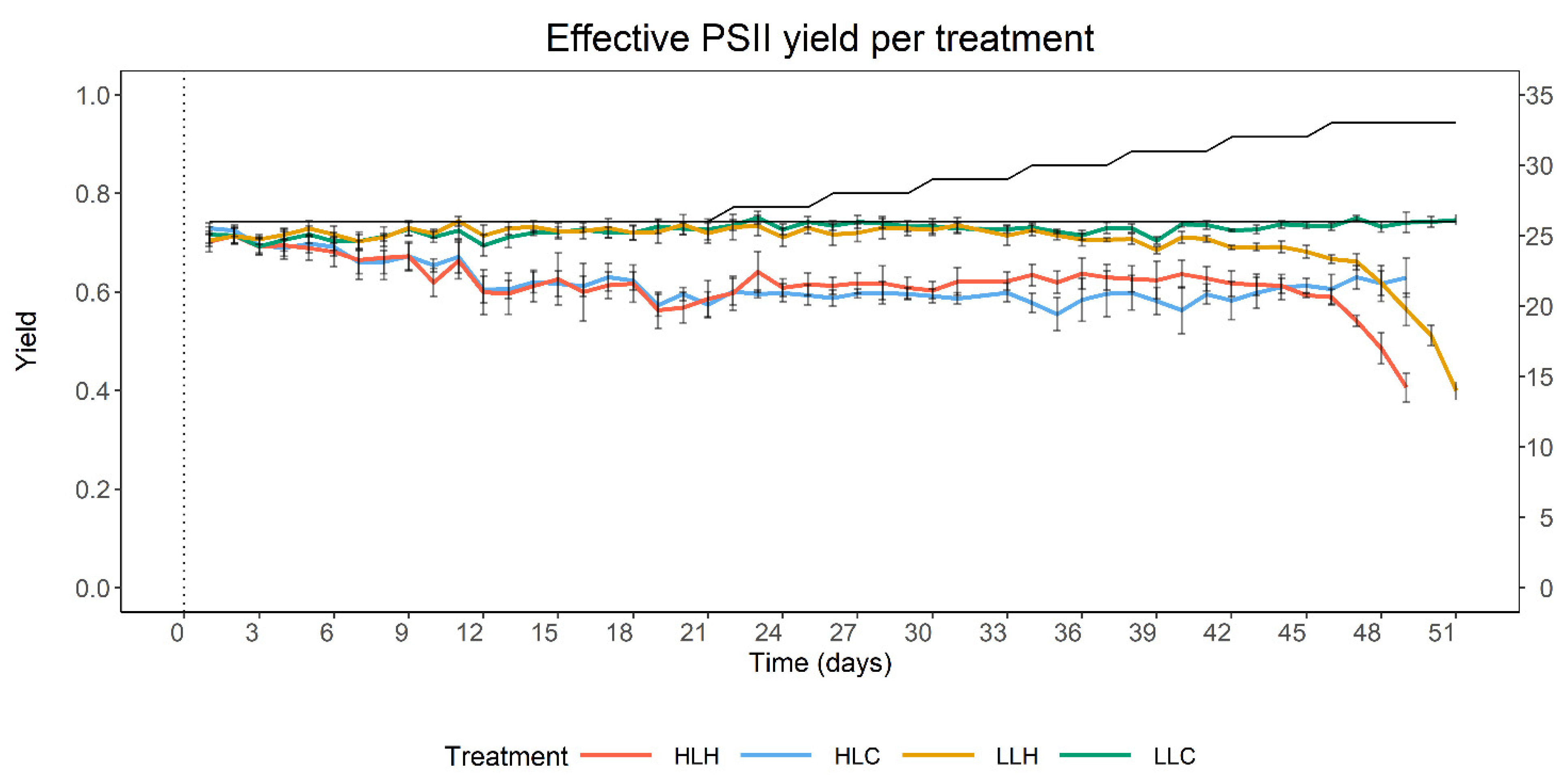
3.1. Effect of Heat and Irradiance on PSII Apparent Yield

Heat and irradiance both affected the apparent photochemical yield of PSII (Figure 2). Immediately after transfer to the experimental system, average values for the apparent PSII yield (ΔF/Fm′) were between 0.69 and 0.73 for all corals. During the acclimation phase, ΔF′/Fm′ for corals in the high irradiance treatments had dropped by approximately 20% to reach stable values around 0.6 at the end of the acclimation phase. In low irradiance treatments, ΔF/Fm′ slightly increased to values around 0.74 during the acclimation phase. These shifts are reflected in the outcomes of the mixed ANOVA that we used to analyze the results of all PSII apparent yield measurements that were done during the entire 51-day experiment: a significant main effect of irradiance on apparent PSII yield was found (Table 1).
After starting the heat ramp on Day 21, ΔF/Fm′ remained stable in the HLH treatment until a temperature of 30 °C was reached. From Day 40 onwards, a slightly decreasing trend in ΔF/Fm′ was observed. In LLH corals, this decreasing trend already commenced at a temperature of 29 °C (Day 34). In both treatments, the mild decreasing trends shifted to a steeper decrease in ΔF/Fm′ when a temperature of 33 °C was reached. This shift started on Day 46 for HLH corals and 1 day later, on Day 47, for LLH corals and usually demarcates the onset of coral bleaching [21,25]. The mixed three-way GLMM (Table 1) confirms that both heat and incubation time significantly affected ΔF/Fm′ and that the two factors interacted significantly. This statistical interaction is reflected by the fact that the decrease in ΔF/Fm′ occurred gradually over time. The three-way interaction factor was also significant (Table 1), indicating that the effect of time on the impact of heat changes under the influence of light. This three-way interaction reflects the observed 1-day delay of the onset of bleaching occurring under the LLH regime when compared to HLH, which is also visible as an interactive trend (p < 0.1) in the effects of temperature and irradiance. To further verify the visually observed tipping points statistically, we compared the regression coefficients (RCs) of the trends in ΔF/Fm′ during the 3 to 4 days before and after the observed tipping points. For both LLH and HLH corals, the post-onset RCs were nearly an order of magnitude higher than the pre-onset RCs, and these differences were highly significant (Table 2, paired t-test), which confirms that the onset of bleaching occurred 1 day earlier in HLH corals than in LLH corals.

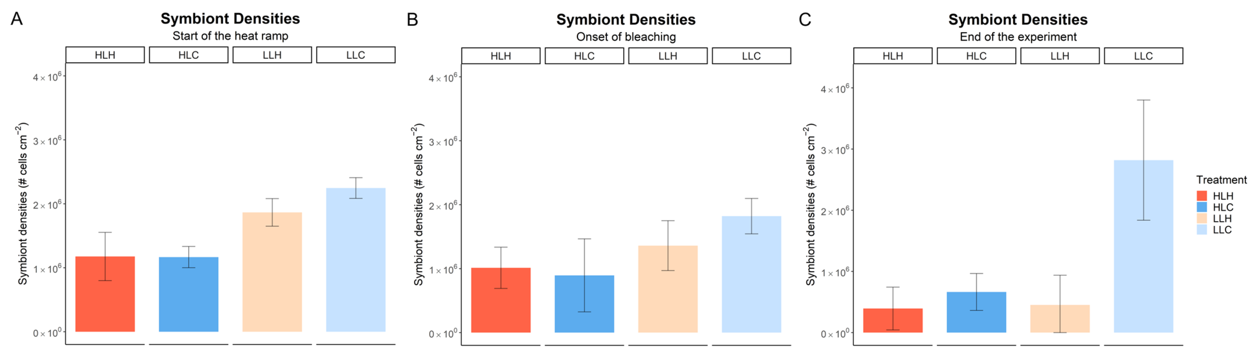
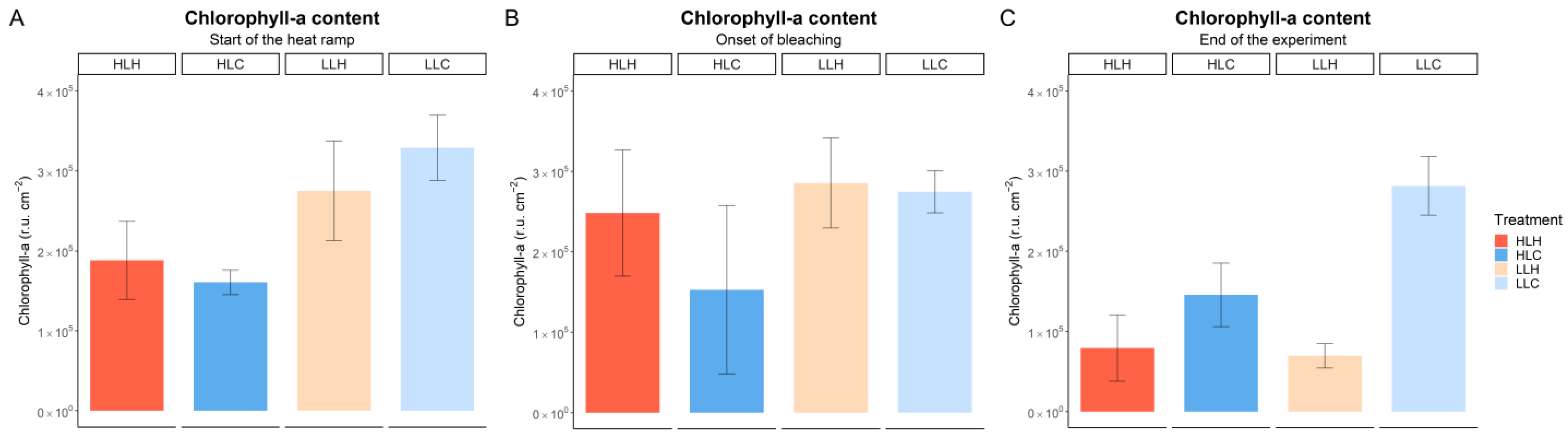
3.2. Effect of Heat and Irradiance on Symbiont Density and Chlorophyll-a Content

After being exposed to the high irradiance regime for 21 days (i.e., before the start of the heat ramp), the HL corals exhibited significantly lower numbers of symbionts per cm2 than corals grown under low irradiance (Figure 3A; Table 3), symbiont densities on average being 43% lower under high irradiance. Correspondingly, the relative chlorophyll-a content per cm2 was also significantly lower (42%) in HL corals compared to LL corals after 21 days of acclimation (Figure 4A; Table 4). Similar trends were observed at the onset of bleaching (Day 47—Figure 3B and Figure 4B): irradiance had a significant effect on both symbiont density and chlorophyll-a content (40% and 28% lower values under high irradiance, respectively). At this timepoint, the effect of heat on these parameters was not significant, and heat did not significantly influence the effect of irradiance (Table 3 and Table 4).
At the end of the experiment, different patterns evolved. Here, heat did affect symbiont densities and chlorophyl-a content in interaction with irradiance (Figure 3C and Figure 4C; Table 3 and Table 4). Heat-exposed corals under high irradiance showed a 41% lower symbiont density and a 46% lower chlorophyll-a content than non-heat-exposed controls. In corals under low irradiance, the differences between heat-exposed and non-heat-exposed corals were more pronounced (84% and 75%, respectively, for symbiont density and chlorophyll-a content).
When expressed as chlorophyll-a content per symbiont cell (Figure S1), data show that chlorophyll-a per cell did not strongly vary among treatments and timepoints.

4. Discussion

In this study, the combined effects of irradiance and heat on the coral–algal symbiosis were studied. Prior to heat stress, corals grown under high irradiance exhibited lower symbiont densities and lower levels of chlorophyll-a per cm2 of coral surface than corals grown under low irradiance. The effective quantum yield of PSII was also lower in corals under high irradiance. These results can be considered as normal photo-acclimation responses. Changes in symbiont densities and chlorophyll-a content can occur within a time frame of a few weeks after translocation of a coral to a position with a different irradiance level [27]. When experimentally relocated from an irradiance regime representing 95% of the irradiance at the water surface to an irradiance level representing 30% of the irradiance at the surface, symbiont densities in colonies of Stylophora pistillata doubled within a time frame of 40 days. Chlorophyll-a content per symbiont cell in these colonies showed a somewhat faster response and increased by 50% within a time frame of a week. These photo-acclimation responses correspond well with the changes in symbiont densities and chlorophyll-a content observed in our study. The lower apparent quantum yield of PSII observed under high irradiance is most likely due to a higher proportion of the incoming irradiance being dissipated as heat through non-photochemical quenching processes (NPQ), which is a mechanism in plants to protect tissue against overexcitation of the photosystems by excess light [28,29]. Most relevant in this respect are the inducible NPQ processes that relate to an increased pH in the lumen of the chloroplast termed qE [29]. Protons that accumulate in the lumen following excess splitting of water in PSII can cause reversible changes to photopigments and other proteins in the reaction center that promote a safe and harmless dissipation of excess excitation energy as heat. Hence, qE is also referred to as “dynamic photoinhibition” in contrast to “chronic photoinhibition”, which causes damage to the photosystems and is likely to be experienced by corals under combined light and heat stress [13]. We conclude that at the end of the acclimation phase, all experimental corals in our experiment represented a healthy condition with respect to their observed photosymbiotic responses.
Heat exposure affected corals grown at low and high irradiance in a comparable way. Under both light regimes, the first signs of inhibition of the photosymbiosis by heat (the point where ΔF/Fm′ starts to show an accelerated decrease—the onset of bleaching) became apparent at a temperature of 33 °C. The fact that symbiont loss occurred after this onset of bleaching suggests that damage to PSII is the root cause of thermal bleaching in symbiotic corals, followed later by a collapse of the symbiosis. This sequence of events contrasts with the frequently mentioned mechanism of expelling healthy symbionts by the coral during bleaching, which was shown to occur in the coral Cyphastrea serailia after exposure to acute heat stress [30]. This discrepancy in the sequence of events indicates that acute heat stress may not be an adequate proxy for naturally occurring summer heat waves that lead to coral bleaching in nature. It must be noted here that different coral species may respond differentially to heat stress.
The predicted synergistic effect of light on thermal bleaching appeared to be moderate. Despite the five-fold higher irradiance, the onset of bleaching started only 1 day earlier in corals grown under high irradiance than in corals grown under low irradiance. This minor contribution of irradiance suggests that the role of photochemically produced reactive oxygen species in augmenting thermal bleaching should not be overemphasized. We conclude that heat-driven damage of PSII proteins [31] rather than oxidative stress caused by impaired photosynthesis [5] is the main driver for bleaching in the studied coral. Some earlier studies corroborate this conclusion [32,33]. Colonies of Acropora millepora exposed to very low light (10 μmol quanta m−2 s−1) showed signs of bleaching earlier than colonies exposed to ambient light [32]. In another study, several species of Acropora were subjected to heat stress in complete darkness [33]. These corals also showed a rapid decrease in photochemical efficiency as a result of exposure to heat, despite the absence of irradiance and hence, photosynthesis. However, it must be noted that these studies were executed either under acute heat stress [33] or by applying a short-term (5 days) heat ramp [32]. As pointed out above, results obtained under acute and/or rapidly increasing heat stress may not fully resemble the response of corals to natural heatwaves. Hence, to conclusively test the oxidative stress hypothesis, future studies should compare the effect of heat on corals in the presence and absence of irradiance while applying a gradual increase in temperature, resembling a natural summer heatwave. Such studies should include irradiance levels higher than those applied in our study. Under irradiance levels resembling the upper few meters of the ocean (i.e., 1000–1500 μmol quanta m−2 s−1), effects of light might be more exacerbated and thus lead to results that are more comparable with earlier experiments [21], in which a much stronger interaction between irradiance and heat was reported than in the current study.
The effect of heat on the photosymbiosis observed in our study likely relates to instability/deformation of structural components of the photosystem. Processes associated with heat-induced damage to the photosystem include denaturation of proteins [34], instability of lipids in the thylakoid membrane [35], and decreased repair rates of structural proteins such as D1 [31]. The latter two processes have a direct connection to photosynthesis, since both lead to an uncoupling of the light and dark reactions in photosynthesis and thus promote the formation of ROS [36]. Since we found only a weak association of heat and light stress, the possible mechanism of heat-induced, irreversible denaturation of photosynthetic proteins is likely at play as well, and thus of high interest for further investigation. If protein denaturation within the symbiont photosystem turns out to be the main driver of coral bleaching, symbiont-specific thermal thresholds for coral bleaching may exist that cannot easily be modulated by other factors. Below these thresholds, long exposure to sublethal temperatures may also lead to bleaching through interactions with other factors such as UVR- and light-mediated oxidative stress. Long-term experimental exposure (i.e., multiple months instead of multiple weeks) to various temperatures should be applied to test these hypotheses, but to date, such studies have been scarce [37]. These future investigations should not only focus on bleaching itself, but also include the potential of the corals to regenerate after the different stages of the bleaching process (e.g., at the onset of bleaching, after the loss of symbionts), which may shed further light on the nature of the mechanisms of thermal bleaching. In addition, long-term incubations can also be used to study the adaptive capacity of the coral holobiont.
It should be noted that conditions in experimental studies such as those reported here often differ from natural circumstances. For example, natural sunlight includes ultraviolet radiation (UVR) that was not present in the spectrum of the artificial lights used in this study. UVR is directly absorbed by PSII and can, as such, cause damage to the photosystem [38]. Indeed, UVR was reported to interact with heat in causing coral bleaching [39]. This detrimental effect of UVR is considered limited to shallow reefs only [40], which corroborates our finding that visible light at moderately high levels is not contributing strongly to thermal bleaching. Nevertheless, multiple studies have shown that in the field, shading can protect corals from thermal bleaching (reviewed in [15]). It is of interest to assess to what extent these observations relate to high levels of UVR rather than high levels of visible light, since these two potential stressors always occur simultaneously in the field. In addition, the beneficial effect of (artificial) shading is considered highly species specific [15]. This specificity may not only relate to differences among the coral host and its symbiotic algal partner, but also to differences in the coral microbiome, which can largely affect the thermotolerance of the coral holobiont [41].
The observed experimental bleaching threshold temperature of 33 °C in this study is consistent with earlier experiments on another morphotype of S. pistillata in our laboratory [22], and corroborates observations from the Gulf of Aqaba (GoA), where the experimental corals originated. The GoA is considered an area where corals have a relatively high thermotolerance [42]. In an experiment that applied a gradual heat ramp similar to the one in the current study, corals obtained from the GoA did not bleach below a temperature of 34 °C, which is 7 degrees higher than the average summer maximum temperature of 27 °C in the GoA [42]. This 7-degree difference contrasts with observations from most other coral reef environments, where bleaching usually occurs when summer maxima are exceeded by only 1 or 2 °C [43]. Likely, the GoA corals inherited their heat tolerance from their source populations that reside further south in the Red Sea and which experience much higher summer maxima [42]. Interestingly, our experimental corals have been kept in captivity for nearly 40 years at a constantly low temperature of 25–27 °C. The fact that these corals still show a high tolerance to heat suggests that host-associated factors contributing to this trait are heritable and conserved. In addition to host factors, the dominant symbiont type in a coral can also strongly determine the thermostability of the symbiosis [44,45]. The dominant symbiont type in our experimental colonies was classified as Symbiodinium sp., which has also been reported to be the dominant symbiont genus in S. pistillata growing at shallow depths (~5 m) in the GoA [46]. Hence, the symbiosis appears to be stable over a period of nearly 40 years in captivity. As such, these corals may represent a population that can be of interest as source material for the restoration of reefs that are more vulnerable to heat stress than those in the GoA.

Supplementary Materials

The following supporting information can be downloaded at: https://www.mdpi.com/article/10.3390/oceans6020032/s1, Figure S1: Relative Chlorophyll-a content per symbiont cell at three designated timepoints in the experiment; Table S1: Shapiro–Wilk test output for normality on effective PSII yield data; Table S2: Levene’s test output for homogeneity of variances on effective PSII yield data; Table S3: Shapiro–Wilk test output for normality on data for symbiont densities; Table S4: Shapiro–Wilk test output for normality on data for relative chlorophyll-a content; Table S5: Levene’s test output for homogeneity of variances on data for symbiont densities; Table S6: Levene’s test output for homogeneity of variances on data for relative chlorophyll-a content; Table S7: ANOVA on symbiont densities—Start of the heat ramp; Table S8: ANOVA on symbiont densities—Onset of bleaching; Table S9: ANOVA on symbiont densities—End of the experiment; Table S10: ANOVA on relative chlorophyll-a content—Start of the heat ramp; Table S11: ANOVA on relative chlorophyll-a content—Onset of bleaching; Table S12: ANOVA on relative chlorophyll-a content—End of the experiment.

Author Contributions

Conceptualization, R.O. and E.v.V.; methodology, all authors; data analysis, E.v.V., D.S.L.G., B.J.A.v.H. and S.O.; investigation, all authors; data curation, R.O.; writing—original draft preparation, R.O. and E.v.V.; writing—review and editing, all authors; visualization, E.v.V. All authors have read and agreed to the published version of the manuscript.

Funding

This research received no external funding.

Institutional Review Board Statement

Not applicable.

Informed Consent Statement

Not applicable.

Data Availability Statement

The raw data supporting the conclusions of this article will be made available by the authors on request.

Acknowledgments

We thank the staff of Carus for technical support. Robbert-Jan Geertsma, Diede Maas, and Tim Wijgerde provided useful advice throughout the study.

Conflicts of Interest

The authors declare no conflicts of interest.

Abbreviations

The following abbreviations are used in this manuscript:
GLMMGeneralized Linear Mixed Model
GoAGulf of Aqaba
ROSReactive Oxygen Species
HLHHigh Light Heat treatment
HLCHigh Light Control treatment
LLHLow Light Heat treatment
LLCLow Light Control treatment

References

  1. Hoegh-Guldberg, O.; Skirving, W.; Dove, S.G.; Spady, B.L.; Norrie, A.; Geiger, E.F.; Liu, G.; De La Cour, J.L.; Manzello, D.P. Coral reefs in peril in a record breaking year. Science 2023, 382, 1238–1240. [Google Scholar] [CrossRef] [PubMed]
  2. Hughes, T.P.; Anderson, K.D.; Connolly, S.R.; Heron, S.F.; Kerry, J.T.; Lough, J.M.; Baum, J.K.; Berumen, M.L.; Bridge, T.C.; Claar, D.C.; et al. Spatial and temporal patterns of mass bleaching of corals in the Anthropocene. Science 2018, 359, 80–83. [Google Scholar] [CrossRef] [PubMed]
  3. Reimer, J.D.; Peixoto, R.S.; Davies, S.W.; Traylor-Knowles, N.; Short, M.L.; Cabral-Tena, R.A.; Burt, J.A.; Pessoa, I.; Banaszak, A.T.; Winters, R.S.; et al. The Fourth Global Coral Bleaching Event: Where do we go from here? Coral Reefs 2024, 43, 1121–1125. [Google Scholar] [CrossRef]
  4. Van Oppen, M.J.H.; Lough, J.M. Introduction: Coral Bleaching–Patterns, Processes, Causes and Consequences. In Coral Bleaching–Patterns, Processes, Causes and Consequences, 2nd ed.; Van Oppen, M.J.H., Lough, J.M., Eds.; Ecological Studies; Springer: Cham, Switzerland, 2018; Volume 233, pp. 1–8. [Google Scholar]
  5. Lesser, M.P. Oxidative stress causes coral bleaching during exposure to elevated temperatures. Coral Reefs 1997, 16, 187–192. [Google Scholar] [CrossRef]
  6. Weis, V.M. Cellular mechanisms of Cnidarian bleaching: Stress causes the collapse of symbiosis. J. Exp. Biol. 2008, 211, 3059–3066. [Google Scholar] [CrossRef]
  7. Warner, M.E.; Fitt, W.K.; Schmidt, G.W. The effects of elevated temperature on the photosynthetic efficiency of zooxanthellae in hospite from four different species of reef coral: A novel approach. Plant Cell Environ. 1996, 19, 291–299. [Google Scholar] [CrossRef]
  8. Nielsen, D.A.; Petrou, K.; Gates, R.D. Coral bleaching from a single cell perspective. ISME J. 2018, 12, 6. [Google Scholar] [CrossRef]
  9. Saragosti, E.; Tchernov, D.; Katsir, A.; Shaked, Y. Extracellular Production and Degradation of Superoxide in the Coral Stylophora pistillata and Cultured Symbiodinium. PLoS ONE 2010, 5, e12508. [Google Scholar] [CrossRef]
  10. Aro, E.M.; Virgin, I.; Andersson, B. Photoinhibition of photosystem II. Inactivation, protein damage and turnover. Biochim. Biophys. Acta Bioenergy 1993, 1143, 113–134. [Google Scholar] [CrossRef]
  11. Lesser, M.P. Phylogenetic signature of light and thermal stress for the endosymbiotic dinoflagellates of corals (Family Symbiodiniaceae). Limnol. Oceanogr. 2019, 64, 1852–1863. [Google Scholar] [CrossRef]
  12. Nanba, O.; Satoh, K. Isolation of a photosystem II reaction center consisting of D-1 andD-2 polypeptides and cytochrome b-559. Proc. Nat. Acad. Sci. USA 1987, 84, 109–112. [Google Scholar] [CrossRef] [PubMed]
  13. Fitt, W.K.; Brown, B.E.; Warner, M.E.; Dunne, R.P. Coral bleaching: Interpretation of thermal tolerance limits and thermal thresholds in tropical corals. Coral Reefs 2001, 20, 51–65. [Google Scholar] [CrossRef]
  14. Sweet, M.E.; Brown, B.E. Coral responses to anthropogenic stress in the twenty-first century: An ecophysiological perspective. Oceanogr. Mar. Biol. Ann. Rev. 2016, 54, 271–314. [Google Scholar]
  15. Tagliafico, A.; Baker, P.; Kelaher, B.; Ellis, S.; Harrison, D. The effects of shade and light on corals in the context of coral bleaching and shading technologies. Front. Mar. Sci. 2022, 9, 919382. [Google Scholar] [CrossRef]
  16. Brown, B.E.; Downs, C.A.; Dunne, R.P.; Gibb, S.W. Exploring the basis of thermotolerance in the reef coral Goniastrea aspera. Mar. Ecol. Prog. Ser. 2002, 242, 119–129. [Google Scholar] [CrossRef]
  17. Dunne, R.P.; Brown, B.E. The influence of solar radiation on bleaching of shallow water reef corals in the Andaman Sea, 1993–1998. Coral Reefs 2001, 20, 201–210. [Google Scholar] [CrossRef]
  18. Bhagooli, R.; Hidaka, M. Comparison of stress susceptibility of in hospite and isolated zooxanthellae among five coral species. J. Exp. Mar. Biol. Ecol. 2003, 291, 181–197. [Google Scholar] [CrossRef]
  19. Brown, B.E.; Dunne, R.P. Solar radiation modulates bleaching and damage protection in a shallow water coral. Mar. Ecol. Prog. Ser. 2008, 362, 99–107. [Google Scholar] [CrossRef]
  20. Fournie, J.W.; Vivian, D.N.; Yee, S.H.; Courtney, L.A.; Barron, M.G. Comparative sensitivity of six scleractinian corals to temperature and solar radiation. Dis. Aquat. Organ. 2012, 99, 85–93. [Google Scholar] [CrossRef]
  21. Lesser, M.P.; Farrell, J.H. Exposure to solar radiation increases damage to both host tissues and algal symbionts of corals during thermal stress. Coral Reefs 2004, 23, 367–377. [Google Scholar] [CrossRef]
  22. Wijgerde, T.; van Ballegooijen, M.; Nijland, R.; van der Loos, L.; Kwadijk, C.; Osinga, R.; Murk, A.J.; Slijkerman, D. Adding insult to injury: Effects of chronic oxybenzone exposure and elevated temperature on two reef-building corals. Sci. Total Environ. 2020, 733, 139030. [Google Scholar] [CrossRef] [PubMed]
  23. Hume, B.C.C.; Ziegler, M.; Poulain, J.; Pochon, X.; Romac, S.; Boissin, E.; de Vargas, C.; Planes, S.; Wincker, P.; Voolstra, C.R. An improved primer set and amplification protocol with increased specificity and sensitivity targeting the Symbiodinium ITS2 region. PeerJ 2018, 6, e4816. [Google Scholar] [CrossRef] [PubMed]
  24. Wiedenmann, J.; D’Angelo, C.; Smith, E.G.; Hunt, A.N.; Legiret, F.E.; Postle, A.D.; Achterberg, E.P. Nutrient enrichment can increase the susceptibility of reef corals to bleaching. Nat. Clim. Change 2013, 3, 160–164. [Google Scholar] [CrossRef]
  25. Fernandes de Barros Marangoni, L.; Ferrier-Pagès, C.; Rottier, C.; Bianchini, A.; Grover, R. Unravelling the different causes of nitrate and ammonium effects on coral bleaching. Sci. Rep. 2020, 10, 11975. [Google Scholar] [CrossRef]
  26. Tilstra, A.; Wijgerde, T.; Dini-Andreote, F.; Eriksson, B.K.; Falcao Salles, J.; Pen, I.; Osinga, R.; Wild, C. Light induced intraspecific variability in response to thermal stress in the hard coral Stylophora pistillata. PeerJ 2017, 5, e3802. [Google Scholar] [CrossRef]
  27. Titlyanov, E.A.; Titlyanova, E.V.; Yamazato, K.; Van Woesik, R. Photo-acclimation dynamics of the coral Stylophora pistillata to low and extremely low light. J. Exp. Mar. Biol. Ecol. 2001, 263, 211–225. [Google Scholar] [CrossRef]
  28. Enriquez, S.; Borowitzka, M. The Use of the Fluorescence Signal in Studies of Seagrasses and Macroalgae. In Chlorophyll a Fluorescence in Aquatic Sciences: Methods and Applications; Sugget, D.J., Prasil, O., Borowitzka, M., Eds.; Springer: Dordrecht, The Netherlands, 2009; Developments in Applied Phycology 4; pp. 187–208. [Google Scholar]
  29. Niyogi, K.K. Safety valves for photosynthesis. Curr. Opin. Plant Biol. 2000, 3, 455–460. [Google Scholar] [CrossRef]
  30. Ralph, P.J.; Gademann, R.; Larkum, A.W.D. Zooxanthellae expelled from bleached corals at 33_C are photosynthetically competent. Mar. Ecol. Prog. Ser. 2001, 220, 163–168. [Google Scholar] [CrossRef]
  31. Warner, M.E.; Fitt, W.K.; Schmidt, G.W. Damage to Photosystem II in symbiotic dinoflagellates: A determinant of coral bleaching. Proc. Nat. Acad. Sci. USA 1999, 96, 8007–8012. [Google Scholar] [CrossRef]
  32. Rosic, N.; Rémond, C.; Mello-Athayde, M.A. Differential impact of heat stress on reef-building corals under different light conditions. Mar. Environ. Res. 2020, 158, 104947. [Google Scholar] [CrossRef]
  33. Tolleter, D.; Seneca, F.O.; DeNofrio, J.C.; Krediet, C.J.; Palumbi, S.R.; Pringle, J.R.; Grossman, A.R. Coral Bleaching Independent of Photosynthetic Activity. Curr. Biol. 2013, 23, 1782–1786. [Google Scholar] [CrossRef] [PubMed]
  34. Yamane, Y.; Kashino, Y.; Koike, H.; Satoh, K. Effects of high temperatures on the photosynthetic systems in spinach: Oxygen-evolving activities, fluorescence characteristics and the denaturation process. Photosynth. Res. 1998, 57, 51–59. [Google Scholar] [CrossRef]
  35. Tchernov, D.; Gorbunov, M.Y.; de Vargas, C.; Yadav, S.N.; Milligan, A.J.; Häggblom, M.; Falkowski, P.G. Membrane lipids of symbiotic algae are diagnostic of sensitivity to thermal bleaching in corals. Proc. Nat. Acad. Sci. USA 2004, 101, 13531–13535. [Google Scholar] [CrossRef] [PubMed]
  36. Oakley, C.A.; Davy, S.K. Cell biology of coral bleaching. In Coral Bleaching–Patterns, Processes, Causes and Consequences, 2nd ed.; Van Oppen, M.J.H., Lough, J.M., Eds.; Ecological Studies; Springer: Cham, Switzerland, 2018; Volume 233, pp. 189–211. [Google Scholar]
  37. McLachlan, R.H.; Price, J.T.; Solomon, S.L.; Grottoli, A.G. Thirty years of coral heat-stress experiments: A review of methods. Coral Reefs 2020, 39, 885–902. [Google Scholar] [CrossRef]
  38. Keren, N.; Krieger-Liszkay, A. Photoinhibition: Molecular mechanisms and physiological significance. Physiol. Plant. 2011, 141, 1–5. [Google Scholar] [CrossRef]
  39. Courtial, L.; Roberty, S.; Shick, J.M.; Houlbrèque, F.; Ferrier-Pagès, C. Interactive Effects of Ultraviolet Radiation and Thermal Stress on Two Reef-Building Corals. Limnol. Oceanogr. 2017, 62, 1000–1013. [Google Scholar] [CrossRef]
  40. Banaszak, A.T.; Lesser, M.P. Effects of Solar Ultraviolet Radiation on Coral Reef Organisms. Photochem. Photobiol. Sci. 2009, 8, 1276–1294. [Google Scholar] [CrossRef]
  41. Santoro, E.P.; Cárdenas, A.; Villela, H.D.M.; Vilela, C.L.S.; Ghizelini, A.M.; Duarte, G.A.S.; Perna, G.; Saraiva, J.P.; Thomas, T.; Voolstra, C.R.; et al. Inherent differential microbial assemblages and functions associated with corals exhibiting different thermal phenotypes. Sci. Adv. 2025, 11, eadq2583. [Google Scholar] [CrossRef]
  42. Fine, M.; Gildor, H.; Genin, A. A coral reef refuge in the Red Sea. Glob. Change Biol. 2013, 19, 3640–3647. [Google Scholar] [CrossRef]
  43. Strong, A.; Liu, G.; Skirving, W.; Eakin, C.M. NOAA’s Coral Reef Watch program from satellite observations. Ann. GIS 2011, 17, 83–92. [Google Scholar] [CrossRef]
  44. Bhagooli, R.; Hidaka, M. Photoinhibition, bleaching susceptibility and mortality in two scleractinian corals, Platygyra ryukyuensis and Stylophora pistillata, in response to thermal and light stresses. Comp. Biochem. Physiol. A Mol. Integr. Physiol. 2004, 137, 547–555. [Google Scholar] [CrossRef] [PubMed]
  45. Silverstein, R.N.; Cunning, R.; Baker, A.C. Change in algal symbiont communities after bleaching, not prior heat exposure, increases heat tolerance of reef corals. Glob. Change Biol. 2015, 21, 236–249. [Google Scholar] [CrossRef] [PubMed]
  46. Lampert-Karako, S.; Stambler, N.; Katcoff, D.J.; Achituv, Y.; Dubinsky, Z. Effects of depth and eutrophication on the zooxanthella clades of Stylophora pistillata from the Gulf of Eilat (Red Sea). Aquat. Cons. 2008, 18, 1039–1045. [Google Scholar] [CrossRef]
Figure 1. Overview of the experimental set-up consisting of two basins containing eight aquaria each. One basin was kept at the control temperature of 26 °C, and the other basin experienced a heatwave reaching a temperature of 33 °C. Half of the aquaria in each basin experienced a low light intensity (100 μmol quanta m−2 s−1) and the other half a high light intensity (500 μmol quanta m−2 s−1). Each aquarium had an individual waterflow and circulation pump. Both basins had one heater to control the temperature and two circulation pumps to keep the temperature throughout the basin constant. A cooler was used to keep temperature fluctuations minimal. Each aquarium was covered with a lid to prevent evaporation.
Oceans 06 00032 g001
Figure 2. Development of the effective photochemical yield of PSII (ΔF/Fm′) of the experimental corals over time. Error bars indicate standard deviations (n = 4). Colored lines represent the four treatments (HLH = high light heat, HLC = high light control, LLH = low light heat, LLC = low light control). Black lines indicate the ambient temperature over time in the heatwave treatments (increasing line) and the control treatments (flat line).
Oceans 06 00032 g002
Figure 3. Symbiont densities (# cells per cm2 coral surface) in the tissue of the experimental corals at three designated timepoints in the experiment. (A) Start of the heat ramp. (B) Onset of bleaching. (C) End of the experiment. Error bars indicate standard deviations (n = 4). Color coding as in Figure 2.
Oceans 06 00032 g003
Figure 4. Relative chlorophyll-a content (r.u. per cm2 coral surface; r.u. = relative units) in the tissue of the experimental corals at three designated timepoints in the experiment. (A) Start of the heat ramp. (B) Onset of bleaching. (C) End of the experiment. Error bars indicate standard deviations (n = 4). Color coding as in Figure 2.
Oceans 06 00032 g004
Table 1. Output of the generalized linear mixed effect model on data for the apparent photochemical yield of PSII (yield ~ time × temperature treatment × irradiance treatment + (1/tank)). Asterisks Indicate level of significance (** <0.01; *** <0.001).
GLMMEstimatestd. ErrorDFt-Valuep-Value
(intercept)7.11 × 10−19.59 × 10−32.07 × 10−174.12<2 × 10⁻¹⁶ ***
time5.87 × 10−41.82 × 10−47.64 × 10−23.220.00134 **
temperature4.56 × 10−21.36 × 10−22.07 × 10−13.360.00301 **
irradiance−4.32 × 10−21.36 × 10−22.10 × 10⁻¹−3.180.00453 **
time × irradiance−2.79 × 10−32.58 × 10−47.64 × 10⁻²−10.83<2 × 10⁻¹⁶ ***
time × temperature2.57 × 10−32.66 × 10−47.64 × 10−2−9.64<2 × 10⁻¹⁶ ***
irradiance × temperature3.83 × 10−21.92 × 10−22.10 × 10−1−1.990.05981
time × temperature × irradiance−2.52 × 10−33.76 × 10−47.64 × 10−26.704.06 × 10−11 ***
Table 2. Regression coefficients (RC ± standard deviation) of the trends in ΔF/Fm′ during the three (HLH corals) and four (LLH corals) days prior and after the presumed onset of bleaching. p-values show the outcome of the comparison between pre-onset and post-onset RCs (paired t-test).
TreatmentRC Pre-OnsetRC Post-Onsetp-Value
LLH0.007 ± 0.0040.063 ± 0.003>0.0000001
HLH0.01 ± 0.0040.061 ± 0.008>0.00001
Table 3. p-values for one-way ANOVA (first sampling point) and two-way ANOVA (second and third sampling points) on data for symbiont density. Asterisks indicate level of significance (* <0.05; ** <0.01; *** <0.001). Full outcomes of the ANOVAs can be found in Supplemental Information (Tables S7–S9).
Sampling Point:Start of Heat RampOnset of BleachingEnd of Experiment
irradiance1.27 × 10−5 ***0.0174 *0.0092 **
temperaturex0.71750.0001 ***
irradiance × temperaturex0.21410.0263 *
Table 4. p-values for one-way ANOVA (first sampling point) and two-way ANOVA (second and third sampling points) on data for relative chlorophyll-a content. Asterisks indicate level of significance (* < 0.05; ** <0.01; *** <0.001). Full outcomes of the ANOVAs can be found in Supplemental Information (Tables S10–S12).
Sampling Point:Start of Heat RampOnset of BleachingEnd of Experiment
irradiance9.95 × 10−5 ***0.0481 *3.82 × 10−6 ***
temperaturex0.16710.00344 **
irradiance × temperaturex0.26530.00128 **
Disclaimer/Publisher’s Note: The statements, opinions and data contained in all publications are solely those of the individual author(s) and contributor(s) and not of MDPI and/or the editor(s). MDPI and/or the editor(s) disclaim responsibility for any injury to people or property resulting from any ideas, methods, instructions or products referred to in the content.

