Article

Understanding Fish Larvae’s Feeding Biology to Improve Aquaculture Feeding Protocols

by
Laura Ribeiro
1,*,
François Noel Hubert
2,
Vera Rodrigues
3,
Carlos Rojas-Garcia
4 and
Maria Teresa Dinis
5
1
IPMA—Portuguese Institute for Sea and Atmosphere/EPPO—Aquaculture Research Station, Av. Parque Natural da Ria Formosa, s/n, 8700-194 Olhão, Portugal
2
Bivalvia-Mariscos da Formosa, Lda., Piscicultura da Meia-Légua Cova da Onça, Caixa Postal 491A, 8700-177 Olhão, Portugal
3
SPAROS Lda., Área Empresarial de Marim, Lote C, 8700-221 Olhão, Portugal
4
Department of Biological Sciences, University of Bergen, 5008 Bergen, Norway
5
Center of Marine Sciences (CCMAR), Universidade do Algarve, Campus de Gambelas, 8005-139 Faro, Portugal
*
Author to whom correspondence should be addressed.
Oceans 2022, 3(1), 94-113; https://doi.org/10.3390/oceans3010009
Submission received: 30 May 2021 / Revised: 24 February 2022 / Accepted: 7 March 2022 / Published: 21 March 2022
(This article belongs to the Special Issue Fish Larval Biology and Ecology)

Abstract

:
Knowing that food ingestion and digestion are processes under neuroendocrine regulation, it is important to understand how fish larvae regulate these processes, when the digestive system itself is under development. This study aimed to analyze how gilthead seabream (Sparus aurata) larval feeding incidence and intensity were affected by time, light, and water temperature, through the analysis of gut content and how this affected cholecystokinin (CCK) content. Three short-term experiments were done: (A) 27 and 42 days post-hatch (DPH) larvae were analyzed at different intervals for 80 min, after feeding; (B) 38 and 48 DPH larvae were kept under different light intensities (0, 400, 1000 lux) for 30 min; (C) 41 DPH larvae were kept at 17, 19, 25 °C for 30 min. Feeding incidence below 100% was observed for 27 DPH larvae, and for 38 DPH larvae fed at 0 lux. Feeding intensity was several fold higher at older ages. However, the number of prey in the gut was lower at 0 lux, and higher at 25 °C. Overall, no clear pattern was observed for CCK, but at older ages increasing gut content resulted in higher CCK content. In conclusion, until 38 DPH light is important for gilthead sea bream larval feeding ability, and maturing functionality of CCK was only observed at later stages.

Graphical Abstract

1. Introduction

Food availability and efficient digestion provide the necessary energy that enables marine fish larvae to grow and develop faster, increasing the chances of survival [1,2]. The larval period is a transitory period in which fish display high growth rates concomitant with systems differentiation (e.g., organs, tissues), that will enable organisms to undertake the transition from larva to juvenile [3]. The vulnerability during this period is high, due to small larval size, incomplete development, and low energy reserves, among many other factors [4].
Food ingestion and digestion involve several steps that range from food perception and capture by fish larvae, to ingestion and digestion, and absorption of nutrients for energy supply to support organism metabolic needs [5]. Neuroendocrine regulatory mechanisms are essential for the appropriate coordination and integration of these steps, considering the physiological and developmental stages of the organisms, as well as the environmental conditions in which animals find themselves [6,7]. Still, scarce information exists about how these processes are controlled by marine fish larvae [8]. Marine fish larvae differ in digestive strategies when compared to adults. Most marine fish larvae rely on pancreatic digestive enzymes for digestion, the intestinal epithelium is not yet mature, and stomach acidic secretion is also absent [5,9]. These digestive capabilities are reached during the first month of development, varying with species and influenced by abiotic and biotic conditions. Therefore, it is expected that the regulation of the digestive process for fish larvae differs from adults.
In vertebrates, the presence of food in the gastrointestinal tract stimulates the release of several neuroendocrine peptides (e.g., cholecystokinin, gastrin releasing peptide) belonging to the gut–brain regulatory axis, where some of these neuroendocrine peptides may also act as short-term satiety factors, signaling back to the brain to reduce further food intake [10]. Food intake, gut transit, and evacuation are key aspects for the regulation of digestive functions. Knowledge on the factors that control food intake in marine fish larvae is still very incomplete [11], since it is very challenging to work with these early stages. The small size and the difficulty in estimating the food intake, in combination with the diversity of neuroendocrine responses, render the results very difficult to compare among studies and obtain robust conclusions [12].
Light and temperature are external factors known to modulate food intake during larval development [13]. High light intensities (1000–3000 lux) at the water surface are normally recommended for larval rearing of gilthead seabream (Sparus aurata) and seabass (Dicentrachus labrax) until 25 DPH, decreasing thereafter to 500–1000 lux [14]. During the planktivorous phase, marine fish larvae are visual diurnal feeders [15]. Several studies reported that low levels of light reduce food acquisition of fish larvae [16,17,18,19]. Temperature affects multiple metabolic processes such as energy allocation, growth, food intake, digestive function, enzyme activities, or even swimming activity [20,21,22]. Therefore, it is expected that affecting food intake by variation in abiotic factors, such as light and temperature, will cause interference with the neuroendocrine regulation of the digestive process.
The neuropeptide hormone cholecystokinin (CCK) controls gastrointestinal digestion in vertebrates [23], by stimulating pancreatic enzyme secretions, pancreatic growth, gall bladder contractions, and satiety, among other functions [23,24,25]. These functions and the CCK molecules and pathways involved in the regulation of digestion in mammals have been described to be similar among fishes, indicating high conservation of these mechanisms in vertebrates [26]. Still, specific responses in fish may differ from mammals [27], and research to understand endocrine appetite-controlling systems in fish has increased [28]. In adult fish, CCK has an inhibitor (anorexigenic) function on the appetite [7,12,28], promoting decreased food intake when satiated [12]. Research in this area of food intake—appetite of marine fish—has been limited due to biological and technical challenges (e.g., accurate determination of food intake, the use of individual larvae) [28].
For the optimization of feeding strategies and diet formulation during larval rearing of marine fish, the role of CCK in the digestive function and processing capacity of fish larvae has been the subject of several studies. Some studies described the spatio-temporal expression of CCK, both mRNA and the CCK peptide, along the digestive tract [29,30,31,32], identifying the presence of a few CCK cells a few days after mouth opening that increased with fish development. The establishment of a quantification method for CCK in larvae [33] contributed to the in-depth knowledge on the CCK physiological responses [34,35] and contents in different tissues [34]. This technique was used to analyze CCK physiological response triggered by protein-rich solutions [36], dietary levels of protein quality [37], and CCK daily pattern variation [8,38].
Here, we investigated how food intake was affected by the post-feeding time, light intensity, and temperature at certain larval ages, and how the amount of food in the gut may affect CCK levels in fish larvae. We hypothesize that larval development, higher levels of light, and higher temperatures will result in higher food intake, thus triggering a response in CCK production. We used gilthead seabream larvae as our model organism. This sparid species occurs throughout the North East Atlantic Ocean and the Mediterranean Sea, and it can be found in both marine and brackish water environments such as coastal lagoons and estuarine areas, particularly during the initial stages of its life cycle [39]. It has important economic value both within fisheries and aquaculture [40]. The larval stages have been widely studied to establish larval rearing protocols, the physiological and energetic aspects of growth and development, and particularly the ontogeny of the digestive system and functionality until the juvenile stage [41,42,43,44,45,46,47]. For this study, we considered 19–20 °C and under 1000 lux light intensity at the water surface as normal rearing conditions [14]. In addition, fish larvae should exhibit a level of maturation of digestive functions and robustness to support the experimental conditions. Therefore, the period selected for study was between 25 DPH, when maturation of important digestive functions has been achieved, and the juvenile stage around 1 g wet weight (100–120 DPH) [48]. Understanding the factors affecting marine larval fish feeding behavior and digestive processes may help in optimizing feeding protocols for commercial fish species, while providing insights on feeding ecology to improve the comprehension of data collected in the wild.

2. Materials and Methods

2.1. Origin and Husbandry Conditions of Fish Larvae

Fish eggs were obtained by artificial spawning from gilthead seabream breeders adapted to captivity at Viveiros Vila-Nova fish farm (Vila Nova de Milfontes, Portugal) and transported to the University of Algarve. Two spawns were used for the different trials (A and B). Identical quantities of fish eggs (60 g of viable eggs) from each spawn were incubated in 70 L cylindro-conical fiberglass tanks, and upon hatching larvae were distributed in two 200 L cylindro-conical fiberglass tanks (density 120 larvae L−1), connected to a recirculation water system, equipped with UV lights for continuous disinfection. Fish larvae were reared following the standardized procedures of the Aquaculture Research Group from the University of Algarve [47], until 50 DPH. Fish larvae used in the short-term trials were collected from these stock-rearing tanks. Water parameters were monitored daily for temperature (20 ± 0.5 °C), oxygen saturation (>90%), and salinity (35.5 ± 0.5 g L−1), while ammonia and nitrates were monitored on a weekly basis and kept under 0.024 ppm [46]. Photoperiod was set at 14 light:10 dark and light intensity was 1000 lux.
The fish larval feeding protocol consisted of: enriched rotifers DHA Protein Selco (INVE Aquaculture, Dendermonde, Belgium) from 4 to 19 DPH, maintained at a density of 5 rotifers mL−1; newly hatched Artemia nauplii (BE 480, INVE Aquaculture, Belgium) from 15 to 25 DPH, at increasing density from 1 to 10 Artemia nauplii mL−1; and 24 h Artemia metanauplii (RH Artemia cysts, INVE Aquaculture, Belgium) enriched with Super Selco (INVE Aquaculture, Belgium) from 23 DPH onwards, at increasing prey density from 0.5 to 10 Artemia metanauplii mL−1. Fish larvae were fed four times a day (9:00, 12:00, 15:00, 18:00).
At hatching, fish larvae from both spawns exhibited similar dry weight (around 0.05 mg) and during development dry weight growth curves were similar (DWA = 0.0308e0.113x, r2 = 0.955; DWB = 0.0381e0.1001x, r2 = 0.996). Spawn was therefore not used as a variable or random factor. Fish larval growth (total length and dry weight) (Table 1; Figure S1) was determined for individual seabream larvae (n = 30) sampled from stock rearing tanks, on the same days of the experiments. Due to technical problems, the factor temperature was only tested for spawn A.

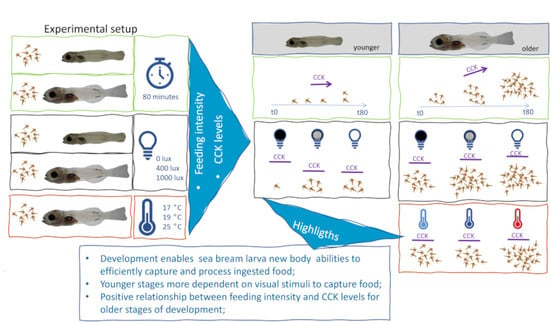
2.2. Experimental Conditions

All animal handling and experimentation was directed by trained scientists certified by the Portuguese National Directory for Veterinary Medicine. The experimental design respected all the procedures to safeguard animal welfare and ensure and extend the application of the 3Rs (reduce, refine, replace). The number of animal’s sacrificed, were the minimum to needed to obtain result with statistical significance.
The experimental set-up for the different trials is described in Figure 1. A total of five experiments were conducted in an acclimatized and lightproof room, and all the treatments were done in triplicate. Experimental units consisted in quadrangular plastic recipients (13 cm wide × 23 cm height), painted with an opaque sandy color, identical to stock rearing tanks to ensure similar feeding behavior, and filled with 3 L of UV-sterilized sea water (salinity: 35.5 ± 0.5 g L−1). Each unit had gentle aeration provided by air stones (aeration increased with development). These experimental units were maintained in water bath trays (dark gray color) to maintain similar water temperature conditions among units (except for the temperature trial). Two linear fluorescent tubes illuminated the whole experimental set-up almost vertically and illuminance at the water surface of the experimental units ranged from 990 to 1030 lux (TES-1335 Light Meter, Taipei, Taiwan). The experimental conditions for light intensity and water temperature trials were tested before fish larvae were transferred. To provide an acclimation period to the experimental conditions and minimize potential effects on feeding behavior, fish larvae were transferred to the experimental units the day before the experiments. At this point, gilthead seabream larvae were collected from the stock rearing tanks, before the last meal, counted, and distributed in the experimental units. The larvae were fasted overnight for a period of about 18 h. On the day of the trial, water parameters were monitored and larvae observed to confirm that no mortality occurred during the night, except for units with limited light or no light, where just after the trial it was possible to monitor those parameters. For all the trials, no mortality was observed.
On the day of the trial, live food was distributed sequentially, with a 5–7 min delay for the different conditions to ensure that sampling was done at the established time after feeding.

2.2.1. Trial 1—Feeding Ability over Time

Larval food ingestion in gilthead seabream was studied at 27 and 42 DPH by quantifying the gut content for a period of 80 min, after being fed. During this period, fish larvae were sampled at 10, 20, 30, 40, and 80 min (total of 15 experimental units). Three additional units (t0) were used to confirm that fish larvae had empty digestive tracts at the beginning of the trial. In total, 540 larvae were used for each age (n = 30 larvae/experimental unit). The fish larvae were fed by hand with Artemia nauplii or enriched Artemia metanauplii, respectively, for 27 DPH and for 42 DPH larvae, at a density of 6 prey mL−1. Temperature was 18.8 ± 0.1 °C and 17.8 ± 0.1 °C, for 27 and 42 DPH larvae, respectively, and oxygen concentration was maintained above 90% air saturation with air stones.

2.2.2. Trial 2—Light Intensity Assay

To assess the effect of light intensity on gilthead seabream feeding ability at 38 and 49 DPH, three light intensities were studied. Several light measurements were taken on the top of the experimental units, before fish larvae were transferred: (i) 1000 lux—values obtained at the water surface from two fluorescent bulbs placed 1 m above the tanks (values ranged between 990 and 1030 lux 9; (ii) 400 lux—light intensity at the water surface was reduced by using a 15 × 15 mm plastic mesh net to cover the experimental units (values ranged between 380 and 390 lux); (iii) 0 lux—experimental units were covered on the top and sides with thick black plastic (doubled sheet) to simulate dark conditions (values ranged between 0.03 and 0.06 lux). Feeding ability for each light condition was analyzed at 10 and 30 min after the meal, resulting in six experimental units for each light condition. In total, 540 gilthead seabream larvae were used (n = 30 in each experimental unit). Gilthead seabream larvae were fed with enriched Artemia instar II at a density of 6 and 10 prey mL−1 at 38 and 49 DPH in each experimental unit. Light conditions were kept constant during trials, so larvae in the covered experimental units were fed through a small tube using a syringe (S2). Light intensity was measured at the water surface with a lux meter (TES-1335 Light Meter). Water temperature was maintained at 18 ± 0.1 °C, and oxygen saturation was maintained above 90%. Gilthead seabream larvae were sampled at 10 and 30 min after being fed, and fish larvae were sampled for gut content analysis.

2.2.3. Trial 3—Temperature

The effect of temperature on feeding ability was studied in gilthead seabream larvae aged 41 DPH. Three temperatures were used, 17 °C, 19 °C, and 25 °C, and feeding ability was assessed at 10 and 30 min after feeding the fish larvae. Six experimental units were used for each temperature, and within a treatment, three units for each time. In total, 18 experimental units were used, and a total of 990 gilthead seabream larvae were distributed across the units (n = 55 larvae). Gilthead seabream larvae from each experimental unit were fed with enriched Artemia metanauplii at 6 prey mL−1. Experimental units for each temperature were grouped in the same tray due to water bath temperature conditions. Room temperature was 17 °C (17.4 ± 0.1 °C, n = 6), being the lowest temperature studied, and the other water baths were warmed up with thermostats up to 19 °C (18.9 ± 0.1 °C, n = 6) and 25 °C (25.2 ± 0.1 °C, n = 6). The oxygen concentration was kept around 80% air saturation (82.2 ± 6%, n = 18). Within each tray, three of the six containers were sampled 10 min after meal delivery, while the remaining three were sampled 30 min following meal delivery.

2.3. Sampling

Fish larvae were sampled from the experimental units by gently pouring the content of each unit into a sieve (250 µm) submersed in water, which was then immediately transferred to a 0.025% MS222 solution (Sigma-Aldrich, Darmstadt, Germany) to anaesthetize the larvae and avoid gut evacuation. Larvae sampled for gut content quantification (n = 15) were preserved in a 4% buffered formaldehyde solution. Larvae sampled for CCK quantification (n = 5) were thoroughly rinsed with distilled water, flash frozen in liquid nitrogen, and stored at −80 °C until analysis. In the temperature trial, a pool of fish larvae (n = 30) was additionally sampled for determination of digestive enzyme activities.

2.4. Analytical

2.4.1. Dry Weight and Total Length

Fish larvae (n = 30) collected from 200 L stock rearing tanks were photographed under a binocular microscope (ZEISS Stemi 2000-C, Zeiss, Jena, Germany) and then rinsed with distilled water, flash frozen in liquid nitrogen, and stored at −80 °C for later freeze drying. Total length was determined from the digital images using ImageJ (Bethesda, MD, USA). Larval dry weight was determined by weighing the freeze-dried larvae on a Sartorius microbalance (±0.001 mg).

2.4.2. Gut Content

For gut content quantification, larvae were dissected under a binocular microscope (ZEISS Stemi 2000-C, Zeiss, Jena, Germany), and the number of prey in the whole gut was counted. For all trials, 15 larvae were used from each experimental unit, giving a total of 45 larvae for each treatment. Fish larva feeding ability was expressed by feeding incidence (FInc) and feeding intensity (FI). FInc represents the percentage of larvae with food in the gut over the total number of larvae analyzed [49]. FI refers to the number of prey quantified in each larva’s gut [50].

2.4.3. CCK Quantification

CCK was quantified through radioimmunoassay kit EURIA-CCK (EuroDiagnostica, Malmö, Sweden) on the body portion of the larvae (head segment discarded) [33]. CCK was extracted with absolute ethanol for 1 h from freeze-dried larvae. Then, samples were centrifuged at 6000× g for 30 min at 4 °C. The supernatant was evaporated until dry using a speed-vac to obtain CCK extracts, and then resuspend with a buffer solution. At this point, samples were ready to be treated according to the manufacturer’s protocol. When performing this assay, specific internal and external controls were carried out.
CCK levels were calculated by standard curve interpolation with seven known concentrations (range: 0 to 25 ρM). A rabbit antiserum to synthetic sulfated CCK 26–33 was added to test tubes which were incubated at 2–8 °C for 46–50 h. After, CCK labeled with iodine (125I-CCK) was added to the tubes and incubated for 94–96 h at 2–8 °C. A double solid phase antibody was then added to the samples and incubated for 1 h at 2–8 °C, followed by a 20 min centrifugation at 3000× g at 4 °C, and then the supernatant was decanted. Samples’ radioactivity was measured using a 2470 WIZARD automatic gamma counter (PerkinElmer, Waltham, MA, USA). CCK was determined individually in 3 larvae from each experimental unit. Results were expressed as ρmol larva−1.

2.4.4. Digestive Enzymes

The activity of digestive enzymes was determined as described for Senegalese sole (Solea senegalensis) [51]. Briefly, a pool of larvae was homogenized for 30 s in 15 volumes (w/v) of cold distilled water. Afterwards, samples were centrifuged (Sigma 4K11, Osterode am Harz, Germany) at 3300× g and 4 °C for 3 min, and the supernatant was sonicated for 10 s (Ultrasonic Homogenizer 4710 Series, Cole Parmer, Cambridgeshire, UK). Trypsin activity was measured using Nα-Benzoyl-DL-arginine-p-nitroanilide (BAPNA) as substrate [52]. Amylase activity was measured using starch as substrate [53]. Alkaline phosphatase activity was measured using p-nitrophenylphosphate (pNPP) as substrate [54]. Leucine aminopeptidase N activity was measured using leucine-nitroanilide as substrate [55]. Enzymatic activities were calculated as substrate μmoles hydrolyzed per minute at 25 °C for trypsin, and 37 °C for alkaline phosphatase and leucine aminopeptidase. Amylase activity was calculated as the equivalent enzymatic activity to hydrolyze 1 mg of starch in 30 min at 37 °C. Enzymatic activities were expressed as total activity, mU larva−1, since analyses were performed on fed larvae, as explained previously. All reagents were of analytical grade and purchased from Sigma-Aldrich (Barcelona, Spain).

2.5. Data Analysis

Descriptive statistics (mean, median, and 25th and 75th percentiles) were calculated to characterize the dataset within each trial. The FIcn was below 100% only for 27 DPH and 38 DPH, respectively, for time of feeding and light intensity short trials. The obtained percentages were transformed by arcsine transformation [56] for homogenization and compared with one-way ANOVA. The normality and homoscedasticity of the different data sets were analyzed for the parameters studied (FI, CCK, and digestive enzymes), using the Kolmogorov–Smirnov test and Levene test. FI did not meet the normality assumption, therefore comparison of these parameters in the different trial conditions was carried out using a non-parametric Kruskal–Wallis test, followed by paired comparisons using a Mann–Whitney U test between different experimental conditions (Table S1) when significant differences were observed. Normality and variance homoscedasticity (Levene test) assumptions were analyzed for CCK and digestive enzyme activities. Variables were transformed using log10 (X + 1) when assumptions were not met. One-way ANOVA was used to compare CCK levels over time within each age, and to compare the values of CCK at each time for both ages, followed by a Tukey post hoc test if differences were observed. Digestive enzymes were analyzed using two-factorial ANOVA considering as factors temperature (three levels), time after meal (two levels), and the interaction between temperature and time after meal. The level of significance was set at α = 0.05. All boxplots were made using the ggplot2 package [57] in R version 3.6.3 (R Core Team, Vienna, Austria, 2020) and statistical analyses were performed using IBM-SPSS Statistics v20 software (IBM Corp., Armonk, NY, USA).

3. Results

3.1. Feeding Ability over Time

Fish larvae from control units (fasted overnight and not fed on the day of the trial), exhibited empty guts both at 27 and 42 DPH (FInc = 0%). These results support that the procedure followed, transfer of fish larvae the previous day and before the last meal, was adequate to have fish larvae with empty guts before the trial. They also indicate that fish larvae from 27 DPH onwards survived after fasting for 18 h and exhibited normal behavior.
FInc varied differently over time for gilthead seabream larvae at both ages. At 27 DPH, FInc ranged from 47 to 67% after 10 min of feeding, reaching mean values of 80% after 20 min. FInc remained around these values until the end of the trial, and no significant differences were obtained (p = 0.315). After 80 min, only one replicate presented 100% FInc, meaning that all fish larvae exhibited at least one prey in the gut. For the other replicates, around 20% exhibited empty guts at 80 min. Gilthead seabream larvae aged 42 DPH exhibited FInc of 100% 10 min after feeding, which was maintained until 80 min.
Gilthead seabream FI (the number of prey in the gut) increased with time of feeding (p < 0.001), but the variation pattern was different between ages. At 27 DPH, the number of prey ingested increased between 10 and 20 min after feeding (p = 0.019), remaining stable until 40 min to increase again at 80 min after feeding (p < 0.001, Figure 2). At 42 DPH, FI increased progressively with time, from a median number of prey in the gut of 126 after 10 min of feeding and reaching 233.7 at 80 min. FI was significantly higher in older larvae for the same period after feeding. For both ages, individual variability of FI was high.
Seabream larvae aged 27 and 42 DPH displayed a different pattern of CCK variation after feeding (Figure 3). At 10 min after feeding, seabream larvae 27 DPH displayed a slight increase in CCK content to return to similar values as determined at the 0 point, when no prey were present in the digestive tract. However, no significant differences were observed for CCK content throughout the analyzed experimental period after feeding (p = 0.189). For seabream larvae aged 42 DPH, CCK content increased slightly during the first 30 min, reaching a plateau maintained until 80 min after feeding. CCK values at 30 min and thereafter were significantly higher compared to the first 20 min after feeding (p = 0.008). Comparing CCK values between ages at each time after feeding, significantly higher values were observed for older fish larvae at 30 min after feeding (p = 0.002). Unfed larvae and larvae at 20 min after feeding at 42 DPH exhibited higher CCK values, on the limit of significance when compared to 27 DPH (p0 = 0.055; p20 = 0.051). Correlation between the median value and CCK content was observed (r = 0.8488; Supplementary Material).

3.2. Light Intensity

FInc was affected by light intensity at 38 DPH, with fish larvae from the dark treatment (0 lux) exhibiting values of 28.9 ± 13.9% and 71.1 ± 27.7%, respectively, at 10 and 30 min after feeding (p10 < 0.001; p30 = 0.180). With increasing levels of light intensity (400 and 1000 lux), FInc reached 100% at both 10 and 30 min of feeding. Gilthead seabream larvae at 49 DPH presented 100% FInc regardless of light treatment, at both 10 and 30 min after feeding.
Gilthead seabream larval FI was significantly affected by light, age, and time after feeding (Figure 4) (p < 0.001).
When compared to fish fed under light, FI was significantly lower for both 38 and 49 DPH gilthead seabream larvae fed in darkness (p < 0.050). For both ages, at 10 min feeding intensity increased with increasing luminosity (p < 0.050), whereas at 30 min significant differences were observed between dark and light treatments (p < 0.050). Despite the similar pattern of variation, feeding intensity was more than 8 times higher for 49 DPH when compared to 38 DPH larvae (p < 0.050).
Gilthead seabream larvae fed under different light conditions exhibited differences in CCK content (Figure 5).
At 38 DPH, similar CCK values were observed for gilthead seabream under different treatments 10 min after feeding. However, higher CCK values were determined after 30 min of feeding (p < 0.001) for fish from the dark treatment when compared with light treatments. Identical to younger larvae, at 49 DPH similar values of CCK were observed among treatments 10 min after feeding. However, at 30 min a different pattern was observed with higher CCK values for gilthead seabream from the 1000 lux treatment (p = 0.032). A significant positive correlation was observed between the median value of prey in gut content and CCK content, with higher values of CCK observed when higher numbers of prey were present in the gut (r = 0.850).

3.3. Temperature

Gilthead seabream larvae from all treatments had prey in the gut, resulting in FInc of 100%.
Water temperature significantly affected fish larval FI (p < 0.001). Seabream larvae maintained at 25 °C exhibited higher numbers of prey in the gut, when compared to seabream larvae from 17 °C and 19 °C groups (Figure 6). This pattern was similar at 10 and 30 min after feeding.
Gilthead seabream larvae fed at different temperatures exhibited similar CCK content (p = 0.911) at 10 and 30 min after being fed (Figure 7). No significant correlation was observed between the median value of prey in the gut of seabream and CCK level.
The activities of trypsin, amylase, and aminopeptidase determined in seabream larvae were similar, regardless of temperature, time after feeding, and the interaction of these factors (p > 0.050; Table S4), despite a slight tendency for higher values with increasing temperature (Figure 8). Only alkaline phosphatase activities were significantly higher (p = 0.001) for seabream larvae fed at a higher temperature (25 °C) when compared to seabream from the lower temperature groups. Time after feeding and the interaction of temperature and time of feeding did not affect the activity of this enzyme.

4. Discussion

4.1. Feeding Ability

The lower feeding incidence observed for younger larvae seems to be explained by less developed capture structures and less experience. Previous studies showed that feeding success increases with marine fish larval growth, and the functional and developmental responses are associated with the maturation of the biological systems (e.g., skeletal, locomotor, digestive, vision, etc.) [5,58,59]. In addition, experience and learning (predator–prey interaction) have been suggested to contribute to capture success [60]. The foraging process is complex, involving search and encounter, strike, capture, and ultimately ingestion of prey by fish larvae [61], and the integration of these steps is suggested to be under neural control [62].
The striking increase of feeding intensity with age suggests a higher energetic demand at this stage of development, which may be related to the acquisition of new competences and the maturation of biological systems and physiological functions involved in capture and digestion, among other processes. Growth and differentiation of new tissues and functionalities are characterized by high energetic costs [2,63]. Therefore, larvae increase the number of prey ingested and/or ingest larger prey, to cope with the growing energetic demands of the larval period, [1,60,64]. Another important result of this study was the high individual variability observed in the quantity of prey in the gut and impact this might have on fish larval development. Considering that prey represent the source of energy for the different metabolic needs of fish larvae, and that basal metabolic costs are similar (e.g., swimming and digestion costs), the individual energetic budget will be strikingly different among fish of the same age. This observation might explain the high size dispersion normally observed during larval fish rearing [28,65].
Despite the increase in feeding intensity by fish larvae during the 80 min of feeding, CCK levels varied differently between the two stages of development. At younger stages, CCK fluctuated during the 80 min, whereas at older stages the increase of CCK levels followed the increase in feeding intensity. The increase in the number of CCK-immunoreactive cells with larval development as described for some marine fish species (herring larvae—Clupea harengus, ayu—Plecoglossus altivelis, turbot—Scophthalmus maximus, Atlantic halibut—Hippoglossus hippoglossus) [29,31,66,67], suggests the enhancement of the regulatory mechanisms with fish development. The interaction between the higher number of cells CCK-immunoreactive cells and the higher quantity of prey in the gut suggests gut distension, known to stimulate local enteric neurons in adult fish [10], and might have contributed to enhancing the CCK response of older stages. On the other hand, the lower number of prey in the gut, or even the empty guts, observed for younger stages might have contributed to the higher variability observed for CCK responses. For younger stages of sea bass (23 DPH) and Senegalese sole larvae (20–32 DPH), no relation was observed for CCK levels and gut fullness [8,38]. However, since a daily pattern of CCK was observed, these authors hypothesized the existence of an endogenous rhythm for CCK independent of food intake [8]. However, in our study the CCK levels in older larvae were dependent on food intake, thus suggesting that maturing functionality of CCK seems to be achieved at later stages for gilthead seabream. Still, caution is needed when establishing the relationship between CCK levels and food intake, since no differentiation between synthesized CCK (stored in neuroendocrine cells) and released CCK (blood) was done due to biological and analytical resolution limitations. Still, CCK levels analyzed in different compartments (head, gut, whole body) for Atlantic herring showed that CCK values in the gut and whole body increased linearly with larval development, whereas head compartment values tended to stabilize, thus suggesting higher circulating levels of CCK with fish development [35]. This observation potentially indicates that the regulatory mechanism becomes more established or optimized at later stages, reinforcing the idea observed in the current study that maturing functionality of CCK is achieved at older stages for seabream larvae.
Apparently, no evidence of CCK functioning as an anorexigenic (satiety) were found at the studied ages since gut content increased during the 80 min experimental period. These results might suggest that feedback mechanism systems and satiety signals originating in the GI tract are not yet functional [28]. Since the quantification of prey and CCK was done in different fish larvae, caution is needed in this assumption due to small size and analytical resolution limitations. From this study, no threshold was observed between unfed and fed larvae, partially due to the variability observed. Still, at later stages clear differences were observed between unfed and fed larvae. This observation is in accordance with the values obtained for herring larvae, where fed larvae exhibited higher values of CCK [35].

4.2. Light Intensity

Gilthead seabream feeding ingestion was clearly depressed under darkness (0 lux treatment—0.03 to 0.06 lux), being more notorious for younger stages. Similar behavior was observed for Atlantic cod (Gadus morhua) that reduced or stopped feeding in darkness (0.165 lux field measurement—0.001 lux conversion) for ages under 34 DPH, but 53 DPH larvae were unaffected [68]. Despite the lower feeding intensity, the feeding incidence mean value after 30 min of feeding reached 71%, indicating fish larvae’s ability to capture and ingest prey in dark conditions. A study analyzing cod response to light changes showed a light threshold of 0.1–0.4 lux, but when feeding on Artemia nauplii feeding incidence was >0 only at 0.4 lux and with larvae aged 9 DPH [69]. In fact, although 0.1 lux is defined as the light limit in foraging models, at older stages (53 DPH) cod larvae continued to attack prey below this limit [68]. Light affects feeding behavior since most marine fish larvae rely on vision for feeding success [70]. These age-related differences are expected since vision is normally incipient at hatching and develops progressively through ontogeny [58]. The most relevant process involved in retinogenesis of gilthead seabream occurs at the late pro-larva phase (phase 3–3.5 mm; mouth opening to 4 DPH) [71]. It involves the emergence of the retinal layers and the appearance of neurochemical profiles in differentiating photoreceptors, amacrine, and ganglion cells, whereas at the juvenile stage the eye is completely mature [71]. The development of rods (eye photoreceptors) later in the ontogeny coincides with increased visual acuity allowing larvae to feed in dark or dim light conditions [72,73]. Rod photoreceptors provide fish with dim light/dark vision, whereas cone photoreceptors (present at hatching) adequate for day/bright light vision coincide with the period when fish larvae are more dependent on light for feeding [72]. As development progresses, eye diameter also increases, providing a wider angle of vision and longer-distance perception [59]. The acuity of vision and other sensorial organs (e.g., olfaction, lateral line) continues to develop with fish growth [5], contributing to improving feeding in darker conditions. Results of this study indicate a turning point in the feeding behavior of gilthead seabream larvae at around 42 days of development. The low values of feeding intensity around 35 DPH reflected the importance of light for gilthead seabream feeding behavior. However, at around 49 DPH feeding ingestion was higher, suggesting that other sensory organs were used to perceive the chemical and mechanical cues elicited by the prey. The enhancement of visual acuity was followed by the maturation of other biological systems (e.g., skeletal, locomotor, digestive, etc.) [5,58,59] and the greater experience in capture of older stages that together contribute to efficient feeding behavior. This suggests that after 49 days (7 weeks) gilthead seabream exhibit a level of development of structures, mechanisms, and functionalities, together with on other sensory organs besides vision, which contributes to overcoming dim light environments.
Age-dependent patterns were also observed between CCK levels and feeding intensity in different light conditions, being more evident at 30 min after feeding. Previous studies with herring larvae described a CCK level increase 15 min after tube-feeding stimulus (force feeding) [36], whereas other study reported CCK release 30 min after feeding with live prey [35]. This suggests that 10 min might have been insufficient for fish larvae to display a CCK response. When fed in darkness, older gilthead larvae (49 DPH) exhibited lower ingestion and lower CCK levels, and younger larvae fed in darkness displayed higher CCK values while exhibiting lower food intake. The different patterns might reflect the maturing functionally of CCK in older larvae, as discussed in the previous section, which is temporally in accordance with the enhancement of visual acuity. Besides the low maturity of younger larvae, the higher CCK values might be related to anticipatory mechanisms of fish larvae, and/or a warning mechanism that they must ingest food, whose presence is perceived but the larvae have limited capacity to capture it. Anticipatory physiological regulation is an adaptive strategy that enables animals to respond faster to physiologic and metabolic challenges [74]. Based on the stimulatory action of CCK levels on the secretion of pancreatic enzymes [75], the higher level of CCK might be a signal to enhance pancreatic secretion to optimize the digestion of a small amount of food. Although under different experimental conditions, a similar response was displayed by seabass larvae, which exhibited a higher CCK value when fed a starch-rich diet (poor in protein) than for the protein-rich diet [37]. Therefore, the higher values of CCK observed when larvae fail to feed properly (adequate number of prey and/or adequate values of protein, if fed) might be a strategy to enhance the digestive mechanism to efficiently digest and/or assimilate important nutrients (e.g., protein). This anticipatory mechanism may only be displayed in certain situations, such as when food is below the minimum energy required to maintain basic metabolism and with unsuccessful foraging movements. Swimming activities of marine fish larvae were described to increase with longer food deprivation, followed after a certain period by a decrease until death [50,76]. How fish larvae regulate these mechanisms is still unclear but a closed link between neural integration of the information from foraging movements and digestion seems to exist.

4.3. Temperature

Although early life stages of marine fish larvae are described to be stenotherm when compared to juveniles and adult stages [63], the temperatures of this study were within the range tolerated by gilthead seabream. Previous studies described that gilthead seabream hatching rates were kept above 90% for a range of temperatures of 16–26 °C, whereas the interval of ideal temperatures for mouth opening fish larvae was 16–24 °C [77]. Although fish can tolerate a wide range of temperatures, there is an optimal temperature, which is species-specific, for growth and development of structure and function [78]. In the present study, higher feeding intensity was observed at the highest temperature, as expected. Although within the tolerable range of temperatures, fish energy requirements exponentially increase with increasing temperatures as described for sockeye (Oncorhynchus nerka) [63]. Temperature is particularly important in poikilothermic organisms, such as fish, for controlling the rates of metabolic processes that govern growth energetics, by directly altering the rates of digestion, gut evacuation, enzyme activity, swimming activity, and general catabolism [20,63]. Still, in the present study similar results were observed for the lower temperature treatments. The existence of thermal range tolerance of 2–4 °C for this species has been described for early stages [77], which might explain the similar feeding intensities observed for 17 and 19 °C treatments, since at these temperatures physiological processes have similar efficiencies, as pointed out for cod larvae [79].
Here, and contrary to previous trials (time after feeding and light), CCK levels were identical for the different temperatures tested regardless of the higher feeding intensity. Comparing the gut content after 30 min of feeding, the number of prey for the highest feeding intensity in the temperature trial was lower when compared to the other trials, suggesting that the quantity was insufficient to stimulate higher levels of CCK. Several studies have shown an increase in digestive enzyme activities with higher food intake [80]. However, for the current study digestive enzyme activities were similar, regardless of the higher number of prey in the gut for gilthead seabream larvae at 25 °C. The secretion of digestive enzymes in fish larvae is stimulated by the quantity and nutritional quality of the food [5,9]. Since the nutritional quality of the food was identical, it might explain the similar digestive activities observed for fish larvae at the different temperatures. In addition, CCK is known to stimulate pancreas secretion with the presence of food in the intestine [10]. In previous studies, a straight relationship was observed between CCK levels and trypsin activity for Atlantic halibut, sea bass, and Senegalese sole [8,34,37,38]. Here, similar levels of CCK were in accordance with the similar values for trypsin activity. Only alkaline phosphatase activity was higher for gilthead seabream fed at higher temperatures. Alkaline phosphatase, although included in the digestive enzyme group, is a ubiquitous enzyme responsible for transport across membranes, and present in several other tissues (e.g., urinary tract, bone), although it is more active in the intestinal epithelium. Higher activity of this enzyme is associated with higher absorption activity, and has been used as a nutritional indicator for larval quality [81]. In the present study, the higher level of activity might indicate a higher level of absorption to fuel the enhanced metabolism due to higher temperature. The higher activity of alkaline phosphatase at higher temperatures, an indicator of more active transport across the membrane, suggests that although the activities of the pancreatic and intestinal enzymes studied were similar, they were more efficient for the same level of activity.

5. Conclusions

The study demonstrated variations in the feeding ability of gilthead seabream larvae under different environmental conditions. The feeding responses of gilthead seabream were enhanced with fish development in accordance with new physical abilities to capture and process ingested food. This study gave us new insights into the hypothesis that younger stages are mainly dependent on visual stimuli to capture food, whereas older larvae can perceive and successfully capture prey even in dark environments, related to eye development. These observations suggest that at around 49 DPH gilthead seabream achieve new biological competences and functionalities. The time interval of 30 min after feeding seems to be adequate to sample for CCK. A positive relationship was established between feeding intensity and CCK levels, which was more consistent at later stages of development. Still, no clear evidence was obtained for the potential regulatory role of CCK in feeding behavior. Having these facts in mind, we can suggest that the gilthead seabream larva is a visual feeder with diurnal feeding habits. Additionally, it was confirmed that naive and small young larvae at 27 DPH have a limited capacity to sustain CCK endocrine control after food ingestion and digestion and this capacity evolves with the larval growth.

Supplementary Materials

The following supporting information can be downloaded at: https://www.mdpi.com/article/10.3390/oceans3010009/s1, Figure S1. Plot of dry weight with total length (A) and standard length and total length (B) for Sparus aurata larvae in the short-term trials (time—27 and 42 DPH; light—38 and 49 DPH; temperature—41 DPH). Figure S2. Experimental set-up for light trial (A); fish larvae were fed using a syringe, to maintain light treatment (B, C). Detail of gilthead seabream post-larvae in experimental unit during sampling. Table S1: Feeding incidence (FInc) of Sparus aurata larvae from different experimental conditions 1. Values indicate the percentage of larvae with food in the gut for each experimental unit (n = 15). Table S2. Kruskal–Wallis non-parametric test statistical analysis of Sparus aurata larvae feeding intensity under different experimental conditions. Bold and italicized values indicate significant differences. Table S3. Paired comparisons using Mann–Whitney U test when significant differences were identified with Kruskal–Wallis test; (A) time after feeding at 27 and 42 days post-hatching (DPH); (B) light levels (0, 400, and 1000 lux) at 38 and 49 DPH, 10 and 30 min after feeding; (C) temperature (17, 19, and 25 °C) at 41 DPH, 10 and 30 min after feeding. Bold and italicized values indicate significant differences. Table S4. Summary of the statistical analysis of one-way ANOVA or two-way ANOVA used for comparisons of CCK levels of Sparus aurata larvae under different experimental conditions: (A) time after feeding at 27 and 42 days post-hatching (DPH); (B) light levels (0, 400, and 1000 lux) at 38 and 49 DPH, 10 and 30 min after feeding; (C) temperature (17, 19, and 25 °C) at 41 DPH, 10 and 30 min after feeding. Bold and italicized values indicate significant differences. Table S5. Two-way ANOVA results for digestive enzyme activities of Sparus aurata fed at different temperatures: T—temperature; t—time in minutes. Bold and italicized values indicate significant differences.

Author Contributions

Conceptualization, L.R.; methodology, L.R.; investigation, F.N.H., V.R., L.R.; resources, C.R.-G., M.T.D.; writing—original draft preparation, F.N.H., L.R.; writing—review and editing, L.R.; project administration, L.R., M.T.D.; funding acquisition, L.R. All authors have read and agreed to the published version of the manuscript.

Funding

This research was funded by FCT (POCI/CVT/58790/2004).

Institutional Review Board Statement

The experiments were realized in 2007, before the implementation of the European directive on animal experimentation and welfare (Directive 2010/63/UE of European Parliament and of the European Union Council Decree-law) and its transposition to Portuguese law (Decree- Law 113/2013). Still, CCMAR endeavor efforts and obtained provisional certification by the Portuguese National Directory for Veterinary Medicine for CCMAR researchers to work with animals. Therefore, animal handling and experimentation was directed by trained scientists. The experimental design respected all the procedures to safeguard animal welfare and ensure and extend the application of the 3Rs (reduce, refine, replace). The number of animal’s sacrificed, were the minimum to needed to obtain result with statistical significance.

Acknowledgments

Deborah Power for using gamma-counter. Pedro Lino for manuscript revision.

Conflicts of Interest

The authors declare no conflict of interest. The funders had no role in the design of the study; in the collection, analyses, or interpretation of data; in the writing of the manuscript, or in the decision to publish the results.

References

  1. Houde, E.D.; Schekter, R.C. Feeding by marine fish larvae: Developmental and functional responses. J. Appl. Phycol. 1980, 5, 315–334. [Google Scholar] [CrossRef]
  2. Pedersen, B.H. The cost of growth in young fish larvae, a review of new hypotheses. Aquaculture 1997, 155, 259–269. [Google Scholar] [CrossRef]
  3. Power, D.; Einarsdóttir, I.E.; Pittman, K.; Sweeney, G.E.; Hildahl, J.; Campinho, M.A.; Silva, N.; Sæle, Ø.; Galay-Burgos, M.; Smáradóttir, H.; et al. The Molecular and Endocrine Basis of Flatfish Metamorphosis. Rev. Fish. Sci. 2008, 16, 95–111. [Google Scholar] [CrossRef]
  4. Fuiman, L.A.; Cowan, J.H. Behavior and recruitment success in fish larvae: Repeatability and covariation of survival skills. Ecology 2003, 84, 53–67. [Google Scholar] [CrossRef]
  5. Rønnestad, I.; Yúfera, M.; Ueberschär, B.; Ribeiro, L.; Saele, Ø.; Boglione, C. Feeding behaviour and digestive physiology in larval fish: Current knowledge, and gaps and bottlenecks in research. Rev. Aquac. 2013, 5, S59–S98. [Google Scholar] [CrossRef]
  6. Nelson, L.E.; Sheridan, M.A. Gastroenteropancreatic hormones and metabolism in fish. Gen. Comp. Endocrinol. 2006, 148, 116–124. [Google Scholar] [CrossRef]
  7. Volkoff, H.; Canosa, L.F.; Unniappan, S.; Cerda-Reverter, J.M.; Bernier, N.; Kelly, S.; Peter, R. Neuropeptides and the control of food intake in fish. Gen. Comp. Endocrinol. 2005, 142, 3–19. [Google Scholar] [CrossRef]
  8. Tillner, R.; Rønnestad, I.; Dhert, P.; Ueberschär, B. The regulatory loop between gut cholecystokinin and tryptic enzyme activitcxuy in sea bass (Dicentrarchus labrax) larvae is influenced by different feeding regimes and trigger substances. Aquaculture 2014, 420, 139–146. [Google Scholar] [CrossRef]
  9. Zambonino-Infante, J.L.; Gisbert, E.; Sarasquete, C.; Navarro, I.; Gutierrez, J.; Cahu, C.L. Ontogeny and physiology of the digestive system of marine fish larvae. In Feeding and Digestive Functions of Fishes; Cyrino, J.E.P., Bureau, D.P., Kapoor, B.G., Eds.; Science Publishers: Enfield, NH, USA, 2008; pp. 281–348. [Google Scholar]
  10. Jönsson, E.; Forsman, A.; Einarsdottir, I.E.; Egnér, B.; Ruohonen, K.; Björnsson, B.T. Circulating levels of cholecystokinin and gastrin-releasing peptide in rainbow trout fed different diets. Gen. Comp. Endocrinol. 2006, 148, 187–194. [Google Scholar] [CrossRef]
  11. Conceição, L.E.C.; Morais, S.; Dinis, M.T.; Ronnestad, I. Tracer Studies in Fish Larvae. In Feeding and Digestive Functions in Fishes; Cyrino, J.E.P., Bureau, D.P., Kapoor, B.G., Eds.; Science Publishers: Enfield, NH, USA, 2008; pp. 349–392. [Google Scholar] [CrossRef] [Green Version]
  12. Hoskins, L.J.; Volkoff, H. The Comparative Endocrinology of Feeding in Fish: Insights and Challenges. Gen. Comp. Endocrinol. 2012, 176, 327–335. [Google Scholar] [CrossRef]
  13. Blaxter, J.H.S. Pattern and Variety in Development. In The Physiology of Developing Fish; Hoar, W.S., Randall, B.J., Eds.; Academic Press: San Diego, CA, USA, 1988; Volume XI-part, pp. 1–58. [Google Scholar]
  14. Moretti, A.; Pedini Fernandez-Criado, M.; Cittolin, G.; Guidastri, R. Manual on Hatchery Production of Seabass and Gilthead Seabream; FAO: Rome, Italy, 1999; Volume 1. [Google Scholar]
  15. Loew, E.R.; Wahl, C.M. Photoreception. In Fish larval Physiolgy; Finn, R.N., Kapoor, B.G., Eds.; Science Publishers: Enfield, NH, USA, 2008; pp. 395–424. [Google Scholar]
  16. Boeuf, G.; Le Bail, P.Y. Does Light Have an Influence on Fish Growth? Aquaculture 1999, 177, 129–152. [Google Scholar] [CrossRef]
  17. Salas-Berrios, F.; Valdés-Aguilera, J.; Landaeta, M.F.; Bustos, C.A.; Pérez-Vargas, A.; Balbontín, F. Feeding Habits and Diet Overlap of Marine Fish Larvae from the Peri-Antarctic Magellan Region. Polar Biol. 2013, 36, 1401–1414. [Google Scholar] [CrossRef]
  18. Vallés, R.; Estévez, A. Light Conditions for Larval Rearing of Meagre (Argyrosomus regius). Aquaculture 2013, 376–379, 15–19. [Google Scholar] [CrossRef]
  19. Blaxter, J.H.S. Development: Eggs and Larvae. In Fish Physiology; Hoar, W.S., Randall, D.J., Eds.; Academic Press: New York, NY, USA, 1969; Volume III, pp. 177–252. [Google Scholar]
  20. Blaxter, J.H.S. The Effect of Temperature on Larval Fishes. Neth. J. Zool. 1992, 42, 336–357. [Google Scholar] [CrossRef] [Green Version]
  21. Garrido, S.; Cristóvão, A.; Caldeira, C.; Ben-Hamadou, R.; Baylina, N.; Batista, H.; Saiz, E.; Peck, M.A.; Ré, P.; Santos, A.M.P. Effect of Temperature on the Growth, Survival, Development and Foraging Behaviour of Sardina Pilchardus Larvae. Mar. Ecol. Prog. Ser. 2016, 559, 131–145. [Google Scholar] [CrossRef] [Green Version]
  22. Moyano, M.; Illing, B.; Peschutter, P.; Huebert, K.B.; Peck, M.A. Thermal Impacts on the Growth, Development and Ontogeny of Critical Speed in Atlantic Herring Larvae. Comp. Biochem. Physiol. A Mol. Integr. 2016, 197, 23–34. [Google Scholar] [CrossRef]
  23. Dockray, G.J. Gastro-Intestinal Hormones II: Gastrin, Cholecystokinin and Secretin. Hormones in Blood; Academic Press: London, UK, 1979; pp. 189–219. [Google Scholar]
  24. Walsh, J.H. Gastrointestinal Hormones. In Physiology of Gastrointestinal Tract; Johnson, L.R., Ed.; Raven Press: New York, NY, USA, 1994; pp. 1–67. [Google Scholar]
  25. Liddle, R.A. Cholecystokinin Cells. Annu. Rev. Physiol. 1997, 59, 221–242. [Google Scholar] [CrossRef] [Green Version]
  26. Canosa, L.F.; Peter, R.E. Effects of Cholecystokinin and Bombesin on the Expression of Preprosomatostatin-Encoding Genes in Goldfish Forebrain. Regul. Pept. 2004, 121, 99–105. [Google Scholar] [CrossRef]
  27. Ronnestad, I.; Rojas-Garcia, C.R.; Skadal, J. Retrograde Peristalsis: A Possible Mechanism for Filling the Pyloric Caeca? J. Fish Biol. 2000, 56, 216–218. [Google Scholar] [CrossRef]
  28. Rønnestad, I.; Gomes, A.S.; Murashita, K.; Angotzi, R.; Jönsson, E.; Volkoff, H. Appetite-Controlling Endocrine Systems in Teleosts. Front. Endocrinol. 2017, 8, 73. [Google Scholar] [CrossRef] [Green Version]
  29. Kamisaka, Y.; Fujii, Y.; Yamamoto, S.; Kurokawa, T.; Rønnestad, I.; Totland, G.K.; Tagawa, M.; Tanaka, M. Distribution of Cholecystokinin-Immunoreactive Cells in the Digestive Tract of the Larval Teleost, Ayu, Plecoglossus Altivelis. Gen. Comp. Endocrinol. 2003, 134, 116–121. [Google Scholar] [CrossRef]
  30. Rønnestad, I.; Kamisaka, Y.; Conceição, L.E.C.; Morais, S.; Tonheim, S.K. Digestive Physiology of Marine Fish Larvae: Hormonal Control and Processing Capacity for Proteins, Peptides and Amino Acids. Aquaculture 2007, 268, 82–97. [Google Scholar] [CrossRef]
  31. Kamisaka, Y.; Drivenes, O.; Kurokawa, T.; Tagawa, M.; Ronnestad, I.; Tanaka, M.; Helvik, J.V. Cholecystokinin MRNA in Atlantic Herring, Clupea Harengus—Molecular Cloning, Characterization and Distribution in the Digestive Tract during the Early Life Stages. Peptides 2005, 26, 385–393. [Google Scholar] [CrossRef]
  32. Webb, K.A.; Khan, I.A.; Nunez, B.S.; Rønnestad, I.; Holt, G.J. Cholecystokinin: Molecular Cloning and Immunohistochemical Localization in the Gastrointestinal Tract of Larval Red Drum, Sciaenops ocellatus (L.). Gen. Comp. Endocrinol. 2010, 166, 152–159. [Google Scholar] [CrossRef]
  33. Rojas-García, C.R.; Rønnestad, I.; Ueberschär, B. Combined Sensitive Analytical Methods for Cholecystokinin Levels and Tryptic Activity in Individual Fish Larvae. J. Exp. Mar. Biol. Ecol. 2001, 265, 101–115. [Google Scholar] [CrossRef]
  34. Rojas-García, C.R.; Ronnestad, I. Cholecystokinin and Tryptic Activity in the Gut and Body of Developing Atlantic Halibut Larvae: Evidence for Participation in the Regulation of Protein Digestion. J. Fish Biol. 2002, 61, 973–986. [Google Scholar] [CrossRef]
  35. Rojas-García, C.R.; Morais, S.; Rønnestad, I. Cholecystokinin (CCK) in Atlantic Herring (Clupea harengus L.)—Ontogeny and Effects of Feeding and Diurnal Rhythms. Comp. Biochem. Physiol. A Mol. Integr. Physiol. 2011, 158, 455–460. [Google Scholar] [CrossRef]
  36. Koven, W.; Rojas-Garcia, C.R.; Finn, R.N.; Tandler, A.; Rønnestad, I. Stimulatory Effect of Ingested Protein and/or Free Amino Acids on the Secretion of the Gastro-Endocrine Hormone Cholecystokinin and on Tryptic Activity, in Early-Feeding Herring Larvae, Clupea Harengus. Mar. Biol. 2002, 140, 1241–1247. [Google Scholar]
  37. Cahu, C.L.; Ronnestad, I.; Grangier, V.; Zambonino-Infante, J.L. Expression and Activities of Pancreatic Enzymes in Developing Sea Bass Larvae (Dicentrarchus labrax) in Relation to Intact and Hydrolysed Dietary Protein: Involvement of Cholecystokinin. Aquaculture 2004, 238, 295–308. [Google Scholar] [CrossRef] [Green Version]
  38. Navarro-Guillén, C.; Rønnestad, I.; Jordal, A.E.O.; Moyano, F.J.; Yúfera, M. Involvement of Cholecystokinin (CCK) in the Daily Pattern of Gastrointestinal Regulation of Senegalese Sole (Solea senegalensis) Larvae Reared under Different Feeding Regimes. Comp. Biochem. Physiol. Part A Mol. Integr. Physiol. 2017, 203, 126–132. [Google Scholar] [CrossRef]
  39. Hanel, R.; Tsigenopoulos, C.S. Phylogeny, Evolution and Taxonomy of Sparids with Some Notes on Their Ecology and Biology. In Sparidae, Biology and Aquaculture of Gilthead Sea Bream and other Species; Pavlidis, M.A., Mylonas, C.C., Eds.; Wiley-Blackwell: Oxford, UK, 2011; pp. 51–73. [Google Scholar]
  40. Basurco, B.; Lovatelli, A.; García, B. Current Status of Sparidae Aquaculture. In Sparidae, Biology and Aquaculture of Gilthead Sea Bream and other Species; Pavlidis, M.A., Constantinos, C., Eds.; Wiley-Blackwell: Oxford, UK, 2011; pp. 1–50. [Google Scholar]
  41. Yúfera, M.; Pascual, E.; Polo, A.; Sarasquete, M.C. Effect of Starvation on the Feeding Ability of Gilthead Seabream (Sparus aurata L.) Larvae at First Feeding. J. Exp. Mar. Biol. Ecol. 1993, 169, 259–272. [Google Scholar] [CrossRef]
  42. Parra, G.; Yúfera, M. Feeding, Physiology and Growth Responses in First-Feeding Gilthead Seabream (Sparus aurata L.) Larvae in Relation to Prey Density. J. Exp. Mar. Biol. Ecol. 2000, 243, 1–15. [Google Scholar] [CrossRef]
  43. Yúfera, M.; Fernández-Diaz, C.; Pascual, E. Feeding Rates of Gilthead Seabream (Sparus aurata), Larvae on Microcapsules. Aquaculture 1995, 134, 257–268. [Google Scholar] [CrossRef]
  44. Moyano, F.J.; Díaz, M.; Alarcón, F.J.; Sarasquete, C. Characterization of Digestive Enzyme Activity during Larval Development of Gilthead Seabream (Sparus aurata). Fish Physiol. Biochem. 1996, 15, 121–130. [Google Scholar] [CrossRef] [PubMed]
  45. Sarasquete, M.C.; Polo, A.; Yufera, M. Histology and Histochemistry of the Development of the Digestive System of Larval Gilthead Seabream, Sparus aurata L. Aquaculture 1995, 130, 79–92. [Google Scholar] [CrossRef]
  46. Parra, G.; Yúfera, M. Tolerance Response to Ammonia and Nitrite Exposure in Larvae of Two Marine Fish Species (Gilthead Seabream Sparus aurata L. and Senegal Sole Solea Senegalensis Kaup). Aquac. Res. 1999, 30, 857–863. [Google Scholar] [CrossRef]
  47. Rocha, R.J.; Ribeiro, L.; Costa, R.; Dinis, M.T. Does the Presence of Microalgae Influence Fish Larvae Prey Capture? Aquac. Res. 2008, 39, 362–369. [Google Scholar] [CrossRef]
  48. Yúfera, M.; Fernández-Díaz, C.; Vidaurreta, A.; Cara, J.B.; Moyano, F.J.; Yufera, M.; Fernandez-Diaz, C.; Vidaurreta, A.; Cara, J.B.; Moyano, F.J. Gastrointestinal PH and Development of the Acid Digestion in Larvae and Early Juveniles of Sparus aurata (Pisces: Teleostei). Mar. Biol. 2004, 144, 863–869. [Google Scholar] [CrossRef]
  49. Sassa, C.; Kawaguchi, K. Larval Feeding Habits of Diaphus Garmani and Myctophum Asperum (Pisces: Myctophidae) in the Transition Region of the Western North Pacific. Mar. Ecol. Prog. Ser. 2004, 278, 279–290. [Google Scholar] [CrossRef] [Green Version]
  50. Yin, M.C.; Blaxter, J.H.S. Feeding Ability and Survival during Starvation of Marine Fish Larvae Reared in the Laboratory. J. Exp. Mar. Biol. Ecol. 1987, 105, 73–83. [Google Scholar] [CrossRef]
  51. Ribeiro, L.; Zambonino-Infante, J.L.; Cahu, C.; Dinis, M.T. Development of Digestive Enzymes in Larvae of Solea Senegalensis, Kaup 1858. Aquaculture 1999, 179, 465–473. [Google Scholar] [CrossRef]
  52. Tseng, H.C.; Grendell, J.H.; Rothman, S.S. Food, Duodenal Extracts, and Enzyme Secretion by the Pancreas. Am. J. Physiol. 1982, 243, G304–G312. [Google Scholar] [CrossRef] [PubMed]
  53. Métais, P.; Bieth, J. Détermination de l’a-Amylase Par Une Microtechnique. Ann. Biol. Clin. 1968, 26, 133–142. [Google Scholar]
  54. Bessey, O.A.; Lowry, O.H.; Brock, M.J. A Method for the Rapid Determination of Alkaline Phosphatase with Five Cubic Millimeters of Serum. J Biol. Chem. 1946, 164, 321–329. [Google Scholar] [CrossRef]
  55. Maroux, S.; Louvard, D.; Baratti, J. The Aminopeptidase from Hog-Intestinal Brush Border. Biochmica. Biophys. Acta 1973, 321, 282–295. [Google Scholar] [CrossRef]
  56. Sokal, R.R.; Rohlf, F.J. Biometry, 3rd ed.; W.H. Freeman and Company: New York, NY, USA, 1995. [Google Scholar]
  57. Wickham, H. Ggplot2: Elegant Graphics for Data Analysis; Springer: New York, NY, USA, 2016. [Google Scholar]
  58. Pittman, K.; Yúfera, M.; Pavlidis, M.; Geffen, A.J.A.J.; Koven, W.; Ribeiro, L.; Zambonino-Infante, J.L.J.L.; Tandler, A. Fantastically Plastic: Fish Larvae Equipped for a New World. Rev. Aquac. 2013, 5, S224–S267. [Google Scholar] [CrossRef] [Green Version]
  59. Herbing, I.H. Development of Feeding Structures in Larval Fish with Different Life Histories: Winter Flounder and Atlantic Cod. J. Fish Biol. 2001, 59, 767–782. [Google Scholar] [CrossRef]
  60. Hunter, J.R. Swimming and Feeding Behaviour of Larval Anchovy Engraulis Mordax. Fish. Bull. 1972, 70, 821–838. [Google Scholar]
  61. Von Herbing, I.H.; Gallager, S.M. Foraging Behavior in Early Atlantic Cod Larvae (Gadus morhua) Feeding on a Protozoan (Balanion sp.) and a Copepod Nauplius (Psedodiaptomus sp.). Mar. Biol. 2000, 136, 591–602. [Google Scholar] [CrossRef]
  62. Evans, B.I.; Browman, H.I. Variation in the Development of the Fish Retina. Am. Fish. Soc. Symp. 2004, 40, 145–166. [Google Scholar]
  63. Brett, J.R.; Groves, T.D.D. Physiological Energetics. In Fish Physiology; Hoar, W.S., Randall, D.J., Brett, J.R., Eds.; Academic Press: London, UK, 1979; Volume VIII, pp. 279–352. [Google Scholar]
  64. Parra, G.; Yúfera, M. Comparative Energetics during Early Development of Two Marine Fish Species, Solea Senegalensis (Kaup) and Sparus aurata (L.). J. Exp. Biol. 2001, 204, 2175–2183. [Google Scholar] [CrossRef] [PubMed]
  65. Castanho, S.; Califano, G.; Soares, F.; Costa, R.; Mata, L.; Pousão-Ferreira, P.; Ribeiro, L. The Effect of Live Feeds Bathed with the Red Seaweed Asparagopsis Armata on the Survival, Growth and Physiology Status of Sparus aurata Larvae. Fish Physiol. Biochem. 2017, 300, 371–1054. [Google Scholar] [CrossRef]
  66. Reinecke, M.; Muller, C.; Segner, H. An Immunohistochemical Analysis of the Ontogeny, Distribution and Coexistence of 12 Regulatory Peptides and Serotonin in Endocrine Cells and Nerve Fibers of the Digestive Tract of the Turbot, Scophthalmus maximus (Teleostei). Anat. Embryol. 1997, 195, 87–102. [Google Scholar] [CrossRef] [PubMed]
  67. Kamisaka, Y.; Totland, G.K.; Tagawa, M.; Kurokawa, T.; Suzuki, T.; Tanaka, M.; Ronnestad, I. Ontogeny of Cholecystokinin-Immunoreactive Cells in the Digestive Tract of Atlantic Halibut, Hippoglossus Huppoglossus, Larvae. Gen. Comp. Endocrinol. 2001, 123, 31–37. [Google Scholar] [CrossRef] [PubMed]
  68. Vollset, K.W.; Folkvord, A.; Browman, H.I. Foraging Behaviour of Larval Cod (Gadus morhua) at Low Light Intensities. Mar. Biol. 2011, 158, 1125–1133. [Google Scholar] [CrossRef] [PubMed] [Green Version]
  69. Ellertsen, B.; Solemdal, P.; Stromme, T.; Tilseth, S.; Westgard, T.; Moksness, E.; Oiestad, V. Some Biological Aspect of Cod Larvae (Gadus morhua L.). Fisk. Dir. Skr. Ser. Hav. Unders. 1980, 17, 29–47. [Google Scholar]
  70. Blaxter, J.H.S. Development of Sense Organs and Behaviour in Teleost Larvae with Special Reference to Feeding and Predation Avoidancele. Transations Am. Fish. Soc. 1986, 115, 98–114. [Google Scholar]
  71. Pavón-Muñoz, T.; Bejarano-Escobar, R.; Blasco, M.; Martín-Partido, G.; Francisco-Morcillo, J. Retinal Development in the Gilthead Seabream Sparus aurata. J. Fish Biol. 2016, 88, 492–507. [Google Scholar] [CrossRef]
  72. Valen, R.; Eilertsen, M.; Edvardsen, R.B.; Furmanek, T.; Rønnestad, I.; van der Meeren, T.; Karlsen, Ø.; Nilsen, T.O.; Helvik, J.V. The Two-Step Development of a Duplex Retina Involves Distinct Events of Cone and Rod Neurogenesis and Differentiation. Dev. Biol. 2016, 416, 389–401. [Google Scholar] [CrossRef] [Green Version]
  73. Helvik, J.V.; Drivenes, Ø.; Næss, T.H.; Fjose, A.; Seo, H.-C. Molecular Cloning and Characterization of FIve Opsin Genes from the Marine FLatfish Atlantic Halibut (Hippoglossus hippoglossus). Vis. Neurosci. 2001, 18, 767–780. [Google Scholar] [CrossRef]
  74. Power, M.L.; Schulkin, J. Anticipatory Physiological Regulation in Feeding Biology. Handb. Behav. Food Nutr. 2011, 50, 829–844. [Google Scholar]
  75. Einarsson, S.; Davies, P.S.; Talbot, C. Effect of Exogenous Cholecystokinin on the Discharge of the Gallbladder and the Secretion of Trypsin and Chymotrypsin from the Pancreas of the Atlantic Salmon, Salmo salar L. Comp. Biochem. Physiol. C Pharmacol. Toxicol. Endocrinol. 1997, 117, 63–67. [Google Scholar] [CrossRef]
  76. Wyatt, T. Some Effects of Food Density on the Growth and Behaviour of Plaice Larvae. Mar. Biol. 1972, 14, 210–216. [Google Scholar] [CrossRef]
  77. Polo, A.; Yúfera, M.; Pascual, E. Effects of Temperature on Egg and Larval Development of Sparus aurata L. Aquaculture 1991, 92, 367–375. [Google Scholar] [CrossRef]
  78. Rombough, P.J. The Effects of Temperature on Embryonic and Larval Development. In Global Warming; Wood, C.M., McDonald, D.G., Eds.; Cambridge University Press: Cambridge, UK, 1997; pp. 177–224. [Google Scholar] [CrossRef]
  79. Jordaan, A.; Kling, L.J. Determining the Optimal Temperature Range for Atlantic Cod (Gadus morhua) during Early Life. In Proceedings of the 26th Annual Larval Fish Conference, Bergen, Norway, 22–26 July 2002. [Google Scholar]
  80. Hofer, R. The Adaptation of Digestive Enzymes to Temperature, Season and Diet in Roach, Rutilus Rutilus and Rudd Scardinius Erythrophthalmus. Proteases. J. Fish Biol. 1979, 15, 373–379. [Google Scholar] [CrossRef]
  81. Ribeiro, L.; Zambonino-Infante, J.L.; Cahu, C.; Dinis, M.T. Digestive Enzymes Profile of Solea Senegalensis Post Larvae Fed Artemia and a Compound Diet. Fish. Physiol. Biochem. 2002, 27. [Google Scholar] [CrossRef]
Figure 1. Experimental set-up for the different conditions tested with Sparus aurata larvae.
Oceans 03 00009 g001
Figure 2. Feeding intensity of Sparus aurata larvae, fasted (t0) and after being fed (10 to 80 min) with live prey, at 27 and 42 days post-hatching (DPH). Different letters represent significant differences among times after feeding (Kruskal–Wallis, p < 0.05, followed by paired comparisons using Mann–Whitney U test, p < 0.05), whereas the asterisk represents differences between 27 and 42 DPH larvae at each time after feeding (Mann–Whitney U, p < 0.05). Boxplot: black horizontal line represents the median, white the interquartile range, delimited below and above by 1st and 3rd quartile, respectively, the extreme lines below and above (whiskers) represent minimum and maximum values (excluding outliers), respectively, and the black dots represent outliers.
Oceans 03 00009 g002
Figure 3. Variation in CCK levels of Sparus aurata larvae fasted (t0) and after being fed (10 to 80 min) with live prey, at 27 and 42 days post-hatching (DPH). Different letters represent significant differences among times after feeding, whereas the asterisks represent differences between 27 and 42 DPH larvae at each time after feeding (ANOVA, p < 0.05). Boxplot: black horizontal line represents the median, white the interquartile range, delimited below and above by 1st and 3rd quartile, respectively, the extreme lines below and above (whiskers) represent minimum and maximum values (excluding outliers), respectively, and the black dots represent outliers.
Oceans 03 00009 g003
Figure 4. Feeding intensity (FI) of Sparus aurata larvae, aged 38 and 49 days post-hatching (DPH), 10 and 30 min after being fed under different light conditions. Different letters represent significant differences at different times after feeding (Kruskal–Wallis, p < 0.05, followed by paired comparisons using Mann–Whitney U test, p < 0.05), whereas asterisks represent differences between 10 and 30 min for each treatment and each age (Mann–Whitney U, p < 0.05). Boxplot: black horizontal line represents the median, the lower and upper white boxes represent 1st and 3rd quartile, the inferior and superior extremes of whiskers represent minimum and maximum values, respectively, and the black dots represent outliers.
Oceans 03 00009 g004
Figure 5. Variation in CCK levels in Sparus aurata larvae, aged 38 and 49 days post-hatching (DPH), 10 and 30 min after being fed under different light conditions. Different letters represent significant differences among light treatments, whereas asterisks represent differences between 10 and 30 min for each treatment and age (ANOVA, p < 0.05). Boxplot: black horizontal line represents the median, the lower and upper white boxes represent 1st and 3rd quartile, the inferior and superior extremes of whiskers represent minimum and maximum values, respectively, and the black dots represent outliers.
Oceans 03 00009 g005
Figure 6. Feeding intensity of Sparus aurata larvae aged 41 days post-hatching (DPH), after 10 and 30 min of being fed at 17, 19, and 25 °C. Different letters represent significant differences among temperatures (Kruskal–Wallis, p < 0.05, followed by paired comparisons using Mann–Whitney U test, p < 0.05), whereas asterisk represents differences between 10 and 30 min for each temperature (Mann–Whitney U, p < 0.05). Boxplot: black horizontal line represents the median, the lower and upper whites boxes represent 1st and 3rd quartile, the inferior and superior extremes of whiskers represent minimum and maximum values, respectively, and the black dots represent outliers.
Oceans 03 00009 g006
Figure 7. Variation in CCK levels in Sparus aurata larvae aged 41 days post-hatching, at 10 and 30 min after being fed at different water temperatures. Boxplot: black horizontal line represents the median, the lower and upper white boxes represent 1st and 3rd quartile, the inferior and superior extremes of whiskers represent minimum and maximum values, respectively, and the black dots represent outliers.
Oceans 03 00009 g007
Figure 8. Digestive enzyme activities in Sparus aurata larvae, 10 and 30 min after being fed at different water temperatures at 41 days post-hatching (DPH). Different letters represent significant differences among temperatures (two-way ANOVA—temperature, time, temperature ∗ time). Values represent mean ± S.D.
Oceans 03 00009 g008
Table 1. Length and weight (mean ± standard deviation, n = 30) of Sparus aurata larvae used in the feeding, light intensity, and temperature experimental trials.
SpawnTrialDPHDW (mg)TL (mm)SL (mm)
AFeeding over time270.47 ± 0.228.02 ± 1.187.1 ± 0.81
ALight intensity381.56 ± 0.5810.96 ± 1.059.21 ± 0.82
ATemperature415.37 ± 1.3413.20 ± 1.3911.06 ± 1.12
BFeeding over time423.05 ± 1.4512.08 ± 1.0810.02 ± 0.92
BLight intensity494.41 ± 1.6313.22 ± 0.9910.99 ± 0.81
Publisher’s Note: MDPI stays neutral with regard to jurisdictional claims in published maps and institutional affiliations.

