Article

Digital Dentistry and Imaging: Comparing the Performance of Smartphone and Professional Cameras for Clinical Use

1
Department of Prosthetic Dentistry, Faculty of Dental Medicine, Saint-Joseph University of Beirut, Beirut 1107 2180, Lebanon
2
Department of Restorative and Esthetic Dentistry, Faculty of Dental Medicine, Saint-Joseph University of Beirut, Beirut 1107 2180, Lebanon
3
Department of Digital Dentistry, AI, and Evolving Technologies, Faculty of Dental Medicine, Saint-Joseph University of Beirut, Beirut 1107 2180, Lebanon
4
Department of Biomaterials and Bioengineering, INSERM UMR_S 1121, University of Strasbourg, 67000 Strasbourg, France
5
Department of Endodontics and Conservative Dentistry, Faculty of Dental Medicine, University of Strasbourg, 67000 Strasbourg, France
6
Dental Materials Laboratory, Academic Area of Dentistry, Autonomous University of Hidalgo State, San Agustín Tlaxiaca 42160, Mexico
7
Faculty of Dentistry, University of Tours, 37032 Tours, France
8
Division of Education, Ethics, Health, Faculty of Medicine, University of Tours, 37044 Tours, France
9
Department of Orthodontics, Faculty of Dental Medicine, Saint-Joseph University of Beirut, Beirut 1107 2180, Lebanon
10
Craniofacial Research Laboratory, Faculty of Dental Medicine, Saint-Joseph University of Beirut, Beirut 1107 2180, Lebanon
11
Department of Developmental Sciences, Orthodontic Division, Beirut Arab University, Beirut 1107 2809, Lebanon
12
Department of General Dentistry, Medical University of Lodz, 92-213 Lodz, Poland
13
Pôle de Médecine et Chirurgie Bucco-Dentaire, Hôpital Civil, Hôpitaux Universitaire de Strasbourg, 67000 Strasbourg, France
14
Department of Restorative Sciences, Faculty of Dentistry, Beirut Arab University, Beirut 115020, Lebanon
*
Authors to whom correspondence should be addressed.
These authors contributed equally to this work.
Prosthesis 2025, 7(4), 77; https://doi.org/10.3390/prosthesis7040077
Submission received: 21 May 2025 / Revised: 20 June 2025 / Accepted: 26 June 2025 / Published: 2 July 2025

Abstract

Background: Digital dental photography is increasingly essential for documentation and smile design. This study aimed to compare the linear measurement accuracy of various smartphones and a Digital Single-Lens Reflex (DSLR) camera against digital models obtained by intraoral and desktop scanners. Methods: Tooth height and width from six different casts were measured and compared using images acquired with a Canon EOS 250D DSLR, six smartphone models (iPhone 13, iPhone 15, Samsung Galaxy S22 Ultra, Samsung Galaxy S23 Ultra, Samsung Galaxy S24, and Vivo T2), and digital scans obtained from the Helios 500 intraoral scanner and the Ceramill Map 600 desktop scanner. All image measurements were performed using ImageJ software (National Institutes of Health, Bethesda, MD, USA), and statistical analysis was conducted using one-way analysis of variance (ANOVA) with Tukey’s post hoc test (α = 0.05). Results: The results showed no significant differences in measurements across most imaging methods (p > 0.05), except for the Vivo T2, which showed a significant deviation (p < 0.05). The other smartphones produced measurements comparable to those of the DSLR, even at distances as close as 16 cm. Conclusions: These findings preliminary support the clinical use of smartphones for accurate dental documentation and two-dimensional smile design, including the posterior areas, and challenge the previously recommended 24 cm minimum distance for mobile dental photography (MDP). This provides clinicians with a simplified and accessible alternative for high-accuracy dental imaging, advancing the everyday use of MDP in clinical practice.

1. Introduction

In contemporary healthcare, photography has assumed a significant role, particularly in the realm of documentation [1,2]. Within the field of dentistry, the utilization of photographic imagery has brought a new perspective to daily clinical practice. Beyond its educational utility, photography serves multiple functions, including aiding in treatment planning, monitoring treatment progression, documenting cases, facilitating evaluation, supporting communication, enriching publications, enhancing educational presentations, boosting marketing efforts, capturing artistic photographs, serving insurance documentation purposes, and fulfilling legal requirements [3,4,5,6].
The digital smile design (DSD) process enables a meticulous examination of the patient’s facial and dental characteristics. It facilitates a step-by-step exploration of numerous crucial elements that could have been inadvertently omitted during the clinical assessment, photographic analysis, or evaluation of study models [7]. This systematic approach utilizes photography and software analysis to predict aesthetic results and create visually appealing smiles [7,8]. Throughout the procedure, Digital Single-Lens Reflex (DSLR), mirrorless, or smartphone cameras are used to take different photographs of the patient. The gathered photographic data has been characterized as an “objective and efficient communication tool among dentist, patient, and technician,” that is valuable for the purposes of smile design and mock-up techniques [9,10].
The increasing preference among dentists for smartphone cameras over professional DSLR or mirrorless cameras is driven by the ease of access and user-friendly features of mobile devices [11]. In fact, photographers using DSLR cameras can control and adjust key elements such as aperture, exposure time, and International Organization of Standardization (ISO) sensitivity with DSLR and mirrorless cameras, all in accordance with ISO standards. These specific settings and characteristics define the procedures for capturing a photograph but make the entire process more complex and require a long learning curve [12]. For healthcare practitioners, practicality is of paramount importance, especially when it comes to procedures that are routinely performed. In terms of feasibility, mobile dental photography (MDP) offers distinct advantages over photography with DSLR or mirrorless cameras due to the shorter learning curve associated with smartphones, their significantly lighter weight, and their cost-effectiveness [12].
Additionally, there has been a rapid and significant advancement in mobile camera technology. Many new smartphones are now equipped with dual and even triple camera setups, providing access to functionalities that were traditionally exclusive to DSLR and mirrorless cameras [6]. On one hand, mobile phones simplify the picture-taking process [13], but on the other, if the user lacks knowledge of how to manipulate the camera correctly, it can result in image distortion, which underlines the importance of following standardized rules to obtain accurate photographs [14]. Smartphone cameras automatically adjust to different conditions, allowing users to capture images with ease, which can be both beneficial and limiting at the same time, since the operator is limited by the default lens settings [12].
A previous study conducted by Moussa et al. [15] confirmed that pictures taken with a smartphone camera do not cause any dimensional distortion when taken from a 24 cm distance compared to scans and pictures taken with a DSLR camera. Since then, multiple smartphones have been launched with camera setups reaching up to 200 megapixels (MP). Hence, it would be valuable to investigate whether the 24 cm rule remains applicable to newer, more advanced smartphones across various brands and generations. Such a comparison is particularly relevant, as manufacturers employ different camera sensors, lens configurations, and proprietary image-processing algorithms, all of which may influence photographic accuracy.
Therefore, the purpose of this study was to compare the linear measurements of plaster models photographed with a DSLR camera and five smartphones cameras with the linear measurements of digital models obtained with a desktop scanner and an intraoral scanner. Two null hypotheses were tested: (1) there is no statistically significant difference between the linear measurements of teeth derived from digital images of plaster models taken with a DSLR camera, multiple different smartphone cameras, a desktop scanner, or an intraoral scanner, and (2) the distance between the new generation of smartphone cameras and the object in a photograph does not have a significant impact on the linear measurements.

2. Materials and Methods

A sample of six maxillary casts belonging to patients previously treated at the orthodontics department of Saint-Joseph University of Beirut within the age range of 18 to 30 years, possessing unaltered maxillary dentition, were carefully selected for this research endeavor. Exclusion criteria comprise individuals exhibiting gingival recessions, orthodontic irregularities, or prosthetic restorations in their maxillary teeth. All enrolled participants willingly provided written informed consent for the utilization of their clinical records in educational and research activities. The research protocol was conducted in strict adherence to ethical guidelines, and it was approved by the Institutional Review Board of Saint-Joseph University of Beirut (FMD-221; ref. #USJ-2024-289). The experimental procedure followed in this study is based on a protocol described in a previous study [15]. Minor adjustments were made to adapt the protocol to the specific research objectives, as detailed below.

2.1. Protocol of the Study

The dimensions obtained using the desktop scanner and the intraoral scanner were treated as benchmark references due to the well-established precision of this technology [16,17,18]. In fact, the dimensions obtained from the photographs with the different smartphones will be evaluated and compared to the reference values of the digital scans. Evaluation of linear measurement distortion was conducted in relation to tooth position, involving a comparison of the width and height measurements of various teeth (central, lateral, canine, first and second premolars) in images acquired with different devices. Additionally, this investigation involved a comparison of measurements in photographs taken at varying distances between the smartphone camera and the plaster model. Linear measurements of dental images captured from plaster models were assessed based on the following variables: (1) the imaging device employed, which include a DSLR (Canon 250D with a 100 mm macro lens) and the cameras of six different smartphones, i.e., iPhone 13 and iPhone 15 (Apple, Cupertino, CA, USA), Samsung Galaxy S22 Ultra, Samsung Galaxy S23 Ultra, Samsung Galaxy S24 (Samsung, Suwon, Republic of Korea), and Vivo T2 (Vivo, Dongguan, China); and (2) the varying distances between the smartphone camera and the object, namely 16 cm, 20 cm, 24 cm, 28 cm, and 32 cm. These distances were selected to reflect the typical range used in clinical photography for DSD and intraoral imaging, representing both close-up captures and more conventional portrait distances. Evaluating this range allowed for the assessment of how camera-to-object proximity influences measurement accuracy under realistic clinical conditions. The sample size (n = 6 per group) was determined based on the methods of a previous study [15] that evaluated the linear measurements of plaster models photographed using DSLR and smartphone cameras. A one-way analysis of variance (ANOVA) design with six independent groups was assumed. The calculation was based on a minimum detectable difference in means (effect size) of 0.8 mm, a standard deviation of 0.05 mm, a significance level (α) of 0.05, and a statistical power of 0.80 (80%). These values were selected to ensure the ability to detect clinically meaningful differences between devices with a low risk of type I and type II errors. The sample size calculation was performed using a power analysis module in SigmaPlot 14.0 (Systat Software, Inc., San Jose, CA, USA).

2.2. Model Preparation Before Data Collection

Every tooth spanning from the right second premolar to the left second premolar was marked with two reference lines—one vertical and one horizontal. These lines, approximately 0.3 mm in thickness, were meticulously drawn on each tooth using the same pen. The vertical lines extended from the zenith point, running perpendicularly to the midpoint of the incisal edge of incisors or to the cusp tip of the canine and premolar teeth. Meanwhile, the horizontal lines were positioned at the height of contour for each tooth, which is the line encircling the latter at its greatest bulge. These lines were drawn by one operator to ensure maximum consistency. In order to calculate the dimensions consistently on a standardized scale, it was essential to incorporate a fixed dimension in all photographs of the plaster casts. To achieve this, a known width of 1 cm, obtained from a pre-made rectangular sticker, was employed as the constant reference dimension for scale calibration. A sticker of the specified width was affixed at the central position of the base of each cast in its frontal view.

2.3. Model Scanning

For the control group, the initial scanning of the models was performed using the Ceramill Map 600 device (Amann Girrbach, Koblach, Austria). Additional scans were captured using the Helios 500 (Eighteeth, Changzhou, China) intraoral scanner. The full arch scanning strategy recommended by the manufacturer for the Helios 500 intra oral scanner consists of three sweeps, i.e., occlusal, lingual, and buccal, for the lower arch. Meanwhile, for the upper arch, it follows a slightly different sequence consisting of an occlusal sweep, followed by a buccal and palatal sweep in the same sequence in order to ensure good data coverage for all surfaces. To facilitate a comparison of linear measurements between the scanned three-dimensional (3D) model and two-dimensional (2D) photographs, the 3D scan was transformed into a 2D presentation. This transformation involved selecting a frontal view of the scan that matched the frontal view presented in the photographs. To achieve this, a transparent 2D square with gridlines dividing it into thirds was overlaid onto the scan. The choice of the frontal view was made when the edges of the scanned model aligned with the square and when the base of the model was aligned with the lower horizontal gridline.

2.4. DSLR Photographs

The plaster cast models were placed on a fixed stand with a black background, and the DSLR camera was mounted on a tripod, ensuring that both the lens and the models were aligned at the same vertical level. A total of twenty-six photographs were captured for each model: one using the DSLR camera (Canon 250D with a 100 mm macro lens) and five using each smartphone at varying distances, with identical settings for all shots. The image dimensions were selected to maintain a 1:1 aspect ratio, which equates to equal width and height on the screen, resulting in a square picture size. Gridlines, dividing the screen into thirds, were overlaid to assist in aligning the images. The central focal point of each device was directed towards the incisor point, with additional focal points aimed at the canine tips. To ensure consistency and standardization, all casts were captured by a single operator. The DSLR camera settings were maintained consistently, with a shutter speed of 1/125 and an aperture of F-22. The distance between the camera and the models was meticulously adjusted to ensure that the edges of the cast fit perfectly within the 1:1 camera frame, while keeping the cast in sharp focus. All images were captured in the same room, isolated from external light sources, using two softboxes to provide uniform continuous illumination.

2.5. Smartphone Photographs

All smartphones were positioned on a tripod at the same vertical level as the models. Notably, all the phones exhibited a feature wherein the shutter speed and aperture size were automatically adjusted in response to the distance between the camera and the cast. As these settings could not be manually controlled across all devices, the study was conducted using default automatic modes to ensure consistency and reflect typical clinical use. To ensure consistency, the edges of the cast were required to align within the square camera frame on the phone’s screen. A series of five photographs were captured at distinct distances, which were established following a preliminary pilot study, as shown in Figure 1: 16 cm, 20 cm, 24 cm, 28 cm, and 32 cm.

2.6. Collection of Measurements from the Photographs

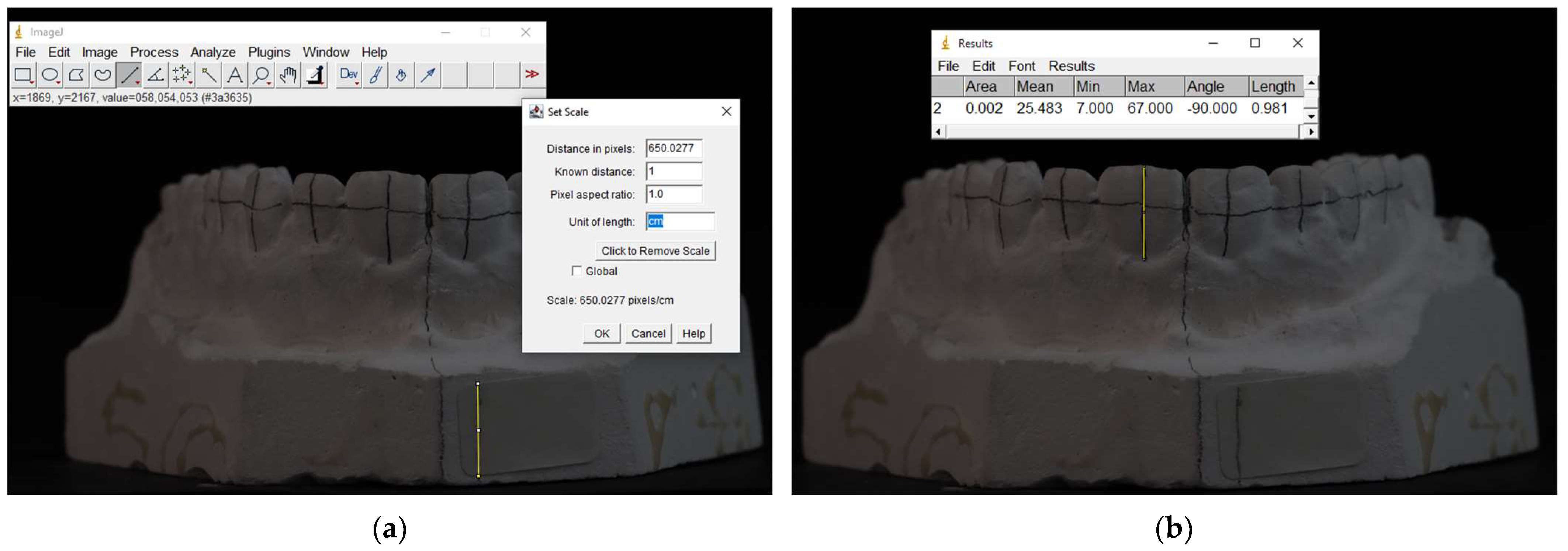
Subsequently, all photographs, in addition to the chosen frontal view of each scanned model, underwent an evaluation using the free software ImageJ (U.S. National Institutes of Health in Bethesda, Bethesda, MD, USA) known for its precision to 0.01 mm. The initial step involved marking the cm length of the reference sticker on each photograph using the software. This dimension was translated into pixel measurements, and a scale was established to automatically convert subsequent measurements into the specified length unit (cm).
Following this, with reference to the previously drawn vertical and horizontal lines, the heights and widths of the teeth were marked, and their measurements were recorded utilizing the Measure Analyze tool from ImageJ (Version 1.54p), as illustrated in Figure 2. Consistency was maintained, as measurements were obtained in a uniform sequence for all the photographs, commencing with the right second premolar and concluding with the left second premolar.

2.7. Statistical Analysis

Data normality was verified using the Shapiro–Wilk test, and homoscedasticity was confirmed using Levene’s test. Statistical analyses were conducted at a significance level of α = 0.05, using Prism 10 (macOS version 10.4.1). The width and height of each tooth were analyzed separately using one-way ANOVA and Tukey’s post hoc test.

3. Results

The measurements of teeth captured with the DSLR camera and all tested smartphone cameras—except for the Vivo T2—at all distances aligned with the scan results, showing no significant differences.
A comparison of tooth height measurements across different photographs revealed no statistically significant differences across all distances (p > 0.05) for the iPhone 13 and 15, Samsung Galaxy S22, S23, and S24, as well as the Vivo T2, as presented in Table 1, Table 2, Table 3, Table 4, Table 5 and Table 6.
Similarly, for tooth width measurements, no statistically significant differences were observed for the iPhone 13; iPhone 15; and Samsung Galaxy S22, S23, and S24 (p > 0.05), as shown in Table 7, Table 8, Table 9, Table 10 and Table 11. However, statistically significant differences were observed for teeth 15 and 14 when using the Vivo T2 smartphone across all distances (p < 0.05), as shown in Table 12.

4. Discussion

This study demonstrated that both DSLR and smartphone cameras yielded comparable measurements for the frontal view of the scanned model for both width and height measurements. The first null hypothesis—stating that there is no statistical difference between DSLR and smartphone measurements—is supported for newer generations of iPhones and all recent Samsung Galaxy S series models but not for VIVO T2. The second null hypothesis—proposing that the distance between the smartphone camera and the object (ranging from 16 cm to 32 cm) does not significantly affect linear measurements—is also accepted for these devices. These results emphasize the advantages of using newer-generation smartphones with advanced camera systems to obtain accurate dental images without linear distortions. Consequently, the previously recommended minimum distance of 24 cm is no longer necessary for a DSD including the premolars.
Specifically, for height measurements across all teeth, no statistically significant differences were observed between the two device types when compared to the reference values from the scans (Table 1, Table 2, Table 3, Table 4, Table 5 and Table 6). These findings align with the findings of a previous study by Moussa et al. [15], which also reported no significant differences in height measurements at various distances for the examined smartphone.
However, differences emerged in regards to width measurements, where some smartphone models exhibited discrepancies compared to the DSLR measurements, while others showed no significant variation. This suggests that measurement accuracy depends on the phone’s brand and generation.
Regarding the iPhone 13, no statistically significant differences were detected in tooth measurements at the 16 cm and 20 cm distances (Table 7). However, a difference was noted at 24 cm for several teeth. In contrast, the iPhone 15 exhibited no statistically significant differences in tooth measurements at any distance, reflecting a marked improvement in the accuracy of MDP with newer generations of smartphones (Table 8).
For the Samsung Galaxy S series, no statistically significant differences in tooth width measurements were found across three consecutive generations—Samsung Galaxy S22, S23, and S24 (Table 9, Table 10 and Table 11). This consistency highlights the high imaging accuracy of Samsung Galaxy S series cameras, reinforcing their reliability as a tool for MDP without dimensional distortions, identifying it as a valuable and accurate tool for DSD and documentation.
Notably, previous studies [15,16], recommend maintaining a minimum distance of 24 cm for DSDs that include the premolars and a minimum distance of 16 cm for DSDs excluding the premolars in order to avoid the barrel effect, which can distort premolar width measurements. This limitation, however, appears to have been mitigated in newer iPhone and Samsung Galaxy S models, as no significant image distortion was observed at closer distances. Hence, photographs for DSDs including the premolars can now be taken at a distance as close as 16 cm.
The Vivo T2, however, displayed width measurement variations for premolars, specifically for teeth 14 and 15, at all distances, while teeth 24 and 25 showed no significant differences (Table 12). This unusual finding may be attributed to variations in imaging sensor quality, lens misalignment, or software processing issues potentially leading to one-sided image distortion. While this remains a hypothesis, it highlights the variability that may exist across different smartphone models, even within the same generation. These deviations suggest that certain devices might apply internal distortion correction or cropping that impacts linear accuracy. To better understand the origin of such discrepancies, future studies should consider performing device-specific validation tests that isolate hardware-related factors (such as lens alignment and sensor architecture) from software-based influences (such as image processing pipelines or artificial intelligence enhancements).
Moreover, no statistically significant difference was observed in tooth measurements obtained from 2D photographs derived from Helios 500 3D scans compared to those captured using the Ceramill Map 600 desktop scanner. This supports the use of the desktop scan as a consistent and dependable control group.
The wide use of smartphone cameras by dental professionals is due to their convenience, since smartphones are more accessible and economically feasible alternatives to DSLR cameras. Their compact design and ease of use, coupled with their ability to produce high-quality images and videos, make them a popular choice [12].
Photographs play a crucial role in the assessment and analysis of smile aesthetics, since they allow for the incorporation of both patient and clinician preferences [1,2]. More specifically, frontal dental photographs are significantly important during treatment planning for aesthetic cases, as they offer the possibility of obtaining accurate measurements of tooth sizes and proportions. Advances in photographic analysis and processing techniques have enhanced the ability to define ideal thresholds for tooth shapes, dimensions, and soft tissue proportions [3,4]. For example, average values for the widths of maxillary teeth in the frontal view are usually utilized to create aesthetically pleasing smiles [17].
Apart from structural analysis, digital imaging has also found its place for use in color measurements to facilitate better communication between dentists and laboratory technicians [12]. The variability of an observer can take place in color matching, but when it comes to digital shade selection, photographs prove reliable within clinically acceptable thresholds. All camera types—DSLR, mirrorless, and smartphone—can produce equally reliable results under appropriate lighting conditions [18,19]. Furthermore, the incorporation of accessories like polarizing filters contributes to improved shade matching, while the utilization of cross-polarizing filters on smartphone cameras has demonstrated an ability to yield images with greater color standardization [20,21].
Additionally, the integration of technology into aesthetic dentistry has spurred the evolution of digital approaches to smile design. DSD and similar systems have become indispensable tools, offering advanced software resources for treatment planning and predicting outcomes through dental photographs [22,23]. A variety of smile design systems, including SmileFy application, Cara Smile, Rebel Simplicity, Planmeca Romexis Smile Design, Aesthetic DSD, Smile Designer Pro, and VisagiSMile, provide tailored methodologies to analyze a patient’s smile and develop comprehensive treatment plans for achieving optimal aesthetic results [24,25,26].
In this study, the selected cameras were chosen for their availability, image quality, and widespread use. The Canon EOS 250D was included due to its proven reliability for tooth dimension measurements, further emphasizing its relevance for dental applications.
This study examined the accuracy of 2D photographs captured by different devices and evaluated the reliability of these photographs based on the type of device used and its distance from the subject. It is well-established that as the lens moves closer to a subject, distortion—particularly at the edges of the image—increases [27]. In this study, the Canon EOS 250D, equipped with a 100 mm macro lens, required a longer working distance to maintain focus, which minimized distortion. Conversely, the smartphone camera, with its capability to automatically adjust camera settings, was tested at various distances to assess its performance.
From a documentation perspective, regardless of the device used or the distance to the subject, all photographs demonstrated efficiency and provided reliable information. There was no statistically significant difference between the recordings captured by the DSLR and the smartphone cameras, confirming the suitability of both devices for clinical documentation [19].
Concerning the precision of measurements, the results showed no significant difference in the height and width measurements of the teeth. Height and width measurements obtained with both the DSLR and smartphone cameras, for all distances evaluated, were in accordance with those obtained from the intraoral scanner and desktop scanner. This accuracy was maintained for all teeth, independent of their position in the dental arch. These results show the reliability of both devices in capturing clinically accurate dental photographs. Such stability suggests that both camera types prove to be reliable in acquiring accurate measurements in this region, proving them suitable for smile design applications [25,28].
While the present study focused on technical validation, its findings have direct clinical implications. To facilitate practical application, clinicians can use a simple technique to ensure an appropriate distance from the patient. By initially switching to 2× zoom on the smartphone, the practitioner is forced to step back in order to capture the full smile, naturally increasing the camera-to-subject distance beyond 16 cm. Once this position is achieved, the zoom can be returned to 1× if desired, without altering the practitioner’s distance. This method provides an intuitive way to respect the recommended photographic parameters and can easily be integrated into DSD workflows or communication protocols with dental laboratories. Additionally, the demonstrated accuracy of smartphone photography supports its potential use in teledentistry and remote diagnostics, enabling reliable case documentation and preliminary assessments in virtual settings.
Clinical studies exploring the variables addressed in this article remain limited. To enhance understanding, randomized controlled clinical trials are necessary to assess the accuracy of digital photography—encompassing both DSLR and smartphone cameras—regarding their precision and trueness. Currently, there is little information about the use of smartphones in dental photography, despite their increasing prevalence. Additionally, the role of varying camera sensors, whether in DSLR, mirrorless, or smartphone devices, presents a critical factor that may significantly impact smile evaluations in frontal view photographs [28].
Even if the results of this study can be universally applied to all recent and future generations of iPhone and Samsung Galaxy S series smartphones, it is important to keep track of new developments. In fact, Apple has announced that it will stop using Sony Sensors for the iPhone cameras in 2026; instead, it will use newly developed three-layer stacked sensors created by Samsung on the iPhone 18, which will make future studies on this matter interesting. With the rapid evolution of technology, future research involving a wider range of brands and models is essential to confirm or possibly challenge this hypothesis. This highlights the need for further studies to address these gaps and establish standardized protocols.
In this study, smartphone settings were left on automatic mode to reflect real-world clinical use. However, this automation may introduce variability due to differences in image processing algorithms, focal length adjustments, and software-based distortion correction across brands. These factors could influence measurement consistency. Future findings should explore standardized or manual settings, where possible, and include a technical analysis of each device’s imaging behavior to enhance reproducibility and transparency.
The present study has focused on maxillary casts, as measurements of tooth dimensions based on photographs are predominantly used in the planning of smiles and particularly, in maxillary dentition. However, due to the great importance of exact measurements of tooth dimensions in orthodontic treatment, future studies should be carried out with mandibular casts to make these findings more clinically applicable. Moreover, more complex scenarios—such as cases involving prosthetic restorations, periodontal compromise, or malocclusion—may present additional challenges that influence photographic accuracy and standardization. Future investigations should therefore incorporate mandibular casts and a broader range of clinical conditions to enhance the relevance and generalizability of the proposed protocol. Furthermore, all measurements were conducted on plaster models under controlled lighting and dry conditions, which do not fully replicate the complexity of real clinical environments. In vivo factors such as variable lighting, saliva, patient movement, and the presence of soft tissues may influence image quality and measurement accuracy. Future research should include validation under clinical conditions to assess the reproducibility and reliability of these findings in everyday dental practice. Another limitation of this study is the use of 2D images to assess 3D structures, which may not fully capture anatomical depth and curvature. Additionally, although this study was based mainly on dimensional accuracy, other critical aspects of camera reliability within the new generations of smartphones should be considered in future studies regarding aesthetic dentistry, such as color matching and color analysis. Expanding this study to incorporate more brands and models of smartphones would provide a more holistic understanding of their functioning. In addition, the accuracy of measurements taken from facial photographs may further improve how photography is implemented in the assessment of patients. Although the number of devices included in this study was supported by a power analysis, the relatively small sample size remains a limitation. This may affect the generalizability of the findings to the wide range of smartphones currently available on the market. Future studies with larger and more diverse samples are needed to confirm the broader clinical applicability of the proposed standardized protocol. Furthermore, while this approach provides valuable insight into the dimensional accuracy of different smartphone cameras, it does not encompass other essential parameters relevant to clinical photography, such as spatial resolution, lighting consistency, and color fidelity—all of which play a significant role in diagnosis, treatment planning, and communication. The exclusion of these variables represents another limitation of the present study. Future research should aim to investigate these parameters systematically to establish a more comprehensive understanding of smartphone camera performance in clinical dentistry.
All in all, it is important to emphasize several key considerations for future research and clinical application. First, the practical implications for clinics without access to DSLR cameras are significant, as smartphones offer a cost-effective alternative for dental imaging. Second, assessing reproducibility in real clinical environments, where factors like varying light, saliva, and soft tissues come into play, is essential to validate the protocol’s robustness. Third, the variability between smartphone brands and generations must be thoroughly evaluated to standardize imaging practices. Fourth, expanding the discussion beyond frequently cited sources such as Moussa et al. [15] by comparing these results with a broader selection of more up-to-date literature will strengthen contextual understanding. Fifth, it is necessary to acknowledge the limitations of this study, notably the exclusive use of plaster models and the lack of in vivo validation, which restrict direct clinical applicability. Finally, the potential for teledentistry and remote diagnostics using mobile photography represents an exciting avenue that warrants further exploration to enhance remote patient care. With ongoing improvements in camera technology, the review of newer models of DSLR, mirrorless, and smartphone cameras will be needed to keep pace with improvements in resolution, lens quality, and image processing capabilities. Changes like these can only serve to improve imaging protocols and enable greater integration of photography into mainstream dental practice.

5. Conclusions

Within the limitations of this study, the findings confirm that the Canon 250D DSLR camera and the recent generations of Apple and Samsung smartphones tested during this study produce tooth measurements consistent with scan results, regardless of shooting distance. Distortion remained minimal for both width and height for all teeth ranging from tooth 15 to tooth 25, even at a close range of 16 cm, reinforcing the suitability of smartphones for clinical applications like documentation and DSD, and this technology is currently accessible to every dentist owning one of these smartphones, facilitating workflows and allowing for more predictable outcomes. These results highlight that dentists can primarily rely on DSLR and the latest Apple and Samsung smartphone models for accurate linear measurements, even for distances closer than 24 cm, ensuring precision in dental assessments. Further research involving additional smartphone brands, as well as in vivo validation under real clinical conditions, would provide a more comprehensive understanding of mobile photography’s reliability and expand its applicability in everyday practice.

Author Contributions

Conceptualization, O.H., R.B., C.E.C.-S., A.O. and L.H.; methodology, O.H., R.B., C.E.C.-S. and L.H.; software, O.H., L.H., R.B., C.E.C.-S., K.K. and C.M.; validation, O.H., A.O., L.H., R.B., K.K., N.N., C.E.C.-S. and C.M.; formal analysis, O.H., R.B., N.N., N.K., Y.H., M.L.-S. and L.H.; investigation O.H., A.O., M.L.-S., Y.H., N.K., R.B. and L.H.; resources, O.H., R.B., M.L.-S. and L.H.; data curation, O.H. and N.K.; writing—original draft preparation, O.H., R.B. and L.H.; writing—review and editing, O.H. and L.H.; visualization, O.H., L.H., R.B., C.M., K.K., N.K. and C.E.C.-S.; supervision, L.H., N.K. and R.B.; project administration, L.H.; funding acquisition, Y.H. All authors have read and agreed to the published version of the manuscript.

Funding

This research received no external funding.

Institutional Review Board Statement

This study was approved by the Ethics Committee of the Faculty of Dental Medicine at the Saint Joseph University of Beirut, Lebanon (FMD-221; ref. #USJ-2024-*289).

Informed Consent Statement

Written informed consent has been obtained from the patient(s) to publish this paper. The research protocol was conducted in strict adherence to ethical guidelines, and it was approved by the Institutional Review Board of Saint-Joseph University of Beirut (FMD-221; ref. #USJ-2024-289).

Data Availability Statement

The data presented in this study are available upon reasonable request from the author (O.H.).

Conflicts of Interest

The authors declare no conflicts of interest.

References

  1. Patussi, E.G.; Poltronieri, B.C.G.; Ottoni, R.; Bervian, J.; Lisboa, C.; Corazza, P.H. Comparisons between photographic equipment for dental use: DSLR cameras vs. smartphones. Rev. Fac. Odontol. UPF 2019, 24, 198–203. [Google Scholar] [CrossRef]
  2. Terry, D.; Snow, S.; McLaren, E. Contemporary dental photography: Selection and application. Compend. Contin. Educ. Dent. 2008, 29, 432. [Google Scholar] [PubMed]
  3. Chen, J.W.; Hobdell, M.H.; Dunn, K.; Johnson, K.A.; Zhang, J. Teledentistry and its use in dental education. J. Am. Dent. Assoc. 2003, 134, 342–346. [Google Scholar] [CrossRef] [PubMed]
  4. Kalpana, D.; Rao, S.J.; Joseph, J.K.; Kurapati, S.K.R. Digital dental photography. Indian J. Dent. Res. 2018, 29, 507. [Google Scholar]
  5. Desai, V.; Bumb, D. Digital dental photography: A contemporary revolution. Int. J. Clin. Pediatr. Dent. 2013, 6, 193–196. [Google Scholar] [CrossRef]
  6. Hardan, L.; Moussa, C. Mobile dental photography: A simple technique for documentation and communication. Quintessence Int. 2020, 51, 510–518. [Google Scholar]
  7. Coachman, C.; Calamita, M.; Ricci, A. Digital smile design. In Ronald E. Goldstein’s Esthetics in Dentistry; John Wiley & Sons Ltd.: Hoboken, NJ, USA, 2018; pp. 84–111. [Google Scholar]
  8. Ahmad, I. Digital dental photography. Part 2: Purposes and uses. Br. Dent. J. 2009, 206, 459–464. [Google Scholar] [CrossRef]
  9. Gonzaga, C.; Garcia, P.; da Costa, R.; Calgaro, M.; Ritter, A.; Correr, G.; da Cunha, L. Digital smile design and mock-up technique for esthetic treatment planning with porcelain laminate veneers. J. Conserv. Dent. 2018, 21, 455–458. [Google Scholar] [CrossRef]
  10. McLaren, E.A.; Goldstein, R.E. The Photoshop smile design technique. Compend. Contin. Educ. Dent. 2018, 39, e17–e20. [Google Scholar]
  11. Zhang, C.; Fan, L.; Chai, Z.; Yu, C.; Song, J. Smartphone and medical application use among dentists in China. BMC Med. Inform. Decis. Mak. 2020, 20, 213. [Google Scholar] [CrossRef]
  12. Hardan, L. Protocols for Mobile Dental Photography with Auxiliary Lighting; Quintessence Publishing: Chicago, IL, USA, 2020. [Google Scholar]
  13. Majumder, S.; Deen, M.J. Smartphone sensors for health monitoring and diagnosis. Sensors 2019, 19, 2164. [Google Scholar] [CrossRef] [PubMed]
  14. Lee, S.; Kim, B.; Lee, J.; Sasian, J. Accurate determination of distortion for smartphone cameras. Appl. Opt. 2014, 53, H1–H6. [Google Scholar] [CrossRef] [PubMed]
  15. Moussa, C.; Hardan, L.; Kassis, C.; Bourgi, R.; Devoto, W.; Jorquera, G.; Panda, S.; Fadel, R.A.; Cuevas-Suárez, C.E.; Lukomska-Szymanska, M.; et al. Accuracy of dental photography: Professional vs. smartphone cameras. Biomed. Res. Int. 2021, 2021, 3910291. [Google Scholar] [CrossRef] [PubMed]
  16. Mackenzie, L.; Sharland, M. Technique Tips: Mobile Dental Photography (MDP). Dent. Update 2020, 47, 884–885. [Google Scholar] [CrossRef]
  17. Jorquera, G.J.; Atria, P.J.; Galán, M.; Feureisen, J.; Imbarak, M.; Kernitsky, J.; Cacciuttolo, F.; Hirata, R.; Sampaio, C.S. A Comparison of Ceramic Crown Color Difference Between Different Shade Selection Methods: Visual, Digital Camera, and Smartphone. J. Prosthet. Dent. 2022, 128, 784–792. [Google Scholar] [CrossRef]
  18. Lazar, R.; Culic, B.; Gasparik, C.; Lazar, C.; Dudea, D. The Accuracy of Dental Shade Matching Using Cross-Polarization Photography. Int. J. Comput. Dent. 2019, 22, 343–351. [Google Scholar]
  19. Antar, F.; Zebouni, E. Comparative review of DSLR cameras and smartphones in dental photography: Indications and limitations. Int. Arab. J. Dent. 2018, 9, 107–115. [Google Scholar]
  20. Sampaio, C.S.; Atria, P.J.; Hirata, R.; Jorquera, G. Variability of Color Matching with Different Digital Photography Techniques and a Gray Reference Card. J. Prosthet. Dent. 2019, 121, 333–339. [Google Scholar] [CrossRef]
  21. Cunha, L.F.D.; Fernandes, A.B.F.; Baechtold, M.S.; Correr, G.M.; Gonzaga, C.C. Cara Smile: Use of Planning Software to Facilitate Esthetic Dental Treatment in a Case. Indian. J. Dent. Res. 2019, 30, 964. [Google Scholar] [CrossRef]
  22. Jafri, Z.; Ahmad, N.; Sawai, M.; Sultan, N.; Bhardwaj, A. Digital smile design: An innovative tool in aesthetic dentistry. J. Oral. Biol. Craniofac. Res. 2020, 10, 194–198. [Google Scholar] [CrossRef]
  23. Gorripati, J.P.; Godbole, S.D. Creating smiles with digital workflow and matrix transfer technique in customizing personality-based smile designing. Int. Arab. J. Dent. 2024, 15, 199–206. [Google Scholar] [CrossRef]
  24. Omar, D.; Duarte, C. The application of parameters for comprehensive smile esthetics by digital smile design programs: A review of literature. Saudi Dent. J. 2018, 30, 7–12. [Google Scholar] [CrossRef] [PubMed]
  25. Iliev, G.V.; Romeo, G. Harmony of smile design in the facial context. Int. J. Esthet. Dent. 2020, 15, 92–106. [Google Scholar]
  26. Bourgi, R.; Qaddomi, M.; Hardan, L.; Tohme, H.; Corbani, K.; Abou Isber, S.; Daher, E.A.; Nassar, N.; Kharouf, N.; Haikel, Y. Gingival Contouring and Smile Makeover through Digital Planning and 3D Guidance. J. Clin. Med. Res. 2025, 6, 6208. [Google Scholar] [CrossRef]
  27. Morris, R.S.; Hoye, L.N.; Elnagar, M.H.; Atsawasuwan, P.; Galang-Boquiren, M.T.; Caplin, J.; Viana, G.C.; Obrez, A.; Kusnoto, B. Accuracy of Dental Monitoring 3D digital dental models using photograph and video mode. Am. J. Orthod. Dentofac. Orthop. 2019, 156, 420–428. [Google Scholar] [CrossRef]
  28. Sajjadi, S.H.; Khosravanifard, B.; Moazzami, F.; Rakhshan, V.; Esmaeilpour, M. Effects of three types of digital camera sensors on dental specialists’ perception of smile esthetics: A preliminary double-blind clinical trial. J. Prosthodont. 2016, 25, 675–681. [Google Scholar] [CrossRef]
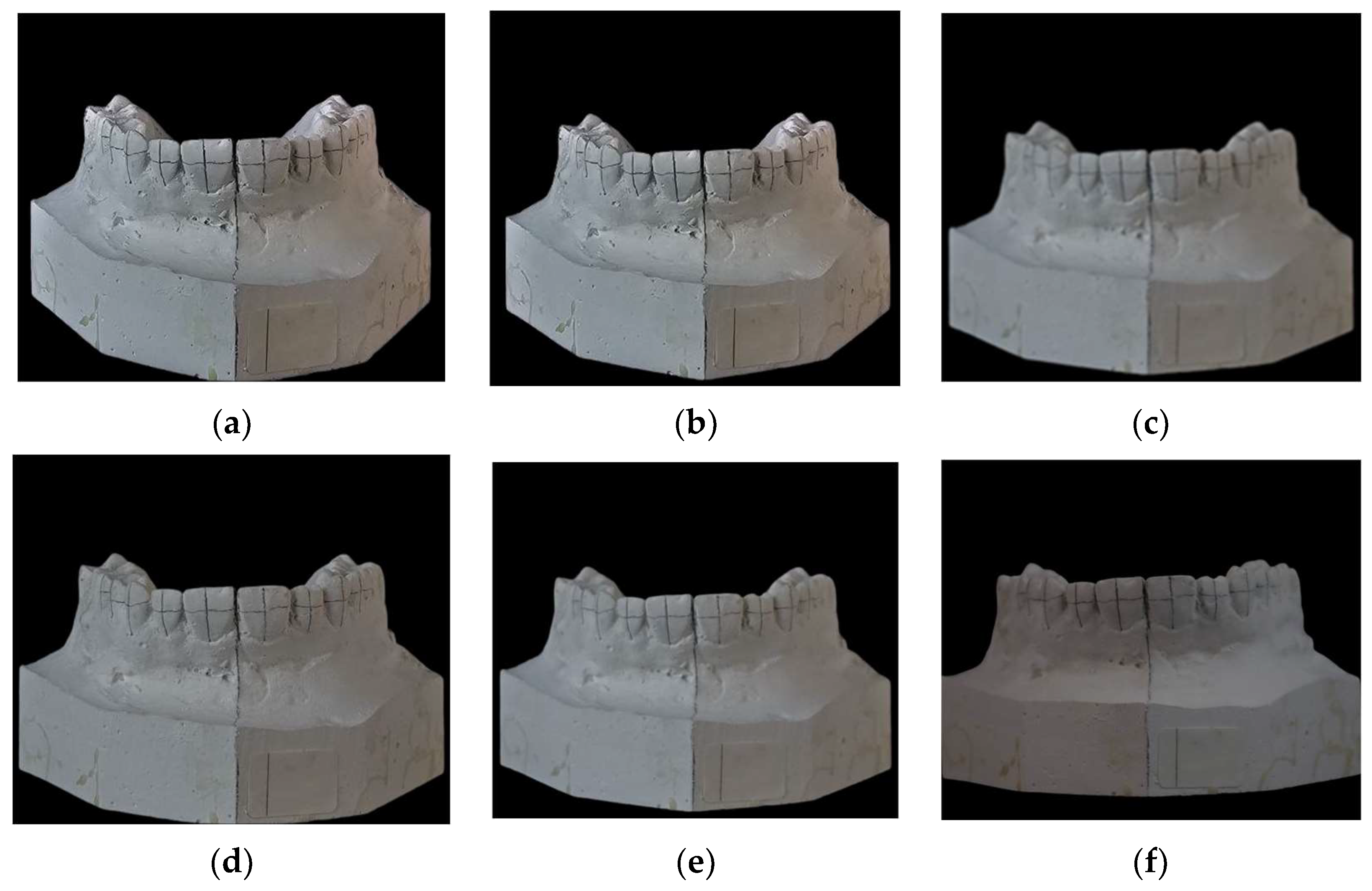
Figure 1. Photographs of the same dental plaster model taken with the Samsung Galaxy S24 smartphone at varying distances and compared with a photograph taken by a DSLR camera (Canon EOS 250D with 100 mm macro lens). The distances from the model to the camera lens for the smartphone images are as follows: (a) 16 cm, (b) 20 cm, (c) 24 cm, (d) 28 cm, and (e) 32 cm. (f) Image captured using the Canon DSLR camera, serving as the reference standard.
Prosthesis 07 00077 g001
Figure 2. Example of setting a scale and measuring tooth heights and widths using ImageJ tools. (a) Setting a scale. (b) Measuring tooth height.
Prosthesis 07 00077 g002
Table 1. Summary of the tooth heights measured using iPhone 13 with different devices and distances.
Device
Teeth EighteethDesktopCanoniPhone 13
16 cm
iPhone 13
20 cm
iPhone 13
24 cm
iPhone 13
28 cm
iPhone 13
32 cm
150.61 (0.09)0.60 (0.09)0.57 (0.08)0.56 (0.08)0.55 (0.08)0.55 (0.07)0.53 (0.06)0.56 (0.07)
140.68 (0.09)0.66 (0.09)0.62 (0.1)0.64 (0.11)0.63 (0.11)0.62 (0.10)0.62 (0.11)0.63 (0.12)
130.85 (0.15)0.83 (0.12)0.79 (0.17)0.82 (0.19)0.80 (0.17)0.79 (0.16)0.81 (0.15)0.82 (0.18)
120.77 (0.19)0.75 (0.16)0.74 (0.19)0.74 (0.20)0.74 (0.20)0.73 (0.19)0.73 (0.19)0.75 (0.19)
110.92 (0.12)0.90 (0.11)0.90 (0.12)0.91 (0.14)0.90 (0.14)0.88 (0.13)0.89 (0.12)0.92 (0.13)
210.95 (0.14)0.94 (0.12)0.91 (0.14)0.91 (0.15)0.91 (0.15)0.87 (0.15)0.90 (0.14)0.91 (0.15)
220.80 (0.13)0.77 (0.10)0.78 (0.12)0.76 (0.13)0.76 (0.13)0.75 (0.13)0.76 (0.12)0.78 (0.13)
230.86 (0.16)0.85 (0.15)0.85 (0.19)0.85 (0.20)0.84 (0.20)0.83 (0.20)0.84 (0.20)0.86 (0.20)
240.67 (0.11)0.66 (0.09)0.64 (0.11)0.62 (0.11)0.62 (0.10)0.61 (0.12)0.62 (0.10)0.64 (0.10)
250.59 (0.08)0.58 (0.08)0.54 (0.07)0.53 (0.07)0.52 (0.06)0.51 (0.06)0.53 (0.06)0.52 (0.08)
Note: Differences in lines were not statistically significant (p > 0.05).
Table 2. Summary of the tooth heights measured using iPhone 15 with different devices and distances (cont.).
Device
Teeth EighteethDesktopCanoniPhone 15
16 cm
iPhone 15
20 cm
iPhone 15
24 cm
iPhone 15
28 cm
iPhone 15
32 cm
150.61 (0.09)0.60 (0.09)0.57 (0.08)0.58 (0.06)0.55 (0.06)0.56 (0.09)0.55 (0.10)0.56 (0.08)
140.68 (0.09)0.66 (0.09)0.62 (0.1)0.67 (0.08)0.65 (0.10)0.64 (0.09)0.63 (0.09)0.64 (0.06)
130.85 (0.15)0.83 (0.12)0.79 (0.17)0.84 (0.10)0.81 (0.11)0.82 (0.10)0.81 (0.12)0.84 (0.10)
120.77 (0.19)0.75 (0.16)0.74 (0.19)0.71 (0.20)0.75 (0.18)0.73 (0.18)0.74 (0.16)0.91 (0.18)
110.92 (0.12)0.90 (0.11)0.90 (0.12)0.92 (0.19)0.89 (0.14)0.89 (0.15)0.88 (0.15)0.91 (0.14)
210.95 (0.14)0.94 (0.12)0.91 (0.14)0.91 (0.20)0.90 (0.16)0.90 (0.16)0.88 (0.17)0.89 (0.13)
220.80 (0.13)0.77 (0.10)0.78 (0.12)0.78 (0.19)0.76 (0.14)0.75 (0.15)0.76 (0.14)0.77 (0.12)
230.86 (0.16)0.85 (0.15)0.85 (0.19)0.86 (0.27)0.85 (0.22)0.84 (0.22)0.84 (0.22)0.86 (0.19)
240.67 (0.11)0.66 (0.09)0.64 (0.11)0.66 (0.16)0.63 (0.14)0.63 (0.12)0.63 (0.11)0.64 (0.09)
250.59 (0.08)0.58 (0.08)0.54 (0.07)0.53 (0.08)0.55 (0.07)0.53 (0.1)0.55 (0.11)0.54 (0.07)
Note: Differences in lines were not statistically significant (p > 0.05).
Table 3. Summary of the tooth heights measured using S22 with different devices and distances (cont.).
Device
Teeth EighteethDesktopCanonS22 16 cmS22 20 cmS22 24 cmS22 28 cmS22 32 cm
150.61 (0.09)0.60 (0.09)0.57 (0.08)0.54 (0.08)0.51 (0.10)0.56 (0.09)0.57 (0.09)0.59 (0.06)
140.68 (0.09)0.66 (0.09)0.62 (0.1)0.61 (0.08)0.60 (0.14)0.64 (0.14)0.64 (0.12)0.68 (0.13)
130.85 (0.15)0.83 (0.12)0.79 (0.17)0.79 (0.20)0.80 (0.22)0.84 (0.21)0.81 (0.20)0.80 (0.18)
120.77 (0.19)0.75 (0.16)0.74 (0.19)0.74 (0.24)0.76 (0.26)0.76 (0.23)0.77 (0.24)0.74 (0.21)
110.92 (0.12)0.90 (0.11)0.90 (0.12)0.90 (0.16)0.89 (0.19)0.92 (0.18)0.92 (0.15)0.93 (0.14)
210.95 (0.14)0.94 (0.12)0.91 (0.14)0.90 (0.18)0.91 (0.22)0.92 (0.20)0.92 (0.16)0.93 (0.17)
220.80 (0.13)0.77 (0.10)0.78 (0.12)0.76 (0.16)0.76 (0.18)0.79 (0.17)0.78 (0.14)0.79 (0.12)
230.86 (0.16)0.85 (0.15)0.85 (0.19)0.83 (0.23)0.84 (0.27)0.87 (0.24)0.87 (0.23)0.85 (0.21)
240.67 (0.11)0.66 (0.09)0.64 (0.11)0.61 (0.13)0.63 (0.15)0.64 (0.12)0.65 (0.13)0.66 (0.1)
250.59 (0.08)0.58 (0.08)0.54 (0.07)0.52 (0.05)0.51 (0.05)0.52 (0.07)0.54 (0.06)0.58 (0.08)
Note: Differences in lines were not statistically significant (p > 0.05).
Table 4. Summary of the tooth heights measured using S23 with different devices and distances (cont.).
Device
Teeth EighteethDesktopCanonS23 16 cmS23 20 cmS23 24 cmS23 28 cmS23 32 cm
150.61 (0.09)0.60 (0.09)0.57 (0.08)0.52 (0.10)0.54 (0.09)0.54 (0.09)0.53 (0.08)0.54 (0.08)
140.68 (0.09)0.66 (0.09)0.62 (0.1)0.62 (0.15)0.63 (0.11)0.64 (0.13)0.63 (0.13)0.62 (0.12)
130.85 (0.15)0.83 (0.12)0.79 (0.17)0.83 (0.22) 0.79 (0.16)0.83 (0.18)0.80 (0.17)0.79 (0.20)
120.77 (0.19)0.75 (0.16)0.74 (0.19)0.75 (0.25)0.73 (0.21)0.74 (0.21)0.73 (0.22)0.73 (0.21)
110.92 (0.12)0.90 (0.11)0.90 (0.12)0.88 (0.20)0.89 (0.15)0.89 (0.16)0.89 (0.16)0.89 (0.15)
210.95 (0.14)0.94 (0.12)0.91 (0.14)0.91 (0.19)0.90 (0.16)0.90 (0.17)0.90 (0.16)0.90 (0.16)
220.80 (0.13)0.77 (0.10)0.78 (0.12)0.76 (0.17)0.75 (0.13)0.77 (0.14)0.76 (0.14)0.75 (0.13)
230.86 (0.16)0.85 (0.15)0.85 (0.19)0.83 (0.25)0.83 (0.22)0.84 (0.22)0.82 (0.22)0.75 (0.13)
240.67 (0.11)0.66 (0.09)0.64 (0.11)0.64 (0.13)0.61 (0.10)0.64 (0.10)0.64 (0.11)0.62 (0.12)
250.59 (0.08)0.58 (0.08)0.54 (0.07)0.50 (0.10)0.50 (0.06)0.52 (0.08)0.53 (0.09)0.50 (0.09)
Note: Differences in lines were not statistically significant (p > 0.05).
Table 5. Summary of the tooth heights measured using S24 with different devices and distances (cont.).
Device
Teeth EighteethDesktopCanonS24 16 cmS24 20 cmS24 24 cmS24 28 cmS24 32 cm
150.61 (0.09)0.60 (0.09)0.57 (0.08)0.54 (0.09)0.58 (0.09)0.56 (0.06)0.55 (0.08)0.55 (0.09)
140.68 (0.09)0.66 (0.09)0.62 (0.1)0.64 (0.14)0.63 (0.14)0.67 (0.12)0.63 (0.12)0.64 (0.14)
130.85 (0.15)0.83 (0.12)0.79 (0.17)0.80 (0.2)0.80 (0.2)0.81 (0.17)0.80 (0.17)0.81 (0.17)
120.77 (0.19)0.75 (0.16)0.74 (0.19)0.73 (0.25)0.74 (0.24)0.72 (0.21)0.75 (0.24)0.72 (0.23)
110.92 (0.12)0.90 (0.11)0.90 (0.12)0.91 (0.15)0.90 (0.16)0.91 (0.15)0.91 (0.14)0.88 (0.16)
210.95 (0.14)0.94 (0.12)0.91 (0.14)0.91 (0.16)0.91 (0.17)0.92 (0.16)0.91 (0.15)0.88 (0.16)
220.80 (0.13)0.77 (0.10)0.78 (0.12)0.77 (0.14)0.76 (0.14)0.58 (0.12)0.54 (0.08)0.53 (0.10)
230.86 (0.16)0.85 (0.15)0.85 (0.19)0.82 (0.22)0.83 (0.22)0.84 (0.22)0.84 (0.23)0.83 (0.21)
240.67 (0.11)0.66 (0.09)0.64 (0.11)0.63 (0.11)0.63 (0.10)0.65 (0.11)0.64 (0.11)0.64 (0.12)
250.59 (0.08)0.58 (0.08)0.54 (0.07)0.51 (0.04)0.52 (0.08)0.54 (0.07)0.52 (0.08)0.54 (0.10)
Note: Differences in lines were not statistically significant (p > 0.05).
Table 6. Summary of the tooth heights measured using Vivo with different devices and distances (cont.).
Device
Teeth EighteethDesktopCanonVivo 16 cmVivo 20 cmVivo 24 cmVivo 28 cmVivo 32 cm
150.61 (0.09)0.60 (0.09)0.57 (0.08)0.47 (0.14)0.51 (0.14)0.55 (0.09)0.57 (0.08)0.55 (0.08)
140.68 (0.09)0.66 (0.09)0.62 (0.1)0.62 (0.13)0.61 (0.07)0.63 (0.12)0.63 (0.10)0.62 (0.12)
130.85 (0.15)0.83 (0.12)0.79 (0.17)0.80 (0.19)0.79 (0.18)0.80 (0.19)0.81 (0.17)0.79 (0.19)
120.77 (0.19)0.75 (0.16)0.74 (0.19)0.73 (0.20)0.73 (0.21)0.75 (0.21)0.74 (0.19)0.72 (0.20)
110.92 (0.12)0.90 (0.11)0.90 (0.12)0.88 (0.14)0.88 (0.14)0.90 (0.14)0.89 (0.13)0.88 (0.14)
210.95 (0.14)0.94 (0.12)0.91 (0.14)0.89 (0.16)0.91 (0.16)0.91 (0.14)0.90 (0.14)0.90 (0.16)
220.80 (0.13)0.77 (0.10)0.78 (0.12)0.76 (0.14)0.77 (0.14)0.76 (0.12)0.75 (0.12)0.75 (0.15)
230.86 (0.16)0.85 (0.15)0.85 (0.19)0.83 (0.22)0.86 (0.21)0.86 (0.22)0.84 (0.21)0.84 (0.20)
240.67 (0.11)0.66 (0.09)0.64 (0.11)0.62 (0.11)0.64 (0.11)0.63 (0.10)0.63 (0.11)0.62 (0.12)
250.59 (0.08)0.58 (0.08)0.54 (0.07)0.51 (0.06)0.54 (0.08)0.53 (0.07)0.54 (0.09)0.52 (0.09)
Note: Differences in lines were not statistically significant (p > 0.05).
Table 7. Summary of the tooth widths measured using iPhone 13 with different devices and distances.
Device
Teeth EighteethDesktopCanoniPhone 13
16 cm
iPhone 13
20 cm
iPhone 13
24 cm
iPhone 13
28 cm
iPhone 13
32 cm
150.19 (0.01)0.19 (0.01)0.23 (0.13)0.18 (0.14)0.16 (0.13)0.49 (0.21) *0.12 (0.02) *0.12 (0.02) *
140.29 (0.02)0.29 (0.02)0.30 (0.13)0.22 (0.03)0.21 (0.03)0.57 (0.21) *0.19 (0.02) *0.21 (0.03)
130.46 (0.05)0.45 (0.05)0.48 (0.15)0.41 (0.07)0.41 (0.07)0.75 (0.24) *0.39 (0.07)0.40 (0.07)
120.55 (0.06)0.56 (0.06)0.54 (0.08)0.52 (0.08)0.52 (0.08)0.72 (0.21) *0.51 (0.07)0.51 (0.08)
110.85 (0.1)0.84 (0.10)0.82 (0.06)0.87 (0.10)0.87 (0.10)0.89 (0.12)0.83 (0.09)0.85 (0.10)
210.85 (0.1)0.85 (0.11)0.83 (0.04)0.90 (0.11)0.90 (0.11)0.89 (0.14)0.87 (0.1)0.89 (0.1)
220.53 (0.07)0.52 (0.07)0.53 (0.08)0.54 (0.09)0.54 (0.09)0.73 (0.15) *0.56 (0.1)0.54 (0.08)
230.43 (0.06)0.43 (0.06)0.48 (0.17)0.43 (0.04)0.42 (0.04)0.82 (0.022) *0.43 (0.04)0.42 (0.06)
240.26 (0.05)0.26 (0.05)0.30 (0.13)0.27 (0.05)0.26 (0.05)0.56 (0.18) *0.28 (0.04)0.28 (0.05)
250.17 (0.05)0.17 (0.05)0.24 (0.12)0.18 (0.04)0.18 (0.04)0.46 (0.16) *0.19 (0.05)0.20 (0.04)
* Differences were statistically significant when compared with results for the controls (p > 0.05).
Table 8. Summary of the tooth widths measured using iPhone 15 with different devices and distances (cont.).
Device
Teeth EighteethDesktopCanoniPhone 15
16 cm
iPhone 15
20 cm
iPhone 15
24 cm
iPhone 15
28 cm
iPhone 15
32 cm
150.19 (0.01)0.19 (0.01)0.23 (0.13)0.15 (0.04)0.13 (0.03)0.14 (0.03)0.15 (0.02)0.18 (0.03)
140.29 (0.02)0.29 (0.02)0.30 (0.13)0.23 (0.04)0.26 (0.08)0.23 (0.04)0.24 (0.04)0.25 (0.04)
130.46 (0.05)0.45 (0.05)0.48 (0.15)0.42 (0.09) 0.41 (0.07)0.43 (0.06)0.44 (0.07)0.46 (0.07)
120.55 (0.06)0.56 (0.06)0.54 (0.08)0.55 (0.12)0.52 (0.09)0.52 (0.09)0.52 (0.08)0.54 (0.08)
110.85 (0.1)0.84 (0.10)0.82 (0.06)0.89 (0.16)0.85 (0.13)0.86 (0.12)0.85 (0.11)0.87 (0.10)
210.85 (0.1)0.85 (0.11)0.83 (0.04)0.92 (0.17)0.89 (0.13)0.89 (0.12)0.87 (0.12)0.90 (0.10)
220.53 (0.07)0.52 (0.07)0.53 (0.08)0.57 (0.13)0.55 (0.10)0.55 (0.10)0.54 (0.10)0.55 (0.09)
230.43 (0.06)0.43 (0.06)0.48 (0.17)0.44 (0.07)0.43 (0.04)0.44 (0.05)0.42 (0.05)0.43 (0.03)
240.26 (0.05)0.26 (0.05)0.30 (0.13)0.22 (0.04)0.23 (0.04)0.27 (0.05)0.27 (0.05)0.27 (0.04)
250.17 (0.05)0.17 (0.05)0.24 (0.12)0.14 (0.05)0.14 (0.04)0.19 (0.05)0.19 (0.05)0.19 (0.04)
Note: Differences in lines were not statistically significant (p > 0.05).
Table 9. Summary of the tooth widths measured using S22 with different devices and distances (cont.).
Device
Teeth EighteethDesktopCanonS22 16 cmS22 20 cmS22 24 cmS22 28 cmS22 32 cm
150.19 (0.01)0.19 (0.01)0.23 (0.13)0.12 (0.02) 0.13 (0.02)0.13 (0.03)0.15 (0.02)0.12 (0.03)
140.29 (0.02)0.29 (0.02)0.30 (0.13)0.20 (0.04)0.21 (0.03)0.23 (0.04)0.23 (0.04)0.20 (0.04)
130.46 (0.05)0.45 (0.05)0.48 (0.15)0.40 (0.06)0.38 (0.11)0.42 (0.08)0.42 (0.09)0.38 (0.08)
120.55 (0.06)0.56 (0.06)0.54 (0.08)0.51 (0.10)0.51 (0.11)0.53 (0.10)0.54 (0.09)0.53 (0.07)
110.85 (0.1)0.84 (0.10)0.82 (0.06)0.84 (0.13)0.88 (0.26)0.86 (0.12)0.87 (0.11)0.86 (0.10)
210.85 (0.1)0.85 (0.11)0.83 (0.04)0.88 (0.13)0.87 (0.16)0.90 (0.12)0.89 (0.11)0.92 (0.11)
220.53 (0.07)0.52 (0.07)0.53 (0.08)0.51 (0.12)0.51 (0.12)0.55 (0.09)0.54 (0.09)0.59 (0.10)
230.43 (0.06)0.43 (0.06)0.48 (0.17)0.39 (0.07)0.39 (0.06)0.42 (0.04)0.43 (0.05)0.47 (0.05)
240.26 (0.05)0.26 (0.05)0.30 (0.13)0.21 (0.04) 0.22 (0.04)0.27 (0.05)0.29 (0.04)0.31 (0.04)
250.17 (0.05)0.17 (0.05)0.24 (0.12)0.13 (0.04)0.14 (0.04)0.20 (0.04)0.20 (0.04)0.23 (0.05)
Note: Differences in lines were not statistically significant (p > 0.05).
Table 10. Summary of the tooth widths measured using S23 with different devices and distances (cont.).
Device
Teeth EighteethDesktopCanonS23 16 cmS23 20 cmS23 24 cmS23 28 cmS23 32 cm
150.19 (0.01)0.19 (0.01)0.23 (0.13)0.17 (0.02)0.16 (0.02)0.18 (0.04)0.17 (0.04)0.16 (0.03)
140.29 (0.02)0.29 (0.02)0.30 (0.13)0.23 (0.04)0.23 (0.05)0.26 (0.05)0.24 (0.04)0.29 (0.12)
130.46 (0.05)0.45 (0.05)0.48 (0.15)0.44 (0.09)0.42 (0.07)0.46 (0.09)0.44 (0.08)0.42 (0.08)
120.55 (0.06)0.56 (0.06)0.54 (0.08)0.57 (0.11)0.52 (0.07)0.56 (0.11)0.55 (0.11)0.55 (0.09)
110.85 (0.1)0.84 (0.10)0.82 (0.06)0.90 (0.15) 0.86 (0.10)0.87 (0.13)0.86 (0.12)0.85 (0.13)
210.85 (0.1)0.85 (0.11)0.83 (0.04)0.92 (0.16)0.88 (0.11)0.87 (0.12)0.87 (0.12)0.87 (0.12)
220.53 (0.07)0.52 (0.07)0.53 (0.08)0.55 (0.14)0.55 (0.09)0.55 (0.10)0.53 (0.09)0.52 (0.11)
230.43 (0.06)0.43 (0.06)0.48 (0.17)0.42 (0.07)0.42 (0.04)0.41 (0.04)0.42 (0.04)0.41 (0.04)
240.26 (0.05)0.26 (0.05)0.30 (0.13)0.22 (0.04)0.22 (0.04)0.26 (0.03)0.26 (0.04)0.26 (0.05)
250.17 (0.05)0.17 (0.05)0.24 (0.12)0.13 (0.05)0.14 (0.04)0.18 (0.04) 0.18 (0.04)0.19 (0.04)
Note: Differences in lines were not statistically significant (p > 0.05).
Table 11. Summary of the tooth widths measured using S24 with different devices and distances (cont.).
Device
Teeth EighteethDesktopCanonS24 16 cmS24 20 cmS24 24 cmS24 28 cmS24 32 cm
150.19 (0.01)0.19 (0.01)0.23 (0.13)0.12 (0.03)0.13 (0.04)0.12 (0.04)0.17 (0.04)0.55 (0.10)
140.29 (0.02)0.29 (0.02)0.30 (0.13)0.22 (0.04)0.22 (0.06)0.23 (0.04)0.26 (0.05)0.26 (0.04)
130.46 (0.05)0.45 (0.05)0.48 (0.15)0.42 (0.06)0.41 (0.10)0.40 (0.05)0.45 (0.10)0.44 (0.07)
120.55 (0.06)0.56 (0.06)0.54 (0.08)0.55 (0.10)0.54 (0.11)0.53 (0.08)0.55 (0.09)0.53 (0.09)
110.85 (0.1)0.84 (0.10)0.82 (0.06)0.88 (0.13)0.87 (0.13)0.85 (0.13)0.87 (0.12)0.86 (0.11)
210.85 (0.1)0.85 (0.11)0.83 (0.04)0.93 (0.15)0.92 (0.13)0.92 (0.13)0.89 (0.11)0.87 (0.13)
220.53 (0.07)0.52 (0.07)0.53 (0.08)0.56 (0.12)0.55 (0.11)0.58 (0.12)0.54 (0.08)0.53 (0.10)
230.43 (0.06)0.43 (0.06)0.48 (0.17)0.44 (0.06)0.44 (0.06)0.48 (0.08)0.42 (0.04)0.41 (0.05)
240.26 (0.05)0.26 (0.05)0.30 (0.13)0.23 (0.04)0.23 (0.04)0.31 (0.05)0.27 (0.04)0.27 (0.05)
250.17 (0.05)0.17 (0.05)0.24 (0.12)0.13 (0.04)0.14 (0.04)0.22 (0.04)0.18 (0.04)0.18 (0.05)
Note: Differences in lines were not statistically significant (p > 0.05).
Table 12. Summary of the tooth widths measured using Vivo with different devices and distances (cont.).
Device
Teeth EighteethDesktopCanonVivo 16 cmVivo 20 cmVivo 24 cmVivo 28 cmVivo 32 cm
150.19 (0.01)0.19 (0.01)0.23 (0.13)0.11 (0.02) *0.10 (0.02) *0.12 (0.02) *0.13 (0.03) *0.14 (0.04) *
140.29 (0.02)0.29 (0.02)0.30 (0.13)0.20 (0.04) *0.20 (0.03) *0.22 (0.04) *0.23 (0.04) *0.23 (0.04) *
130.46 (0.05)0.45 (0.05)0.48 (0.15)0.40 (0.08)0.39 (0.06)0.42 (0.07)0.41 (0.06)0.42 (0.07)
120.55 (0.06)0.56 (0.06)0.54 (0.08)0.50 (0.10)0.50 (0.09)0.51 (0.08)0.52 (0.08)0.52 (0.08)
110.85 (0.1)0.84 (0.10)0.82 (0.06)0.85 (0.12)0.84 (0.12)0.86 (0.10)0.85 (0.09)0.85 (0.11)
210.85 (0.1)0.85 (0.11)0.83 (0.04)0.89 (0.11)0.92 (0.11)0.91 (0.11)0.90 (0.10)0.85 (0.12)
220.53 (0.07)0.52 (0.07)0.53 (0.08)0.55 (0.09)0.57 (0.09)0.56 (0.10)0.56 (0.09)0.54 (0.09)
230.43 (0.06)0.43 (0.06)0.48 (0.17)0.43 (0.05)0.46 (0.05)0.45 (0.04)0.43 (0.04)0.44 (0.04)
240.26 (0.05)0.26 (0.05)0.30 (0.13)0.22 (0.04)0.22 (0.04)0.28 (0.05)0.29 (0.03)0.28 (0.05)
250.17 (0.05)0.17 (0.05)0.24 (0.12)0.13 (0.04)0.26 (0.27)0.21 (0.05)0.20 (0.03)0.19 (0.04)
* Differences were statistically significant when compared with results for the controls (p > 0.05).
Disclaimer/Publisher’s Note: The statements, opinions and data contained in all publications are solely those of the individual author(s) and contributor(s) and not of MDPI and/or the editor(s). MDPI and/or the editor(s) disclaim responsibility for any injury to people or property resulting from any ideas, methods, instructions or products referred to in the content.