Article

Load-Bearing Capacity of Lithium Silicate Derivates Applied as Ultra-Thin Occlusal Veneers on Molars

1
Clinic of Reconstructive Dentistry, Center for Dental Medicine, University of Zurich, CH-8032 Zurich, Switzerland
2
Center of Dental Medicine, University of Zürich, CH-8032 Zurich, Switzerland
3
Clinic of Clinic of Masticatory Disorders and Dental Biomaterials, Center for Dental Medicine, University of Zurich, CH-8032 Zurich, Switzerland
*
Author to whom correspondence should be addressed.
Prosthesis 2025, 7(2), 43; https://doi.org/10.3390/prosthesis7020043
Submission received: 28 January 2025 / Revised: 7 April 2025 / Accepted: 8 April 2025 / Published: 16 April 2025

Abstract

:
Purpose: This study aimed to evaluate the load-bearing capacity of three different millable lithium silicate derivatives compared with lithium disilicate ceramic when used as ultra-thin occlusal veneers on eroded molars. The null hypothesis stated that there would be no significant differences in load-bearing capacity (Fmax). Material and Methods: Four groups were tested: three groups with lithium silicate derivatives—“Celt” (Celtra, Dentsply Sirona, Bensheim, Germany), “Vita” (Vita Suprinity PC, Vita Zahnfabrik, Bad Säckingen, Germany), and “Nice” (n!ce, Straumann, Basel, Switzerland)—and a control group with lithium disilicate ceramic, “Emax” (IPS e.max CAD, Ivoclar Vivadent) (n = 20 per group). Extracted molars (n = 80) were prepared to simulate erosion and restored with occlusal veneers designed and milled by using CAD/CAM technology. After thermo-mechanical aging, the specimens were subjected to static load testing until fracture. Failure types were recorded and analyzed. Statistical evaluation included the Wilcoxon rank-sum test for group comparisons and Weibull distribution modeling to assess fracture probabilities. Results: Thermo-mechanical aging caused restoration debonding in three specimens from the “Nice” and “Celt” groups, resulting in fatigue resistance of 100% for “Emax” and “Vita”, 90% for “Celt”, and 95% for “Nice”. The mean Fmax values ranged from 892 N to 2087 N, with the “Vita” group demonstrating the highest values. Significant differences in stress values were observed among groups (p < 0.05). Cohesive failure was the most frequent failure mode. Conclusions: All tested lithium silicate derivatives demonstrated high load-bearing capacity and are suitable for ultra-thin occlusal veneers on eroded molars. Cohesive failures dominated, indicating reliable material performance and stable bonding under load.

1. Introduction

The erosive wear of dentition is increasingly recognized as a public health concern [1]. The prevalence of dental erosion in adults, as reported in the literature, ranges widely from 4% to 82% [2]. This variability is compounded by the fact that erosion is an irreversible condition that accumulates over time, leading to higher prevalence with advancing age [3]. Modern treatment concepts aim to preserve as much tooth structure as possible, focusing on minimally invasive approaches that primarily replace lost dental tissue [4,5].
As minimally invasive restorative approaches, both direct and indirect methods can be applied and recommended. The decision as to which treatment and materials to use must be based on the extent of tooth substance loss [6]. Over recent decades, advancements in prosthetic materials have driven a paradigm shift in the management of eroded teeth using fixed restorations. Traditional preparation techniques relying on mechanical retention often involve significant removal of healthy tooth structure, which can lead to irreversible damage, including pulpal exposure and a 10% loss in vitality with each intervention [7,8,9]. In contrast, indirect minimally invasive restorative approaches have emerged as effective treatment options for worn dentition [5].
Among these, occlusal veneers made from lithium disilicate have demonstrated a high durability under the substantial mechanical stresses present in the posterior region in vitro [10,11,12,13]. These restorations are typically fabricated by using heat pressing or CAD/CAM milling techniques [14,15]. Recently, alternative CAD/CAM materials, such as lithium silicate derivatives, have been introduced, showing promising potential as substitutes for lithium disilicate ceramics [16].
To date, no study has comprehensively evaluated the performance of ultra-thin occlusal veneers fabricated from commercially available millable lithium silicate derivatives when bonded to eroded molars. Therefore, this study aimed to investigate the load-bearing capacity of three different millable lithium silicate derivatives when used as occlusal veneers for eroded molars. The null hypothesis proposed that there would be no significant difference in the load-bearing capacity (Fmax) among the tested groups compared with commonly used lithium disilicate ceramics.

2. Materials and Methods

2.1. Sample Size Calculation and Group Allocation

This study adhered to the CRIS guidelines [17]. Four groups were formed, consisting of three test groups and one control group, with 20 specimens per group (n = 20) (Table 1). The test groups comprised millable lithium silicate derivative ceramics, while the control group included millable lithium disilicate ceramic. Sample size estimation was based on mean Fmax values from a prior study [18], calculated by using G*Power 3.1 (Heinrich Heine University, Düsseldorf, Germany). A two-tailed t-test with 19 samples per group provided 95% power (α = 0.05). To ensure robustness, this study included 20 samples per group.
The test groups consisted of the following millable lithium silicate derivates: “Nice” (N!ce, Straumann AG, Basel, Switzerland; flexural strength: 350 MPa), “Celt” (Celtra Duo, Dentsply Sirona, Hanau-Wolfgnag, Germany; flexural strength: 370 MPa), “Vita” (VITA Suprinity PC, Vita Zahnfabrik, Bad Säckingen, Germany; flexural strength: 420 MPa).
The control group included lithium disilicate ceramic: “Emax” (IPS e. max CAD PrograMill; Ivoclar Vivadent, Schaan, Liechtenstein; flexural strength: 530 MPa).

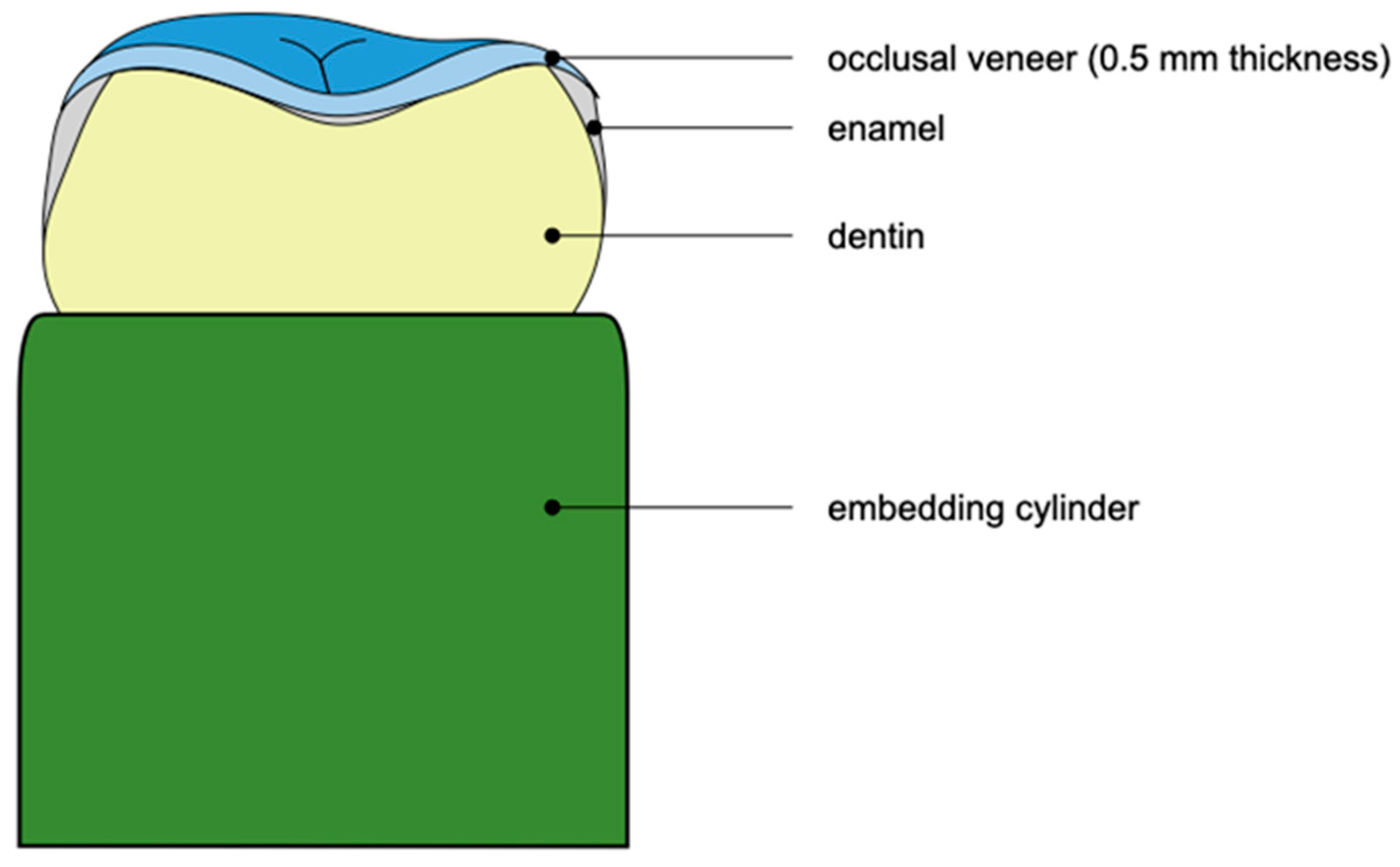
2.2. Specimen Preparation

Eighty extracted human molars were embedded in chemically polymerized resin (Technovit 4071, Kulzer, Wasserburg, Germany) by using additively manufactured hollow cylinders (Med610, Stratasys, Rechovot, Israel). In order to simulate substance loss due to erosion, the occlusal enamel was removed (WS Flex 18 C P80–P2500, Hermes Schleifwerkzeuge, Hamburg, Germany; LaboPol-21, Struers, Ballerup, Denmark). Final preparation involved smoothing sharp edges with diamond burs (Intensiv SA, Montagnola, Switzerland). Specimens were stored in 0.5% Chloramin T throughout the study procedures and randomly assigned to groups. The teeth used in this study were extracted for reasons unrelated to our research. Prior to extraction, all donors provided written informed consent for their teeth to be used in this study, in line with the regulations established by the National Federal Council. Ethical protocols were rigorously followed, and the anonymization of the data was conducted in full compliance with State and Federal laws [19,20,21].

2.3. Scanning Procedure and Digital Restoration Design

The prepared teeth were scanned with a desktop scanner (DOF FreedomHD, Seoul, Republic of Korea). Occlusal veneers were digitally designed by using CAD software (Exocad version 3.2 Elefsina, Darmstadt, Germany) with a standardized thickness of 0.5 mm (Figure 1).

2.4. Fabrication of Restorations

The designs were transferred into milling software (Programill CAM 2022) and milled by using a 5-axis milling machine (Programill PM7, Ivoclar Vivadent, Schaan, Liechtenstein). The milled restorations were cleaned and polished. The groups “Vita” and “Emax” were additionally crystallized in a ceramic furnace (Programat EP 5010, Ivoclar Vivadent, Schaan, Liechtenstein), as indicated by the manufacturer. “Vita” input time: 400 °C; heating rate: 55 °C/min; final temperature: 840 °C with a holding time of 8 min. “Emax” input time: 400 °C; heating rate: 60 °C/min; final temperature: 840 °C with a holding time of 10–15 min.

2.5. Cementation of Restorations

The inner surfaces of all veneers were etched with 5% hydrofluoric acid (IPS Ceramic Etching Gel, Ivoclar Vivadent, Schaan, Liechtenstein) for 20 s, rinsed, and air-dried. A silane coupling agent (Monobond Plus, Ivoclar Vivadent, Schaan, Liechtenstein) was applied for 60 s. Teeth were etched with 37% phosphoric acid (Total Etch, Ivoclar Vivadent, Schaan, Liechtenstein) for 30 s, rinsed, and air-dried. Conditioning involved dentin primer and bond application (Syntac Primer/Bond, Ivoclar Vivadent, Schaan, Liechtenstein), followed by photo-polymerization (20 s, 1200 mW/cm2) with an LED light (Bluephase PowerCure, Ivoclar Vivadent, Schaan, Liechtenstein). Veneers were cemented by using photo-poylmerized resin cement (Variolink Esthetic LC, Ivoclar Vivadent, Schaan, Liechtenstein) in six directions for 40 s each (1200 mW/cm2).

2.6. Aging of Specimens

The ageing process of the specimens was simulated with a custom-made chewing simulator where 1,200,000 chewing cycles were applied with a force of 49 N at a load frequency of 1.67 Hz in a perpendicular direction to the occlusal surface. Thermal ageing (5–55 °C, dwell time 120 s) was performed in addition to the chewing cycles.

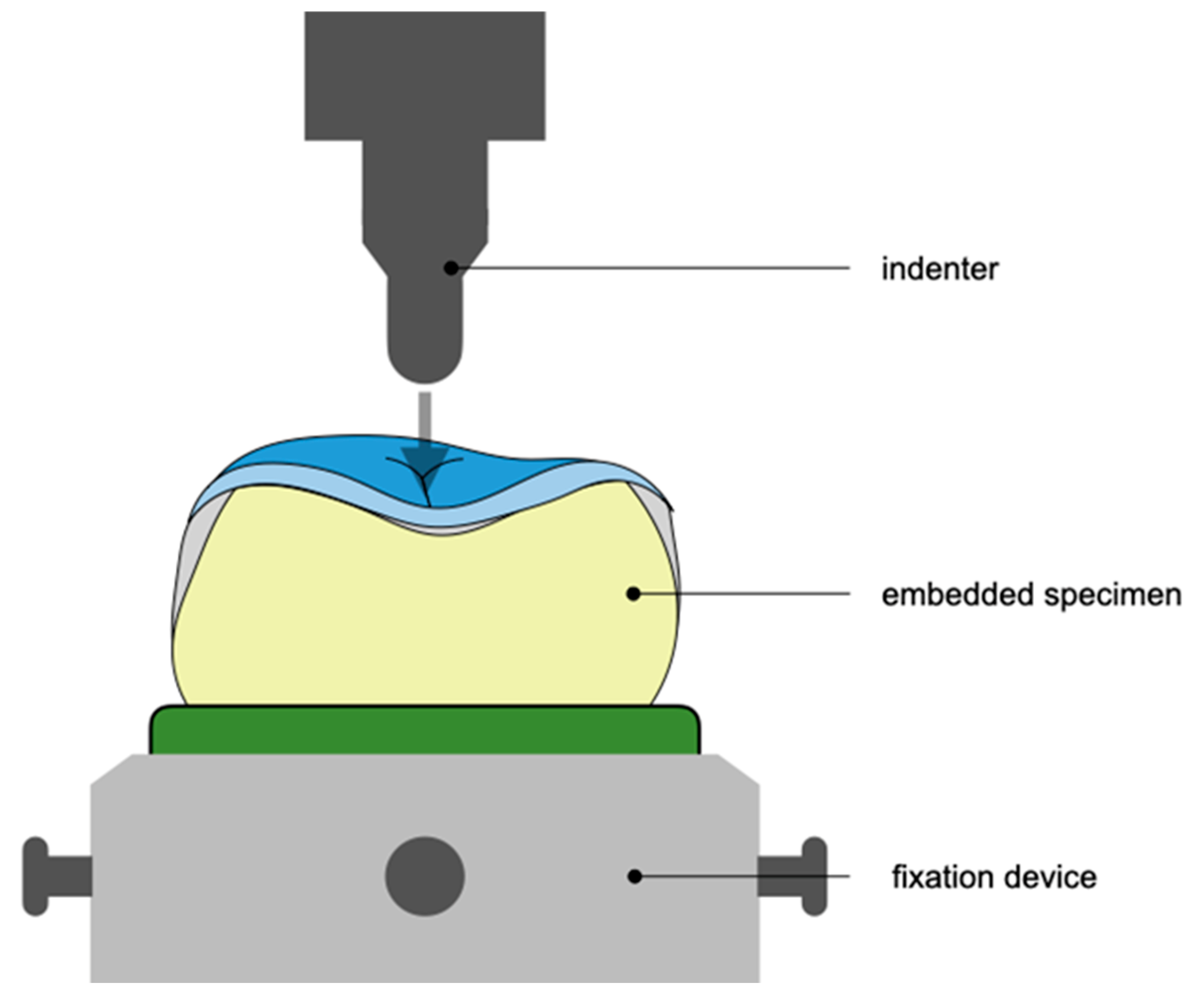
2.7. Static Loading of Specimens

The cemented occlusal veneers were fractured in a universal testing machine (Zwick, Roell Z010, Zwick, Ulm, Germany). An axial load was applied perpendicular to the occlusal plane at 1 mm/min until failure (Figure 2). The load required for failure (Fmax) was measured. Fracture types were categorized as (1) score 0: no visible fracture; (2) score 1: cohesive fracture within the restoration; (3) score 2: cohesive fracture of the restoration; (4) score 3: fracture of the restoration–cement–tooth complex [11]. Fracture types were assessed by using a stereomicroscope (Leica DFC300 FX; 10× magnification; Wetzlar, Germany).

2.8. Statistical Analysis

The load-bearing capacity of three types of millable lithium silicate derivates and one lithium disilicate as occlusal veneers on eroded molars were assessed by testing 20 specimens of each material type for maximum force tolerance (Fmax) until failure. Means, medians, standard deviations, quartiles, and the minimum and maximum values were used to describe the measured values. Statistical comparisons among materials were performed by using the non-parametric Wilcoxon rank-sum test, with p-values adjusted according to Holm’s method. Failure scores, a categorical variable, were summarized by counts and proportions [22,23].
Under the assumption of a Weibull distribution, a parametric approach was explored. Several models were compared: “global” (no difference in materials), “scale” (each material has a different scale but shares the same shape parameter), “shape” (each material has a different shape but shares the same scale parameter), and “scale and shape” (each material has its independent scale and shape parameter). The likelihood ratio tests indicated differences in scale parameters among materials but no significant differences in shape parameters. Diagnostic plots confirmed the validity of the model assumptions [24,25].
All statistical analyses and plots were computed with R statistical software (R version 4.2.3 (2023-03-15 ucrt)–“Shortstop Beagle”) [26], and the significance level α was set to 5%.

3. Results

3.1. Fatigue Resistance

Out of all tested specimens, three failed during artificial aging procedures. These failures were due to the debonding of the restoration and were observed in two specimens from the “Celt” group and one from the “Nice” group. Consequently, the fatigue resistance was 100% for the “Emax” and “Vita” groups, 90% for the “Celt” group, and 95% for the “Nice” group.

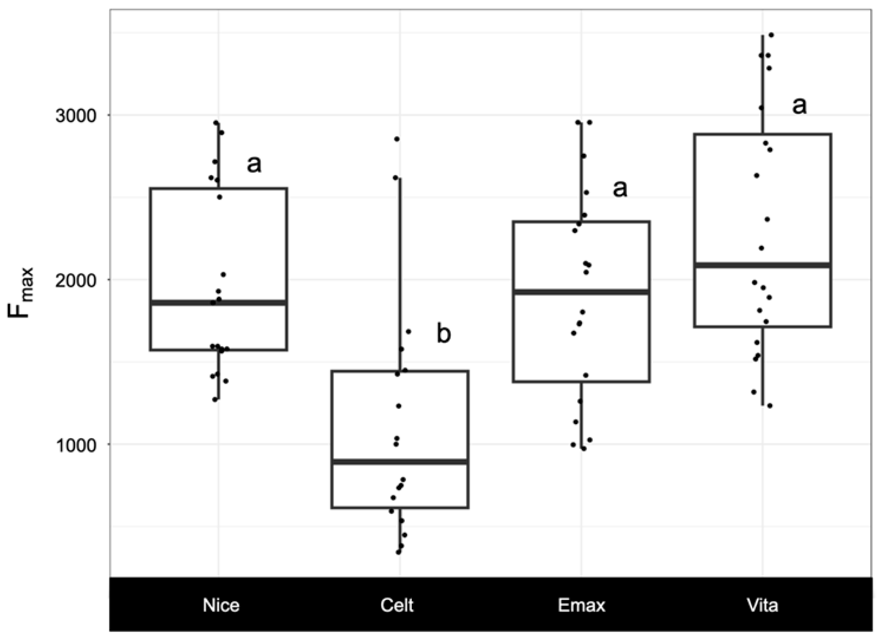
3.2. Load-Bearing Capacity Fmax

The “Vita” group exhibited the highest median Fmax value at 2087 N (Table 2, Figure 3). The median Fmax values for the test groups ranged from 892 N to 2087 N. Statistically significant differences were observed between the “Celt” group and the other groups (“Nice”, “Emax”, and “Vita”) (p < 0.005).

3.3. Failure Types

Failure type 2 was the most commonly observed failure mode across all groups (Table 3). Failure type 3 followed as the second most common type. Notably, the “Nice” group did not exhibit any instances of failure type 1.

4. Discussion

The purpose of this study was to evaluate the load-bearing capacity and fatigue resistance of three milled lithium silicate derivative materials and one milled lithium disilicate material used as ultra-thin occlusal veneers on eroded molars. The results showed that the “Nice”, “Emax”, and “Vita” groups had statistically significant differences in load-bearing capacity in comparison with the “Celt” group. Supporting their suitability for restorative applications under high occlusal loads, the results indicate that most of the materials showed excellent fatigue resistance with minimal failure after thermo-mechanical ageing.
Three restorations failed during artificial aging, primarily due to the debonding of the restoration, with two failures in the “Celt” group and one in the “Nice” group. The high fatigue resistance observed in the “Emax” and “Vita” groups suggests superior adhesive performance and material stability. Although the bonding protocol and preparation process were standardized across all groups, variations in substrate properties, material chemistry, or adherence to the bonding protocol may explain the differences in debonding rates. The consistent preparation and the random allocation of specimens in all groups to simulate erosive defects implies that substrate variations likely did not significantly impact outcomes. Previous studies have noted higher tendency for debonding when ceramics are bonded to dentin rather than enamel [27]. Despite these differences, all tested materials demonstrated viability for dental restorations, with most specimens enduring the simulated aging process without failure. This highlights the mechanical durability of ultra-thin lithium disilicate veneers, which can withstand typical cyclic stresses and temperature fluctuations within the oral cavity over extended periods [28,29,30].
All tested materials showed high load-bearing capacities, exceeding typical masticatory forces, including those in patients with bruxism [28,31]. The null hypothesis proposed that there would be no significant difference in the load-bearing capacity (Fmax) among the tested groups was rejected. The “Celt” group demonstrated significantly lower Fmax values compared with the other groups. The observed differences may be attributed to variations in the chemical composition and structural properties of the materials. The “Celt” material, a lithium silicate derivative reinforced with 10% zirconia, is supposed to strengthen the ceramic matrix by interrupting crack propagation [32]. However, the lower crystalline filler volume in “Celt” compared with “Emax” may reduce its mechanical strength [33]. Additionally, differences in the milling process—such as the use of partially sintered blocks in the “Celt” group versus fully sintered blocks in “Emax”—may influence material properties and load-bearing capacity. The findings align with previous research comparing “Celt” and “Vita” on titanium implant abutments, which also found significant differences in load-bearing capacity [34].
The materials in this study showed similar load-bearing capacity to lithium disilicate restorations, as shown in previous research [11]. However, both lithium silicate derivatives and lithium disilicate appear to have lower load-bearing capacity when applied as ultra-thin occlusal veneers on eroded molars [35]. Cohesive fractures were the most frequently observed failure type across all groups, consistent with previous studies [11,36,37]. Cohesive fractures occur through the ceramic, indicating the material’s robustness under high loads. Complete fractures of the restoration–cement–tooth complex were observed only at high loads, highlighting the structural integrity of the restorative materials. No minor fractures were observed, which would typically require only repair rather than complete replacement. This finding underscores the clinical reliability of these materials, minimizing technical complications and improving patient outcomes.
Future studies with larger sample sizes could better capture material-specific effects on mechanical performance. Additionally, this in vitro study may not fully replicate the complex biomechanical and environmental conditions in the oral cavity. Saliva, temperature fluctuations, and other intraoral factors could influence the performance and longevity of these materials. Finding that high in vitro forces are required for failure suggests that ultra-thin lithium disilicate veneers remain resilient under typical oral forces, even under extreme conditions. The high mechanical loads required to fracture the specimens suggest that forces beyond those encountered in normal mastication—including up to 800 N in patients with bruxism—are required for failure [28]. Nevertheless, little is known about how these in vitro results can be transferred into a real-world clinical scenario. Although a recent clinical study indicates good and stable results of ultra-thin occlusal veneers up to three years [38], this warrants further investigations in clinical studies and in particular in bruxing patients.

5. Conclusions

All tested materials demonstrated high fracture and fatigue resistance, making them suitable for ultra-thin occlusal veneers under high masticatory forces. Differences in performance were linked to variations in composition and material properties, with cohesive fractures being the most common failure type. Despite limitations, the study confirms the robustness of these materials, warranting in vivo validation for clinical application.

Author Contributions

L.F.: Investigation and Writing—original draft. L.W.: Investigation, Formal analysis, and Writing—review and editing. D.W.: Formal analysis and Writing—review and editing. M.Ö.: Investigation, Formal analysis, and Writing—review and editing. A.I.: Methodology, Funding acquisition, Conceptualization, Writing—original draft, and Writing—review and editing. All authors have read and agreed to the published version of the manuscript.

Funding

This research was funded by the Clinic of Clinic of Masticatory Disorders and Dental Biomaterials and Clinic of Reconstructive Dentistry, Center for Dental Medicine, University of Zurich, Switzerland and by the Swiss Society for Reconstructive Dentistry (SSRD).

Institutional Review Board Statement

Not applicable.

Informed Consent Statement

Not applicable.

Data Availability Statement

The data presented in this study are available on request from the corresponding author.

Conflicts of Interest

The authors declare no competing interests.

References

  1. Lussi, A.; Carvalho, T.S. Erosive tooth wear: A multifactorial condition of growing concern and increasing knowledge. Monogr. Oral Sci. 2014, 25, 1–15. [Google Scholar] [PubMed]
  2. Jaeggi, T.; Lussi, A. Prevalence, incidence and distribution of erosion. Monogr. Oral Sci. 2006, 20, 44–65. [Google Scholar] [PubMed]
  3. Lee, A.; He, L.H.; Lyons, K.; Swain, M.V. Tooth wear and wear investigations in dentistry. J. Oral Rehabil. 2012, 39, 217–225. [Google Scholar] [CrossRef] [PubMed]
  4. Bazos, P.; Magne, P. Bio-emulation: Biomimetically emulating nature utilizing a histo-anatomic approach; structural analysis. Eur. J. Esthet. Dent. 2011, 6, 8–19. [Google Scholar] [PubMed]
  5. Loomans, B.; Opdam, N.; Attin, T.; Bartlett, D.; Edelhoff, D.; Frankenberger, R.; Benic, G.; Ramseyer, S.; Wetselaar, P.; Sterenborg, B.; et al. Severe tooth wear: European consensus statement on management guidelines. J. Adhes. Dent. 2017, 19, 111–119. [Google Scholar]
  6. Ioannidis, A.; Pitta, J.; Panadero, R.A.; Chantler, J.G.M.; Villar, S.F.; Fonseca, M.; Lozano, J.G.; Hämmerle, C.H.F.; Hey, J.; Karasan, D.; et al. Rehabilitation Strategies and Occlusal Vertical Dimension Considerations in the Management of Worn Dentitions: Consensus Statement From SSRD, SEPES, and PROSEC Conference on Minimally Invasive Restorations. J. Esthet. Restor. Dent. 2025, 1–5. [Google Scholar] [CrossRef]
  7. Edelhoff, D.; Sorensen, J.A. Tooth structure removal associated with various preparation designs for anterior teeth. J. Prosthet. Dent. 2002, 87, 503–509. [Google Scholar] [CrossRef]
  8. Kontakiotis, E.G.; Filippatos, C.G.; Stefopoulos, S.; Tzanetakis, G.N. A prospective study of the incidence of asymptomatic pulp necrosis following crown preparation. Int. Endod. J. 2015, 48, 512–517. [Google Scholar] [CrossRef]
  9. Valderhaug, J.; Jokstad, A.; Ambjørnsen, E.; Norheim, P.W. Assessment of the periapical and clinical status of crowned teeth over 25 years. J. Dent. 1997, 25, 97–105. [Google Scholar] [CrossRef]
  10. Albelasy, E.H.; Hamama, H.H.; Tsoi, J.K.H.; Mahmoud, S.H. Fracture resistance of cad/cam occlusal veneers: A systematic review of laboratory studies. J. Mech. Behav. Biomed. Mater. 2020, 110, 103948. [Google Scholar] [CrossRef]
  11. Zumstein, K.; Fiscalini, L.; Al-Haj Husain, N.; Evci, E.; Özcan, M.; Ioannidis, A. Load-bearing capacity of pressable lithium disilicates applied as ultra-thin occlusal veneers on molars. J. Mech. Behav. Biomed. Mater. 2022, 136, 105520. [Google Scholar] [CrossRef] [PubMed]
  12. Paqué, P.N.; Gantner, C.; Mätzener, K.J.; Özcan, M.; Ioannidis, A. Load-bearing capacity, internal accuracy and time-efficiency of heat-pressed, milled and 3D-printed lithium disilicate ultra-thin occlusal veneers. Dent. Mater. 2024, 40, 1602–1610. [Google Scholar] [CrossRef] [PubMed]
  13. Jurado, C.A.; Lee, D.; Ramirez, P.; Cortes-Treviño, D.A.; Tsujimoto, A. Fracture Resistance of Chairside CAD/CAM Lithium Disilicate-reinforced Ceramic Occlusal Veneers with and Without Margin and Full Coverage Crowns. Oper. Dent. 2024, 49, 84–90. [Google Scholar] [CrossRef] [PubMed]
  14. Mörikofer, N.; Benic, G.I.; Park, J.M.; Özcan, M.; Hüsler, J.; Ioannidis, A. Relationship between internal accuracy and load-bearing capacity of minimally invasive lithium disilicate occlusal veneers. Int. J. Prosthodont. 2021, 34, 365–372. [Google Scholar] [CrossRef]
  15. Alkadi, L.; Ruse, N.D. Fracture toughness of two lithium disilicate dental glass ceramics. J. Prosthet. Dent. 2016, 116, 591–596. [Google Scholar] [CrossRef]
  16. Traini, T.; Sinjari, B.; Pascetta, R.; Serafini, N.; Perfetti, G.; Trisi, P.; Caputi, S. The zirconia-reinforced lithium silicate ceramic: Lights and shadows of a new material. Dent. Mater. J. 2016, 35, 748–755. [Google Scholar] [CrossRef]
  17. Krithikadatta, J.; Gopikrishna, V.; Datta, M. CRIS Guidelines (Checklist for Reporting In-vitro Studies): A concept note on the need for standardized guidelines for improving quality and transparency in reporting in-vitro studies in experimental dental research. J. Conserv. Dent. 2014, 17, 301–304. [Google Scholar] [CrossRef]
  18. Maeder, M.; Pasic, P.; Ender, A.; Özcan, M.; Benic, G.I.; Ioannidis, A. Load-bearing capacities of ultra-thin occlusal veneers bonded to dentin. J. Mech. Behav. Biomed. Mater. 2019, 95, 165–171. [Google Scholar] [CrossRef]
  19. Human Research Ordinance (810.301), Art. 30. Available online: https://www.fedlex.admin.ch/eli/cc/2013/642/en (accessed on 3 March 2025).
  20. World Medical Association (WMA). Declaration of Helsinki—Ethical Principles for Medical Research Involving Human Subjects. In Proceedings of the 64th WMA General Assembly, Fortaleza, Brazil, 16–19 October 2013. [Google Scholar]
  21. Human Research Act (810.30), Art. 2 and 32, Human Research Ordinance (810.301), Art. 25. Available online: https://www.fedlex.admin.ch/eli/cc/2013/642/en (accessed on 3 March 2025).
  22. Pohlert, T. The Pairwise Multiple Comparison of Mean Ranks Package (PMCMR). R Package. 2014. Available online: https://CRAN.R-project.org/package=PMCMR (accessed on 3 March 2025).
  23. Graves, S.; Piepho, H.; Selzer, L. multcompView: Visualizations of Paired Comparisons. R Package Version 0.1-8. 2019. Available online: https://CRAN.R-project.org/package=multcompView (accessed on 3 March 2025).
  24. Delignette-Muller, M.L.; Dutang, C. fitdistrplus: An R Package for Fitting Distributions. J. Stat. Softw. 2015, 64, 1–34. [Google Scholar] [CrossRef]
  25. Rigby, R.A.; Stasinopoulos, D.M. Generalized additive models for location, scale and shape, (with discussion). Appl. Statist. 2005, 54 Pt 3, 507–554. [Google Scholar]
  26. R Core Team. R: A Language and Environment for Statistical Computing; R Foundation for Statistical Computing: Vienna, Austria, 2021; Available online: https://www.R-project.org/ (accessed on 3 March 2025).
  27. Gurel, G.; Sesma, N.; Calamita, M.A.; Coachman, C.; Morimoto, S. Influence of enamel preservation on failure rates of porcelain laminate veneers. Int. J. Periodont. Restor. Dent. 2013, 33, 31–39. [Google Scholar] [CrossRef] [PubMed]
  28. Bates, J.F.; Stafford, G.D.; Harrison, A. Masticatory function—A review of the literature. 1. The form of the masticatory cycle. J. Oral Rehabil. 1975, 2, 281–301. [Google Scholar] [CrossRef] [PubMed]
  29. DeLong, R.; Douglas, W.H. An artificial oral environment for testing dental materials. IEEE Trans. Biomed. Eng. 1991, 38, 339–345. [Google Scholar] [CrossRef] [PubMed]
  30. Steiner, M.; Mitsias, M.E.; Ludwig, K.; Kern, M. In vitro evaluation of a mechanical testing chewing simulator. Dent. Mater. 2009, 25, 494–499. [Google Scholar] [CrossRef]
  31. Edmonds, H.M.; Glowacka, H. The ontogeny of maximum bite force in humans. J. Anat. 2020, 237, 529–542. [Google Scholar] [CrossRef]
  32. Mavriqi, L.; Valente, F.; Murmura, G.; Sinjari, B.; Macrì, M.; Trubiani, O.; Caputi, S.; Traini, T. Lithium disilicate and zirconia reinforced lithium silicate glass-ceramics for CAD/CAM dental restorations: Biocompatibility, mechanical and microstructural properties after crystallization. J. Dent. 2022, 119, 104054. [Google Scholar] [CrossRef]
  33. Kelly, J.R.; Benetti, P. Ceramic materials in dentistry: Historical evolution and current practice. Aust. Dent. J. 2011, 56 (Suppl. S1), 84–96. [Google Scholar] [CrossRef]
  34. Weyhrauch, M.; Igiel, C.; Scheller, H.; Weibrich, G.; Lehmann, K.M. Fracture Strength of Monolithic All-Ceramic Crowns on Titanium Implant Abutments. Int. J. Oral Maxillofac. Implants 2016, 31, 304–309. [Google Scholar] [CrossRef]
  35. Ioannidis, A.; Bomze, D.; Hämmerle, C.H.F.; Hüsler, J.; Birrer, O.; Mühlemann, S. Load-bearing capacity of CAD/CAM 3D-printed zirconia, CAD/CAM milled zirconia, and heat-pressed lithium disilicate ultra-thin occlusal veneers on molars. Dent. Mater. 2020, 36, e109–e116. [Google Scholar] [CrossRef]
  36. Gierthmuehlen, P.C.; Jerg, A.; Fischer, J.B.; Bonfante, E.A.; Spitznagel, F.A. Posterior minimally invasive full-veneers: Effect of ceramic thicknesses, bonding substrate, and preparation designs on failure-load and -mode after fatigue. J. Esthet. Restor. Dent. 2022, 34, 145–153. [Google Scholar] [CrossRef]
  37. Ioannidis, A.; Muhlemann, S.; Ozcan, M.; Husler, J.; Hammerle, C.H.F.; Benic, G.I. Ultra-thin occlusal veneers bonded to enamel and made of ceramic or hybrid materials exhibit load-bearing capacities not different from conventional restorations. J. Mech. Behav. Biomed. Mater. 2019, 90, 433–440. [Google Scholar] [CrossRef]
  38. Schlichting, L.H.; Resende, T.H.; Reis, K.R.; Raybolt Dos Santos, A.; Correa, I.C.; Magne, P. Ultrathin CAD-CAM glass-ceramic and composite resin occlusal veneers for the treatment of severe dental erosion: An up to 3-year randomized clinical trial. J. Prosthet. Dent. 2022, 128, e1–e158. [Google Scholar] [CrossRef]
Figure 1. Schematic sketch of cross-section of cemented overlay on molar.
Prosthesis 07 00043 g001
Figure 2. Schematic sketch of cross-section of cemented overlay on molar embedded in a cylinder during static loading.
Prosthesis 07 00043 g002
Figure 3. Box plots for the Fmax values of all the groups—Nice, Celt, Emax and Vita. Statistically significant differences are indicated by different letters (compact letter display method).
Prosthesis 07 00043 g003
Table 1. Restorative materials and respective compositions for the control and the test groups provided by the manufacturer.
GroupRestorative MaterialChemical CompositionFlexural Strength (MPa)
NiceLithium aluminosilicate ceramic reinforced with lithium disilicate (N!ce, Straumann AG, Basel, Switzerland)SiO2(64–70); Li2O(10.5–12.15); Al2O3(10.5–11.5); Na2O(1–3); K2O(0–3); P2O5(3–8); ZrO2(0–0.5); CaO(1–2); Coloring oxides (0–9)350 MPa
CeltZirconia-reinforced lithium silicate (Celtra Duo, Dentsply Sirona, Hanau-Wolfgang, Germany)SiO2(58); Li2O(18.5); ZrO2(10.1); P2O5(5); Ce2O3(2); Al2O3(1.9); TbO2(1)370 MPa
EmaxLithium disilicate ceramic (IPS e.max CAD PrograMill; Ivoclar Vivadent, Schaan, Liechtenstein)SiO2(57–80); Li2O(11–19); K2O(0–13); P2O5(0–11); ZrO2(0–8); ZnO(0–5); Al2O3(0–5); MgO (0–5); Coloring oxides (0–8)530 MPa
VitaZirconia-reinforced glass ceramic (VITA Suprinity PC, Vita Zahnfabrik, Bad Säckingen, Germany)SiO2(56–64); Li2O(15–21); K2O(1–4); P2O5(3–8); Al2O3(1–4); ZrO2(8–12); Ce2O3(0–4); La2O3 (0.1); Pigments (0–6)420 MPa
Table 2. Fmax values with means ± standard deviations (SDs), medians and ranges from minimum (min) to maximum (max) of all the groups—Nice, Celt, Emax, and Vita.
GroupMean ± SDMedianRange (Min to Max)
Nice1968 ± 56318591271 to 2953
Celt1118 ± 721892343 to 2854
Emax1910 ± 6411924973 to 2955
Vita2298 ± 74420871234 to 3486
Table 3. Fracture scores in percentage (%) of all the groups—Nice, Celt, Emax, and Vita.
GroupnScore 0 (%)Score 1 (%)Score 2 (%)Score 3 (%)
Nice1900955
Celt1805905
Emax20058015
Vita200106525
Disclaimer/Publisher’s Note: The statements, opinions and data contained in all publications are solely those of the individual author(s) and contributor(s) and not of MDPI and/or the editor(s). MDPI and/or the editor(s) disclaim responsibility for any injury to people or property resulting from any ideas, methods, instructions or products referred to in the content.

