Article

Dental Prostheses Materials: Corrosion Behavior of Co-Cr-W Alloys Processed by SLM Technique

by
Elena-Raluca Baciu
1,
Livia Bobu
2,*,
Ramona Cimpoeșu
3,
Dana Gabriela Budală
1,
Roxana-Ionela Vasluianu
1,
Gabriela Luminița Gelețu
2,
Costin Iulian Lupu
1,
Anca Vițalariu
1 and
Alice Murariu
2
1
Department of Implantology, Removable Dentures, Dental Technology, Faculty of Dental Medicine, “Grigore T. Popa” University of Medicine and Pharmacy, 700115 Iasi, Romania
2
Department of Surgery, Faculty of Dental Medicine, “Grigore T. Popa” University of Medicine and Pharmacy, 700115 Iasi, Romania
3
Faculty of Materials Science and Engineering, “Gheorghe Asachi” Technical University, 700259 Iasi, Romania
*
Author to whom correspondence should be addressed.
Prosthesis 2025, 7(2), 27; https://doi.org/10.3390/prosthesis7020027
Submission received: 27 January 2025 / Revised: 28 February 2025 / Accepted: 1 March 2025 / Published: 4 March 2025
(This article belongs to the Special Issue Advancements in Adhesion Techniques and Materials in Prosthodontics)

Abstract

:
Background/Objectives: Corrosion resistance of dental alloys, indicated by metal ion release, is a significant concern. The aim of this study was to investigate the impact of various sandblasting methods on the corrosion resistance of Co-Cr-W alloy samples in an artificial saliva solution. Materials and Methods: Co-Cr-W samples, obtained by additive manufacturing (selective laser melting) and subjected to different processes, were evaluated using electrochemical corrosion tests on potentiostat equipment in Carter–Brugirard artificial saliva. All measurements were conducted at a temperature of 37 °C. The morphology of the samples was analyzed using scanning electron microscopy to highlight changes in roughness and the effects of the electrolyte solution on the surface of the samples. Results: The corrosion current density values decreased as surface processing increased, dropping from an average of 38.5 μA/cm2 in non-sandblasted samples (group A) to 0.74 μA/cm2 in samples sandblasted with both Al2O3 and glass particles (group C), while the corrosion rate of group A (434.8 mm/year) was significantly higher than that of group C (8.08 mm/year). Conclusions: The results demonstrated different behaviors between sandblasted and non-sandblasted samples, which could be attributed to surface morphology differences influencing the corrosion process.

1. Introduction

The CAD/CAM-based processing inherent in SLM (selective laser melting), DMLM (direct metal laser melting), and LPBF (laser powder bed fusion) technologies allows for the creation of metal components with complex geometries [1,2,3,4].
Recognized as one of the most innovative dental technologies, selective laser melting (SLM) is particularly well-suited for prosthetic dentistry due to its versatility. It enables the production of dental constructions from a wide range of materials, including thermoplastic polymers, waxes, metals and alloys, ceramics, and thermoplastic composites [5,6]. In SLM processing, alloy powder particles are melted using a laser beam with varying energy and power levels. The physical, chemical, and mechanical properties of the solidified metal bath from the liquid state are significantly influenced by the technical processing parameters [7,8,9].
Dental alloys encompass a wide range of compositions, including those based on gold, silver, palladium, nickel, cobalt, iron, and titanium. Cobalt-based alloys, commonly used for removable partial denture (RPD) and metal crown frameworks, are produced using SLM machines without compromising the quality of either the alloy or the restoration [10,11]. These alloys, typically containing Cr, Mo, W, Si, and Mn, readily interact with the biological environment of the oral cavity.
Traditional lost-wax casting involves multiple steps that often introduce imperfections, porosity, and high costs [2,3,4,12]. Advancements in additive manufacturing simplify the process, addressing these challenges while improving restoration quality and reducing expenses. SLM offers significant advantages over traditional fabrication methods by producing a unique microstructure with minimal internal porosity, enhanced internal fitting, and bond strengths comparable to porcelain. A key benefit of SLM is its ability to significantly reduce the internal porosity of Co-Cr dental alloys compared to cast methods, achieved through its precise and controlled manufacturing parameters. This reduction effectively resolves porosity-related issues, such as diminished mechanical properties and increased corrosion susceptibility [13].
Dental alloys interact with the oral environment, potentially causing localized, extensive, or systemic changes such as metallic taste, tooth discoloration, allergic reactions, and even cancer. The corrosion process, influenced by the alloy’s composition and environmental factors, can vary in rate and severity. Research emphasizes the importance of alloy composition, highlighting that casting structures, alloying element proportions, and processing techniques determine the release of products into gum tissues. These released ions can trigger adverse effects like gingival swelling, inflammation, mucosal discomfort, lichenoid reactions, and allergies [14,15,16]. To minimize these risks, exploring advanced processing methods to improve corrosion resistance is essential.
Recent studies have focused on the corrosion resistance of additively manufactured Co-Cr dental alloys produced using selective laser melting (SLM) [17,18,19,20]. The corrosion resistance of these materials is determined by interfacial interactions between the electrolyte and the material’s surface. Advancements in processing techniques are critical for optimizing the performance and biocompatibility of SLM dental alloys.
The objective of this study was to examine the impact of various sandblasting methods on the corrosion resistance of Co-Cr-W alloy samples in an artificial saliva solution. To address this, we proposed the following null hypothesis: there is no significant difference in the corrosion behavior of Co-Cr-W alloy samples produced through SLM processing and finished using two different sandblasting techniques when subjected to electrochemical corrosion in Carter–Brugirard artificial saliva.

2. Materials and Methods

2.1. Specimens’ Fabrication

The fabrication process began with the design of the specimens using 3D computer-aided design (CAD) software, specifically Meshmixer (version 3.5.0, Autodesk, San Rafael, CA, USA). Once the designs were completed, they were exported and sent to a dental laboratory for fabrication via 3D printing, utilizing a Co-Cr-W alloy powder (Starbond CoS55, S&S Scheftner, Mainz, Germany) in the selective laser melting (SLM) process, performed on the SLM 50 equipment (Realizer GmbH, Borchen, Germany). The chemical composition of the alloy used in this process was 59.2 wt% Co, 27.5 wt% Cr, 9.1 wt% W, 4 wt% Mo, 0.20 wt% other elements (C, Fe, Mn), with no Ni, Be, or Ga content.
The SLM 50 system maintained a consistent metal powder layer thickness of 25 μm (Figure 1), with operational parameters including a laser power of 70 W, a scanning rate of 1000 mm/s, and an exposure time of 20 μs [21]. All specimens were fabricated in strict accordance with the manufacturer’s guidelines, resulting in cylindrical specimens with dimensions of 10 mm in diameter and 4 mm in thickness.
Following the specimen fabrication, three experimental groups, each consisting of 10 specimens, were prepared as follows:
  • Group A: Non-sandblasted samples.
  • Group B: Samples sandblasted with alumina (Al2O3) particles.
  • Group C: Samples successively sandblasted with alumina (Al2O3) particles followed by glass particles.
The alumina particles used had a granulation of F100, with dimensions ranging between 106 and 150 μm (working pressure of 3.5–4 bar), while the glass particles were spherical with diameters of 70–110 μm (working pressure of 2.5 bar). The sandblasting process was carried out under controlled pressure, with 4 bar applied for the alumina and 2.5 bar for the glass particles. A specialized Renfert Basic Eco sandblasting unit (Renfert, Hilzingen, Germany), equipped with ceramic nozzles appropriate for the size of the particles, was employed for this procedure.
After surface pretreatments, the specimens underwent ultrasonic cleaning in acetone, ethanol, and deionized water for 15 min. They were then dried using oil-free compressed air, steam-sterilized at 121 °C for 30 min, and placed in a drying oven at 65 °C for 24 h [22].

2.2. Microstructural Characterization and Chemical Composition Analysis

For microstructural characterization and chemical composition analysis, the Co-Cr-W alloys were examined by using a scanning electron microscope type Vega Tescan LMH II (Tescan, Brno, Czech Republic), SE detector, the emission current was between 0.5 pA and 500 nA, and the acceleration voltage was 30 kV.
The chemical composition determinations were made using the energy dispersive detector for X-rays QUANTAX -Bruker EDS detector (5–6 kcps input signal, 15.5 mm working distance, and Esprit 2.2 software) on five different areas.
The surface of the samples was analyzed using the SEM Tescan software’s (version 3.5.0.0) light intensity variation feature, measured in analog-to-digital units (ADU). These dimensionless units were compared to assess the effects of the different processing stages on the material’s surface.

2.3. Electrochemical Corrosion Testing

In the electrochemical corrosion test, one side of each sample (n = 30) was designated as the test surface, while the remaining sides were embedded in epoxy resin. Electrochemical impedance spectroscopy (EIS) was conducted using a Volta Lab21 potentiostat (Radiometer Analytical SAS, Lyon, France), controlled by a computer and Volta Master 4 software (Radiometer Analytical SAS, Lyon, France). The experimental setup involved a three-electrode system, where the Co-Cr alloy specimens served as the working electrode. A saturated calomel electrode was used as the reference electrode, and a platinum electrode functioned as the counter electrode. The working electrode was incorporated into a Teflon holder, exposing a surface area of 0.2 cm2.
The specimens were immersed in Carter–Brugirard artificial saliva, which contained 700 mg/L NaCl, 1200 mg/L KCl, 260 mg/L KH2PO4, 330 mg/L KSCN, 190 mg/L Na2HPO4·H2O, 1500 mg/L NaHCO3, and 130 mg/L CH4N2O, with a pH of 7.6 immediately after preparation [23]. The immersion was conducted at a constant temperature of 37.0 °C.
To evaluate the corrosion resistance of the three sample groups, both linear and cyclic polarization curves were recorded. Linear voltammetry was performed with a scan rate of 1 mV/s across a potential range of −600 mV to +600 mV. The corrosion potential (Ecorr), Tafel slopes (ba and bc), polarization resistance (Rp), corrosion current density (Jcorr), and corrosion rate (CR) were determined using the Tafel extrapolation method. Electrochemical Impedance Spectroscopy (EIS) spectra were recorded over a frequency range of 10−2 Hz to 105 Hz, with an applied alternating potential signal of 10 mV in amplitude. Additionally, the open-circuit potential of the samples was measured for 6000 s prior to the polarization measurements.

2.4. Statistical Analysis

The statistical analysis was conducted using IBM SPSS software version 26.0 for Windows (SPSS Inc., Chicago, IL, USA). The normality of the variables was evaluated through the Kolmogorov-Smirnov and Shapiro–Wilk tests. Group comparisons were carried out using the Kruskal–Wallis test, with a significance threshold set at p ≤ 0.05.

3. Results

3.1. Microstructure and Chemical Composition

The powder analysis shown in Figure 2a highlighted the morphological characteristics of the particles used for selective laser melting (SLM), as observed through scanning electron microscopy (SEM). The 3D profile of the powders revealed a uniform particle distribution and appropriate agglomeration, which were essential for ensuring an efficient SLM process (Figure 2a).
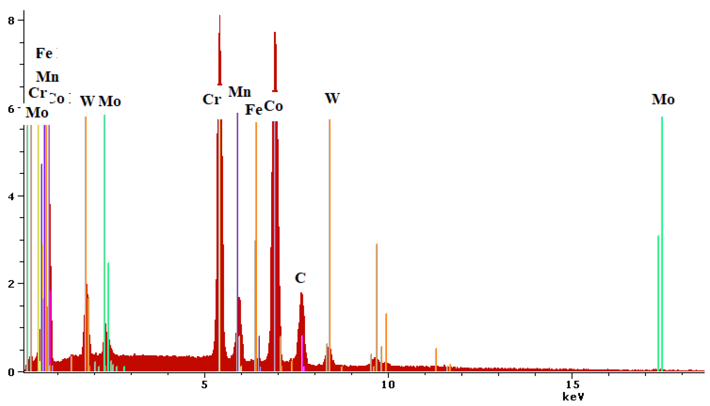
The chemical composition of the powder and samples, presented in Table 1 and Figure 3, was determined based on 20 measurements.
In the initial, non-sandblasted state, remnants of powder particles from the 3D printing process were visible on the surface (Figure 4a). However, after processing (Figure 4b,c), no residual particles remained, indicating a significant modification of the surface. The surface condition of the experimental samples also revealed clear differences in surface roughness. For instance, Group A exhibited ADU values ranging from 130 to 180, while Group B showed values between 110 and 160 ADU, and Group C displayed a range from 80 to 160. Additionally, the wider asperities in group A indicated a larger surface area exposed to the environment compared to groups B and C (Figure 4a,b).

3.2. Electrochemical Corrosion Analysis

Linear and cyclic potentiometry are effective techniques for analyzing the passive region, evaluating a material’s passivation potential in specific environments, assessing stability, and determining passivation quality based on the degradation rate. Figure 5a presents the linear polarization curves, while Figure 5b shows the cyclic polarization curves for three representative samples from the tested groups.
The main parameters of the corrosion process derived from the Tafel extrapolation are shown in Table 2 and Figure 6.
The corrosion current density (Jcorr) values showed a decreasing trend as surface processing increased, dropping from an average of 38.5 μA/cm2 in group A to 0.74 μA/cm2 in group C. Similarly, the corrosion rate of non-sandblasted samples was 434.8 mm/year, significantly higher than that of Al2O3 particle-sandblasted samples (156.1 mm/year) and even more so compared to samples sandblasted with both Al2O3 and glass particles (8.08 mm/year). This difference can be attributed to the non-homogeneous structure inherent to the 3D printing process.
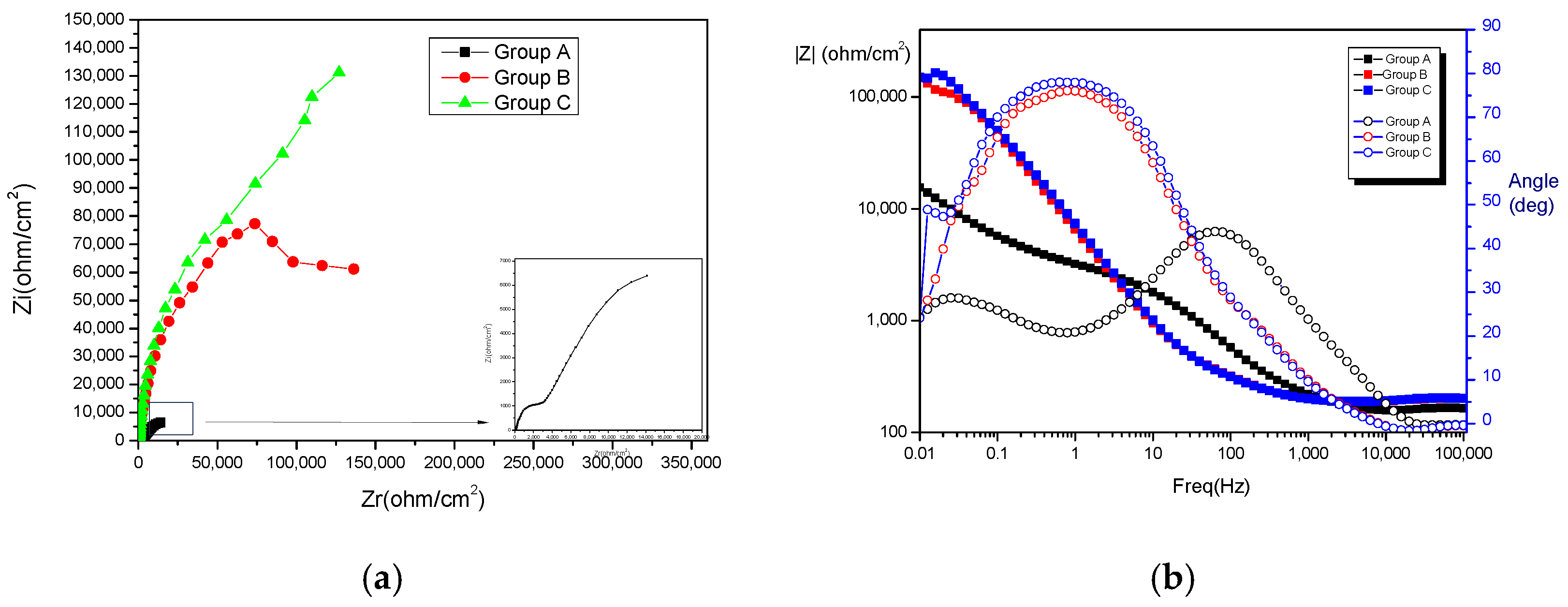
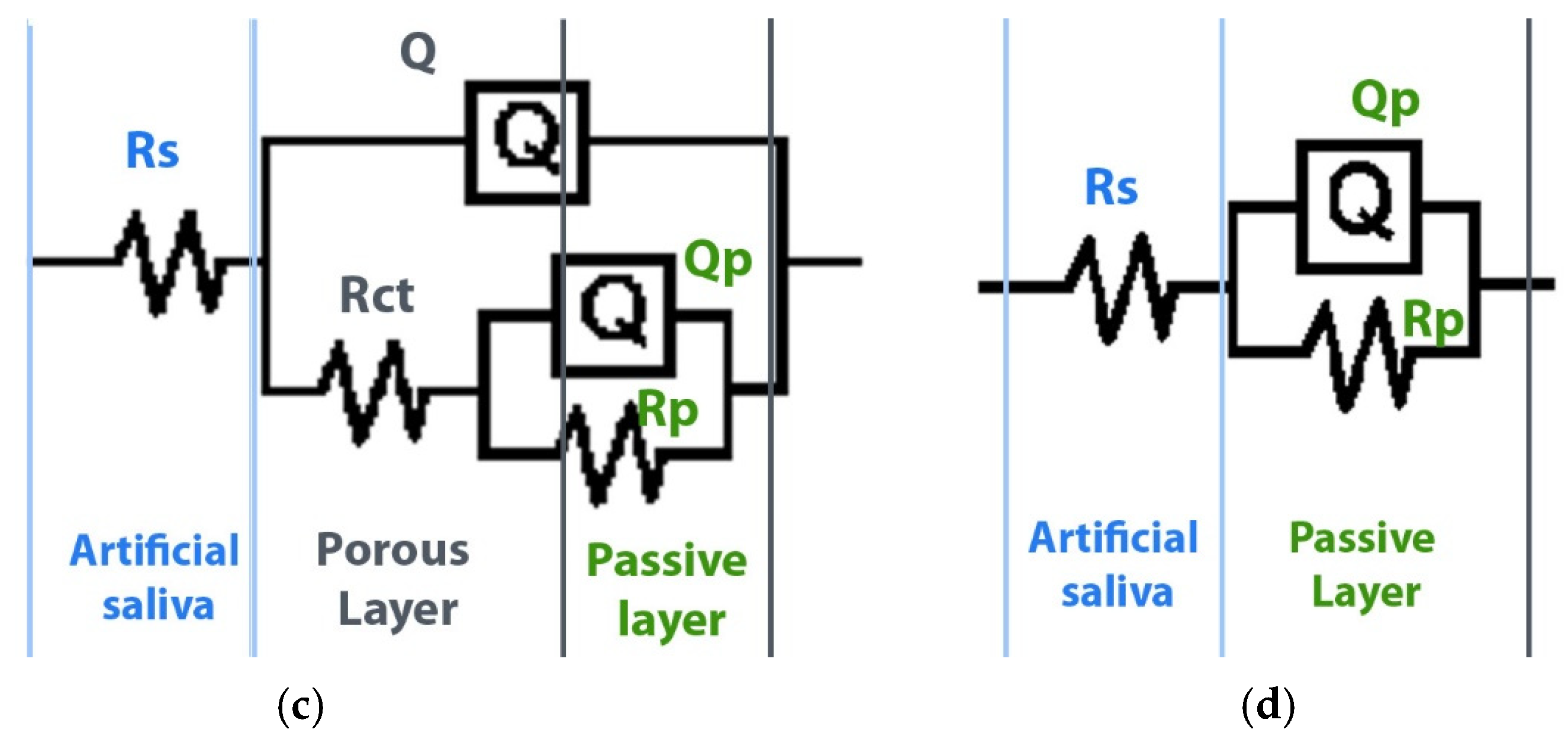
EIS tests were conducted to evaluate the influence of potential pores or micro-holes on the surface of the samples and their impact on corrosion behavior. Figure 7a presents the Nyquist plot for the three groups of samples recorded in Carter–Brugirard artificial saliva electrolyte solution, revealing a distinct difference in behavior between the two sandblasted samples and the non-sandblasted sample. Additionally, the Bode diagrams in Figure 7b illustrates resistance as a function of frequency. For samples in groups B and C, within the low and medium frequency ranges, the spectra exhibit a linear slope as the frequency decreases, with phase angle values approaching 80°. Figure 7c shows the equivalent circuit model used to describe the electrochemical behavior of Group A samples in artificial saliva, while Figure 7d represents the model for Groups B and C.
The analysis of the impedance plots was performed by fitting the data using ZSimpWin software(version 3.22), applying two equivalent circuit models, as shown in Figure 7c,d. The equivalent circuit parameters obtained for Groups A, B, and C are presented in Table 3. For Group A (non-sandblasted samples), the equivalent electrical circuit used consists of an ohmic solution resistance (Rs) in series with a parallel arrangement of a constant phase element (CPE) and a resistor. The CPE was introduced to represent a non-ideal capacitor due to surface heterogeneities and layer porosity. It includes two parameters: Q, which characterizes capacitance, and n, an exponent related to frequency dispersion, with values typically n < 1. The charge transfer resistance (Rct) exhibited a relatively high value, indicating a low corrosion rate.
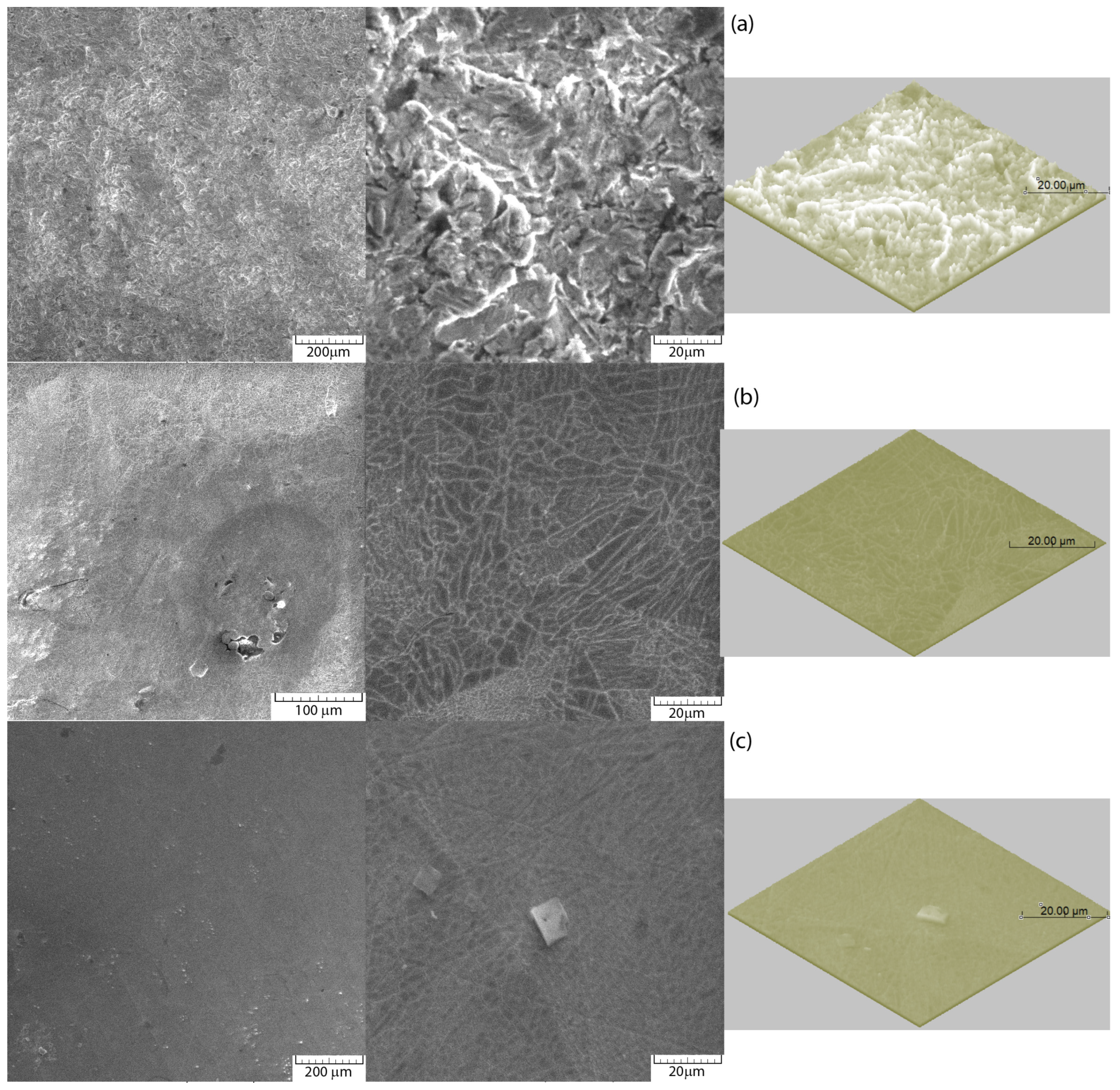
Corrosion occurred predominantly at the boundaries between the particles. In the non-sandblasted samples, pores of varying sizes, formed between the metal powder particles, were observed, while samples from groups B and C exhibited smaller pores, indicating a more compact surface structure (Figure 8a–c).

4. Discussion

In the oral cavity’s dynamic environment, dental alloys face challenges such as pH fluctuations, bacterial biofilms, and salivary enzymes. These factors, along with metabolites produced by bacteria—such as sulfides and organic acids—can alter the corrosion behavior of alloys by affecting oxygen concentration, salinity, and acidity [11,13,14]. The corrosion resistance of alloys is influenced by their composition, microstructure (single- or multi-phase), and the chosen processing and surface treatments [15]. Proper alloy selection and manufacturing techniques are therefore critical to ensuring long-term biocompatibility and minimizing adverse reactions [15,16].
SLM has been shown to enhance the corrosion resistance of Co-Cr dental alloys by improving microstructural homogeneity and reducing porosity, a key factor in corrosion susceptibility. EDS spectral analysis confirmed that the metallic material used for 3D printing is a Co-Cr alloy, widely employed in fabricating fixed prostheses. Comprising primarily Co and Cr at 84%, with strengthening elements such as W and Mo 13% (with W in higher proportion), the alloy exhibited a highly homogeneous microstructure with no significant porosity. This microstructural consistency, typical of Co-Cr alloys produced through selective laser melting (SLM), is attributed to the precise solidification process inherent in this manufacturing technique.
However, this inherent advantage of Co-Cr alloys is highly dependent on the manufacturing and post-processing techniques applied. Studies have demonstrated that surface treatments, such as sandblasting with alumina particles, further improve surface characteristics and corrosion resistance. Successive sandblasting with alumina and glass particles yielded optimal results, significantly enhancing the alloy’s corrosion resistance and reducing susceptibility to pitting and crevice corrosion [24]. The results showed that surface morphology, altered by sandblasting treatments, significantly influenced corrosion behavior, leading to the rejection of the null hypothesis regarding electrochemical behavior.
The findings support established theories in material science and electrochemistry by illustrating how surface morphology influences electrochemical reactions, enhances the stability of passive oxide films, reduces residual stresses, and improves wettability (lower contact angle). These factors collectively contribute to reduced corrosion rates and enhanced performance in demanding environments [21,25,26,27,28,29]. A recent systematic review emphasized the critical role of pretreatment with airborne-particle abrasion using Al2O3 in enhancing surface wettability and improving micromechanical retention between metal alloys and acrylic resin in removable partial dentures. The size of abrasive particles (e.g., 50 μm, 110 μm, and 250 μm) significantly affects the outcome, with larger particles creating rougher and more extensive contact surfaces. This effect is attributed to the crystalline structure of metals, where the interaction of abrasive particles with the metal’s grain boundaries influences the resulting surface texture. Additionally, the combination of airborne-particle abrasion with aluminum oxide particles and primers containing 10-methacryloyloxydecyl dihydrogen phosphate (10-MDP) monomer has been shown to further increase bond strength between heat-polymerized acrylic resin denture base materials and metal RPD framework [30].
For instance, Kanevo et al. [31] found that mirror-finishing using alumina particles, particularly for base-metal alloys, was shown to be the most effective method, and recommended for assessing corrosion resistance. Porojan et al. [32] conducted a comparative study on samples with different surface morphologies (polished and sandblasted with 150 μm aluminum oxide particles) produced by various additive technologies (SLS/SLM), concluding that surface quality after finishing significantly impacts corrosion resistance in acidic (pH = 2) or alkaline (pH = 10) environments, while it is less critical in neutral pH conditions. In contrast, Al Jabbari et al. [33] found that conventional finishing with dental burs after grinding and polishing produced the smoothest surface and highest corrosion resistance in Ringer’s solution, followed by electrodischarge machining with Cu electrodes and kerosene, milling with a CAD/CAM device, and sandblasting with Al2O3 grains, which resulted in the roughest surface and lowest corrosion resistance.
In the case of the samples under study, the linear potentio-dynamic polarization curve in artificial saliva solution with pH = 7.6 indicated instantaneous passivation without any classical activation/passivation transition. This behavior was attributed to the formation of a protective passive layer on the surface. Thus, it was observed that, following the immersion of the Co-Cr-W alloy in an electrolytic solution, the corrosion process was controlled by the anodic process (spontaneous formation of a passive layer made of metal oxides). All the plots (see Figure 5b) exhibited a passive region, followed by pit initiation at the pitting potential (Epit) of approximately 1000 mV. A current hysteresis loop was then formed, which was characteristic of pitting attack. When the applied potential exceeded Epit, the current density increased noticeably, indicating the breakdown of the passive film and the propagation of pitting corrosion. Once the passive film was locally dissolved, a pit formed, initiating metal dissolution.
The formation of the oxide film could be explained by the anodic and cathodic processes of Cr corrosion. While Co dissolved easily, Cr formed a stable and dense Cr2O3 oxide film on the surface. This Cr2O3 layer acted as a barrier, further inhibiting the release of Co ions. The improved corrosion resistance was primarily attributed to the fine microstructure obtained through the SLM process, which minimized carbide precipitation and the formation of phases on the surface.
The main anode and cathode reactions during passivation were as follows:
Cr → Cr2+ + 2e
O2 + 2H2O + 4e → 4OH
Cr2+ + 2OH → Cr(OH)2;
Cr(OH)2 → CrO + H2O
The oxide layer that formed was passive, and the highly stable characteristic of chromium oxide allowed it to become enriched through further oxidation.
Group C, which underwent sandblasting with both alumina (106–150 μm) and glass particles (70–110 μm), exhibited the highest corrosion resistance, with a corrosion rate of 8.08 μm/year compared to 434.8 μm/year for non-sandblasted samples. This improvement was corroborated by reduced corrosion current density and increased polarization resistance values. The findings align with previous studies, which highlighted the superior electrochemical behavior of dental alloys processed through modern techniques like SLM compared to traditional casting or milling methods [10,32].
The EIS tests revealed that while all spectra exhibited a near-capacitive response with incomplete semicircles at low frequencies, the sample in group C showed a distinct behavior, indicative of a compact Cr2O3 passive film. This was evidenced by the presence of a vertical line in the Nyquist plot, suggesting enhanced corrosion resistance due to the formation of a stable protective layer. Furthermore, the Bode phase angle plots showed values between −75° and −80° for groups B and C, indicating a capacitive response associated with a surface layer acting as a barrier. This suggested that sandblasting, particularly in group C, promoted the formation of a protective oxide film, improving corrosion resistance compared to the non-sandblasted sample.
From the presented data, it was observed that the Rs value did not change significantly across the three sample types, as the stability of the exposed surface to the electrolyte remained largely unchanged. However, a different behavior was noted for the passive layer, with its resistance (Rp) increasing in Group B and Group C samples compared to Group A. Additionally, analysis of the CPE parameters revealed a clear difference in the capacitance values of the oxide layer between Groups B and C and Group A. This difference may be attributed to variations in the thickness, compactness, and passivity of the oxide layer, which directly influence its capacitive properties.
Furthermore, Co-Cr alloys are inherently more electropositive than Ni-Cr alloys, which enhances their corrosion resistance in neutral environments such as artificial saliva [17]. This electropositivity ensures better passive film stability, reducing ion release and improving biocompatibility. These results underscore the clinical importance of selecting appropriate alloys and implementing advanced manufacturing techniques and surface treatments for prosthodontic applications, ultimately enhancing their long-term performance in the oral environment.
The SEM 2D and 3D profiles, recorded before and after corrosion, reveal the presence of a chromium oxide layer. In the case of samples from groups B and C, this layer appears particularly compact and smooth, indicating its protective nature. The formation of this oxide layer aligns with the corrosion mechanism, as it serves as a barrier between the sample surface and the electrolyte, effectively reducing metal dissolution and enhancing corrosion resistance. The increased compactness and uniformity of the oxide layer in sandblasted samples suggest that surface modification plays a crucial role in improving the stability and integrity of the passive film.
The surface analysis of corrosion traces, conducted using VegaTescan software version 3.5.0.0 and validated through ImageJ 1.5, provided significant insights into the corrosion behavior of the samples. Approximately 50 corrosion traces were measured, with a particularly high number observed in sample A, indicating a greater susceptibility to corrosion. The corroded areas initially formed in the nanometric range; however, only those that expanded to micrometric dimensions were characterized based on surface imaging. Sample A, exhibiting the lowest corrosion resistance as determined by linear potentiometry and Tafel analysis, displayed corrosion traces ranging from 5 to 10 μm, with an average of 6 μm and a standard deviation of 1 μm. Notably, no deep corrosion traces exceeding 1 μm in depth were observed in any of the samples. In contrast, samples B and C demonstrated significantly smaller corroded surface areas, measuring 175 μm2 and 225 μm2, respectively, compared to the 1250 μm2 recorded for sample A. The smaller and more uniform corrosion traces in samples B and C suggest improved resistance, likely due to the presence of a more protective surface layer. Meanwhile, the larger deviations in corrosion trace dimensions in sample A can be attributed to surface variations, including morphological, chemical, and structural differences.
While the findings of this study demonstrate the significant impact of sandblasting treatments on the corrosion resistance, several limitations must be considered. Firstly, the study exclusively focuses on Co-Cr-W alloys processed via SLM and sandblasting as a surface pretreatment. Additionally, the testing environment utilized artificial saliva, which, although a standard in corrosion studies, does not fully replicate the dynamic and complex conditions of the oral cavity. Factors such as pH fluctuations, varying temperatures, and the presence of bacterial biofilms and salivary enzymes could further influence corrosion behavior and should be examined in future studies. The study’s short-term corrosion testing also limits its ability to predict long-term performance.
Future studies should focus on exploring different surface pretreatments methods, simulating complex oral environments, conducting long-term corrosion testing, and evaluating the impact of surface treatments on mechanical properties and bond strength. Additionally, linking these findings to clinical outcomes, such as prosthesis longevity, patient comfort, and biocompatibility, will provide a more comprehensive understanding of the practical implications of these treatments.

5. Conclusions

Within the limitations, the findings demonstrate that sandblasting treatments, particularly with alumina and glass particles, significantly enhance the corrosion resistance of Co-Cr-W alloys processed via SLM. Additionally, based on the oxide film capacitance results and impedance values at the lowest frequency, which indicate the barrier properties of the oxide layer, these treatments were found to promote the formation of a thicker, more stable chromium oxide layer, offering greater resistance to degradation and superior corrosion protection in aggressive environments.
As a result, these improvements contribute to increased longevity, enhanced biocompatibility, and reduced maintenance needs for dental prostheses. Furthermore, these advancements not only enhance the durability and safety of restorations but also provide valuable insights that can support the development of industry standards and protocols for manufacturing dental prosthetic applications using SLM and advanced surface treatments.

Author Contributions

Conceptualization, E.-R.B. and R.C.; methodology, R.C. and G.L.G.; validation, E.-R.B. and A.M.; investigation, E.-R.B. and R.C.; resources, R.-I.V. and C.I.L.; writing—original draft preparation, E.-R.B. and R.C.; writing—review and editing, L.B.; visualization, D.G.B.; supervision, A.V.; project administration, A.M. All authors have read and agreed to the published version of the manuscript.

Funding

This research received no external funding.

Institutional Review Board Statement

Not applicable.

Informed Consent Statement

Not applicable.

Data Availability Statement

The original contributions presented in the study are included in the article, further inquiries can be directed to the corresponding author.

Conflicts of Interest

The authors declare no conflicts of interest.

References

  1. Xin, X.Z.; Chen, J.; Xiang, N.; Wei, B. Surface properties and corrosion behavior of Co-Cr alloy fabricated with selective laser melting technique. Cell Biochem. Biophys. 2013, 67, 983–990. [Google Scholar] [CrossRef] [PubMed]
  2. Revilla-León, M.; Gómez-Polo, M.; Park, S.H.; Barmak, A.B.; Özcan, M. Adhesion of veneering porcelain to cobalt-chromium dental alloys processed with casting, milling, and additive manufacturing methods: A systematic review and meta-analysis. J. Prosthet. Dent. 2022, 128, 575–588. [Google Scholar] [CrossRef] [PubMed]
  3. Antanasova, M.; Kocjan, A.; Kovac, J.; Zuzek, B.; Jevnikar, P. Influence of thermo-mechanical cycling on porcelain bonding to cobalt-chromium and titanium dental alloys fabricated by casting, milling, and selective laser melting. J. Prosthodont. Res. 2018, 62, 184–194. [Google Scholar] [CrossRef] [PubMed]
  4. Limmahakhun, S.; Oloyede, A.; Sitthiseripratip, K.; Xiao, Y.; Cheng, Y. Stiffness and strength tailoring of cobalt chromium graded cellular structures for stress-shielding reduction. Mater. Des. 2017, 114, 633–641. [Google Scholar] [CrossRef]
  5. Kassapidou, M.; Stenport, V.F.; Hjalmarsson, L.; Johansson, C.B. Cobalt-chromium alloys in fixed prosthodontics in Sweden. Acta Biomater. Odontol. Scand. 2017, 3, 53–62. [Google Scholar] [CrossRef]
  6. Soares, F.M.D.S.; Santana, A.I.D.C.; dos Santos, L.B.F.; Gomes, P.A.M.C.; Monteiro, E.D.S.; Coimbra, M.E.R.; Elias, C.N. Influence of oral pH Environment in the Corrosion Resistance of Cr-Co-Mo alloy Used for Dentistry Prosthetic Components. Mater. Res. 2019, 22, e20190330. [Google Scholar] [CrossRef]
  7. Sarantopoulos, D.M.; Beck, K.A.; Holsen, R.; Berzins, D.W. Corrosion of CoCr and NiCr dental alloys alloyed with palladium. J. Prosthet. Dent. 2011, 105, 35–43. [Google Scholar] [CrossRef]
  8. Akbar, M.; Brewer, J.M.; Grant, M.H. Effect of chromium and cobalt ions on primary human lymphocytes in vitro. J. Immunotoxicol. 2011, 8, 140–149. [Google Scholar] [CrossRef]
  9. Vaicelyte, A.; Janssen, C.; Le Borgne, M.; Grosgogeat, B. Cobalt–Chromium Dental Alloys: Metal Exposures, Toxicological Risks, CMR Classification, and EU Regulatory Framework. Crystals 2020, 10, 1151. [Google Scholar] [CrossRef]
  10. Savencu, C.E.; Costea, L.V.; Dan, M.L.; Porojan, L. Corrosion Behaviour of Co-Cr Dental Alloys Processed by Alternative CAD/CAM Technologies in Artificial Saliva Solutions. Int. J. Electrochem. Sci. 2018, 13, 3588–3600. [Google Scholar] [CrossRef]
  11. Hao, Y.; Huang, X.; Zhou, X.; Li, M.; Ren, B.; Peng, X.; Cheng, L. Influence of Dental Prosthesis and Restorative Materials Interface on Oral Biofilms. Int. J. Mol. Sci. 2018, 19, 3157. [Google Scholar] [CrossRef] [PubMed]
  12. Strub, J.R.; Rekow, E.D.; Witkowski, S. Computer-aided design and fabrication of dental restorations: Current systems and future possibilities. J. Am. Dent. Assoc. 2006, 137, 1289–1296. [Google Scholar] [CrossRef]
  13. Lavanya, M. A Brief Insight into Microbial Corrosion and its Mitigation with Eco-friendly Inhibitors. J. Bio Tribo Corros. 2021, 7, 125. [Google Scholar] [CrossRef]
  14. Musa, A.Y.; Behazin, M.; Wren, J.C. Potentiostatic Oxide Growth Kinetics on Ni-Cr and Co-Cr Alloys: Potential and pH De-pendences. Electrochim. Acta 2015, 162, 185–197. [Google Scholar] [CrossRef]
  15. Nierlich, J.; Papageorgiou, S.N.; Bourauel, C.; Hültenschmidt, R.; Bayer, S.; Stark, H.; Keilig, L. Corrosion behavior of dental alloys used for retention elements in prosthodontics. Eur. J. Oral Sci. 2016, 124, 287–294. [Google Scholar] [CrossRef] [PubMed]
  16. Mystkowska, J.; Niemirowicz-Laskowska, K.; Łysik, D.; Tokajuk, G.; Dąbrowski, J.R.; Bucki, R. The Role of Oral Cavity Biofilm on Metallic Biomaterial Surface Destruction–Corrosion and Friction Aspects. Int. J. Mol. Sci. 2018, 19, 743. [Google Scholar] [CrossRef] [PubMed]
  17. Hancu, V.; Comaneanu, R.M.; Coman, C.; Filipescu, A.G.; Ghergic, D.L.; Cotrut, M.C. In Vitro Studies Regarding the Corrosion Resistance of NiCr and CoCr Types Dental Alloys. Rev. Chim. 2014, 65, 706–709. [Google Scholar]
  18. Wen, C. Structural Biomaterials: Properties, Characteristics, and Selection; Woodhead Publishing: Sawston, UK, 2021; pp. 33–59, 103–122. [Google Scholar]
  19. Mercieca, S.; Conti, M.C.; Buhagiar, J.; Camilleri, J. Assessment of corrosion resistance of cast cobalt- and nickel-chromium dental alloys in acidic environments. J. Appl. Biomater. Funct. Mater. 2018, 16, 47–54. [Google Scholar] [CrossRef]
  20. Ren, X.W.; Zeng, L.; Wei, Z.M.; Xin, X.Z.; Wei, B. Effects of multiple firings on metal-ceramic bond strength of Co-Cr alloy fabricated by selective laser melting. J. Prosthet. Dent. 2016, 115, 109–114. [Google Scholar] [CrossRef]
  21. Baciu, E.-R.; Cimpoeșu, R.; Vițalariu, A.; Baciu, C.; Cimpoeșu, N.; Sodor, A.; Zegan, G.; Murariu, A. Surface Analysis of 3D (SLM) Co–Cr–W Dental Metallic Materials. Appl. Sci. 2021, 11, 255. [Google Scholar] [CrossRef]
  22. Xing, X.; Hu, Q.; Liu, Y.; Wang, Y.; Cheng, H. Comparative analysis of the surface properties and corrosion resistance of Co-Cr dental alloys fabricated by different methods. J. Prosthet. Dent. 2022, 127, 497.e1–497.e11. [Google Scholar] [CrossRef] [PubMed]
  23. Bechir, F.; Bataga, S.M.; Ungureanu, E.; Vranceanu, D.M.; Pacurar, M.; Bechir, E.S.; Cotrut, C.M. Experimental Study Regarding the Behavior at Different pH of Two Types of Co-Cr Alloys Used for Prosthetic Restorations. Materials 2021, 14, 4635. [Google Scholar] [CrossRef] [PubMed]
  24. Kim, H.R.; Jang, S.H.; Kim, Y.K.; Son, J.S.; Min, B.K.; Kim, K.H.; Kwon, T.Y. Microstructures and mechanical properties of Co-Cr dental alloys fabricated by three cad/cam-based processing techniques. Materials 2016, 9, 596. [Google Scholar] [CrossRef]
  25. Lee, M.; Zakiyuddin, A.; Lee, K.; Park, C.J. Electrochemical characterization of passive film and corrosion in Co-rich high entropy alloys in Ringer’s solution. J. Mater. Res. Technol. 2024, 32, 649–660. [Google Scholar] [CrossRef]
  26. Konieczny, B.; Szczesio-Wlodarczyk, A.; Sokolowski, J.; Bociong, K. Challenges of Co–Cr Alloy Additive Manufacturing Methods in Dentistry—The Current State of Knowledge (Systematic Review). Materials 2020, 13, 3524. [Google Scholar] [CrossRef]
  27. Yung, K.C.; Wang, W.J.; Xiao, T.Y.; Choy, H.S. Laser polishing of additive manufactured Co-Cr components for controlling their wettability characteristics. Surf. Coat. Technol. 2018, 351, 89–98. [Google Scholar] [CrossRef]
  28. Qin, L.; Wu, H.; Guo, J.; Feng, X.; Dong, G.; Shao, J.; Zeng, Q.; Zhang, Y.; Qin, Y. Fabricating hierarchical micro and nano structures on implantable Co-Cr-Mo alloy for tissue engineering by one-step laser ablation. Colloid. Surf. B 2018, 161, 628–635. [Google Scholar] [CrossRef] [PubMed]
  29. Wang, W.J.; Yung, K.C.; Choy, H.S.; Xiao, T.Y.; Cai, Z.X. Effects of laser polishing on surface microstructure and corrosion resistance of additive manufactured Co-Cr alloys. Appl. Surf. Sci. 2018, 443, 167–175. [Google Scholar] [CrossRef]
  30. Pereira, A.L.C.; Mendonça, L.M.; Troconis, C.C.M.; Barão, V.A.R.; Porto Carreiro, A.D.F. Which metal surface treatment improves the bond strength between metal alloys and acrylic resin in removable partial dentures? A systematic review. J. Prosthet. Dent. 2023, 15, S0022-3913(23)00688-1. [Google Scholar] [CrossRef]
  31. Kaneko, T.; Hattori, M.; Hasegawa, K.; Yoshinari, M.; Kawada, E.; Oda, Y. Influence of finishing on the electrochemical properties of dental alloys. Bull. Tokyo Dent. Coll. 2000, 41, 49–57. [Google Scholar] [CrossRef]
  32. Porojan, L.; Birdeanu, M.; Savencu, C.; Porojan, S. Surface characteristics and corrosion properties of Co-Cr dental alloys processed by Laser-based Methods. Rev. Chim. 2017, 28, 2538–2541. [Google Scholar] [CrossRef]
  33. Al Jabbari, Y.S.; Ntasi, A.; Gaintatzopoulou, M.; Mueller, W.D.; Eliades, G.; Sherij, E.S.M.; Zinelis, S. Elemental, morphological, and corrosion characterization of different surface states of Co-Cr alloy for prosthodontic applications. Int. J. Electrochem. Sci. 2016, 11, 2982–2993. [Google Scholar] [CrossRef]
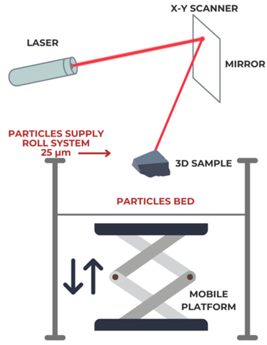
Figure 1. The schematic representation of the SLM equipment and the ingot produced from Co-Cr-W powders, highlighting the principle of selective laser melting.
Prosthesis 07 00027 g001
Figure 2. SEM images of Co-Cr-W powder (a) 2D; (b) 3D profile.
Prosthesis 07 00027 g002
Figure 3. The SEM–EDS spectrum showing elemental composition in list mode.
Prosthesis 07 00027 g003
Figure 4. SEM images of the experimental samples: (a) non-sandblasted sample; (b) Al2O3 particle-sandblasted sample; (c) Al2O3 particle and glass particle-sandblasted sample.
Prosthesis 07 00027 g004
Figure 5. Linear and cyclic potentiometry of three selected experimental samples (A—non-sandblasted sample, B—Al2O3 particle-sandblasted sample, C—Al2O3 particle and glass particle-sandblasted sample): (a) Tafel diagram; (b) cyclic potentiometry.
Prosthesis 07 00027 g005
Figure 6. Electrochemical corrosion parameters across experimental groups.
Prosthesis 07 00027 g006
Figure 7. Measured impedance plots for samples in groups A, B, and C: (a) Nyquist diagrams and (b) Bode plots, and equivalent circuit models (c) for group A and (d) for groups B and C.
Prosthesis 07 00027 g007aProsthesis 07 00027 g007b
Figure 8. SEM images (2D and 3D profile) of Co-Cr-W samples, at different magnification 500× and 2000×, after the electrochemical corrosion process: (a) non-sandblasted sample; (b) Al2O3 particle-sandblasted sample; (c) Al2O3 particle and glass particle-sandblasted sample.
Prosthesis 07 00027 g008
Table 1. Chemical composition of the powder and samples obtained by EDS.
Chemical Composition [Mass %]
ElementPowderSampleEDS Error
Co59.0559.381.1
Cr25.0225.990.8
W9.519.540.5
Mo3.53.480.5
Simax. 1max. 1.160.1
Other elements: C, Fe, Mnmax. 1.50.430.1
Table 2. Mean values of electrochemical corrosion parameters obtained after immersion of the tested alloy in Carter–Brugirard artificial saliva at 37 °C.
Electrochemical Corrosion ParametersGroups
A (n = 10)B (n = 10)C (n = 10)p-Value
Eoc (mV)/mean (SD)−179.6 (17.5)−146.9 (13.8)−117.6 (10.9)p < 0.001 *
Ecorr (mV)/mean (SD)−331.6 (28.1)−163.5 (19.6)−63.6 (7.2)p < 0.001 *
Jcorr (μA/cm2)/mean (SD)38.5 (5.1)13.5 (1.5)0.74 (0.03)p < 0.001 *
βa (mV)/mean (SD)543.7 (42.2)424.7 (35.6)1021.03 (58.2)p < 0.001 *
βc (mV)/mean (SD)−440.7 (68.1)−622.6 (73.5)−40.1 (3.2)p < 0.001 *
Rp (kΩ.cm2)/mean (SD)1.6 (0.1)5.5 (0.6)82.03 (6.7)p < 0.001 *
CR (μm/year)/mean (SD)434.8 (41.6)156.1 (16.9)8.08 (0.8)p < 0.001 *
* p < 0.05 (Kruskal-Wallis test; A—non-sandblasted sample; B—Al2O3 particle-sandblasted sample; C—Al2O3 particle and glass particle-sandblasted sample; Eoc—open circuit potential; Ecorr—corrosion potential; Jcorr—corrosion current density; βa—anodic slope; βc—cathodic slope; Rp—polarization resistance, CR—corrosion rate.
Table 3. The equivalent circuit parameters obtained for the study groups.
GroupsRs
Ohm.cm2
CPERp
Ohm.cm2
CPERct
Ohm.cm2
Qp × 10−5
Ssn/cm2
nQ1 × 10−5
Ssn/cm2
n
A155.91.9330.71310.933.530.6620,420
B204.33.1890.8319,920---
C203.62.8980.8430,970---
Rs—solution resistance; CPE—constant phase element; Rp—passive layer resistance; Qp—constant phase element for the passive layer; Q1—constant phase element for the porous layer.
Disclaimer/Publisher’s Note: The statements, opinions and data contained in all publications are solely those of the individual author(s) and contributor(s) and not of MDPI and/or the editor(s). MDPI and/or the editor(s) disclaim responsibility for any injury to people or property resulting from any ideas, methods, instructions or products referred to in the content.