Systematic Review

Clinical Efficacy of Anterior Ceramic Materials in Resin-Bonded Fixed Dental Prostheses with Different Bridge Designs—A Systematic Review and Meta-Analysis

by
Nutsongsak Panyasuksri
1,
Pattarika Angkasith
1,*,
Apichai Yavirach
1,
Pisaisit Chaijareenont
1,
Surasak Saokaew
2,3,4 and
Sukrit Kanchanasurakit
4,5,6,*
1
Department of Prosthodontics, Faculty of Dentistry, Chiang Mai University, Chiang Mai 50200, Thailand
2
Division of Social and Administrative Pharmacy (SAP), Department of Pharmaceutical Care, School of Pharmaceutical Sciences, University of Phayao, Phayao 56000, Thailand
3
Center of Excellence in Bioactive Resources for Innovative Clinical Applications, Chulalongkorn University, Bangkok 10330, Thailand
4
Unit of Excellence on Clinical Outcomes Research and IntegratioN (UNICORN), School of Pharmaceutical Sciences, University of Phayao, Phayao 56000, Thailand
5
Division of Clinical Pharmacy, Department of Pharmaceutical Care, School of Pharmaceutical Sciences, University of Phayao, Phayao 56000, Thailand
6
Division of Pharmaceutical Care, Department of Pharmacy, Phrae Hospital, Phrae 54000, Thailand
*
Authors to whom correspondence should be addressed.
Prosthesis 2025, 7(2), 41; https://doi.org/10.3390/prosthesis7020041
Submission received: 19 February 2025 / Revised: 27 March 2025 / Accepted: 1 April 2025 / Published: 9 April 2025
(This article belongs to the Special Issue Advancements in Adhesion Techniques and Materials in Prosthodontics)

Abstract

:
Background: Resin-bonded fixed dental prostheses (RBFDPs) have become an increasingly popular minimally invasive solution for replacing missing anterior teeth. However, their long-term success is influenced by factors such as bridge design and material selection. Methods: This systematic review and meta-analysis aimed to assess the impact of different bridge designs on the clinical performance and failure risks of anterior ceramic RBFDPs. A comprehensive search of electronic databases was conducted to identify clinical studies published in English up to January 2025. Nine studies, including randomized controlled trials, cohort studies, and clinical trials, met the inclusion criteria. Results: The analysis revealed that single-retainer RBFDPs demonstrated lower failure rates than two-retainer models (RR: 0.36, 95% CI: 0.17–0.75). Among failure types, fractures were significantly less common in cantilever designs compared to two-retainer designs (RR: 0.25, 95% CI: 0.09–0.73), while no significant differences were observed in debonding rates (OR: 0.40, 95% CI: 0.015–1.10). Conclusions: In terms of material selection, all-ceramic RBFDPs in cantilever designs exhibited significantly lower failure rates (RR: 0.12, 95% CI: 0.03–0.43), whereas metal-ceramic RBFDPs showed no significant difference in failure rates based on bridge design (RR: 0.56, 95% CI: 0.21–1.53). These findings suggest that all-ceramic cantilever RBFDPs may offer superior long-term outcomes, highlighting the importance of precise preparation and cementation protocols for clinical success.

1. Introduction

The absence of teeth adversely affects an individual’s quality of life, primarily impacting physical aspects such as challenges in eating and social dimensions due to communication difficulties with others. Within restorative dentistry, the potential for minimally invasive treatment has grown considerably due to the integration of adhesive techniques and restorative materials that mimic the characteristics of natural teeth [1]. Resin-bonded fixed dental prostheses (RBFDPs) hold a pivotal position in the realm of fixed prosthetic rehabilitation. The strategy for replacing individual missing anterior teeth with this material has roots dating back to the 1970s [2]. RBDFPs have undergone substantial refinements in design, materials, and preparation techniques over the years, all aimed at enhancing their overall clinical durability [3]. With continuous progress advancements in dental materials, cements, and clinical protocols, RBFDPs are now considered a long-term treatment alternative rather than just a temporary solution.
In today’s dental practice, RBFDPs serve as a viable substitute for implant-supported restorations in situations where restorations are contraindicated, extensive surgical procedures are undesirable, or the available space of restoration is ill suited for implant placement. This option is particularly beneficial not only for patients whose age does not align with implant suitability due to insufficient bone, the lack of keratinized mucosa or other anatomical limitations but also for those who prefer not to undergo implant therapy [4,5]. Additionally, RBFDPs are ideal when immediate restoration on the same day is required, as they can be efficiently fabricated using chairside CAD-CAM systems [6]. However, the selection of RBFDPs is contradicted in patients who exhibit any of the following conditions: parafunctional habits, such as bruxism; an unfavourable occlusal scheme, such as a deep bite; a long edentulous span; treatment plans requiring an increased occlusal vertical dimension (OVD); and a loss of posterior support [4,7,8]. Regular follow-ups are essential for long-term success, as the functional loads, particularly during protrusion and lateral movements induced by differential abutments, can result in the de-cementation of such sensitive prosthetics as RBFDPs.
As reported in the previous literature, survival rates of ceramic materials including metal–ceramics and all-ceramics materials using RBFDPs vary widely from 62% to 100% with a minimum average observation time of five years [9,10]. The most frequent causes regarding the absolute failures include debonding, which is the most common [11,12,13], followed by the fracture of the prosthesis framework [14,15,16], loss due to trauma, secondary caries and periodontal problems [14]. Therefore, it is crucial to understand the role of framework design and material properties in minimizing these risks. The survival rate of RBFDPs vary according to concept of tooth preparation, manufacturing procedures, prosthesis design and materials and adhesive cementation protocols [17].
Comparisons between anterior RBFDPs with single-retainer cantilever and two-retainer fixed–fixed designs have been conducted using various materials, including two studies on all-ceramic RBFDPs and three on metal–ceramics [16]. Notably, the framework design did not significantly influence the performance of metal restorations. However, no conclusive evidence exists to determine which ceramic material is inherently superior for RBFDPs, particularly in anterior applications [16]. This uncertainty arises from the diverse range of ceramic materials used in RBFDP fabrication, each offering unique properties and advantages [17].
With the evolution of materials and technologies in dental prosthesis over the years, multiple clinical studies [2,17,18] and systematic reviews [19] consolidate the outcomes of RBFDPs featuring diverse designs that are often grouped together as a single entity, preventing a thorough examination of the performance of distinct design elements. An evidence-based assessment of the clinical performance of RBFDPs with one or two retainers is still lacking, particularly due to the scarcity of high-quality RCTs. Additionally, the failure rates of different types of ceramic RBFDP materials and designs remain inconclusive [16]. Therefore, the aim of this article was to conduct a systematic update of the existing literature, aiming to assess the long-term outcome of different framework designs of anterior ceramic materials in RBFDPs and how they influence the clinical longevity of these restorations and to assess possible factors that may contribute to risk of failures or complications. Based on these objectives, it was hypothesized that there would be no differences in clinical performance between cantilever anterior ceramic RBFDPs and two-wing RBFDPs.

2. Materials and Methods

2.1. Information Sources

The systematic review and meta-analysis adhered strictly to the guidelines established by the Preferred Reporting Items for Systematic Reviews and Meta-Analyses (PRISMA) statement [20]. This research was registered as CRD420234463981 on the International Prospective Register of Systematic Reviews (PROSPERO). The systematic review formulated the research question utilizing the PICOT format. Patients with missing anterior teeth were subjected to treatment with RBFDPs (P), while those receiving cantilever ceramic materials for RBFDPs constituted the intervention group (I). The control group comprised patients with missing anterior teeth treated with two-retained fixed–fixed RBFDPs within the same study (C). This study aimed to report mechanical or biological incidences (O) with at least a 2-year follow-up period.

2.2. Search Strategy

To identify relevant studies, a systematic search was conducted across key scientific databases including PubMed, Embase, Scopus, Cochrane Library, and Web of Science, covering the period from 1967 to January 2025. The focus was specifically on clinical studies involving human subjects, utilizing Medical Subject Headings (MeSH) when applicable. Additionally, the bibliographies of related articles were thoroughly examined. The search strategy involved using keywords such as [fixed dental prostheses OR resin bonded bridge OR adhesive bridge OR acid etched bridge] AND [ceramic*] with minor adjustments made based on the specific requirements of each database. There were no restrictions placed on study design or language. Additionally, supplementary searches were carried out within the reference lists of included studies to avoid overlooking any relevant articles (Supplementary File Table S1).

2.3. Inclusion Criteria

(1)
Studies presenting clinical characteristics with quantitative outcomes, including survival rates, failure rates, debonding incidents, fractures, and other biological complications.
(2)
Articles of any study design, such as randomized controlled trials (RCTs) and observational studies.
(3)
Studies evaluating cantilever or fixed–fixed RBFDPs with sufficient clinical outcome data.

2.4. Exclusion Criteria

(1)
Animal studies, in vitro studies, case reports, case series, systematic reviews, and meta-analyses.
(2)
Studies lacking intervention with cantilever or fixed–fixed RBFDPs or providing insufficient details on clinical outcomes.
(3)
Articles reporting multiple publications on the same patient cohort or with a follow-up duration of less than two years.

2.5. Selection Process

The screening process involved two investigators, NP and PA, who conducted an independent screening of each title, abstract, and full-text article to identify potentially eligible studies. Any discrepancies were resolved through discussions involving a third investigator, AY. Subsequently, NP and PA independently reviewed all extracted data from the studies, including the study design, mean follow time (month), prosthesis design, manufacturing protocols (materials and surface treatment), clinical considerations (tooth preparation, isolation method, and bonding system) and details of the outcome (failure: debonding vs. fracture).

2.6. Study Risk of Bias Assessment

Individual study quality was assessed independently using the Newcastle–Ottawa Scale (NOS), a comprehensive tool for cohort studies. This evaluation encompassed crucial aspects like patient selection and group comparability and were meticulously scrutinized, assigning higher scores to those demonstrating superior quality [21]. For randomized controlled trials (RCTs), a bias assessment followed established guidelines outlined in the Cochrane Handbook for Systematic Reviews of Interventions [22].

2.7. Statistical Analysis

The odds ratio (OR) and 95% confidence interval (CI) for each study were computed using the Cochrane Collaboration Review Manager (Rev Man, Version 5). The combined pool effects were determined using a fixed-effect model. The heterogeneity assessment involved Cochran’s Q statistic and I2 with Cochran’s Q statistic using an alpha value of 0.10 to designate heterogeneity amongst trials for each analysis. The heterogeneity level was classified as high (I2 > 75%), moderate (25–75%), or low (<25%) [23]. In the case of heterogeneity, efforts were made to explore potential sources of heterogeneity. Publication bias was examined using Egger’s test and funnel plot analysis [24], with a p-value less than 0.05 indicating publication bias. If publication bias was detected, the trim-and-fill method was implemented [25].

2.8. Sensitivity and Subgroup Analysis

In order to assess the strength of our analysis, sensitivity analysis was utilized to account for unmeasured confounding factors. Furthermore, subgroup analysis was carried out based on the materials employed (all-ceramic and metal–ceramic). Specialized data science software, specifically Stata/SE14.0, was employed to conduct statistical analyses. Statistical significance was acknowledged at the 0.05 threshold.

3. Results

3.1. Search Results and Characteristics of Studies Included

During the initial search across all databases, a total of 2446 articles were obtained. Among these, 573 duplicates were identified and subsequently removed. The screening process involved assessing titles and abstracts, resulting in the exclusion of 1702 studies that were deemed irrelevant to our research objective. After a thorough evaluation of the full texts, a total of nine studies were deemed suitable for inclusion in the meta-analysis (Figure 1). The significant characteristics and outcomes extracted from the included articles are consolidated and presented in Table 1 and Table 2. Of the nine articles, three were randomized controlled trials (RCTs) [26,27,28], two were prospective cohort studies [29,30], three were retrospective cohort studies [18,31,32], and one was a clinical trial [33]. The mean follow-up time of the included studies was 5.4 years. All studies compared at least two designs of RBFDPs and defined outcomes of failure as debonding or fracture.

3.2. Quality Assessment

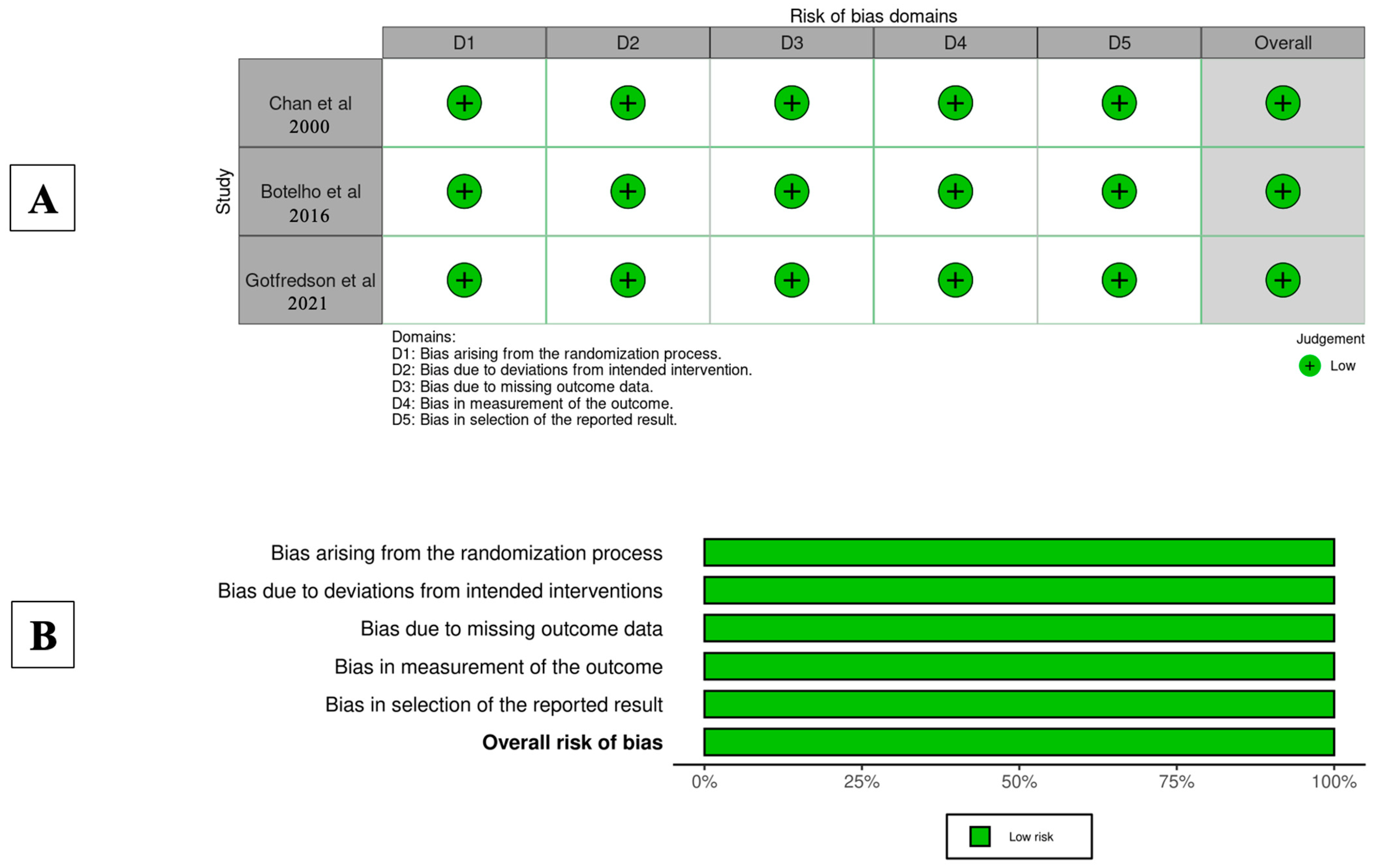
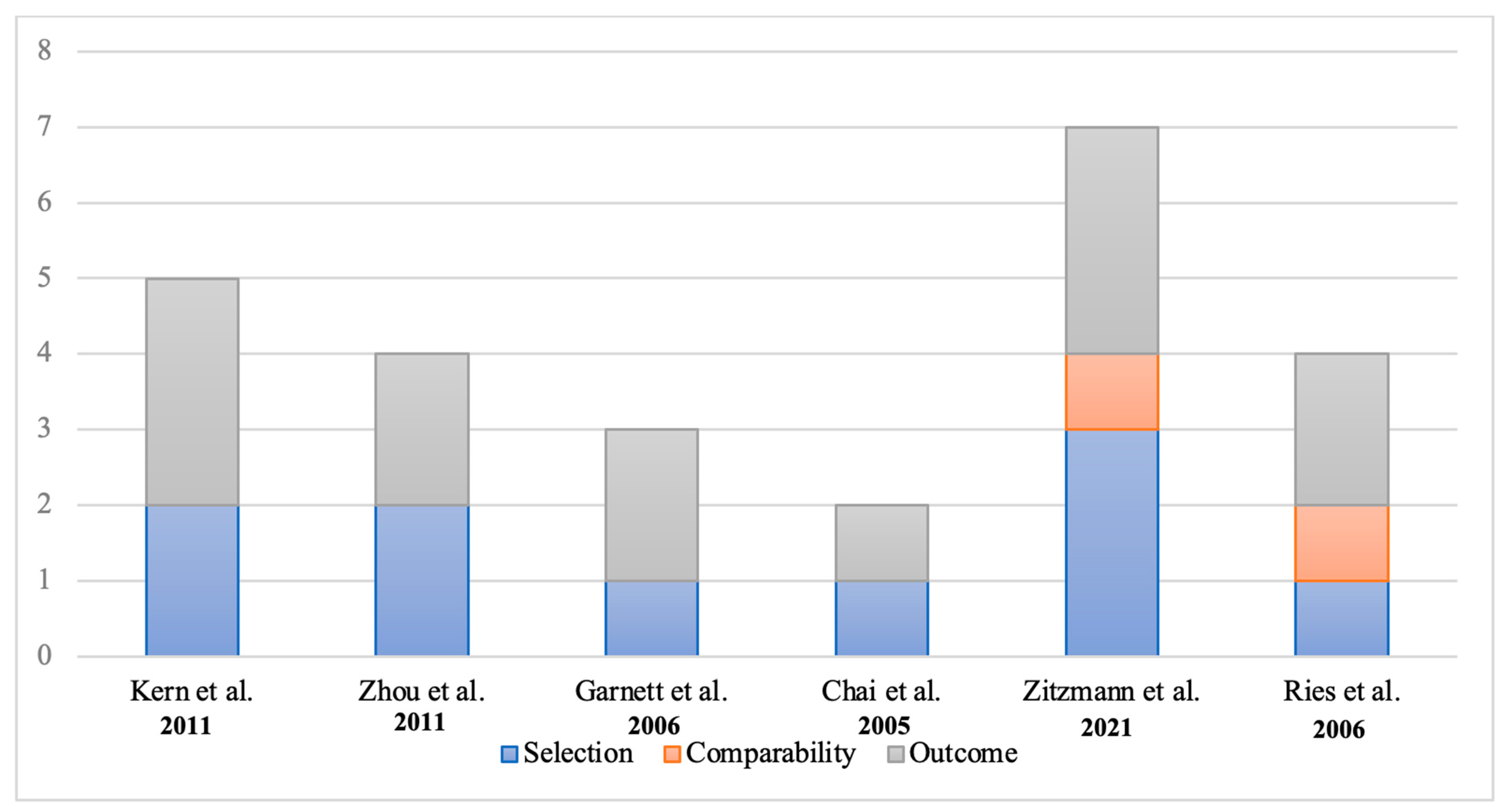
The risk of bias in the randomized controlled trial (RCT) was minimal, as the study provided comprehensive information on randomization procedures, deviations from the intended intervention, blinding, outcome measurement, and other potential sources of bias (Figure 2). In cohort studies, the Newcastle–Ottawa scores varied from 2 to 7 stars, indicating a range of bias risks from low to high. Notably, only two cohort studies detailed baseline control measures for patient selection (Figure 3).

3.3. Synthesis of Results

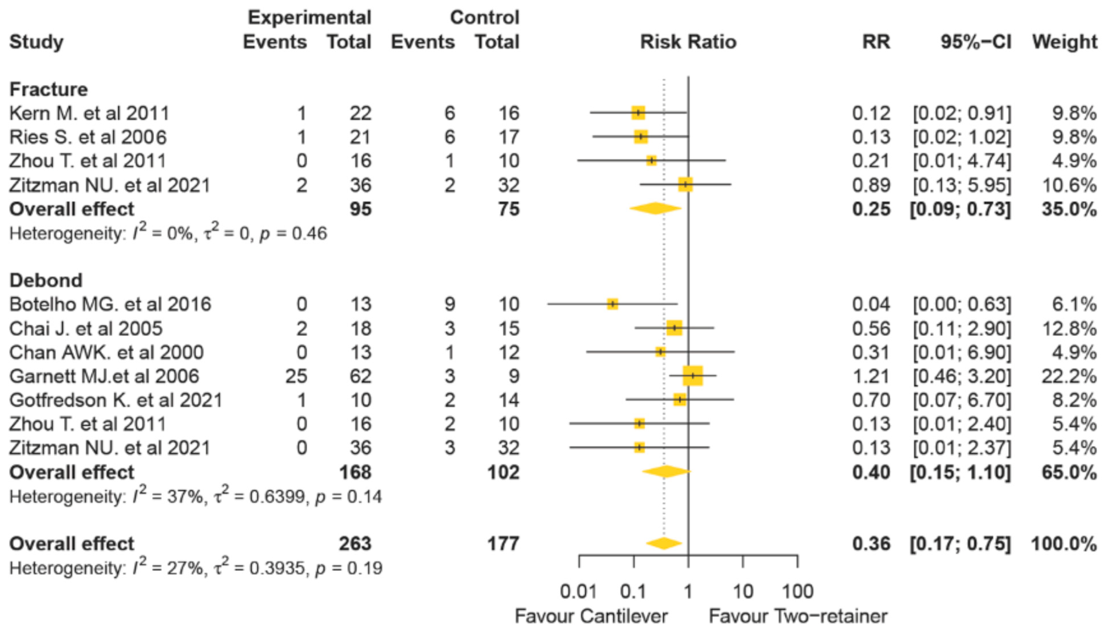
The results of the overall failure rates showed that cantilever RBFDPs [23,24,25,26,27,29,30] exhibited a lower failure rate in eight of the nine included studies. Cantilever RBFDPs exhibited significantly lower failure rates compared to two-retainer fixed–fixed RBFDPs (RR 0.36, 95% CI 0.17–0.75), as indicated in Figure 4. The heterogeneity among studies did not suggest significant variation (I2 = 27%).

3.4. Sensitivity and Subgroup Analyses

The analysis of fracture rates and debonding rates in resin-bonded fixed dental prostheses (RBFDPs) revealed interesting findings. Among the nine studies included, four reported a lower fracture rate compared to two-retainer RBFDPs, aligning with an overall significantly lower fracture rate compared to two-retainer fixed–fixed RBFDPs (RR 0.25, 95% CI 0.09–0.73), with no significant heterogeneity among studies (I2 = 0%) [26,27,28,30]. In terms of debonding rates, six studies found no instances of debonding in cantilever RBFDPs [24,25,26,27,28,30], while two studies reported no debonding in two-retainer fixed–fixed RBFDPs [30,33]. However, no significant difference in debonding rates was observed between two designs, as shown in Figure 4 (RR 0.40, 95% CI 0.15–1.10). The heterogeneity among the studies did not suggest substantial variation (I2 = 37%).
The investigation into failures across various configurations for both metal–ceramic and all-ceramic resin-bonded fixed dental prostheses (RBFDPs) revealed notable insights. In terms of metal–ceramic RBFDPs, comparable failure rates were identified between cantilever and two-retainer fixed–fixed designs (RR 0.56, 95% CI 0.21–1.53, Table 3), with no significant heterogeneity across studies (I2 = 29.5%). Conversely, when focusing on all-ceramic RBFDPs, three out of nine studies indicated lower failure rates for cantilever designs [29,30,33]. Cantilever RBFDPs exhibited significantly lower failure rates compared to their two-retainer fixed–fixed counterparts (RR 0.12, 95% CI 0.03–0.43, Table 3), with negligible heterogeneity among studies (I2 = 0%). Regarding debonding rates, metal–ceramic RBFDPs showed no significant difference between cantilever and two-retainer fixed–fixed designs (RR 0.60, 95% CI 0.21–1.70) [18,23,24,25,29]. The heterogeneity among studies did not suggest substantial variation (I2 = 31.6%, Figure 4). Notably, one study documented bond loss in two two-retainer fixed–fixed RBFDPs but none in the cantilever designs [33]; Similarly, another study reported no instances of debonding with two different designs of all-ceramic RBFDPs [5,33]. However, due to the limited number of studies included, statistical analysis was not conducted to assess the influence of factors such as prosthesis materials, luting cements, and tooth preparation retention.

3.5. Publication Bias of Included Studies

The assessment conducted to detect publication bias revealed no substantial evidence of bias. This was evidenced by the symmetrical funnel plot and the non-significant results from Egger’s tests across all resin-bonded fixed dental prostheses (RBFDPs) and their outcomes (Supplementary File Figures S1 and S2).

4. Discussion

The systematic review and meta-analysis were conducted to examine the existing clinical data regarding the long-term clinical outcomes and complications associated with RBFDPs. Our research has revealed a limitation in the previous literature, specifically in terms of the quantity and heterogeneity of clinical studies related to this type of RBFDP. The diversity among the studies makes it challenging to perform a comparative analysis, encompassing factors such as prosthesis design, surface preparation, and the success rate of the cement used. Furthermore, the evidence found from the prior literature primarily stems from prospective and retrospective cohort studies, with a notable absence of randomized controlled trials (RCTs) and a limited sample size in some cases. RBFDPs serve as an alternative to traditional bridges, particularly when supporting teeth are healthy. Recent advancements in ceramic materials have improved their mechanical properties and esthetics, increasing the popularity of ceramic RBFDPs. These provide a conservative solution for replacing missing anterior teeth, especially in cases with strong abutments and minimal occlusal contact or when implants are not an option [4,5]. The advantages of RBFDPs include preserving tooth structure, strong bonding, colour stability, thermal expansion compatibility with enamel, and excellent esthetics [34]. Over recent years, a significant amount of information on RBFDPs has been published. The conclusions drawn from our systematic review are rooted in the analysis of nine studies, which collectively investigated more than 300 RBFDPs with either metal–ceramic or all-ceramic materials. The results indicate a higher survival rate of cantilever RBFDPs in comparison with cantilever RBFDPs and two-wing RBFDPs. This finding is consistent with both clinical and in vitro studies, which included follow-ups at 5 and 10 years, demonstrating a higher clinical success rate for the cantilever design (73.9% and 92.3%) in contrast to the two-retainer lingual design (73.9% and 94.4%). These findings further support the rejection of the null hypothesis, indicating that the cantilever design is a more favourable option for anterior tooth replacement (p < 0.05). Interestingly, after a 15-year follow-up, they only reported data for lingual cantilever RBFDPs [4,30,35]. Their findings concluded that all-ceramic cantilever RBFDPs are a viable alternative for anterior tooth replacement [4,10,23,26,28,29,30]. The main reason for the higher failure rate of two-retainer fixed–fixed RBFDPs might appear unsurprising when considering the mechanical aspects of tooth movement. Variations in the functional activity of the stomatognathic system of abutment teeth can impact abutment teeth within two-retainers RBFDPs when the jaw moves laterally during chewing. These movements create shear and torque forces on both pontics and connections, leading to potential outcomes such as fractures, debonding, or stress at the interface of the resin RBFDPs [4,33,36,37]. To further support this idea, 3D finite element analysis was conducted and highlighted a notable rise in stress concentration specifically within the abutment teeth and connectors of two-wing RBFDPs when compared to their one-wing counterparts during periods of functional loading [38]. On the other hand, the periodontal apparatus’s natural shock-absorbing capability has the potential to counteract occlusal forces applied to the bridgework within the cantilever group. In this case, the bridge moves harmoniously with the abutment tooth, aligning in both direction and intensity during occlusal function. Consequently, this alignment may lead to a reduction in stress concentration at the interface between the cement and metal [28,30].
Regarding material selection, Zitzmann conducted a study examining the long-term performance of both metal–ceramic and all-ceramic RBFDPs. These findings are crucial in understanding the factors influencing RBFDP success. The study revealed a 5-year survival rate of 90.6% for metal–ceramic RBFDPs and 79.6% for all-ceramic RBFDPs, aligning closely with previously documented rates in the literature: 91.3% for metal–ceramic RBFDPs and 100% for zirconia frameworks [31,32]. One of the primary risk factors for RBFDPs failure pertains to the presence of multiple pontics within a restoration and the vitality status of the abutment teeth [31]. It is postulated that fractures occurring in three-unit restorations are primarily attributable to shear forces acting upon the abutments, especially in the context of curved RBFDP designs. Additionally, the failure of teeth with negative pulp often necessitates tooth extraction [39]. Furthermore, a comprehensive literature review exploring various materials used in RBFDPs highlighted the most prevalent issues encountered in practice. These issues were predominantly related to debonding, particularly when zirconia ceramics were employed. In contrast, fractures were more frequently associated with the use of glass-infiltrated alumina and lithium disilicate ceramics [40]. Notably, RBFDPs fabricated from glass-infiltrated alumina particularly with a two-retainer design exhibited a high tendency toward fractures when compared to the use of glass ceramics [30]. The choice of materials plays a pivotal role in RBFDP performance. Opting for glass ceramics offers inherent benefits, including simplified, less error-prone technician processing and natural translucency, while In-Ceram necessitates additional veneering [33]. Nevertheless, cantilever RBFDPs using all-ceramic materials demonstrated a lower failure rate than two-retainer ones, whereas metal–ceramics showed no significant difference.
Regarding tooth preparation, the consensus among studies, except for two from a retrospective design, favoured a tooth preparation strategy that minimized dentin exposure [23,24,25,26,27,28,30]. Botelho and Chan suggested that creating proximal grooves and additional rests could enhance retention [27,28]. In contrast, Kern and Zhou disagreed with this perspective, arguing that such modifications would solely establish a definite seat for restoration without contributing to mechanical retention [29,30]. However, to improve the overall survival rate of RBFDPs, it is recommended to perform tooth preparation that includes retention features like proximal boxes, grooves and pinholes [9,29]. The specific prerequisites for fabricating different types of materials are needed. A palatal clearance of 0.7 mm is necessary for zirconia, while a retainer thickness of 1 mm is required for lithium disilicate [31]. In the case of metal–ceramic materials, occlusal reduction from 0.4 to 0.6 mesially and distally for the groove is needed [31,41]. Nevertheless, the preparation is contingent upon factors such as the space required for prosthesis extension and the minimum thickness of the prosthesis material. The availability of inter-arch space for prosthesis emerges as a pivotal factor in determining the necessary extent of the preparation.
In cases of missing lateral incisors, Garnett conducted a study comparing cantilevers from central incisors and canines, showing better survival rates for central incisors. However, there was no significant difference in survival between the two cantilever designs [18]. Nevertheless, when determining the cantilevered tooth, it is crucial to assess the amount of the remaining tooth structure.
Prosthetic challenges, specifically related to documentation issues, were consistently tackled in the cited articles [18,23,24,25,26,27,28,30]. Cementation techniques have evolved over the years, adapting to material advancements and emphasizing internal surface and abutment treatments. Panavia resin cement has been commonly used [18,23,24,25,26,27,29], but Variolink, Clearfil, and RelyX have also been cited for their cement-like properties [30,31]. These alternatives need to be examined in detail to understand their suitability for addressing the challenges. Initial challenges, such as the high failure rates in resin-bonded FPDs, stemmed from adhesive bond weakening due to hydrolysis. This weakening compromised the durability and stability of prosthetic restorations, leading to concerns about their long-term success. Efforts to address this challenge involved incorporating retentive preparations. However, persistent obstacles arose, particularly in cases involving metal frameworks [33,42]. These frameworks created complexities due to specific material interactions and required specialized approaches for successful adhesive bonding [33,42,43]. Nevertheless, the clinical results of RBFDPs using different types of resin cement showed no significant difference during the initial 5-year period [18]. Therefore, the selection of different resin cements might not play a significant role in determining the risk of RBFDP failures in clinical practice [44].
Seven out of nine studies performed cementation in a dry clinical environment. Among the studies, five employed a rubber dam, one used cotton rolls, and one employed both a rubber dam and cotton rolls to control saliva contamination [18,27,28,30,31,33]. Instances where a rubber dam was used, no debonding was shown compared to those using only cotton rolls [18]. Hence, the application of a rubber dam is essential to achieve a successful bond with etched enamel.
To enhance the micromechanical retention of metal framework RBFDPs, various techniques have been proposed. It is crucial to consider these methods based on the specific material being used. For instance, air particle abrasion is recommended for metal framework RBFDPs [28,32]. When dealing with materials like lithium disilicate (E-max) or Feldspathic porcelain, a 5% hydrofluoric acid (HF) conditioning of surfaces is advised. If silane is applied, optional adhesive use can further enhance bonding efficiency [45]. For In-Ceram alumina or zirconia, a multi-step approach is recommended. Initiating with tribochemical silica coating or air abrasion using 50 um alumina particles at 1–2.5 bar sets the foundation. Subsequently, applying primers containing a specialized phosphate monomer, specifically 10-methacryloyloxydecyl dihydrogen phosphate (10-MDP), ensures a strong bond with zirconia and resin cement [43,44,45,46,47,48]. The pivotal aspect of the 10-methacryloyloxydecyl dihydrogen phosphate (10-MDP) monomer lies in its unique composition, featuring both a phosphate group and a methacrylate group. In vitro studies have also demonstrated that using MDP primers significantly increases bond strength with zirconia due to their higher MDP concentration compared to MDP resin cement [49]. This composition enables them to form robust bonds with zirconia and resin cement, ensuring the durability of the restoration. Extensive research consistently demonstrates the superiority of MDP-primed zirconia bonds. In contrast to other priming agents or untreated zirconia, 10-MDP-primed zirconia exhibits significantly enhanced bond strength, ensuring the long-term stability of the restoration [50,51,52,53,54,55,56]. In summary, the integration of 10-methacryloyloxydecyl dihydrogen phosphate (10-MDP) in adhesive formulations stands as a pivotal recommendation.
There are several limitations of the existing clinical data in this systematic review and meta-analysis. Firstly, variations in clinician skills, techniques employed and materials used may have influenced the success and survival rates due to the lack of standardization in these factors. Secondly, differences in patient-related factors, including age, oral hygiene, personal habits, and dietary habits may further complicate outcome evaluation. Future research could explore whether one-wing or two-wing designs are more suitable when ceramic materials are used in RBFDPs for posterior teeth.

5. Conclusions

  • Success of RBFDPs: Resin-bonded fixed dental prostheses (RBFDPs) show high long-term cumulative survival rates, confirming their effectiveness as minimally invasive restorations.
  • Design Preference: RBFDPs with a single retainer in the anterior region are preferred, outperforming two-retainer designs.
  • Material Comparison: All-ceramic materials deliver superior esthetic results and higher survival rates compared to metal–ceramic options.
  • Recommended Protocols:
    Utilize conservative preparation techniques like proximal grooves and rests to enhance retention.
    Follow specific clearance measurements for materials.
    Strictly adhere to manufacturer guidelines for resin cement usage.
  • Material-Specific Recommendations
    For zirconia: use air abrasion followed by an MDP primer to provide the most durable bond to zirconia ceramics.
    For lithium disilicate: use 5% HF etching followed by silane application.

Supplementary Materials

The following supporting information can be downloaded at: https://www.mdpi.com/article/10.3390/prosthesis7020041/s1.

Author Contributions

Study concept and design: N.P., P.A., A.Y., P.C., S.S. and S.K.; acquisition of data: N.P. and P.A.; analysis and interpretation of data: N.P., P.A., A.Y., S.S. and S.K.; drafting of the manuscript: N.P.; critical revision of the manuscript: N.P., P.A., A.Y., P.C. and S.K. All authors have read and agreed to the published version of the manuscript.

Funding

This work was supported Chiang Mai University, as well as partially supported by the University of Phayao and the Thailand Science Research and Innovation Fund (Fundamental Fund 2025, Grant No. 5017/2567). The funding source had no role in the study design, collection, analysis, and interpretation of data.

Institutional Review Board Statement

The systematic review and meta-analysis did not require ethics approval as it entailed the aggregation and analysis of data from pre-existing studies. The authors ensured compliance with pertinent guidelines from the EQUATOR Network (https://www.equator-network.org access on 27 March 2025) during this research project.

Data Availability Statement

This study contains the original contributions, which are detailed in the article/Supplementary Materials. For any further inquiries, please contact the corresponding authors.

Acknowledgments

We thank Chiang Mai University and the University of Phayao for their financial support of this research project.

Conflicts of Interest

The authors declare no conflicts of interest.

List of Abbreviations

CIConfidence interval
HFHydrofluoric acid
MDP Methacryloyloxydecyl dihydrogen phosphate
OROdds ratio
OVDOcclusal vertical dimension
RBFDPsResin-bonded fixed dental prostheses
RCTRandomized controlled trial

References

  1. Edelhoff, D.; Liebermann, A.; Beuer, F.; Stimmelmayr, M.; Güth, J.-F. Minimally invasive treatment options in fixed prosthodontics. Quintessence Int. 2016, 47, 207–216. [Google Scholar] [CrossRef] [PubMed]
  2. Zalkind, M.; Ever-Hadani, P.; Hochman, N. Resin-bonded fixed partial denture retention: A retrospective 13-year follow-up. J. Oral Rehabil. 2003, 30, 971–977. [Google Scholar] [CrossRef] [PubMed]
  3. Pjetursson, B.E.; Tan, W.C.; Tan, K.; Brägger, U.; Zwahlen, M.; Lang, N.P. A systematic review of the survival and complication rates of resin-bonded bridges after an observation period of at least 5 years. Clin. Oral Implant. Res. 2008, 19, 131–141. [Google Scholar] [CrossRef]
  4. Kern, M. Clinical long-term survival of two-retainer and single-retainer all-ceramic resin-bonded fixed partial dentures. Quintessence Int. 2005, 36, 141–147. [Google Scholar]
  5. Kern, M.; Cleser, R. Cantilevered all-ceramic, resin-bonded fixed partial dentures: A new treatment modality. J. Esthet. Restor. Dent. 1997, 9, 255–264. [Google Scholar] [CrossRef] [PubMed]
  6. Jurado, C.A.; Tsujimoto, A.; Watanabe, H.; Villalobos-Tinoco, J.; Garaicoa, J.L.; Markham, M.D.; Barkmeier, W.W.; Latta, M.A. Chair-side CAD/CAM fabrication of a single-retainer resin bonded fixed dental prosthesis: A case report. Restor. Dent. Endod. 2020, 45, e15. [Google Scholar] [CrossRef]
  7. Olin, P.S.; Hill, E.M.E.; Donahue, J.L. Clinical evaluation of resin-bonded bridges: A retrospective study. Quintessence Int. 1991, 22, 873–877. [Google Scholar]
  8. Hansson, O.; Bergström, B. A longitudinal study of resin-bonded prostheses. J. Prosthet. Dent. 1996, 76, 132–139. [Google Scholar] [CrossRef]
  9. Behr, M.; Rammelsberg, P.; Handel, G.; Leibrock, A.; Stich, W.; Rosentritt, M. Adhesive-fixed partial dentures in anterior and posterior areas. Results of an on-going prospective study begun in 1985. Clin. Oral Investig. 1998, 2, 31–35. [Google Scholar] [CrossRef]
  10. Sailer, I.; Bonani, T.; Brodbeck, U.; Hammerle, C. Retrospective Clinical Study of Single-Retainer Cantilever Anterior and Posterior Glass-Ceramic Resin-Bonded Fixed Dental Prostheses at a Mean Follow-up of 6 Years. Int. J. Prosthodont. 2013, 26, 443–450. [Google Scholar] [CrossRef]
  11. Thoma, D.S.; Sailer, I.; Ioannidis, A.; Zwahlen, M.; Makarov, N.; Pjetursson, B.E. A systematic review of the survival and complication rates of resin-bonded fixed dental prostheses after a mean observation period of at least 5 years. Clin. Oral Implant. Res. 2017, 28, 1421–1432. [Google Scholar] [CrossRef] [PubMed]
  12. Santos, M.C.; Azevedo, L.; Fonseca, P.; Viana, P.C.; Araújo, F.; Villarinho, E.; Fernandes, G.V.O.; Correia, A. The Success Rate of the Adhesive Partial Fixed Prosthesis after Five Years: A Systematic Review. Prosthesis 2023, 5, 282–294. [Google Scholar] [CrossRef]
  13. Gratton, D.R.; Jordan, R.E.; Teteruck, W.R. Resin-bonded bridges: The state of the art. Ont. Dent. 1983, 60, 9–11, 13–16, 18–19. [Google Scholar] [PubMed]
  14. Sasse, M.; Eschbach, S.; Kern, M. Randomized clinical trial on single retainer all-ceramic resin-bonded fixed partial dentures: Influence of the bonding system after up to 55 months. J. Dent. 2012, 40, 783–786. [Google Scholar] [CrossRef]
  15. Dundar, M.; Ozcan, M.; Comlekoglu, E.; Gungor, M.A. A preliminary report on short-term clinical outcomes of three-unit resin-bonded fixed prostheses using two adhesive cements and surface conditioning combinations. Int. J. Prosthodont. 2010, 23, 353–360. [Google Scholar]
  16. Wei, Y.-R.; Wang, X.-D.; Zhang, Q.; Li, X.-X.; Blatz, M.B.; Jian, Y.-T.; Zhao, K. Clinical performance of anterior resin-bonded fixed dental prostheses with different framework designs: A systematic review and meta-analysis. J. Dent. 2016, 47, 1–7. [Google Scholar] [CrossRef]
  17. Tezulas, E.; Yildiz, C.; Evren, B.; Ozkan, Y. Clinical procedures, designs, and survival rates of all-ceramic resin-bonded fixed dental prostheses in the anterior region: A systematic review. J. Esthet. Restor. Dent. 2018, 30, 307–318. [Google Scholar] [CrossRef]
  18. Garnett, M.J.; Wassell, R.W.; Jepson, N.J.; Nohl, F.S. Survival of resin-bonded bridgework provided for post-orthodontic hypodontia patients with missing maxillary lateral incisors. Br. Dent. J. 2006, 201, 527–534. [Google Scholar] [CrossRef]
  19. Botelho, M. Design principles for cantilevered resin-bonded fixed partial dentures. Quintessence Int. 2000, 31, 613–619. [Google Scholar]
  20. Page, M.J.; McKenzie, J.E.; Bossuyt, P.M.; Boutron, I.; Hoffmann, T.C.; Mulrow, C.D.; Shamseer, L.; Tetzlaff, J.M.; Akl, E.A.; Brennan, S.E.; et al. The PRISMA 2020 statement: An updated guideline for reporting systematic reviews. BMJ 2021, 372, 71. [Google Scholar] [CrossRef]
  21. Stang, A. Critical evaluation of the Newcastle-Ottawa scale for the assessment of the quality of nonrandomized studies in meta-analyses. Eur. J. Epidemiol. 2010, 25, 603–605. [Google Scholar] [CrossRef]
  22. Higgins, J.P.T.; Thomas, J.; Chandler, J.; Cumpston, M.; Li, T.; Page, M.J.; Welch, V.A. Cochrane Handbook for Systematic Reviews of Interventions; John Wiley & Sons: Chichester, UK, 2019. [Google Scholar] [CrossRef]
  23. Grant, J.; Hunter, A. Measuring inconsistency in knowledgebases. J. Intell. Inf. Syst. 2006, 27, 159–184. [Google Scholar] [CrossRef]
  24. van Dalen, A.; Feilzer, A.J.; Kleverlaan, C.J. The influence of surface treatment and luting cement on in vitro behavior of two-unit cantilever resin-bonded bridges. Dent. Mater. 2005, 21, 625–632. [Google Scholar] [CrossRef] [PubMed]
  25. Duval, S.; Tweedie, R. Trim and fill: A simple funnel-plot-based method of testing and adjusting for publication bias in meta-analysis. Biometrics 2000, 56, 455–463. [Google Scholar] [CrossRef] [PubMed]
  26. Gotfredsen, K.; Alyass, N.; Hagen, M. A 5-Year Randomized Clinical Trial on 3-Unit Fiber-Reinforced vs 3- or 2-Unit Metal-Ceramic Resin-Bonded Fixed Dental Prostheses. Int. J. Prosthodont. 2021, 34, 703–711. [Google Scholar] [CrossRef]
  27. Botelho, M.G.; Chan, A.W.; Leung, N.C.; Lam, W. Long-term evaluation of cantilevered versus fixed–fixed resin-bonded fixed partial dentures for missing maxillary incisors. J. Dent. 2016, 45, 59–66. [Google Scholar] [CrossRef] [PubMed]
  28. Chan, A.W.K.; Barnes, I.E. A prospective study of cantilever resin-bonded bridges: An initial report. Aust. Dent. J. 2000, 45, 31–36. [Google Scholar] [CrossRef]
  29. Zhou, T.; Wang, X.; Zhang, G. All-ceramic resin bonded fixed partial denture made of IPS hot-pressed casting porcelain restore anterior missing teeth: A three years clinical observation. Beijing Da Xue Xue Bao Yi Xue Ban = J. Peking Univ. Health Sci. 2011, 43, 77–80. [Google Scholar]
  30. Kern, M.; Sasse, M. Ten-year survival of anterior all-ceramic resin-bonded fixed dental prostheses. J. Adhes. Dent. 2011, 13, 407–410. [Google Scholar] [CrossRef]
  31. Zitzmann, N.U.; von Büren, A.; Glenz, F.; Rohr, N.; Joda, T.; Zaugg, L.K. Clinical outcome of metal- and all-ceramic resin-bonded fixed dental prostheses. J. Prosthodont. Res. 2021, 65, 243–248. [Google Scholar] [CrossRef]
  32. Chai, J.; Chu, F.C.S.; Newsome, P.R.H.; Chow, T.W. Retrospective survival analysis of 3-unit fixed-fixed and 2-unit cantilevered fixed partial dentures. J. Oral Rehabil. 2005, 32, 759–765. [Google Scholar] [CrossRef] [PubMed]
  33. Ries, S.; Wolz, J.; Richter, E.-J. Effect of design of all-ceramic resin-bonded fixed partial dentures on clinical survival rate. Int. J. Periodontics Restor. Dent. 2006, 26, 143–149. [Google Scholar]
  34. Galiatsatos, A.A.; Bergou, D. Clinical evaluation of anterior all-ceramic resin-bonded fixed dental prostheses. Quintessence Int. 2014, 45, 9–14. [Google Scholar] [CrossRef]
  35. Kern, M. Fifteen-year survival of anterior all-ceramic cantilever resin-bonded fixed dental prostheses. J. Dent. 2017, 56, 133–135. [Google Scholar] [CrossRef] [PubMed]
  36. Koutayas, S.O.; Kern, M.; Ferraresso, F.; Strub, J.R. Influence of framework design on fracture strength of mandibular anterior all-ceramic resin-bonded fixed partial dentures. Int. J. Prosthodont. 2001, 15, 223–229. [Google Scholar]
  37. Walker, M.P.; Spencer, P.; Eick, J.D. Effect of simulated resin-bonded fixed partial denture clinical conditions on resin cement mechanical properties. J. Oral Rehabil. 2003, 30, 837–846. [Google Scholar] [CrossRef] [PubMed]
  38. Piva, A.M.D.; Tribst, J.P.; Saavedra, G.S.; Souza, R.O.; de Melo, R.M.; Borges, A.L.; Özcan, M. Short communication: Influence of retainer configuration and loading direction on the stress distribution of lithium disilicate resin-bonded fixed dental prostheses: 3D finite element analysis. J. Mech. Behav. Biomed. Mater. 2019, 100, 103389. [Google Scholar] [CrossRef]
  39. Yoshida, T.; Kurosaki, Y.; Mine, A.; Kimura-Ono, A.; Mino, T.; Osaka, S.; Nakagawa, S.; Maekawa, K.; Kuboki, T.; Yatani, H.; et al. Fifteen-year survival of resin-bonded vs. full-coverage fixed dental prostheses. J. Prosthodont. Res. 2019, 63, 374–382. [Google Scholar] [CrossRef]
  40. Chen, J.; Cai, H.; Ren, X.; Suo, L.; Pei, X.; Wan, Q. A Systematic Review of the Survival and Complication Rates of All-Ceramic Resin-Bonded Fixed Dental Prostheses. J. Prosthodont. 2018, 27, 535–543. [Google Scholar] [CrossRef]
  41. Aggstaller, H.; Beuer, F.; Edelhoff, D.; Rammelsberg, P.; Gernet, W. Long-term clinical performance of resin-bonded fixed partial dentures with retentive preparation geometry in anterior and posterior areas. J. Adhes. Dent. 2008, 10, 301–306. [Google Scholar]
  42. Rammelsberg, P.; Pospiech, P.; Gernet, W. Clinical factors affecting adhesive fixed partial dentures: A 6-year study. J. Prosthet. Dent. 1993, 70, 300–307. [Google Scholar] [CrossRef]
  43. Miettinen, M.; Millar, B.J. A review of the success and failure characteristics of resin-bonded bridges. Br. Dent. J. 2013, 215, E3. [Google Scholar] [CrossRef]
  44. Hussey, D.; Linden, G. The clinical performance of cantilevered resin-bonded bridgework. J. Dent. 1996, 24, 251–256. [Google Scholar] [CrossRef] [PubMed]
  45. Nogueira, I.d.O.; Pereira, C.N.d.B.; Abreu, L.G.; Diniz, I.M.; de Magalhães, C.S.; da Silveira, R.R. Do different protocols affect the success rate or bond strength of glass-ceramics repaired with composite resin? A systematic review and meta-analysis. J. Prosthet. Dent. 2023. [Google Scholar] [CrossRef] [PubMed]
  46. Creugers, N.; Snoek, P.; Hof, M.V.; Käuyser, A. Clinical performance of resin-bonded bridges: A 5-year prospective study. Part III: Failure characteristics and survival after rebonding. J. Oral Rehabil. 1990, 17, 179–186. [Google Scholar] [CrossRef]
  47. Özcan, M.; Vallittu, P.K. Effect of surface conditioning methods on the bond strength of luting cement to ceramics. Dent. Mater. 2003, 19, 725–731. [Google Scholar] [CrossRef] [PubMed]
  48. Strasser, T.; Preis, V.; Behr, M.; Rosentritt, M. Roughness, surface energy, and superficial damages of CAD/CAM materials after surface treatment. Clin. Oral Investig. 2018, 22, 2787–2797. [Google Scholar] [CrossRef]
  49. Lehmann, F.; Kern, M. Durability of Resin Bonding to Zirconia Ceramic Using Different Primers. J. Adhes. Dent. 2009, 11, 479–483. [Google Scholar]
  50. Blatz, M.B.; Sadan, A.; Kern, M. Resin-ceramic bonding: A review of the literature. J. Prosthet. Dent. 2003, 89, 268–274. [Google Scholar] [CrossRef]
  51. Wegner, S.M.; Kern, M. Long-term resin bond strength to zirconia ceramic. J. Adhes. Dent. 2000, 2, 139–147. [Google Scholar]
  52. Tanaka, R.; Fujishima, A.; Shibata, Y.; Manabe, A.; Miyazaki, T. Cooperation of Phosphate Monomer and Silica Modification on Zirconia. J. Dent. Res. 2008, 87, 666–670. [Google Scholar] [CrossRef] [PubMed]
  53. Blatz, M.B.; Chiche, G.; Holst, S.; Sadan, A. Influence of surface treatment and simulated aging on bond strengths of luting agents to zirconia. Quintessence Int. 2007, 38, 745–753. [Google Scholar] [PubMed]
  54. Shokry, M.; Al-Zordk, W.; Ghazy, M. Retention strength of monolithic zirconia crowns cemented with different primer-cement systems. BMC Oral Health 2022, 22, 187. [Google Scholar] [CrossRef]
  55. Kukiattrakoon, B.; Jampa, T.; Sanohkan, S.; Larpboonphol, N.; Sae-Yib, T.; Manoppan, S. The effect of various primers on shear bond strength of zirconia ceramic and resin composite. J. Conserv. Dent. 2013, 16, 499–502. [Google Scholar] [CrossRef] [PubMed]
  56. Steiner, R.; Heiss-Kisielewsky, I.; Schwarz, V.; Schnabl, D.; Dumfahrt, H.; Laimer, J.; Steinmassl, O.; Steinmassl, P. Zirconia Primers Improve the Shear Bond Strength of Dental Zirconia. J. Prosthodont. 2020, 29, 62–68. [Google Scholar] [CrossRef]
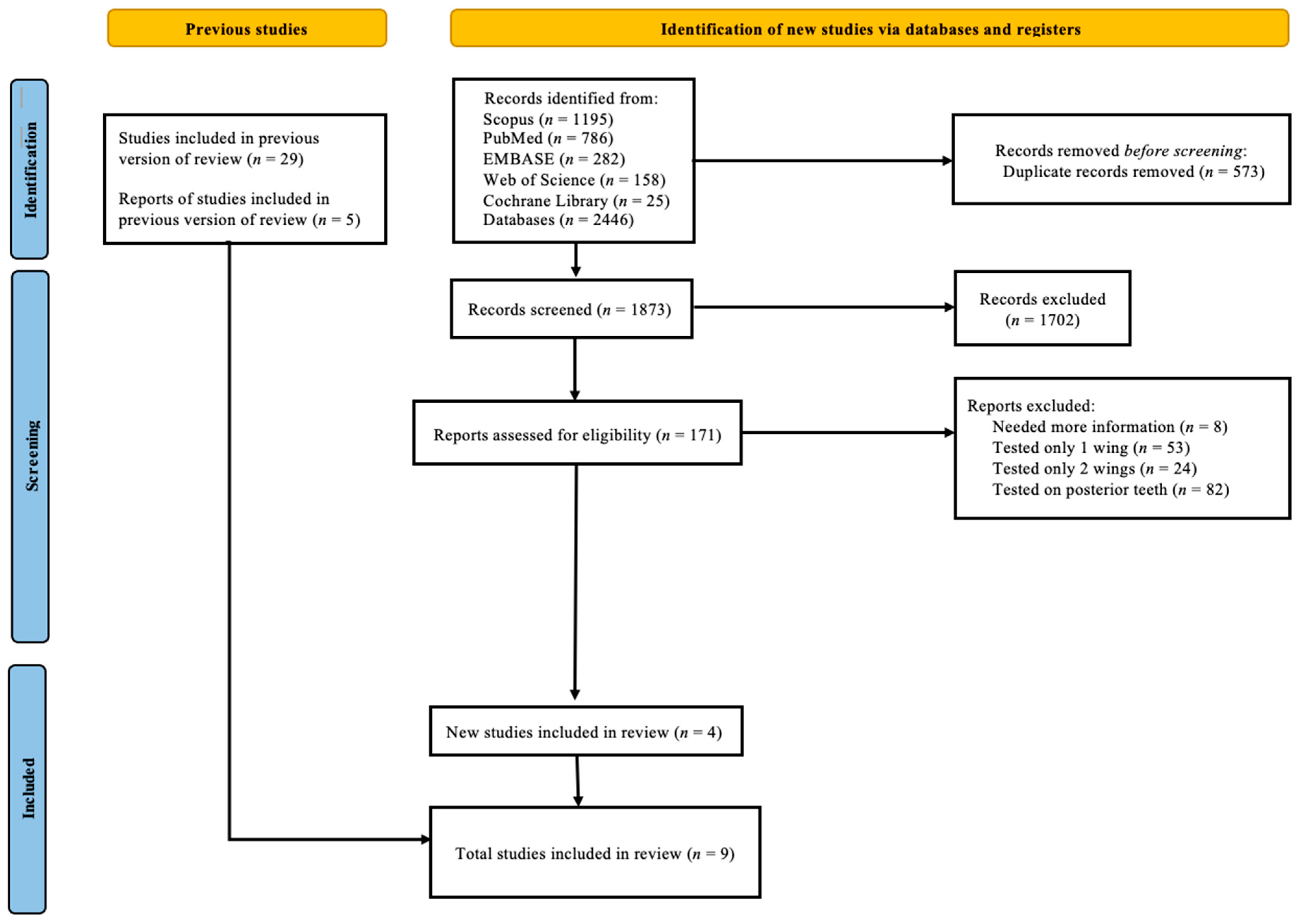
Figure 1. The PRISMA flowchart of the study selection process.
Prosthesis 07 00041 g001
Figure 2. The quality of the bias assessment of the included studies according to the Cochrane risk-of-bias tool (RoB 2.0). (A) Risk-of-bias summary for each included study [26,27,28]. (B) Risk-of-bias graph for included studies.
Prosthesis 07 00041 g002
Figure 3. A summary of the quality of the included studies, using the Newcastle–Ottawa Scale [18,29,30,31,32,33].
Prosthesis 07 00041 g003
Figure 4. Forest plot of failure rate of cantilever vs. two-retainer RBFDP designs, including both fracture [29,30,31,33] and debonding rates [18,26,27,28,29,31,32].
Prosthesis 07 00041 g004
Table 1. Study characteristics of included studies of experimental study.
Experimental Study 3 Papers
Study
(Study
Design)
Mean
Follow Up Time (Month)
Prosthesis
Design
Manufacturing ProcedureClinical ConsiderationsFailures
MaterialsSurface TreatmentTooth PreparationIsolation MethodBonding
System
DebondFractureOthers
Gotfredson 2021 (RCTs) [26]36Cantilever = 10
Fixed-Fixed = 14
Metal-
ceramic
Electrolyte etching of cast metal
(18% HCl for 10 min)
Vertical, approximal groove
A half circular prep at cingulum limited removal of enamel
Not reportedPanavia EXCantilever = 1
Fixed-fixed = 1
Cantilever = 0
Fixed-Fixed = 0
0
1 (esthetics)
Botelho et al 2016 (RCTs) [27]216.5Cantilever = 13
Fixed-Fixed = 10
Metal
ceramic
Sandblasted with 50 um alumina particles at pressure of 520 kPaBroad coverage of enamel
Marginal ridge and cingulum rests on each abutment
Rest seat and proximal groove
Rubber damPanaviaCantilever = 0
Fixed-fixed = 8
Cantilever = 0
Fixed-Fixed = 0
0
1 (perio)
Chan et al 2000 (RCTs) [28]35 (1 wing)
33 (2 wings)
Cantilever = 13
Fixed-fixed = 12
Metal-
ceramic
Grit blasted with 50 um alumina particles at pressure of 520 kPaBroad coverage of enamel, supragingival margins, marginal ridge and cingulum rests on each abutment
Some with retention form (proximal grooves and additional tests)
Rubber damPanaviaCantilever = 0
Fixed-fixed = 1
Cantilever = 0
Fixed-Fixed = 0
0
0
Table 2. Study characteristics of included studies of observational study.
Observational Study 6 Papers
StudyMean Follow up Time (Month)Prosthesis
Design
Manufacturing ProcedureClinical ConsiderationsFailures
MaterialsSurface TreatmentTooth PreparationIsolation MethodBonding
System
DebondFracture
Zhou et al 2011 (Prospective Cohort) [29]41.3Cantilever = 16
Fixed-fixed = 10
Empress II, e.max Press, Ivoclar Vivadent2% HF 69 sNo retention formCotton rollVariolink IICantilever = 0
Fixed-fixed = 2
Cantilever = 0
Fixed-fixed = 1
Kern et al 2011 (Prospective Cohort) [30]111.1 (1 wing)
120.2 (2 wings)
Cantilever = 22
Fixed-fixed = 16
16 In ceram Alumina (fixed-fixed)
14 In ceram Alumina
8 In ceram Zirconia
Tribochemically silica coated and silanated
(2 wings)
Air abrasion (1 wing)
A lingual veneer, a groove on the cingulum, a small proximal box
(2 mm × 1 mm × 0.5 mm)
Rubber damPanavia TCCantilever = 0
Fixed-fixed = 0
Cantilever = 1
Fixed-fixed = 7
Zitzmann et al 2021 (Retrospective Cohort) [31]56.1Cantilever = 36
Fixed-Fixed = 32
Metal-Ceramic
(1 wing = 15, 2 wings = 22)
All-ceramic
(1 wing = 21, 2 wings = 10)
18 LiSi etched
5% HF 20 s
14 Zirconia
Metal – tribochemical silica coating + ceramic primer/silane
No undercut
Shallow Groove at cingulum
Slight proximal wrap around
Clear margin
Thickness zirconia 0.7 mm
Thickness LiSi 1 mm
In general, oral thickness = 0.5 mm (Metal-ceramic)
Rubber damResin Cement Cantilever = 0
Fixed-fixed = 3
Cantilever = 2
Fixed-Fixed = 2
Garnett et al 2006 (Retrospective Cohort) [18]59.3Cantilever = 62
Fixed-fixed =9
Metal-ceramic
Non-perforated Ni-Cr sandblasted with 50-250 um alumina + chemically adhesive resin
Not reported69 cases no report of extent of preparationRubber dam Panavia 21Cantilever = 25
Fixed-fixed = 3
Cantilever = 0
Fixed-fixed = 0
Chai et al 2005
(Retrospective Cohort) [32]
29Cantilever = 18
Fixed-fixed = 15
Metal-ceramic
(metal framework use base-metal alloy – Ni 72%, Cr20%, Mo8%) -> air abraded with 50 um alumina -> prep with carborumdum stone & tungsten carbide bur) -> clean in water bath
Air-abraded with 50 um alumina particlesAbutment prep create a single path of insertion
Support from cingulum rests
Stability and retention from maximum palatal coverage by metal framework
Not reportedPanaviaCantilever = 2
Fixed-fixed = 3
Cantilever = 0
Fixed-fixed = 0
Ries 2006
(Historical Clinical Trials) [33]
21.2 (1 wing)
15.1 (2 wings)
Cantilever = 21
Fixed-fixed = 17
All ceramics
26 Empress II
12 e.max Ivoclar Vivadent
5% HF + silane
(monobond S, Ivoclar)
Preparation followed guidelines for proposed of PFM involving minimal veneer prep of lingual aspect, groove on cingulum and shallow approximal preparation
No exposure of dentin
Rubber damNot reportedCantilever = 0
Fixed-fixed = 0
Cantilever = 1
Fixed-fixed = 6
Table 3. Sensitivity and subgroup analyses.
Failures
NRisk Ratio
(95% CI)
Models
Fixed effects Model90.32 (0.19–0.55)
Random effects model90.36 (0.17–0.75)
Materials
All-ceramic30.12 (0.03–0.43)
Metal-Ceramic50.56 (0.21–1.53)
Both10.89 (0.13–5.95)
Rubber dam
Yes50.20 (0.07–0.56)
No10.09 (0.01–1.62)
Preparation Deign (Groove)
Yes70.30 (0.14–0.67)
No20.48 (0.04–5.40)
Study Design
Observational60.43 (0.17–1.07)
Experimental30.23 (0.04–1.28)
Disclaimer/Publisher’s Note: The statements, opinions and data contained in all publications are solely those of the individual author(s) and contributor(s) and not of MDPI and/or the editor(s). MDPI and/or the editor(s) disclaim responsibility for any injury to people or property resulting from any ideas, methods, instructions or products referred to in the content.