Article

Experience Using a New High-Density Polyethylene-Based Porous Orbital Implant: Explorative 1-Year Performance and Safety Results

by
Antonio Manuel Garrido-Hermosilla
1,2,3,4,*,
Irene Martínez-Alberquilla
5,6,
María Concepción Díaz-Ruiz
1,2,3,
Raquel Monge-Carmona
3,7,
Mariola Méndez-Muros
3,8,
Víctor Sánchez-Margalet
9,
Estanislao Gutiérrez-Sánchez
3,4,10,
María Isabel Relimpio-López
2,3,10 and
Enrique Rodríguez-de-la-Rúa-Franch
2,4,10
1
Oculoplastics-Orbit Unit, Department of Ophthalmology, Virgen Macarena University Hospital, Avenida Doctor Fedriani 3, 41009 Seville, Spain
2
National Reference Unit for Intraocular Tumors, Ocular Oncology Unit, Department of Ophthalmology, Virgen Macarena University Hospital, 41009 Seville, Spain
3
Research Unit, Department of Ophthalmology, Virgen Macarena University Hospital, 41009 Seville, Spain
4
Ophthalmology Area, Department of Surgery, Faculty of Medicine, University of Seville, 41009 Seville, Spain
5
Optometry and Vision Department, Faculty of Optics and Optometry, Universidad Complutense de Madrid, 28037 Madrid, Spain
6
Clinical and Experimental Eye Research Group, Faculty of Optics and Optometry, Universidad Complutense de Madrid, 28037 Madrid, Spain
7
Department of Physics of Condensed Matter, Optics Area, University of Seville, 41009 Seville, Spain
8
Diabetes and Thyroid Units, Department of Endocrinology and Nutrition, Virgen Macarena University Hospital, 41009 Seville, Spain
9
Department of Medical Biochemistry and Molecular Biology and Immunology, Faculty of Medicine, University of Seville, 41009 Seville, Spain
10
Retina Unit, Department of Ophthalmology, Virgen Macarena University Hospital, 41009 Seville, Spain
*
Author to whom correspondence should be addressed.
Prosthesis 2024, 6(6), 1400-1409; https://doi.org/10.3390/prosthesis6060101
Submission received: 2 September 2024 / Revised: 21 November 2024 / Accepted: 26 November 2024 / Published: 27 November 2024
(This article belongs to the Special Issue Innovative Prosthetic Devices Applied to the Human Body)

Abstract

:
OBJECTIVES: To describe our experience with a new high-density porous polyethylene orbital implant post-enucleation and evisceration and comment on their performance and safety. METHODS: Patients with an indication of enucleation or evisceration with orbital implantation were included in a prospective study. All patients were implanted with OCULFIT orbital implants (AJL Ophthalmic S.A.) and followed up over 12 months. Anatomical and functional parameters, motility, and aesthetic appearance were evaluated. Patient satisfaction on a scale of 0 (very bad) to 5 (excellent), complications and success rates were reported. RESULTS: Overall, 16 enucleated and 17 eviscerated eyes were analyzed. Orbital implant motility was good for 93.8% and 100% of enucleated and eviscerated patients, respectively. No shortened fornixes were found after external prosthesis placement, and palpebral fissure, orbital volume, and lower eyelid laxity were symmetric with the contralateral eye for the vast majority. The aesthetic appearance was good for 87.5% and 100% of enucleated and eviscerated patients at the last visit with no differences between groups. Excellent satisfaction was reported for 100% and 94.1% of enucleated and eviscerated patients. Anatomical and functional success rates were 78.8% and 81.8%, respectively. No exposure, infection or complications related to surgery were reported during the follow-up. CONCLUSIONS: After 1 year of follow-up, OCULFIT orbital implants provide excellent patient satisfaction and performance in terms of motility and symmetry with the contralateral eye after external prosthesis placement with no complications related to the surgical procedure. No differences between enucleated and eviscerated eyes were found, and aesthetic results were remarkable for both groups. Conclusions should be interpreted with caution due to the small sample size.

1. Introduction

Orbital implants are used after the removal of an eye (either through enucleation or evisceration) to restore the volume of the empty socket, maintain motility following the placement of an external prosthesis, and enhance aesthetic appearance [1,2].
The manufacturing of new materials for orbital implants aims to reduce surgical and postoperative complications as well as to enhance performance after implantation. The evolution of materials for orbital implants in ophthalmic reconstructive surgery has witnessed a transition from historical substances like wood, silver or bone to contemporary options [3]. In the late 1980s, hydroxyapatite (HA) from sea coral was introduced as a biocompatible, non-toxic, non-allergenic material with a chemical and porous structure similar to bone tissue [4], becoming the most commonly used material in anophthalmic surgery [5]. Following this, several porous orbital implants were developed as competitors to HA, such as synthetic HA, aluminum oxide, and a variety of porous polyethylene-type implants [6].
Porous polyethylene, a synthetic material known for its biocompatibility, fibrovascular growth, high tensile strength and malleability [3,7], is currently used by surgeons in anophthalmic cavity surgeries as an alternative to HA or aluminum oxide [8]. Although these proposed advantages sounded promising, the initial wave of enthusiasm with porous implants has been tempered over time as an increasing number of surgeons recognize the touted advantages (decreased migration, extrusion, or infection) have little scientific support, being associated with numerous risks and complications [6]. Evidence suggests that porous polyethylene implants are just as effective as non-porous implants [9]. The only real advantage comes when pegging the porous orbital implant, which is seldom performed any longer.
In recent years, new versions of this material are being launched into the market. OCULFIT orbital implant (AJL Ophthalmic S.A., Vitoria, Basque Country, Spain) is a new high-density porous polyethylene implant with additional biopolymers besides high-density polyethylene.
To date, this implant has been evaluated in rabbits to study its safety and biocompatibility [10], but there is little knowledge regarding its clinical performance. Therefore, this study aimed to describe our experience with OCULFIT orbital implants after 1 year of follow-up and comment on their performance and safety.

2. Materials and Methods

2.1. Study Design and Population

This was a prospective study including patients from the Hospital Universitario Virgen Macarena, Seville (Spain). Inclusion criteria were an indication of enucleation or evisceration with orbital implantation. Exclusion criteria included severe orbital infection or severe trauma with the possibility of orbital infection. Signed informed consent was obtained from all patients. The study conformed to the principles of the Declaration of Helsinki and was approved by the hospital ethics committee. The participants in this study were also included in a separate study with a different objective [11].

2.2. Surgical Technique

Enucleation was performed in the usual way: peritomy 360°, which was followed by intermuscular dissection, presuturing and section of rectus muscles (oblique muscles without presuturing), and section of the optic nerve. The implant was inserted into the anophthalmic cavity using the injector (Figure 1), and the muscles were subsequently sutured using the existing holes and tunnels on the anterior surface of the implant. This approach ensures stable fixation of the muscles to the implant. Vicryl 6-0 resorbable sutures were used.
Once all the remaining rectus muscles had been fixed, Tenon’s capsule and conjunctiva were closed in separate layers using Vicryl 6-0 and Vicryl 7-0, respectively. Those cases that underwent multiple previous surgeries or were administered prior to ophthalmic brachytherapy also had a dermal fat graft (harvested from the abdominal area) implanted to improve the coverage of the polyethylene sphere, reducing the risk of exposure of the orbital implant in the postoperative period. Evisceration was performed following the four-petal evisceration technique [12]. All procedures were performed by the same surgeon (AM.G.H) from 2021 to 2022.

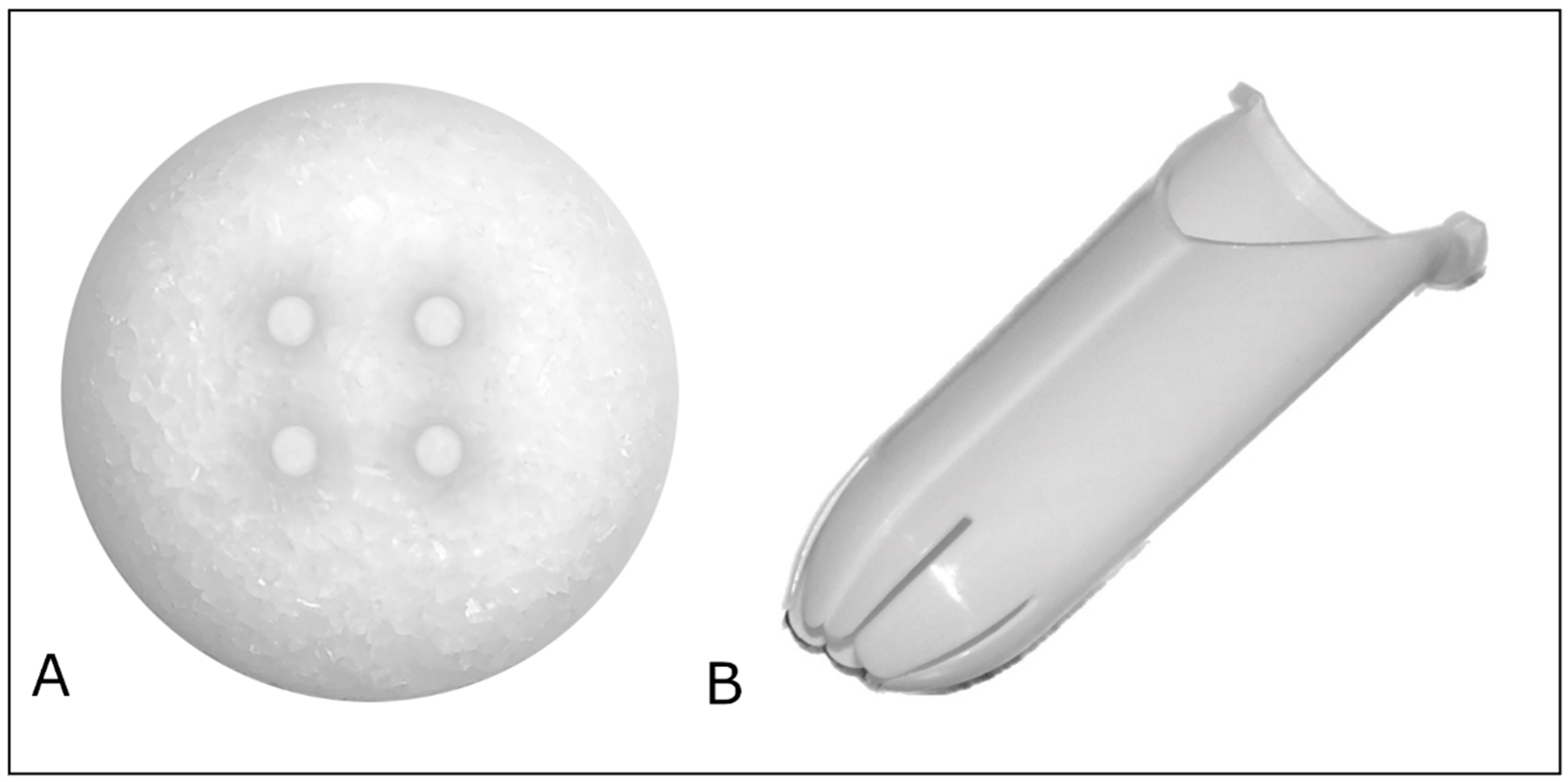
2.3. Orbital Implants

OCULFIT orbital implants (AJL Ophthalmic S.A., Vitoria, Basque Country, Spain) are manufactured from high-density porous polyethylene (Figure 1). OCULFIT orbital implants contain different grain size polyethylene in the front and back sides of the implant, producing different pore sizes and porosity. They are available in sphere diameters from 12 mm to 23 mm. OCULFIT specifications are described in Table 1. OCULFIT implants have been implanted in rabbits, showing no remarkable clinical complications and minor inflammatory response [10].

2.4. Clinical Evaluation

After the orbital implantation, follow-up visits were established as follows: Visit 1 (from 1 to 3 months postoperatively), Visit 2 (from 3 to 6 months), and Visit 3 (12 months).
OCULFIT performance was registered by the same examiner (the surgeon) after external prosthesis adjustment through several parameters: aesthetic appearance (good/poor/bad), external prosthesis motility (good/limited/null), orbital volume (symmetric/asymmetric), ocular protrusion (measured with Hertel exophthalmometer), superior palpebral sulcus depression (mild/moderate/severe), upper eyelid elevator muscle function (worse/same as contralateral eye), palpebral fissure (symmetric/asymmetric), fornix condition (wide/shortened) and lower eyelid laxity (symmetric/asymmetric). Patient satisfaction on a scale of 0 (very bad) to 5 (excellent) was recorded for all patients at every visit. Patient satisfaction was evaluated based on several factors, such as comfort, patients’ subjective aesthetic appearance, prosthesis motility, or functional results, to provide a comprehensive overview of patient perspective. Additionally, orbital implant motility without the external prosthesis was also registered (good/limited/null). All parameters (except for orbital implant motility without external prosthesis) were recorded after external prosthesis placement.
The presence of complications, such as pain, postoperative blepharoptosis, exposure, infection, and symptoms of phantom eye syndrome, was registered as well as the presence of metastasis in those cases of enucleation due to tumor.
Success was also evaluated at the end of the follow-up. Anatomical success was defined as those cases presenting all of the following: aesthetic appearance with external prosthesis good or fair + orbital volume with external prosthesis symmetric to the remaining eye + slight or moderate depression of the upper palpebral sulcus + palpebral fissure symmetric to the remaining eye + lower palpebral laxity symmetric to the remaining eye + wide fornices. Functional success was defined as those cases presenting good external prosthesis motility and good orbital implant motility. We acknowledge that the evaluation was based on subjective assessments. Different variables, such as conjunctiva-tenon status, previous surgeries, gender, abdominal dermal fat grafting in surgery, main cause of eye removal, type of intervention, age and diameter of implant, were analyzed to check their effect on anatomical and functional success rates.

2.5. Statistical Analysis
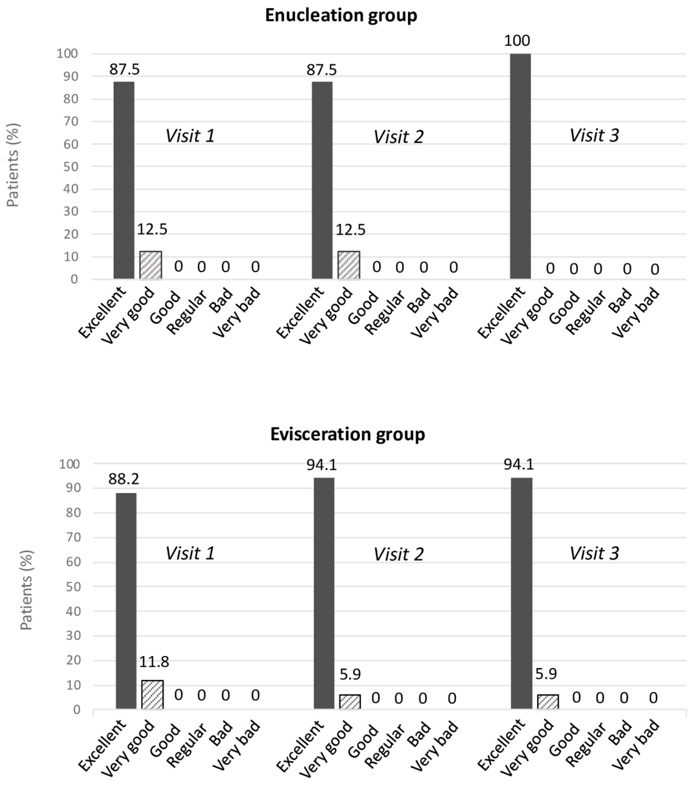
Descriptive and statistical analyses were performed using the IBM SPSS statistics software version 25.0 (SPSS Inc., Chicago, IL, USA). Results were divided into enucleation and evisceration groups. Comparisons between groups were performed using the Student’s t-test for continuous variables and the Chi-square test for categorical variables. Performance results are represented as the number of patients and percentage. Ocular protrusion and patient satisfaction are expressed as mean ± standard deviation. Patient satisfaction is also represented in a bar chart for every visit on a scale from 0 to 5. The effect of different variables on success rates was analyzed using the Chi-square test for categorical variables.
The sample size was calculated using the Granmo v.7.11. sample size and power calculator. The sample size calculation is based on the rate of one of the most serious complications with this type of implant as the primary endpoint, which is the development of an ocular infection. According to previous literature regarding the rate of this complication, which has a value of 2%, and assuming a minimum difference to be detected of 5% and a confidence level of 95%, the minimum number of evaluable eyes would be 29.

3. Results

Thirty-three patients (19 women and 14 men) with a mean age of 65.6 ± 12.0 years (range 30–85) were included in the study. The mean follow-up was 12.0 ± 1.9 months (range 10–19 months; median 12 months). Sixteen of them underwent enucleation in one eye, whereas 17 underwent evisceration. Eighteen right eyes and 15 left eyes were evaluated. The main causes of eye loss were the presence of a tumor (n = 16, 48.5%), postsurgical complications (n = 11, 33.3%), glaucoma (n = 4, 12.1%), infection (n = 1, 3.0%), and others (n = 1, 3.0%). Among those patients with a preoperative tumor, eight presented with spindle cell choroidal melanoma, three presented with epithelioid cell choroidal melanoma, and five presented with mixed-cell type choroidal melanoma. Mean optic nerve resection was 9.75 mm, 8.83 mm, and 5.80 mm, respectively.
The most common preoperative symptoms reported by patients were ocular pain (n = 18), blurred vision (n = 11), secretion or exudation (n = 1), and others (n = 3). Among the 33 patients, 15 spheres with a diameter of 21 mm, 11 with a diameter of 20 mm, 4 with a diameter of 18 mm, 2 with a diameter of 22 mm and 1 with a diameter of 19 mm were implanted. Overall, 9 thick and 7 thin external prostheses were adjusted in the enucleation group, whereas 5 thick and 12 thin prostheses were placed in the evisceration group.
Results in terms of aesthetics, symmetry, and motility are summarized in Table 2. Overall, there were no significant differences after implantation surgery between enucleated and eviscerated eyes. Nearly all patients showed good orbital implant motility without external prostheses. After external prosthesis placement, aesthetic appearance and motility were also good for the most part. The palpebral fissure was symmetric for the vast majority of them (93.8% and 88.2% for enucleated and eviscerated eyes, respectively). No shortened fornixes were found at any visit, and lower eyelid laxity was symmetric with the contralateral eye. External prosthesis motility was good for 68.8% of enucleated eyes and for 94.1% of eviscerated eyes. The operated eye presented a slightly lower ocular protrusion compared to the other eye, but the differences between eyes were not significant between enucleated and eviscerated groups. Superior palpebral sulcus depression was mild in 75% of enucleated cases and in 88.2% of eviscerated cases. The function of the upper eyelid elevator muscle was worse than the contralateral eye in 68.8% of enucleated eyes and in 41.2% of eviscerated eyes in the last visit, but the difference between groups was not significant. At the end of the follow-up, the orbital volume with external prosthesis was symmetric to the contralateral eye in 100% of cases for both groups.
Figure 2 shows patient satisfaction after OCULFIT implantation after external prosthesis placement for both enucleation and evisceration groups. Satisfaction was excellent for both procedures for almost all patients and remained excellent over time. Average patient satisfaction on a scale from 0 to 5 was 4.88 ± 0.34, 4.88 ± 0.34 and 5.00 ± 0.00 in the enucleated group for Visits 1, 2 and 3, respectively, and 4.88 ± 0.33, 4.94 ± 0.24 and 4.94 ± 0.24 in the eviscerated group.
Regarding safety results, complications and visual symptoms were reported. No postoperative complications related to either enucleation/evisceration or OCULFIT implantation were found. During the follow-up period of 12.0 ± 1.9 months (range 10–19 months; median 12 months), two eviscerated eyes (6%) required advancement of levator aponeurosis surgery because of moderate aponeurotic ptosis (7 and 9 months after surgery) and obtaining full recovery with no side effects. One enucleated eye (3%) required horizontal eyelid shortening and lateral canthoplasty surgeries 9 and 12 months after implantation because of recurrent moderate palpebral laxity (floppy eyelid syndrome) due to obstructive sleep apnea. However, these conditions were present before surgery. Six patients (18.2%) experienced symptoms of phantom eye syndrome during the follow-up. At the end of the follow-up, these symptoms disappeared in three patients, improved in one patient, and remained in three patients. One patient suffered from preoperative Charles Bonnet syndrome due to binocular blindness, which did not worsen after the surgery. One patient presented liver metastasis 11 months after surgery.
Table 3 summarizes those cases reported as an anatomical and functional success and the variables that might influence the success rate. Overall, 26 (78.8%) and 27 (81.8%) cases out of 33 eyes were classified as anatomical and functional success, respectively. None of the variables analyzed significantly affected anatomical or functional success rates (all p > 0.05).

4. Discussion

Orbital implants are a pivotal factor in restoring the volume of the lost eye and maintaining the aesthetic appearance and motility in comparison to the contralateral eye [1,2]. New versions of different materials are being launched into the market. OCULFIT is a new high-density porous polyethylene implant with additional biopolymers besides high-density polyethylene. Moreover, this implant contains different grain size polyethylene in the front and back sides, producing different pore sizes and porosity. OCULFIT implants have been evaluated in rabbits, showing no remarkable clinical complications and minor inflammatory response [10]. However, there is still little knowledge regarding its clinical performance in humans. Therefore, this research studies the performance and safety of the OCULFIT orbital implant manufactured in this porous material 1 year after implantation.
In this study with short-term follow-up (12.0 ± 1.9 months; range 10–19 months; median 12 months), the overall results showed excellent performance and safety after OCULFIT implantation. There were no exposures or infections, which are two of the most reported complications in this type of procedure. No complications secondary to evisceration/enucleation or OCULFIT implantation were reported, although 18.2% experienced symptoms of phantom eye syndrome during the follow-up.
On the other hand, the external prosthesis motility depends on the efficiency of transmitting the movement from the implant [13]. Thus, the assessment of implant motility is crucial for the final result. The results of the present study showed good orbital implant motility (without external prosthesis placement) for 100% of eviscerated patients and 93.8% of enucleated patients. Only one patient experienced limited motility.
After external prosthesis placement, 68.8% and 94.1% of enucleated and eviscerated patients reported good motility at the last visit, whereas 31.2% and 5.9% showed limited motility. Although there was a difference between enucleated and eviscerated eyes, this was not statistically significant. No patient showed null motility. Moreover, the aesthetic appearance of this prosthesis was good for all eviscerated patients and 87.5% of enucleated patients. We emphasize that the variables analyzed in this study were subjective, and we acknowledge that the use of objective techniques could have enhanced the rigor of our findings. While subjective assessments provided valuable insights, incorporating objective methods would likely contribute to greater accuracy and reproducibility in future research.
Additionally, anatomical and functional success rates were calculated (Table 3), showing excellent results (78.8% and 81.8%, respectively). Even though none of the variables analyzed significantly affected the success rates (all p > 0.05), patients classified as anatomical failure were 11 years older than those classified as anatomical success. This age difference could suggest that older patients may have more comorbidities or anatomical variations that could potentially influence surgical outcomes.
In addition to complications and implant performance, patient satisfaction is essential after an orbital implant. The enormous impact losing an eye has on patients’ quality of life is well known [14,15]. Therefore, patient satisfaction after implantation should also be taken into account. In the present study, patient satisfaction was assessed at different visits for both enucleated and eviscerated groups. Excellent satisfaction was reported for almost all patients at the last visit (100% and 94.1% for enucleated and eviscerated patients, respectively) with a small percentage showing very good satisfaction. Good, regular, bad, or very bad satisfaction scores were not reported by any patient.
Some investigators have studied the safety and biocompatibility of OCULFIT implants in animal models. Fernandez-Bueno et al. [10] performed an experimental study in rabbits implanted with OCULFIT with a follow-up of 180 days. No remarkable clinical complications were found, neither implant exposure nor infection, observing minor inflammatory response. Ophthalmic tolerance and biocompatibility were also comparable to Medpor implants. When analyzing both implants (OCULFIT and Medpor) from the histological point of view, they found differences between designs. Medpor showed a structure of small spherical granules, whereas OCULFIT had a more compact geometry and presented multiple microgranules. Additionally, both implants showed peripheral ingrowth of host vasculature and soft tissue. Regional tenderness, conjunctival hyperemia and eyelid swelling were similar between materials. It should be taken into account that animal studies cannot be directly extrapolated to humans, so future long-term clinical studies are needed to confirm these findings.
The results of this study showed the overall good performance and safety of OCULFIT implants. However, this study presents some limitations that need to be taken into consideration. The small sample size represents its primary limitation, as it may limit the statistical power of the study and affect the robustness of the conclusions drawn. Additionally, the relatively short follow-up period of one year for anophthalmic cases may not capture long-term outcomes or complications that could arise over time, as many issues may manifest beyond this timeframe. This limitation can also affect patient satisfaction and aesthetic results, which may evolve as the patients continue their recovery. Additionally, the vast majority of the parameters evaluated in this research rely on subjective measurements. While the findings provide valuable information, they should not be considered as a complete validation of the product, since it has not been subjected to sufficiently rigorous testing to guarantee its long-term safety and efficacy. This study should not be interpreted as an endorsement for its widespread use without further, more comprehensive research, including larger-scale clinical trials.
Although the inclusion of a single evaluator with extensive experience in surgeries involving anophthalmic cavities offers notable advantages—such as minimizing inter-rater variability and ensuring consistency and reliability—this approach also presents certain limitations. A broader technical assessment could benefit from the input of a multidisciplinary panel, including experts from diverse fields to provide varied perspectives and reduce potential bias, particularly when the experts are independent. This limitation highlights an important consideration for future research.
In conclusion, OCULFIT implants showed promising results after external prosthesis placement after 1 year of follow-up, providing excellent patient satisfaction and excellent performance in terms of motility and symmetry with the contralateral eye with no complications related to the surgery. Anatomical and functional success rates were high. Aesthetic results after external prosthesis placement were remarkable for both enucleated and eviscerated eyes.

Author Contributions

Conceptualization: A.M.G.-H., E.G.-S., M.I.R.-L. and E.R.-d.-l.-R.-F.; methodology: A.M.G.-H., M.C.D.-R., R.M.-C., M.M.-M. and V.S.-M.; formal analysis: A.M.G.-H. and I.M.-A.; investigation: A.M.G.-H. and I.M.-A.; data curation: A.M.G.-H. and I.M.-A.; writing—original draft: A.M.G.-H. and I.M.-A.; preparation: R.M.-C., M.M.-M. and V.S.-M.; writing—review and editing: A.M.G.-H., I.M.-A., E.G.-S., M.I.R.-L. and E.R.-d.-l.-R.-F. All authors have read and agreed to the published version of the manuscript.

Funding

The APC and manuscript preparations costs were funded by AJL Opthalmic S.A. (Vitoria, Basque Country, Spain).

Institutional Review Board Statement

The study was conducted in accordance with the Declaration of Helsinki and approved by the Ethics Committee of Hospitales Universitarios Virgen Macarena (protocol code 0104-N-21 and date of approval 15 March 2021).

Informed Consent Statement

Informed consent was obtained from all subjects involved in the study.

Data Availability Statement

The data that support the findings of this study are available from the corresponding author upon reasonable request.

Conflicts of Interest

The authors declare no conflicts of interest. Manuscript and publication costs were funded by AJL Opthalmic S.A. (Vitoria, Basque Country, Spain). None of the authors have any contractual relationships, financial interests, or other affiliations with the funding organization.

References

  1. Custer, P.L.; Kennedy, R.H.; Woog, J.J.; Kaltreider, S.A.; Meyer, D.R. Orbital implants in enucleation surgery: A report by the American Academy of Ophthalmology. Ophthalmology 2003, 110, 2054–2061. [Google Scholar] [CrossRef] [PubMed]
  2. Wladis, E.J.; Aakalu, V.K.; Sobel, R.K.; Yen, M.T.; Bilyk, J.R.; Mawn, L.A. Orbital Implants in Enucleation Surgery: A Report by the American Academy of Ophthalmology. Ophthalmology 2018, 125, 311–317. [Google Scholar] [CrossRef] [PubMed]
  3. Moshfeghi, D.M.; Moshfeghi, A.A.; Finger, P.T. Enucleation. Surv. Ophthalmol. 2000, 44, 277–301. [Google Scholar] [CrossRef]
  4. Shah, S.U.; Shields, C.L.; Lally, S.E.; Shields, J.A. Hydroxyapatite orbital implant in children following enucleation: Analysis of 531 sockets. Ophthal. Plast. Reconstr. Surg. 2015, 31, 108–114. [Google Scholar] [CrossRef]
  5. Hornblass, A.; Biesman, B.S.; Eviatar, J.A. Current techniques of enucleation: A survey of 5,439 intraorbital implants and a review of the literature. Ophthal. Plast. Reconstr. Surg. 1995, 11, 77–78. [Google Scholar] [CrossRef]
  6. Jordan, D.R. Porous versus Nonporous Orbital Implants: A 25-Year Retrospective. Ophthalmology 2018, 125, 1317–1319. [Google Scholar] [CrossRef]
  7. Soares, I.P.; França, V.P. Evisceration and enucleation. Semin. Ophthalmol. 2010, 25, 94–97. [Google Scholar] [CrossRef]
  8. Alwitry, A.; West, S.; King, J.; Foss, A.J.; Abercrombie, L.C. Long-term follow-up of porous polyethylene spherical implants after enucleation and evisceration. Ophthal. Plast. Reconstr. Surg. 2007, 23, 11–15. [Google Scholar] [CrossRef] [PubMed]
  9. Schellini, S.A.; El Dib, R.; Limongi, R.M.; Mörschbächer, R. Anophthalmic socket: Choice of orbital implants for reconstruction. Arq. Bras. Oftalmol. 2015, 78, 260–263. [Google Scholar] [CrossRef] [PubMed]
  10. Fernandez-Bueno, I.; Di Lauro, S.; Alvarez, I.; Lopez, J.C.; Garcia-Gutierrez, M.T.; Fernandez, I.; Larra, E.; Pastor, J.C. Safety and Biocompatibility of a New High-Density Polyethylene-Based Spherical Integrated Porous Orbital Implant: An Experimental Study in Rabbits. J. Ophthalmol. 2015, 2015, 904096. [Google Scholar] [CrossRef] [PubMed]
  11. Garrido-Hermosilla, A.; Martínez-Alberquilla, I.; Díaz-Ruiz, M.C.; Monge-Carmona, R.; Méndez-Muros, M.; López-Díaz, Á.; Sánchez-Margalet, V.; Gutiérrez-Sánchez, E.; Relimpio-López, M.I.; Rodríguez-de-la-Rúa-Freanch, E. Long-Term Mental Health after High-Density Polyethylene-Based Porous Orbital Implant in Enucleated and Eviscerated Patients. Prosthesis 2024, 6, 979–988. [Google Scholar] [CrossRef]
  12. Sales-Sanz, M.; Sanz-Lopez, A. Four-petal evisceration: A new technique. Ophthal. Plast. Reconstr. Surg. 2007, 23, 389–392. [Google Scholar] [CrossRef] [PubMed]
  13. Goiato, M.C.; Haddad, M.F.; dos Santos, D.M.; Pesqueira, A.A.; Ribeiro, P.d.P.; Moreno, A. Orbital implants insertion to improve ocular prostheses motility. J. Craniofac. Surg. 2010, 21, 870–875. [Google Scholar] [CrossRef] [PubMed]
  14. Ahn, J.M.; Lee, S.Y.; Yoon, J.S. Health-related quality of life and emotional status of anophthalmic patients in Korea. Am. J. Ophthalmol. 2010, 149, 1005–1011.e1. [Google Scholar] [CrossRef] [PubMed]
  15. Rasmussen, M.L.R.; Ekholm, O.; Prause, J.U.; Toft, P.B. Quality of life of eye amputated patients. Acta Ophthalmol. 2012, 90, 435–440. [Google Scholar] [CrossRef] [PubMed]
Figure 1. Images of the OCULFIT system. (A) OCULFIT implant; (B) OCULFIT injector.
Prosthesis 06 00101 g001
Figure 2. Patient satisfaction after OCULFIT implantation for enucleated and eviscerated groups at every visit. Scale ranged from very bad (0), bad (1), regular (2), good (3), very good (4) and excellent (5) satisfaction.
Prosthesis 06 00101 g002
Table 1. OCULFIT orbital implant specifications.
OCULFIT ORBITAL IMPLANT
Sphere diameter14/16/18–23 mm
MaterialHigh-density porous polyethylene
Porosity45%
Pore size>100 µm
Granules used<400 µm in the previous part
700–100 µm at the rear
Suture channels4 interconnected
SterilizationEthylene Oxide
Expiration5 years
Table 2. Orbital implant performance for enucleated and eviscerated eyes at each visit.
Visit 1Visit 2Visit 3
AESTHETIC APPEARANCE (WITH EXTERNAL PROSTHESIS) [n (%)]
GoodPoorBadGoodPoorBadGoodPoorBad
Enucleation---13 (81.3)3 (18.7)0 (0)14 (87.5)2 (12.5)0 (0)
Evisceration---13 (76.5)4 (23.5)0 (0)17(100)0 (0)0 (0)
p-value-0.740.13
PALPEBRAL FISSURE(WITH EXTERNAL PROSTHESIS) [n (%)]
SymmetricAsymmetricSymmetricAsymmetricSymmetricAsymmetric
Enucleation--14 (87.5)2 (12.5)15 (93.8)1 (6.3)
Evisceration--12 (70.6)5 (29.4)15 (88.2)2 (11.8)
p-value-0.240.58
FORNIX CONDITION [n (%)]
WideShortenedWideShortenedWideShortened
Enucleation16 (100)0 (0)16 (100)0 (0)16 (100)0 (0)
Evisceration17 (100)0 (0)17 (100)0 (0)17 (100)0 (0)
p-value- a- a- a
LOWER EYELID LAXITY [n (%)]
SymmetricAsymmetricSymmetricAsymmetricSymmetricAsymmetric
Enucleation15 (93.8)1 (6.3)15 (93.8)1 (6.3)16 (100)0 (0)
Evisceration16 (94.1)1 (5.9)16 (94.1)1 (5.9)16 (94.1)1 (5.9)
p-value0.970.970.33
EXTERNAL PROSTHESIS MOTILITY [n (%)]
GoodLimitedNullGoodLimitedNullGoodLimitedNull
Enucleation---11 (68.8)5 (31.2)0 (0)11 (68.8)5 (31.2)0 (0)
Evisceration---15 (88.2)2 (11.8)0 (0)16 (94.1)1 (5.9)0 (0)
p-value-0.170.06
OCULAR PROTRUSION (WITH EXTERNAL PROSTHESIS) (mm)
Operated eyeContralateral eyeOperated eyeContralateral eyeDifference between eyesOperated eyeContralateral eyeDifference between eyes
Enucleation--14.59 ± 2.7315.81 ± 2.501.22 ± 0.9315.00 ± 2.3715.63 ± 2.310.63 ± 0.67
Evisceration--14.94 ± 2.5315.19 ± 3.030.29 ± 1.8015.24 ± 2.4815.76 ± 2.760.53 ± 1.07
p-value-0.08 b0.76 b
SUPERIOR PALPEBRAL SULCUS DEPRESSION (WITH EXTERNAL PROSTHESIS) [n (%)]
MildModerateSevereMildModerateSevereMildModerateSevere
Enucleation---12 (75.0)2 (12.5)2 (12.5)12 (75.0)3 (18.8)1 (6.2)
Evisceration---15 (88.2)1 (5.9)1 (5.9)15 (88.2)1 (5.9)1 (5.9)
p-value-0.620.52
UPPER EYELID ELEVATOR MUSCLE FUNCTION (WITH EXTERNAL PROSTHESIS) [n (%)]
Worse than contralateral eyeSame as contralateral eyeWorse than contralateral eyeSame as contralateral eyeWorse than contralateral eyeSame as contralateral eye
Enucleation--11 (68.8)5 (31.2)11 (68.8)5 (31.2)
Evisceration--6 (35.3)11 (64.7)7 (41.2)10 (58.8)
p-value-0.060.11
ORBITAL VOLUME (WITH EXTERNAL PROSTHESIS) [n (%)]
SymmetricAsymmetricSymmetricAsymmetricSymmetricAsymmetric
Enucleation--14 (87.5)2 (12.5)16 (100)0 (0)
Evisceration--15 (88.2)2 (11.8)17 (100)0 (0)
p-value-0.95- a
ORBITAL IMPLANT MOTILITY (WITHOUT EXTERNAL PROSTHESIS) [n (%)]
GoodLimitedNullGoodLimitedNullGoodLimitedNull
Enucleation15 (93.8)1 (6.3)0 (0)15 (93.8)1 (6.3)0 (0)15 (93.8)1 (6.3)0 (0)
Evisceration17 (100)0 (0)0 (0)17 (100)0 (0)0 (0)17 (100)0 (0)0 (0)
p-value0.300.300.30
a no statistics are computed because the variable is a constant. b p-value for the difference between eyes.
Table 3. Analysis of the effect of different variables on anatomical and functional success rates.
VariableANATOMICAL SUCCESSFUNCTIONAL SUCCESS
Success
(n = 26)
Failure
(n = 7)
p-ValueSuccess
(n = 27)
Failure
(n = 6)
p-Value
Conjunctiva-Tenon status
[n, (%)]
  • Optimal
11 (42.3)2 (28.6)0.5110 (37.0)3 (50)0.56
  • Non optimal
15 (57.7)5 (71.4)17 (63.0)3 (50)
Previous surgeries [n, (%)]
  • Yes
16 (61.5)5 (71.4)0.6319 (70.4)2 (33.3)0.09
  • No
10 (38.5)2 (28.6)8 (29.6)4 (66.7)
Gender [n, (%)]
  • Men
12 (46.2)2 (28.6)0.4010 (37.0)4 (66.7)0.18
  • Women
14 (53.8)5 (71.4)17 (63.0)2 (33.3)
Abdominal dermal fat grafting [n, (%)]
  • Yes
10 (38.5)4 (57.1)0.3811 (40.7)3 (50)0.68
  • No
16 (61.5)3 (42.9)16 (59.3)3 (50)
Main cause of eye removal [n, (%)]
  • Surgical complication
9 (34.6)2 (28.6)0.3910 (37.0)1 (16.7)0.44
  • Glaucoma
3 (11.5)1 (14.3)4 (14.8)0 (0.0)
  • Infection
1 (3.9)0 (0.0)1 (3.7)0 (0.0)
  • Tumour
13 (50.0)3 (42.9)11 (40.7)5 (83.3)
  • Other
0 (0.0)1 (14.3)1 (3.7)0 (0.0)
Type of intervention [n, (%)]
  • Enucleation
13 (50.0)3 (42.9)0.7411 (40.7)5 (83.3)0.06
  • Evisceration
13 (50.0)4 (57.1)16 (59.3)1 (16.7)
Age (mean ± SD)63.2 ± 14.274.4 ± 9.80.0665.7 ± 15.065.2 ± 9.50.94
Diameter of implants (mean ± SD)20.3 ± 1.120.1 ± 1.20.6620.4 ± 1.019.8 ± 1.20.24
Disclaimer/Publisher’s Note: The statements, opinions and data contained in all publications are solely those of the individual author(s) and contributor(s) and not of MDPI and/or the editor(s). MDPI and/or the editor(s) disclaim responsibility for any injury to people or property resulting from any ideas, methods, instructions or products referred to in the content.

