Review

Advances in Nanomedicine for Modulating DNA Methylation and Inducing Pyroptosis

1
School of Pharmaceutical Sciences, Capital Medical University, Beijing 100069, China
2
Cancer Hospital Chinese Academy of Medical Sciences, Beijing 100021, China
*
Authors to whom correspondence should be addressed.
These authors contributed equally to this work.
J. Nanotheranostics 2026, 7(2), 14; https://doi.org/10.3390/jnt7020014
Submission received: 28 April 2026 / Revised: 25 May 2026 / Accepted: 30 May 2026 / Published: 5 June 2026
(This article belongs to the Special Issue Feature Review Papers in Nanotheranostics)

Abstract

DNA methylation is a key mechanism in epigenetic regulation and plays a pivotal role in tumor initiation, progression, and therapeutic resistance. We begin by elucidating how the dysregulation of key DNA methylation enzymes in tumors drives concurrent global hypomethylation and cytosine-phosphate-guanine (CpG) island hypermethylation. This aberrant epigenetic landscape promotes tumorigenesis through silencing tumor suppressor genes and triggering abnormal activation of oncogenic signaling pathways. Notably, DNA methylation is intimately linked to cellular pyroptosis. In particular, the hypermethylation-mediated silencing of pyroptosis effector genes represents a critical epigenetic mechanism underlying acquired drug resistance. Targeting DNA methylation with epigenetic drugs offers a novel strategy to resensitize tumors to chemotherapy, radiotherapy, and immunotherapy. Moreover, advances in nanomedicine have yielded smart platforms for the precise administration of epigenetic modulators and combination therapies. These platforms enable a coordinated “epigenetic priming-pyroptosis execution” strategy, which holds promises for reversing therapeutic resistance and remodeling the tumor immune microenvironment. By integrating DNA methylation regulation, pyroptosis mechanisms, and nano-targeted strategies, this review aims to provide a theoretical framework and novel perspectives for developing innovative, epigenetically driven anti-tumor therapies.

1. Introduction

Cancer initiation and progression result from not only accumulated genetic mutations, but also profound dysregulation of epigenetic regulatory networks [1,2]. Among epigenetic modifications, DNA methylation is the earliest discovered and most extensively studied type [3,4,5]. In cancer cells, DNA methylation patterns undergo characteristic remodeling, featuring widespread global hypomethylation alongside focal CpG island hypermethylation [6]. Global hypomethylation promotes genomic instability and aberrant proto-oncogene activation, whereas CpG island hypermethylation silences tumor suppressor genes, endowing cancer cells with malignant traits such as unlimited proliferation, apoptosis resistance, and immune evasion [7].
Pyroptosis is an inflammatory form of programmed cell death [8,9]. Its execution relies on the cleavage of Gasdermin family proteins (e.g., Gasdermin E, GSDME) by activated caspases, leading to the formation of membrane pores [10]. Pivotal studies have revealed that the GSDME gene is silenced in various tumors due to promoter hypermethylation, rendering cancer cells resistant to chemotherapy-induced pyroptosis. This uncovers a novel mechanism by which DNA methylation directly regulates cell death fate and mediates therapeutic resistance [11].
Given the reversible nature of DNA methylation, epigenetic drugs targeting its regulatory pathways, such as the DNMT inhibitor decitabine (DAC), have become important strategies in cancer therapy [12,13]. However, traditional epigenetic drugs face challenges including rapid in vivo metabolism, poor tumor targeting, and limited efficacy as monotherapies. The emergence of nanomedicine offers opportunities to address these obstacles [14,15]. Through the construction of intelligent nano-delivery systems, the co-delivery of epigenetic drugs with chemotherapeutic agents, photosensitizers, or immunomodulators can be achieved. This enables spatiotemporally controlled release within the tumor, realizing a synergistic effect of “epigenetic priming” and “pyroptosis induction-execution”, thereby efficiently killing cancer cells and provoking anti-tumor immune responses [16]. Therefore, a deep understanding of the regulatory networks of DNA methylation in tumorigenesis and pyroptosis, coupled with the development of combination therapeutic strategies leveraging advanced nanotechnology, is of great significance for overcoming the limitations of current therapies and advancing the development of precision oncology. This review will start with the fundamental regulatory mechanisms of DNA methylation, focus on its aberrant patterns in cancer and its interplay with pyroptosis, and summarize recent research progress in nanomedicine-based anti-tumor therapies targeting both DNA methylation and pyroptosis induction.

2. Epigenetic Regulation in Cancer

Epigenetics explores heritable changes in gene expression that occur without altering the DNA sequence itself. Since the concept was first introduced by Conrad Waddington in 1942, this field has undergone significant evolution from theoretical hypothesis to the elucidation of molecular mechanisms [17]. The main areas of epigenetic research include DNA methylation, histone modifications, non-coding RNA regulation, and chromatin remodeling [18]. A fundamental characteristic of epigenetics is the diverse covalent modifications of histones and nucleic acids, which collectively control chromatin structure and gene expression [19]. These epigenetic modifications are reversible and involve the interplay of three key enzyme systems—“writers”, “erasers”, and “readers”. Epigenetic modifications orchestrate gene expression and transcription, critically shaping processes from embryonic development and stem cell differentiation to aging and tumorigenesis, while also offering promising novel therapeutic targets for cancer intervention [20,21]. The cumulative effects of various epigenetic mechanisms ultimately lead to several core cellular dysfunctions, including oncogene activation and tumor suppressor gene inactivation, unlimited proliferative capacity, resistance to cell death, microenvironment remodeling, and metabolic reprogramming. These modifications directly contribute to the hallmarks of cancer, driving tumor initiation, progression, metastasis, and development of therapeutic resistance [22,23,24]. Recent studies have established that the phenotypic and functional dysregulation of cells within the tumor microenvironment (TME)—including cancer cells, immune cells, and stromal cells—is pervasively influenced by upstream reversible epigenetic regulation [25]. This insight broadens the therapeutic potential of epigenetic modulators beyond direct effects on cancer cells to include reprogramming of immune and stromal compartments.

2.1. DNA Methylation

Of all epigenetic modifications, DNA methylation was the first to be identified and has been studied most thoroughly [26,27]. Since its initial discovery in bacteria in 1925, DNA methylation has been extensively investigated across a broad spectrum of organisms, shedding light on its fundamental involvement in gene regulation, development, reproduction, disease pathogenesis, and aging [28]. In 1948, Hotchkiss’s identification of 5-methylcytosine (5mC) in calf thymus DNA marked the beginning of DNA methylation research. Subsequently, in 1975, Holliday and Pugh proposed the hypothesis that DNA methylation might be involved in gene expression regulation. Further, the development of high-throughput sequencing in the 21st century has enabled genome-wide DNA methylation mapping, profoundly advancing research in tumor epigenetics [29].

2.2. Mechanisms of DNA Methylation

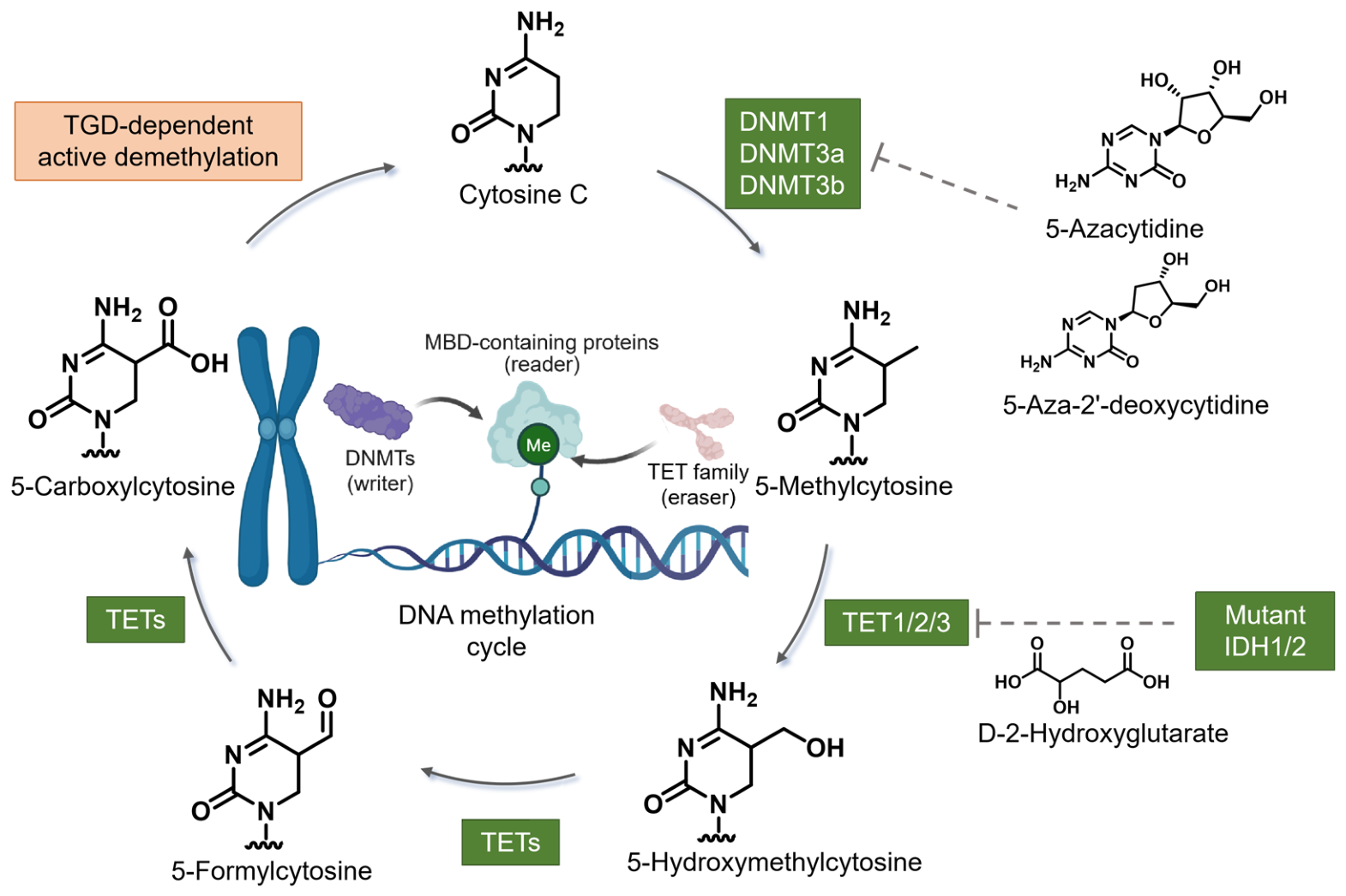
DNA methylation refers to the covalent addition of a methyl group to the 5-carbon position of cytosine residues within CpG dinucleotides to form 5-methylcytosine (5mC) under the catalysis by DNA methyltransferases (DNMTs) [30]. The DNMTs that act on cytosine include DNMT1, DNMT3A, and DNMT3B. Specifically, DNMT1 is primarily responsible for maintaining methylation patterns, ensuring that methyl groups are transferred to the newly synthesized strand during DNA replication; DNMT3A and DNMT3B, on the other hand, are responsible for establishing new methylation patterns. The general reaction pathway involves: (i) nucleophilic attack by a conserved cysteine residue in the DNMT motif IV on the C6 position of the cytosine ring; (ii) transfer of a methyl group from S-adenosylmethionine (SAM) to the C5 position of cytosine; and (iii) proton elimination at C5, with the cysteine leaving, resulting in the formation of 5-methylcytosine (Figure 1). Furthermore, DNA methylation occurs not only in gene promoter regions but is also widespread in regions such as gene enhancers and silenced gene areas. The methylation status directly influences gene expression states, thereby determining biological behaviors including cell differentiation, proliferation, and death [31,32,33].

2.3. Mechanisms of DNA Demethylation

The process of DNA demethylation is mediated by the TET proteins family, which includes TET1, TET2, and TET3. This oxidative reaction does not occur in a single step but rather proceeds through progressive oxidation: TET enzymes first oxidize stable 5mC to 5-hydroxymethylcytosine (5hmC), which is further oxidized to 5-formylcytosine (5fC) and 5-carboxylcytosine (5caC) [34]. Concurrently, TET enzyme genes themselves are commonly mutated in cancer. Therefore, TET-mediated oxidation represents a central nexus between cellular metabolism and epigenetic regulation, ultimately, TET dysregulation constitutes a key driver of epigenetic reprogramming in tumorigenesis [35,36].

2.4. Key Enzymes in DNA Methylation

The epigenetic modification is regulated by specific enzymes, namely “writers”, “erasers”, and “readers”, which collectively construct complex regulatory networks [37].
Writers: DNMTs are the key enzymes responsible for DNA methylation, catalyzing the addition of methyl groups to cytosine residues in DNA to form 5mC. DNMT1 functions as the primary maintained methyltransferase, recognizing hemi-methyl CpG sites and catalyzing the addition of methyl group to the nascent daughter strand during DNA replication. DNMT3A and DNMT3B mediate de novo methylation, establishing new methylation patterns in previously unmethylated region [38]. In cancer cells, overexpression of DNMTs frequently leads to the silencing of critical tumor suppressor genes, thereby promoting tumorigenesis [39].
Readers: MBD family proteins serve as “readers” that specifically bind to methylated DNA, primarily recognizing methylated CpG sites [40]. By altering chromatin structure and recruiting chromatin remodeling complexes, they dynamically modulate chromatin accessibility between accessible and condensed states to precisely regulate gene transcription and expression. In addition, members of the MBD family (such as MBD1, MBD2, and MeCP2) affect cellular phenotypes through regulating chromatin structure and function [41,42,43].
Erasers: The TET family (TET1/2/3) is crucial for DNA demethylation, which progressively oxidize 5mC to 5hmC, 5fC, and 5-carboxylcytosine (5caC). These intermediates are subsequently replaced by unmethylated cytosine through the thymine DNA glycosylase (TDG)-mediated base excision repair pathway, thereby restoring gene expression [44]. By mediating DNA demethylation process, TETs serve as crucial regulators of gene activation, especially in the reactivation of tumor suppressor genes, underscoring the dynamic reversibility of epigenetic methylation modifications [45,46,47].
This intricate network of “writers-erasers-readers” ensures precise regulation of gene expression and plays a critical role in cellular differentiation, development, and tumorigenesis [48,49].

3. Mechanisms of DNA Methylation in Tumorigenesis

3.1. Aberrant Expression of DNMT Family in Tumors

DNA methyltransferases play a dual role in tumorigenesis (Table 1). DNMT1 is highly expressed in various tumors, including colorectal cancer, liver cancer, lung cancer, and breast cancer [50,51,52,53,54,55,56]. Studies have shown that DNMT1 overexpression is closely associated with tumor grade, stage, and prognosis. In colorectal cancer, DNMT1 expression progressively increase with tumor advancement, and patients with high DNMT1 expression exhibit significantly lower 5-year survival rates compared to those with low expression. Mechanistic studies reveal that DNMT1 maintains the hypermethylation status of tumor suppressor gene promoters, continuously inhibiting their expression and promoting tumor cell proliferation and invasion [57]. The aberrant expression patterns of DNMT3A and DNMT3B vary across different tumor types. In acute myeloid leukemia (AML), the mutation frequency of the DNMT3A gene reaches 20–30%, with the R882 mutation being most common, resulting in decreased enzyme activity and abnormal methylation patterns [58]. AML patients with DNMT3A mutations typically have poorer prognosis and higher recurrence rates. Conversely, in solid tumors such as hepatocellular carcinoma, DNMT3A and DNMT3B are often overexpressed, mediating aberrant de novo methylation and leading to silencing of multiple tumor suppressor genes [59].
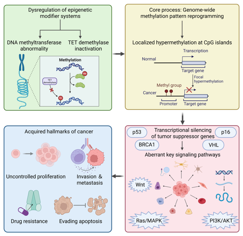
The DNMTs expression is subject to multifaceted regulation across epigenetic, transcriptional, and post-translational levels. At the epigenetic level, it should be noted that, contrary to the silencing observed by many tumor suppressors, DNMT1 is typically overexpressed in cancers; accordingly, its promoter is generally maintained in an unmethylated or hypomethylated state. For example, aberrant promoter hypermethylation of DNMT3A and DNMT3B have been reported in certain cancers, contributing to their transcriptional silencing. By contrast, DNMT1 expressions are more commonly modulated by transcription factors, microRNAs, and post-translational modifications, rather than by promoter methylation. Additionally, long non-coding RNAs can recruit chromatin modifiers to DNMT genomic regions, providing further epigenetic control [60]. At the transcriptional level, a complex network of transcription factors constitutes a sophisticated regulatory system [61,62]. It has demonstrated that bortezomib induces DNA hypomethylation and gene transcriptional silencing in acute myeloid leukemia by disrupting Sp1/NF-κB-dependent DNA methyltransferase activity. Transcription factors such as Sp1, E2F, and NF-κB are known to regulate DNMT gene expression. Specifically, E2F modulates DNMT1 and DNMT3A expression during the cell cycle, and inactivation of the RB tumor suppressor gene leads to dysregulated E2F-mediated transcription [57]. Critically, the tumor suppressor p53 can directly inhibit the transcription of multiple DNMTs; its frequent mutation or inactivation in tumors effectively releases this “brake” on DNMTs, contributing to global hypermethylation. Furthermore, other factors including c-Myc and FOXO3a are also extensively involved in this regulatory network [63,64]. At the post-transcriptional level, microRNAs play a significant negative regulatory role. Among these, the miR-29 family specifically binds to the 3′UTR of DNMT3A and DNMT3B mRNA, inhibiting their translation or promoting their degradation [65]. However, in various tumors such as lung cancer, the expression of miR-29 is often significantly downregulated, leading to aberrant accumulation of DNMT3A/3B proteins, which in turn triggers abnormal methylation silencing of tumor suppressor genes [66,67,68,69]. Similarly, miR-148a and miR-152 exert regulatory effects on DNMT1. At the post-translational modification level, modifications such as ubiquitination and phosphorylation dynamically regulate the stability, activity, and localization of DNMT proteins. For instance, the ubiquitin-proteasome pathway controls the degradation rate of DNMT proteins, while phosphorylation couples DNMT activity with cellular growth signaling pathways [70]. Aberrations in these modifications can similarly affect the functional status of DNMTs within cells (Figure 2).
In summary, transcriptional activation, microRNA inhibition, and post-translational modifications collectively constitute a sophisticated regulatory network governing DNMT expression. In tumors, these regulatory mechanisms are often disrupted, resulting in aberrant DNMT expression [71,72].

3.2. TET Family Dysfunction and Cancer

As key executors of active DNA demethylation, TET family proteins maintain epigenetic homeostasis; consequently, their dysfunction represents a core epigenetic mechanism driving the initiation and progression of various tumors [73]. Across different cancer types, TET proteins are inactivated through distinct mechanisms—including gene mutation, expression silence, or activity inhibition—collectively resulting in decreased genome-wide 5hmC levels, accumulation of aberrant hypermethylation, subsequent silencing of tumor suppressor genes, and reprogramming of cellular fate [74,75].
In hematologic malignancies, loss-of-function mutations in TET2 are particularly prominent. In myeloid neoplasms such as myelodysplastic syndromes, chronic myelomonocytic leukemia, and acute myeloid leukemia, the mutation frequency of the TET2 gene can reach 10–30% [76,77,78]. These mutations directly impair TET2 catalytic activity, leading to significantly reduced 5hmC levels in hematopoietic stem cells and aberrant genome-wide DNA hypermethylation patterns, ultimately disrupting normal hematopoietic differentiation programs and promoting leukemic transformation [79]. Notably, this specific molecular defect holds important clinical translational value: patients carrying TET2 mutations often exhibit better therapeutic responses to demethylating agents (e.g., azacitidine), positioning TET2 mutation status as a potential biomarker for guiding treatment selection [80].
Compared to mutation-driven inactivation in hematologic tumors, downregulation of TET1 expression represents a more common mode of inactivation in solid tumors. Significant reductions in TET1 mRNA and protein levels are frequently observed in various epithelial-derived tumor tissues, including breast, colorectal, and gastric cancers [81,82,83,84]. The underlying mechanism often involves hypermethylation of CpG islands within the TET1 promoter region itself, creating a vicious positive feedback loop where hypermethylation leads to low TET1 expression, and low TET1 expression further exacerbates global hypermethylation”. Functionally, restoration of TET1 expression can reactivate critical tumor suppressor genes such as TIMP2 and TIMP3, effectively inhibiting tumor cell invasion and metastatic capacity [85,86,87].
Furthermore, TET protein function is highly dependent on the tumor cell metabolic microenvironment [88]. As dioxygenases, TET enzymes strictly require α-ketoglutarate as an essential cofactor for their catalytic activity. In tumors such as gliomas carrying IDH mutations, the oncometabolite 2-hydroxyglutarate accumulates aberrantly and competitively inhibits α-ketoglutarate-dependent dioxygenase activity, thereby broadly suppressing TET protein function. It results in a characteristic CpG island hyper-methylator phenotype, profoundly altering cellular gene expression profiles and differentiation states [89].
Recent studies have revealed distinct therapeutic vulnerabilities arising from TET dysfunction that are not shared by tumors with aberrant DNMT activity. For instance, TET2-mutant leukemia cells display downregulation of BRCA1 and LIG4, resulting in reduced homologous recombination and non-homologous end-joining repair activity; consequently, they become dependent on PARP1-mediated alternative non-homologous end-joining and are exquisitely sensitive to PARP inhibitors, whereas DNMT3A-mutant cells are resistant [90]. This differential sensitivity suggests that TET2 mutation status may serve as a predictive biomarker for PARP inhibitor therapy—a vulnerability that is mechanistically distinct from DNMT inhibition. Conversely, in TET2-deficient settings where DNA hypermethylation accumulates, tumor cells may paradoxically become dependent on DNMTs to maintain their aberrant methylation landscape, rendering them hypersensitive to DNMT inhibitors [91]. Moreover, emerging evidence shows that DNMT1 gene deletion can drive TET2 upregulation, which in turn confers resistance to DNMT inhibitors via reactivation of tumor suppressors such as p16 [92]. Taken together, these findings underscore that TET dysfunction and DNMT dysregulation necessitate fundamentally different therapeutic strategies: TET2-deficient tumors may benefit from PARP inhibitors, whereas TET2-upregulated or DNMT1-deleted tumors may require alternative approaches beyond conventional DNMT inhibition. In summary, TET protein dysfunction represents a common feature spanning both hematologic and solid malignancies. Although the modes of inactivation vary by tumor type, they ultimately converge on the common endpoints of DNA hypermethylation and gene silencing. Deeply intertwined with the tumor metabolic microenvironment, this dysfunction constitutes a core component of the epigenetic regulatory network in cancer [93,94].

3.3. Global Hypomethylation and Genomic Instability

In cancer cells, the overall DNA methylation across the genome decreases substantially, often by 20% to 60%. Global hypomethylation primarily occurs in intergenic regions, introns, and repetitive sequences, rather than in CpG island regions [95,96,97,98]. Hypomethylation of repetitive sequences, particularly long interspersed nuclear elements (LINEs) and short interspersed nuclear elements (SINEs), is especially pronounced and can lead to reactivation of these transposable elements, thereby increasing genomic instability [99,100]. Hypomethylation of LINE-1 elements is particularly critical, as it is closely associated with chromosomal rearrangements, gene amplifications, and deletions [101]. In colorectal cancer, LINE-1 methylation levels correlate with microsatellite instability (MSI) status, with hypomethylated tumors frequently exhibiting the chromosomal instability (CIN) phenotype [102,103,104,105]. Hypomethylation of satellite DNA can lead to relaxation of pericentromeric heterochromatin, increasing the risk of chromosomal segregation errors and aneuploidy formation [106]. It has been studied that hypomethylation of SAT2 repetitive sequences is associated with instability in the chromosome 1q12 region, and amplification of this region is observed in various tumors [107].
Beyond inducing structural genomic variations, global hypomethylation can directly relieve epigenetic silence of specific proto-oncogenes, leading to their aberrant activation. Oncogenes such as R-RAS family genes which are normally silenced by DNA methylation in healthy tissues are reactivated with promoter hypomethylation and then promote tumorigenesis [108,109]. For example, in gastric cancer, hypomethylation of the HRAS proto-oncogene promoter results in overexpression, activating downstream MAPK signaling pathways and promoting tumor cell proliferation [110].
In summary, global hypomethylation promotes tumor initiation and progression through dual mechanisms: altering genomic structural stability via effects on repetitive sequences and dysregulating transcriptional programs through aberrant activation of proto-oncogenes. These processes drive synergistically cancer development.

3.4. CpG Island Hypermethylation and Gene Silencing

A key epigenetic abnormality coexisting with global hypomethylation in tumors is the hypermethylation of promoter region CpG islands. Under normal conditions, approximately 60% of human gene promoters containing CpG islands remain unmethylated to ensure normal gene expression [111]. In cancer, specific CpG islands undergo aberrant hypermethylation, leading to transcriptional silencing of associated genes—a phenomenon termed the “CpG island methylator phenotype” (CIMP) [112].
The incidence and characteristics of CIMP vary across different tumor types. CIMP-positive tumors account for approximately 15–20% colorectal cancer. These tumors predominantly occur in the proximal colon and are frequently associated with BRAF V600E mutations and microsatellite instability. CIMP-positive colorectal cancers exhibit hypermethylation of a characteristic set of genes, including CDKN2A, MLH1, and MGMT [112,113,114]. In glioblastoma, the G-CIMP subtype is closely associated with IDH mutations, and patients with this subtype generally have a relatively favorable prognosis [115]. CIMP subtypes have also been identified in breast cancer, characterized by coordinated hypermethylation of multiple genes including the estrogen receptor gene ESR1 [116].
The target genes regulated by CpG island hypermethylation are involved in multiple functional pathways and biological categories. Methylation silencing of tumor suppressor genes such as VHL, BRCA1, and PTEN directly promote tumorigenesis. Methylation of DNA repair genes including MGMT, MLH1, and BRCA1 leads to increased genomic instability [117]. Methylation of cell cycle regulatory genes such as CDKN2A (p16) and CDKN2B results in loss of proliferation control. Methylation of apoptosis-related genes including DAPK and TMS1 confers survival advantages to tumor cells [118]. Methylation of invasion and metastasis-associated genes such as CDH1 (E-cadherin) and TIMP3 promote tumor invasion and metastatic potential [119]. In summary, promoter-specific CpG island hypermethylation constitutes a key regulatory mechanism in tumor development and progression.

3.5. Methylation Silencing of Key Tumor Suppressor Genes

Promoter methylation of CDKN2A is one of the most common epigenetic alterations in human tumors, with high incidence across a broad spectrum of cancer types [120,121]. The CDKN2A gene encodes two tumor suppressor proteins p16INK4a and p14ARF. Despite sharing partial DNA sequences, these two isoforms arise from different reading frames and promoters, resulting in two structurally and functionally distinct proteins from the same gene sequence [122,123,124]. Tumor-specific methylation of p14ARF and p16INK4a genes is detected in 33% and 32% in primary colon cancer, respectively [125]. Highly expressed p16INK4a inhibits CDK4/6 and prevents RB protein phosphorylation; Activated RB persistently binds and suppresses E2F, consequently blocking the cell cycle at the G1 phase [126]. p14ARF acts through the p53 pathway as a key regulator of cellular stress response and apoptosis. Inactivation of the CDKN2A gene or loss of p14ARF leads to excessive MDM2 activity and low p53 expression, rendering cells unable to initiate repair or apoptotic programs upon DNA damage or oncogene activation [127].
In non-small cell lung cancer, CDKN2A methylation frequency is approximately 30–40% and correlates with smoking history and tumor stage. Methylation-positive patients often exhibit higher proliferation indices and poorer prognosis [128]. In the head and neck squamous cell carcinoma, CDKN2A methylation is an adverse prognostic factor, and microRNAs targeting the CDKN2A gene serve as potential prognostic markers [129]. In pancreatic cancer, CDKN2A inactivation is observed in nearly all cases, and individuals carrying pathogenic germline CDKN2A variants have up to a 12.3 fold increased risk of developing pancreatic cancer [130].
Temporal studies of CDKN2A methylation reveal its occurrence in early tumorigenesis. In Barrett’s esophagus, CDKN2A methylation frequency progressively increases with disease advancement, from 5% in normal mucosa to 35% in low-grade dysplasia and 85% in high-grade dysplasia, suggesting its driving role in tumor progression [131]. Similar trends are observed in colorectal adenomas, where CDKN2A methylation progressively increases throughout the adenoma-carcinoma sequence [104]. These findings indicate that CDKN2A methylation may serve as a biomarker for early tumor diagnosis and risk assessment.
As the most important tumor suppressor, p53 function is regulated by complex epigenetic mechanisms. TP53 dysfunction is prevalent in most human malignancies, primarily driven by gene mutations and downregulation of wild type p53 expression. Additionally, p53 activity is negatively regulated by MDM2/MDM4-mediated mechanisms. Given that p53 is nearly universally inactivated in tumors, targeting the p53 pathway has become an important direction for developing novel anti-tumor drugs [132,133,134].
Methylation and p53 mutually regulate each other at the DNA and protein levels, constituting a bidirectional regulatory circuit. DNA methylation of p53 pathway-associated genes abolishes its tumor suppressor activity, while protein methylation finely tunes p53 stability and transcriptional capacity.
At the DNA methylation level, this mechanism typically does not directly methylate the TP53 gene itself, as its promoter is usually maintained in an unmethylated state. Instead, it methylates the promoters of p53 target genes (p21, PUMA, DAPK1) and upstream regulators such as p14, rendering p53 unable to execute its subsequent functions. Studies have shown that epigenetic silencing of p53 downstream target genes in colorectal cancer can impair wild-type p53 function [135]. Research has found that FOXD3 can directly bind to the p53 promoter and enhance its expression; knockdown of p53 attenuates FOXD3-induced apoptosis. Furthermore, promoter hypermethylation of p53 upstream activators (such as PKNOX2) can indirectly inhibit p53 function [136]. Notably, mutant p53 (such as R273H) can alter global DNA methylation patterns in cancer cells and reshape histone methylation profiles by recruiting methyltransferases (including MLL1 and MLL2), thereby driving malignant tumor progression [137].
At the protein level, p53 undergoes methylation modifications at lysine and arginine residues, precisely regulating its function. For example, methylation at the K372 site of the p53 protein mediated by the methyltransferase Set7/9 promotes its acetylation, consequently strengthening protein stability and transcriptional activity [138,139]. The dynamic balance of methylation states p53 functional fate: monomethylating versus demethylation at the same lysine residue can produce opposing effects, either inhibiting or activating p53, respectively. Studies have confirmed that dysregulation of the post-translational modification network is closely associated with the development and progression of various solid tumors and hematological malignancies [140].
The von Hippel-Lindau (VHL) gene is a critical tumor suppressor in clear cell renal cell carcinoma (ccRCC), and its functional loss is frequently observed in sporadic ccRCC. The VHL protein serves as the substrate recognition subunit of an E3 ubiquitin ligase complex to mediate ubiquitination and degradation of hypoxia-inducible factor (HIF) α subunits under normoxic conditions [141]. VHL inactivation leads to aberrant stabilization and accumulation of HIF, persistently activating hypoxia response programs including angiogenesis, glycolysis, and cell proliferation [142].
In addition to gene mutations and deletions, promoter hypermethylation of VHL represents another important mechanism of its inactivation, occurring in approximately 5–19% of sporadic clear cell renal cell carcinomas [143,144,145,146]. VHL methylation is typically mutually exclusive with gene mutations, suggesting functional equivalence between these two mechanisms. Tumors with VHL methylation exhibit molecular characteristics like those with mutations, including upregulation of HIF target genes and enhanced angiogenesis [147,148].
The mechanism of VHL methylation involves multiple epigenetic regulators. Studies have shown that EZH2, a histone methyltransferase, can recruit DNMTs to the VHL promoter, mediating DNA methylation [149]. MicroRNAs such as the miR-92 and miR-200 families may also indirectly regulate VHL expression [150]. In terms of renal cancer treatment, VHL methylation status correlates with response to targeted therapies. Some studies suggest that patients with VHL methylation may exhibit better responses to VEGF/VEGFR inhibitors compared to those with mutations, although this observation requires validation through additional clinical data [151].
BRCA1 is a critical DNA damage repair gene that maintains genomic stability through the homologous recombination repair pathway. Germline mutation carriers of BRCA1 have a significantly increased risk of developing breast and ovarian cancers [118]. In sporadic breast cancer, promoter hypermethylation of BRCA1 represents the primary mechanism of its inactivation, with a frequency of approximately 10–15%, and up to 30% in triple-negative breast cancer (TNBC) [152,153,154,155].
Breast cancers with BRCA1 methylation exhibit distinct clinicopathological features. These tumors typically present as triple-negative (ER/PR/HER2), high-grade, with high proliferation indices, and morphologically resemble basal-like breast cancer. Molecular subtyping studies have revealed that BRCA1-methylated tumors share similar gene expression profiles with BRCA1-mutated tumors, with both classified as the basal-like subtype, suggesting comparable biological characteristics [156,157,158].
BRCA1 methylation significantly impacts treatment response. BRCA1 deficiency leads to impaired homologous recombination repair, rendering tumor cells sensitive to DNA cross-linking agents and PARP inhibitors. Clinical studies have demonstrated that TNBC patients with BRCA1 methylation exhibit higher response rates to platinum-based chemotherapy regimens [159,160,161]. Multiple clinical trials have evaluated the efficacy of PARP inhibitors in BRCA1-methylated breast cancer, and the results indicate that tumors exhibiting BRCA1 promoter hypermethylation respond to PARP inhibitor treatment. Notably, BRCA1 methylation is reversible, and demethylation may occur during treatment, potentially leading to acquired resistance [154,162].

3.6. DNA Methylation-Driven Dysregulation of Key Signaling Pathways

The Wnt/β-catenin signaling pathway plays a critical role in cell proliferation, differentiation, and stemness maintenance, and its aberrant activation serves as a driving event in various tumors. DNA methylation regulates Wnt pathway activity through multiple targets. Methylation silencing of Wnt antagonist genes represents an important mechanism of Wnt pathway activation. The SFRP (secreted frizzled-related protein) family genes encode secreted Wnt receptor antagonists that inhibit pathway activation by competitively binding Wnt ligands [162,163,164]. In colorectal cancer, promoter hypermethylation frequencies of SFRP1, SFRP2, SFRP4, and SFRP5 range from 40% to 90% in breast cancer, SFRP1 methylation frequency is approximately 60% and correlates with tumor grade and prognosis [165]. Functional studies demonstrate that restoration of SFRP expression inhibits tumor cell proliferation, invasion, and stemness [162,163,164,166].
WIF1 (Wnt inhibitory factor 1) is another important Wnt antagonist, and its methylation has been reported in various tumors. In non-small cell lung cancer (NSCLC), mutations in β-catenin and APC genes are uncommon; however, the Wnt signaling pathway plays a significant role in NSCLC cell lines, and inhibition of Wnt signaling reduces cell proliferation [167]. The DKK (Dickkopf) family genes, particularly DKK3, are inactivated through methylation in multiple tumors, relieving inhibition of the Wnt pathway. In hepatocellular carcinoma (HCC), DKK-3 and WIF-1 function as Wnt antagonists and tumor suppressors; however, promoter hypermethylation and reduced mRNA expression of these genes aberrantly activate the Wnt signaling pathway and induce HCC development and progression. Aberrant methylation and reduced expression of DKK-3 and WIF-1 promoters represent important mechanisms in HCC [168,169].
The APC (Adenomatous Polyposis Coli) gene is a critical negative regulator of the Wnt pathway, with its encoded protein promoting β-catenin phosphorylation and degradation through the formation of a destruction complex. Promoter hypermethylation of APC has been reported in various cancers. Research data indicate that in sporadic breast cancer, APC promoter methylation rates range from approximately 30% to 50% and positively correlate with lymph node metastasis [170]. In hepatocellular carcinoma, patients in the APC methylation-positive group exhibit significantly higher mRNA and protein expression levels of β-catenin, c-Myc, and Cyclin D1, compared to the methylation-negative group. APC epigenetic silencing induces β-catenin accumulation in the cytoplasm and its nuclear translocation. The intracellular β-catenin interacts with TCF/LEF to initiate the transcription of target genes including c-Myc and Cyclin D1, which facilitates unlimited proliferation of tumor cells [171,172]. Therefore, APC promoter methylation serves as a potential prognostic biomarker and therapeutic target in cancers such as breast cancer and hepatocellular carcinoma, where restoring APC expression or inhibiting downstream Wnt/β-catenin signaling may effectively and persistently counteract uncontrolled proliferation of malignant cells, thus inhibiting cancer progression, thereby prolonging overall patient survival and significantly enhancing therapeutic efficacy.
The Ras/MAPK pathway is a core signaling axis regulating cell growth, differentiation, and survival. RASSF1A (Ras association domain family 1 isoform A) serves as a critical negative regulator in this pathway cascade, restraining cell growth through inter-action with Ras effector proteins [173]. Beyond Ras signaling, RASSF1A also participates in the Hippo tumor suppressor pathway by binding to MST1 and LATS1, thereby modulating organ size and cell proliferation. RASSF1A silencing mediated by promoter hypermethylation is a common epigenetic signature across lungs, breast, liv-er and other human cancers, and has been recognized as an early diagnostic biomarker for multiple solid tumors [174]. This methylation event occurs frequently in early-stage lesions, facilitating non-invasive early detection.
Clinically, RASSF1A promoter methylation correlates with poor prognosis in several cancers, including non-small cell lung cancer and hepatocellular carcinoma, where it associates with reduced survival and increased metastasis. Restoration of RASSF1A expression via demethylating agents re-sensitizes tumor cells to chemotherapeutics such as paclitaxel and docetaxel, suggesting methylation status as a predictive biomarker for epigenetic therapy response. Given its high frequency and tumor-specific occurrence, detection of RASSF1A methylation in circulating tumor DNA or other liquid biopsies holds promise for real-time monitoring of tumor dynamics and therapeutic efficacy, supporting its clinical utility as a non-invasive epigenetic marker.
Table 1. Mechanisms of DNA Methylation in tumorigenesis.
Regulatory LayerKey MoleculesPrimary AlterationFunctional ConsequenceClinical/Pathological SignificanceRepresentative Cancer Types/ModelsRef.
Dysregulation of the Methylation MachineryDNMT1Transcriptional upregulationMaintains aberrant TSG promoter hypermethylationPan-cancer analysis of DNA methyltransferase family with potential implications in prognosis and immunology in human cancerColorectal, hepatocellular, lung, breast[51,54]
DNMT3ALoss-of-function mutations (e.g., R882H)Disrupted de novo methylation and differentiationPoor prognosis, high relapse risk in AMLAcute Myeloid Leukemia (AML)[53,58]
Transcriptional upregulationPromotes TSG hypermethylationPromotes tumor progressionHepatocellular Carcinoma (HCC)[56]
Regulation of DNMTsOncogenic TF activation (Sp1/E2F/NF-κB); loss of p53 repressionIncreased DNMT expressionLinks oncogenic signaling to epigenetic silencingp53-mutant tumors[64]
miR-29 downregulationDNMT3A/3B accumulationLeads to aberrant TSG methylationLung cancer[65]
Dysregulated PTMs (ubiquitination, phosphorylation)Altered DNMT stability, activity, localizationDisrupted DNMT functionMultiple malignancies[69,70]
TET2Loss-of-function mutationsGlobal 5hmC loss; regional hypermethylationDisrupts hematopoietic differentiation, drives leukemogenesisMyelodysplastic syndromes (MDS), chronic myelomonocytic leukemia (CMML), AML[73]
TET InhibitionIDH1/2 mutationsTET inhibitionLeads to a CpG island methylator phenotypeGlioma, AML (with IDH mutations)[89]
Methylation RemodelingRepetitive Elements Global DNA hypomethylationTE reactivation; genomic instabilityGenomic instability (CIN)Colorectal carcinoma[101]
Oncogenes (HRAS, R-RAS, MAGE family)Promoter-associated hypomethylationOncogene reactivation, proliferative signalingDirectly drives tumorigenesisGastric carcinoma[110]
CpG Island Methylator Phenotype (CIMP)Coordinate CpG island hypermethylationSilencing of specific genes (TSGs, DNA repair)Defines distinct molecular subtypesCIMP-positive colorectal cancer, G-CIMP glioma[113,115]
Silencing of Key Tumor Suppressor GenesCDKN2A (p16/p14)Promoter region hypermethylationp16 loss → RB dysregulation; p14 loss → p53 impairmentCell cycle dysregulation, apoptosis evasion; early diagnostic biomarkerLung, head and neck, pancreatic cancers; Barrett’s esophagus[121,128,129,130,131]
TP53 (p53)MDM2/4 overexpression; TP53 lossLoss of p53 function → impaired DDR, cell cycle, apoptosisGenomic instability, therapy resistance; poor prognosisColorectal, gastric, breast, lung cancers; hematologic malignancies[132,135,138,140]
VHLPromoter region hypermethylationHIF stabilization → hypoxic program activationDrives renal carcinogenesis; linked to VEGFR inhibitor responseSporadic clear cell Renal Cell Carcinoma (ccRCC)[141]
BRCA1Promoter region hypermethylationHRR deficiencyPlatinum/PARP inhibitor sensitivity; resistance mechanismTriple-Negative Breast Cancer (TNBC)[161]
Dysregulation of Key Signaling PathwaysWnt Antagonists (SFRPs, WIF1, DKK3)Promoter region hypermethylationLoss of Wnt inhibitionβ-catenin nuclear accumulation; MYC/CCND1 activationBreast, non-small cell lung (NSCLC), hepatocellular carcinomas[163,164,167,168]
APCPromoter region hypermethylationLoss of β-catenin destruction complexConverges with mutations to activate WntBreast cancer, HCC[170,171]
RASSF1APromoter region hypermethylationRas derepression; Hippo/p53 dysregulationMAPK activationSmall cell lung cancer (SCLC)[175]
PTENPromoter region hypermethylationPI3K-AKT-mTOR hyperactivationGrowth promotion, apoptosis inhibitionEndometrial carcinoma[176]
In small cell lung cancer (SCLC), the methylation rate of RASSF1A reaches up to 56%. Methylation silencing of RASSF1A disrupts its inhibitory effect on Ras activity, resulting in sustained activation of the MAPK pathway [175]. RASSF1 interacts with two components of the Hippo pathway, MST1/2 and LATS1/2, regulating tumor development and organ size [177]. Additionally, it modulates p53-mediated signaling, serving as a DNA damage sensor and assisting in activating cell cycle checkpoints in response to genotoxic stress. Restoration of RASSF1A expression not only inhibits tumor growth but also increases tumor cell sensitivity to microtubule inhibitors such as paclitaxel, providing a rationale for its role as an epigenetic therapeutic target [177,178].
PTEN (Gene of phosphate and tension homology deleted on chromosome ten) is a critical negative regulator of the PI3K/Akt/mTOR signaling pathway. By dephosphorylating PIP3 to PIP2 through its lipid phosphatase activity, PTEN inhibits Akt activation and subsequent downstream signaling [179,180]. Although PTEN inactivation is often attributed to gene mutations or deletions, promoter methylation of PTEN represents an important mechanism of its silencing in various cancers. For instance, in sporadic endometrial cancer, PTEN promoter methylation is present in 38.5% of endometrioid endometrial carcinomas, with studies of 36 samples confirming its occurrence in cancer tissues [176].
PTEN epigenetic silencing results in sustained phosphorylation and activation of Akt, which subsequently drives downstream mTORC1 signaling, promoting protein synthesis, cell growth and proliferation, while inhibiting apoptosis and autophagy processes. Clinical studies indicate that PTEN methylation status correlates with resistance to PI3K inhibitors, with PTEN methylation silencing associated with tumor resistance to PI3K/Akt/mTOR pathway inhibitors. Notably, demethylation treatment using DNA methyltransferase inhibitors can restore PTEN expression and partially reverse tumor cell resistance to targeted drugs, providing experimental evidence for combining epigenetic therapy with targeted therapy [181].

4. DNA Methylation and Pyroptosis

Pyroptosis is a caspase-dependent Gasdermin family protein-mediated inflammatory programmed cell death. Unlike apoptosis, pyroptosis is characterized by cell membrane perforation, osmotic swelling, release of intracellular contents, and a robust inflammatory response, serving as an important immune defense mechanism against pathogen infection [182]. However, its dysregulation is also closely associated with autoimmune diseases, neurodegenerative disorders, and cancer progression [183]. Emerging evidence indicates that epigenetic regulation, particularly DNA methylation, is deeply involved in the initiation and progression of pyroptosis through precise control of the transcriptional activity of key pyroptosis-related genes [9,184]. In recent years, numerous studies have demonstrated that DNA methylation, along with related RNA modifications (such as m6A and m5C), constitutes one of the core mechanisms regulating the expression of critical components in the pyroptosis pathway, including inflammasome sensors, adaptor proteins, and executioner proteins. A comprehensive understanding of the interactive network between DNA methylation and pyroptosis not only helps elucidate novel mechanisms of disease pathogenesis but also provides new insights for developing therapeutic strategies targeting the epigenetic-pyroptosis axis [185,186].

4.1. Overview of the Molecular Mechanisms of Pyroptosis

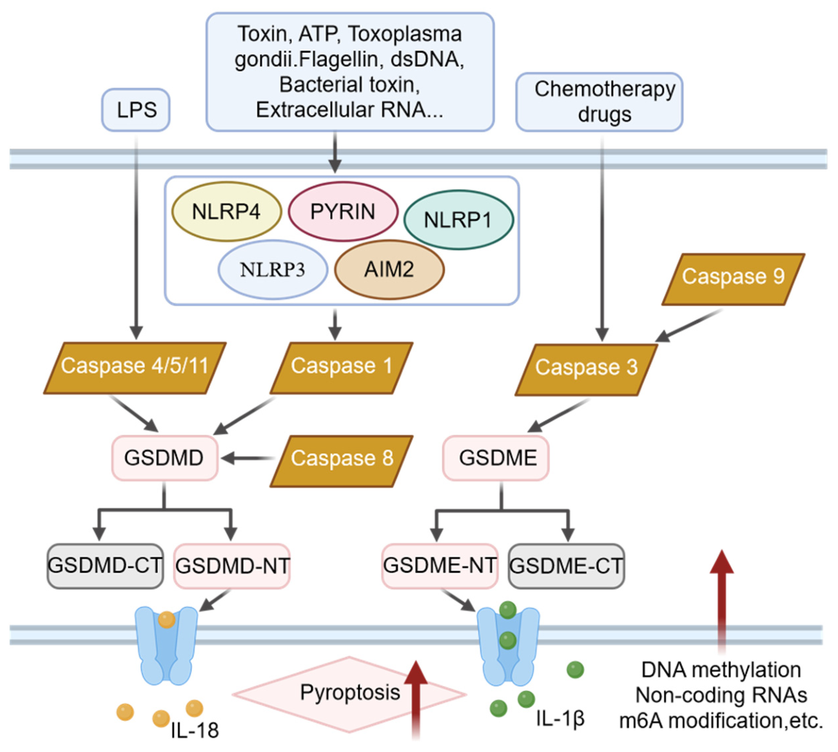
Pyroptosis is primarily activated through canonical and non-canonical pathways, ultimately converging on the cleavage and activation of Gasdermin proteins. The initiation of pyroptosis depends on upstream signals activating inflammatory caspases. As illustrated in Figure 1, various stimuli, including pathogen-associated molecular patterns (PAMPs) such as flagellin and dsDNA, or danger signals such as ATP and extracellular RNA, are recognized by intracellular pattern recognition receptors, subsequently assembling into inflammasome complexes [187,188,189]. The canonical pathway is triggered by inflammasome activation. Five major inflammasomes are involved in the pyroptosis pathway: NLRP3, AIM2, NLRP1, PYRIN, and NLRC4. Inflammasomes are multi-protein complexes that recognize intracellular PAMPs or damage-associated molecular patterns (DAMPs). Taking the most extensively studied NLRP3 inflammasome as an example, its activation recruits and activates caspase-1 [190]. Activated caspase-1 performs dual functions: it cleaves pro-IL-1β and pro-IL-18, facilitating their maturation and release, while simultaneously cleaving Gasdermin D (GSDMD) to liberate its N-terminal domain (GSDMD-NT). The non-canonical pathway is typically activated by intracellular lipopolysaccharide (LPS) directly activating caspase-4/5 in humans or caspase-11 in mice. These caspases can also cleave GSDMD to induce pyroptosis [191,192,193]. Additionally, under certain cell types or stimuli, the apoptotic executioner caspase-3 can cleave Gasdermin E (GSDME). When GSDME is highly expressed, caspase-3 activation shifts the cell death mode from apoptosis to pyroptosis, a switch of significant importance in the anti-tumor effects of chemotherapeutic agents. The released GSDMD-NT fragment translocates to the cell membrane, binds to membrane phospholipids, and oligomerizes to form non-selective transmembrane pores with diameters of 10–20 nm. These pores compromise membrane integrity, leading to ion flux, cellular osmotic swelling, and eventual plasma membrane rupture. Notably, recent studies have delineated pyroptosis into two phases: an early subcellular permeabilization phase (which is reversible) and a late cell lysis phase [194,195]. The latter phase depends on Ninjurin-1 (NINJ1)-mediated plasma membrane rupture, resulting in substantial release of pro-inflammatory cytokines such as IL-1β and IL-18, along with DAMPs, thereby potently amplifying local and systemic inflammatory responses. Regardless of the pathway, Gasdermin pore formation represents the irreversible execution step of pyroptosis and serves as the central hub connecting cell death with inflammatory responses [196]. It is critical to distinguish between two major Gasdermin-mediated pyroptotic pathways relevant to cancer therapy. The canonical inflammasome pathway (caspase-1/GSDMD) is triggered by pathogen-associated or damage-associated molecular patterns, leading to inflammasome assembly, caspase-1 activation, and subsequent GSDMD cleavage. In contrast, the chemotherapy-induced pathway (caspase-3/GSDME) is activated by chemotherapeutic agents such as cisplatin or doxorubicin, which engage the apoptotic executioner caspase-3 [197]. In cells where GSDME is expressed, activated caspase-3 cleaves GSDME, converting the default apoptotic response into pyroptosis (Figure 3). This distinction is therapeutically crucial: while GSDMD-mediated pyroptosis is more relevant to immune cell biology and inflammatory microenvironments, GSDME-mediated pyroptosis is the primary target for drug-based strategies aimed at sensitizing tumors to chemotherapy by epigenetic derepressing of GSDME [198].
Numerous studies have demonstrated that pyroptosis can either promote or inhibit the development and metastasis of various cancers including gastric cancer, BRCA-associated breast cancer, breast cancer, and lung cancer. Cucurbitacin B directly binds to TLR4, activating the NLRP3 inflammasome and pyroptosis, thereby exerting anti-tumor effects in non-small cell lung cancer [199]. Cisplatin activates the MEG3/NLRP3/caspase-1/GSDMD pathway to induce pyroptosis in TNBC, consequently inhibiting tumor growth and metastasis. The tumor suppressor DRD2 promotes macrophage M1 polarization, inhibits the NF-κB signaling pathway, and triggers apoptosis in breast cancer cells, providing novel predictive and therapeutic targets for breast cancer [200,201]. Under hypoxic conditions, formation of the NPD-L1/pStat3 complex increases GSDMC expression in the cancer cells, converting apoptosis to pyroptosis, thereby promoting tumor progression and suppressing anti-tumor immune responses. Epigenetic modifications can also regulate tumor growth and metastasis by modulating pyroptosis-related pathways. Therefore, in-depth analysis of the correlation between DNA methylation and pyroptosis unveils novel insights for developing therapeutic strategies targeting the epigenetic-pyroptosis axis [202].

4.2. Multifaceted Regulation of Pyroptosis by DNA Methylation

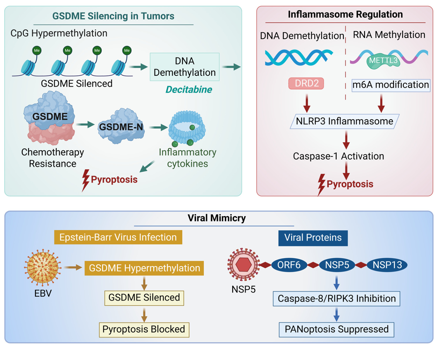
The regulation of pyroptosis by DNA methylation can be understood from three dimensions: (1) direct silencing of the pyroptosis executioner gene GSDME through promoter hypermethylation induces pyroptosis resistance and chemotherapy tolerance in tumor cells; (2) regulation of the expression and stability of inflammasome components such as NLRP3 through methyltransferases (e.g., METTL3) or RNA methylation (e.g., NSUN7-mediated m5C) influences inflammasome assembly and activation; and (3) through “molecular mimicry”, viruses simulate or disrupt host DNA methylation regulatory networks, thus evading or triggering host cell pyroptosis immune responses (Figure 4). This section aims to elucidate the multidimensional regulatory role of DNA methylation in pyroptosis, providing a theoretical basis for epigenetic therapies in related diseases [203,204,205].

4.2.1. GSDME Promoter Hypermethylation

As a key epigenetic modification that silences the expression of the pyroptosis executioner protein GSDME, promoter hypermethylation of the GSDME gene has emerged as an important mechanism underlying acquired drug resistance in tumors. Multiple studies have revealed its core role across various cancer types.
In breast cancer, it has been definitively demonstrated that methylation of the GSDME enhancer region is directly associated with significant downregulation of GSDME expression in the drug-resistant breast cancer cell line MCF-7/Taxol, a mechanism conferring resistance to paclitaxel in tumor cells. Mechanistic validation showed that treatment with the DNA methyltransferase inhibitor decitabine induced GSDME demethylation, restoring its expression, subsequently triggering pyroptosis, and significantly enhancing the chemosensitivity of resistant cells to paclitaxel [205]. From a tumor microenvironment (TME) perspective, studies have shown that estrogen inhibits GSDME-mediated pyroptosis by inducing GSDME promoter methylation and upregulating DNMT1 expression, highlighting the role of this epigenetic modification in the development of drug resistance [206,207].
In prostate cancer, it has been systematically elucidated that GSDME promoter hypermethylation results in its transcriptional silencing, thereby diminishing tumor cell sensitivity to the PARP inhibitor olaparib. Combined treatment with olaparib and decitabine synergistically induced GSDME expression and cleavage activation, activating the caspase-3-dependent pyroptosis pathway, which significantly enhanced anti-tumor efficacy and induced tumor regression. These results substantiate the potential of combination therapy as a viable strategy to overcome drug resistance [208,209].
As a pan-cancer epigenetic marker, GSDME promoter methylation holds significant promise for early tumor detection, molecular subtyping, prognostic assessment, and prediction of treatment response [192,208]. The evidence above indicates that hypermethylation of CpG islands in the GSDME promoter region leads to transcriptional silencing of the pyroptosis executioner protein GSDME, rendering tumor cells unable to effectively activate the caspase-3-dependent pyroptosis pathway. This allows them to evade immunogenic cell death induced by chemotherapeutic agents, ultimately driving the development of acquired resistance. Targeting GSDME methylation status holds promise as an important therapeutic strategy to reverse tumor drug resistance and enhance chemotherapy sensitivity.

4.2.2. Methylation Modifications of Inflammasome Components

NLRP3 inflammasome activation is a two-step process governed by tight regulation. The first, or priming, step involves the transcriptional upregulation of key components such as NLRP3 itself. The second, or activation, step is triggered by specific signaling events that result in inflammasome assembly [210]. This activation subsequently induces pyroptosis, a lytic form of programmed cell death essential for eliminating pathogenic niches and maintaining homeostasis. Therefore, the activation of the inflammasome represents a pivotal upstream event in the initiation of pyroptosis, and its core components are subject to precise epigenetic control, including both DNA and RNA methylation.
DNA methylation directly regulates inflammation-related genes. It has been reported that dopamine receptor D2 (DRD2) functions as a tumor suppressor in breast cancer and its downregulation is associated with promoter hypermethylation. Treatment with demethylating agents can restore DRD2 expression, which subsequently limits NF-κB signaling pathway activity, influences macrophage polarization, and ultimately triggers NLRP3/GSDME-dependent pyroptosis in tumor cells [201,211]. These findings demonstrate that the DNA methylation status of target genes can indirectly yet profoundly modulate inflammasome activation and pyroptosis by regulating the upstream signaling molecules.
DNA methyltransferases catalyze DNA methylation. Notably, their involvement in the regulation of inflammasomes and pyroptosis has emerged as a focal point of recent research. Pyroptosis is a programmed cell death modality induced by inflammasome activation, with its canonical pathway involving caspase-1 activation and subsequent cleavage of GSDMD, ultimately leading to cell membrane perforation and the release of pro-inflammatory cytokines. Emerging evidence indicates that DNMTs participate in inflammasome-mediated pyroptosis regulation across various disease models by modulating the methylation levels of distinct target genes. In mechanistic studies of disease pathogenesis, Haldar et al. first demonstrated that DNMT1 and DNMT3B mediated promoter methylation silence the Ogg1 gene. This event triggers NLRP3 inflammasome activation and ultimately promotes the progression of chemotherapy-induced hemorrhagic cystitis [212]. Huang et al. demonstrated that hypomethylation of the NLRC4, NLRP12, and IL-1β genes in leukocytes from patients with Kawasaki disease leads to their upregulation and subsequent inflammatory responses [213]. Zhong et al. reported that DNMT1 regulates NLRP3 inflammasome activation in atherosclerosis through hypermethylation of the miR-145 promoter [214]. Sun et al. discovered that downregulation of DNMT1 and DNMT3A expression in osteoarthritis results in CtBP hypomethylation and overexpression, consequently activating the NLRP3 inflammasome [215]. In summary, DNMTs play a pivotal regulatory role in inflammatory responses and disease progression by targeting DNA methylation of inflammasome-related genes, thereby offering novel potential therapeutic targets for the prevention and treatment of inflammatory diseases.
Beyond DNA methylation, RNA modifications such as m6A also critically regulate pyroptosis. Multiple studies have revealed the direct regulatory role of METTL3-mediated m6A modification on the NLRP3 inflammasome. A review by Guan et al. systematically summarized that METTL3, a core m6A “writer” enzyme, catalyzes m6A modification of NLRP3 mRNA, enhancing its mRNA stability and/or translation efficiency. This upregulation facilitates NLRP3 inflammasome assembly, caspase-1 activation, and pyroptosis progression [216,217]. Research by Xie et al. further elucidated the underlying mechanism: in an acute soft tissue injury model, METTL3 coordinated with the m6A reader protein YTHDF1 to regulate endothelial cell pyroptosis by enhancing NLRP3 expression. Knockdown of METTL3 significantly reduced both global m6A levels and m6A enrichment on NLRP3 mRNA, suppressing pyroptosis [218]. Although these studies focused on spinal cord ischemia–reperfusion injury and soft tissue injury respectively, they suggest that the “METTL3-m6A-NLRP3” regulatory axis has broad applicability across diverse disease models.
RNA methylation (m5C) directly stabilizes NLRP3 mRNA, thereby regulating pyroptosis. In research on polycystic ovary syndrome (PCOS), Xu et al. discovered that the RNA methyltransferase NSUN7 was upregulated in patient granulosa cells and LPS-treated KGN cells. Through methylated RNA immunoprecipitation (MeRIP) and actinomycin D treatment experiments, they confirmed that NSUN7 directly catalyzes m5C modification at specific sites on NLRP3 mRNA, significantly enhancing its mRNA stability, thereby upregulating NLRP3 protein levels and promoting caspase-1/GSDMD-mediated pyroptosis. Knockdown of NSUN7 inhibited pyroptosis by reducing NLRP3 expression and ameliorated the PCOS model phenotype, establishing a clear “NSUN7-m5C-NLRP3” regulatory axis [219].
In summary, methylation modifications across DNA and RNA levels construct an epigenetic network that orchestrates multilayered, finely tuned regulation of inflammasome activity, thereby maintaining a balanced equilibrium between physiological inflammatory responses and pathological hyperactivation [220,221,222].

4.2.3. Viral Mimicry

Viruses have evolved “molecular mimicry” strategies over long-term evolution, simulating the structure or function of host proteins to hijack cellular processes—including epigenetic regulation—to facilitate their replication or evade immunity. DNA methylation mechanisms represent one of the crucial targets manipulated by viruses [223].
Viruses silence GSDME through DNA methylation. A 2025 study published in Microorganisms provided the first evidence that Epstein–Barr virus (EBV) can directly induce hypermethylation of the host GSDME gene promoter, leading to silencing of the pyroptosis executioner protein. Through transcriptomic analysis of TCGA data, the researchers found that GSDME expression was selectively inhibited in EBV-positive gastric cancer, while other members were upregulated [224]. Further validation in multiple cell lines confirmed that EBV infection significantly reduces GSDME expression through promoter hypermethylation, and this epigenetic silencing could be reversed by the DNA methyltransferase inhibitor 5-azacytidine. Functional experiments demonstrated that although EBV-positive cells retain the ability of caspase-3 to activate GSDME, baseline silencing of GSDME impedes pyroptosis induction and reduces chemotherapy sensitivity. Restoration of GSDME expression reversed this phenotype, suggesting that demethylation therapy may serve as a sensitization strategy for EBV-associated gastric cancer.
Viral-encoded proteins directly target key nodes of the pyroptosis signaling pathway. Research published in Advanced Science by Wang et al. systematically elucidated how coronaviruses (e.g., SARS-CoV-2) finely regulate ZBP1-mediated PANoptosis—a composite form of cell death simultaneously encompassing pyroptosis, apoptosis, and necroptosis—through their encoded NSP5, ORF6, and NSP13 proteins. Specifically, NSP5 and ORF6 directly block the initiation of apoptosis and pyroptosis by binding to caspase-8 and inhibiting caspase-8 activity, while NSP13 inhibits programmed necrosis through competitive binding to RIPK3. This multi-target inhibition strategy enables viruses to effectively curtail host defense mechanisms that limit viral spread through pyroptosis and other pathways. The study also found that coronavirus inhibition of PANoptosis significantly promoted influenza A virus replication and enhanced inflammatory cytokine expression during co-infection, providing mechanistic explanation for the high mortality rates observed clinically during coinfections [225].
Therefore, in the context of viral infection, viral interference and host methylation regulatory mechanisms collectively establish a dynamic interplay network: host cells attempt to initiate defensive pyroptosis through epigenetic reprogramming, precisely regulating methylation modifications of pyroptosis-related genes; meanwhile, viruses achieve immune evasion by inducing GSDME promoter hypermethylation for epigenetic silencing or by directly inhibiting pyroptosis execution molecules through encoded proteins [226]. Future research priorities lie in elucidating how viral proteins specifically target the recruitment and activity regulation of host DNA methylation enzymes (e.g., DNMTs, TETs) at pyroptosis gene loci, thereby elucidating the molecular mechanisms by which viruses manipulate host epigenetic defenses.
This section systematically reviews the multi-layered regulatory correlation between DNA methylation and pyroptosis. Pyroptosis plays a critical role in maintaining organismal homeostasis and defending against pathogen invasion through its specific activation [227,228]. DNA methylation profoundly influences the initiation and execution of pyroptosis across three dimensions: At the execution level, hypermethylation of promoter regions in key pyroptosis effector molecules such as GSDME can directly induce their transcriptional silencing, rendering tumor cells resistant to chemotherapy-induced pyroptosis and constituting an important epigenetic basis for tumor drug resistance [229]. At the activation level, DNA or RNA methylation modifications of core components such as the NLRP3 inflammasome and their upstream signaling networks precisely regulate the cascade amplification threshold of inflammatory signals, thereby influencing the initiation and progression of inflammation-related diseases [230,231]. At the host–pathogen interaction level, viruses have evolved various strategies including molecular mimicry to manipulate pyroptosis fate in reverse—by disrupting host methylation homeostasis or directly targeting pyroptosis pathway nodes—thereby achieving immune evasion [232,233]. These three levels of regulatory mechanisms are interconnected, collectively revealing the central position of DNA methylation within the pyroptosis regulatory network. Based on these insights, therapeutic strategies targeting DNA methylation demonstrate significant potential. For example, demethylating agents such as 5-azacytidine can restore GSDME expression to sensitize chemotherapy; development of specific METTL3 or NSUN7 inhibitors may attenuate excessive inflammatory responses; and designing blockers against viral epigenetic mimicry proteins could restore normal host pyroptosis immune defenses [234,235]. However, current research still faces challenges: How do DNA methylation and other epigenetic modifications such as histone modifications and chromatin remodeling coordinately regulate pyroptosis? The detailed molecular map of viral manipulation of host methylation remains to be elucidated. In the future, integrating multi-omics technologies, epigenetic editing tools (such as dCas9-DNMT3A/TET1), and advanced disease models will enable more precise dissection of the dynamic changes and functions of DNA methylation in pyroptosis, advancing epigenetic therapies targeting this pathway from bench to bedside. It should be noted that while numerous studies report correlations between DNA methylation at specific loci (e.g., GSDME, NLRP3) and pyroptosis-related phenotypes, functional causality has been firmly established only in a subset of these cases, primarily through DNMT inhibitor treatment, genetic knockdown/rescue, or CRISPR/dCas9-based epigenetic editing. Future studies are encouraged to move beyond associational analyses to definitive mechanistic validation.

5. Advances in Nanomedicine for Targeting DNA Methylation and Inducing Pyroptosis

In recent years, epigenetic drugs including DNA methyltransferase inhibitor (DNMTi) have achieved remarkable efficacy in certain hematologic and solid tumors. Various epigenetic agents have been demonstrated to increase tumor sensitivity to conventional treatments such as chemotherapy, radiotherapy, photodynamic therapy, and immunotherapy (Table 2). GSDME is a key protein in chemotherapy-induced pyroptosis but is not expressed or is expressed at low levels in most tumor cells due to promoter region hypermethylation, resulting in GSDME silencing across various cancer types [236,237]. Therefore, epigenetic drugs that inhibit GSDME gene methylation can upregulate GSDME expression in tumor cells, thereby triggering pyroptosis.

5.1. Epigenetic Drugs Sensitize Chemotherapy

Chemotherapy, as a cornerstone of cancer treatment, relies on cytotoxic drugs that are essential for curbing tumor progression. However, systemic toxicity due to lack of targeting and development of tumor drug resistance remains major clinical challenges. Advanced drug delivery strategies, particularly those leveraging nanotechnology, offer effective pathways to enhance chemotherapeutic efficacy while reducing side effects by improving tumor-targeted accumulation and intelligent controlled release of drugs. At the mechanistic level, various chemotherapeutic agents such as doxorubicin (DOX) can induce caspase-3-mediated apoptosis. Recent studies have further revealed that activated caspase-3 can cleave GSDME protein, thereby converting classical apoptosis into inflammatory programmed cell death pyroptosis. Therefore, it provides a novel theoretical framework for reshaping the cytotoxic effects of chemotherapy through GSDME modulation. In tumor cells, hypermethylation of the GSDME gene leads to deficiency in the key GSDME protein required for caspase-3-induced pyroptosis, resulting in drug resistance [195,249,250].
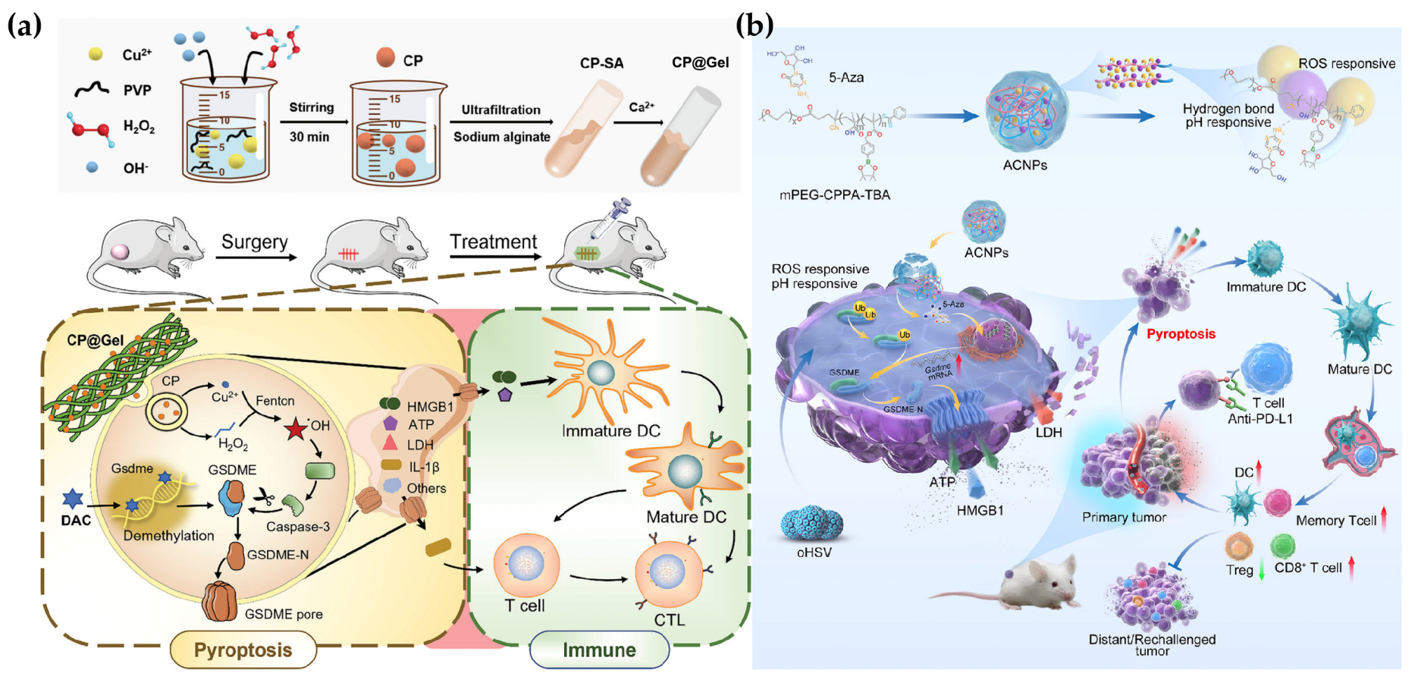
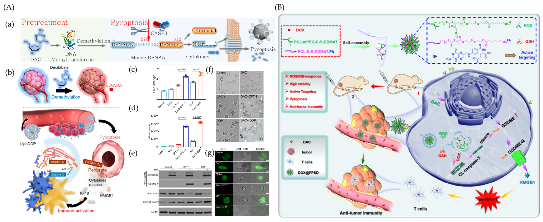
To overcome GSDME silencing-mediated chemotherapy resistance, Fan et al. developed a liposome-based combination therapy strategy (Lipo-DDP) designed for spatiotemporally coordinated delivery of the DNA methyltransferase inhibitor DAC and the chemotherapeutic agent cisplatin. The core of this strategy lies in the sequential action: DAC encapsulated in liposomes first acts on tumor cells, inducing demethylation of the GSDME promoter and restoring its expression, thereby “priming” the cells for pyroptosis; subsequently, co-delivered cisplatin activates caspase-3, which cleaves the “primed” GSDME protein, efficiently triggering pyroptosis. In vitro experiments confirmed that this combination therapy significantly elevated levels of pyroptosis characteristic proteins, release of the pro-inflammatory cytokine IL-1β, and extracellular release of HMGB1—a key indicator of immunogenic cell death (ICD). By temporally regulating epigenetic “priming” and drug “execution”, this work harnesses pyroptosis to enhance the immunostimulatory effects of chemotherapy, offering new solutions for tumor immunotherapy [238].
Addressing the challenge of limited pyroptosis due to GSDME downregulation in breast cancer treatment, researchers developed a novel folate (FA)-modified, glutathione (GSH)/reactive oxygen species (ROS) dual-responsive nanocarrier (FPSD NP) for targeted co-delivery of the chemotherapeutic agent DOX and the DNMTs inhibitor DAC. This strategy aims to induce pyroptosis through a synergistic “epigenetic priming-chemotherapy execution” mechanism. Specifically, DAC upregulates GSDME expression in 4T1 breast cancer cells through demethylation, creating conditions for pyroptosis; subsequently, DOX delivered via FPSD NP activates caspase-3, which cleaves GSDME to trigger typical pyroptotic cell death. In vivo and in vitro experimental results demonstrated that the combination therapy (DAC + DOX@FPSD NPs) effectively inhibited tumor growth through inducing pyroptosis, characterized by cell swelling and membrane pore formation, reduced expression of the proliferation marker Ki67, and increased cell death. Furthermore, the therapy elicited significant anti-tumor immune responses, manifested as increased infiltration of CD3+/CD4+/CD8+T cells in the tumor microenvironment, the release of pro-inflammatory factors, and the exposure of ICD-associated molecules (Figure 5). In summary, this intelligent nano-delivery system provides a promising strategy for targeted pyroptosis-based breast cancer therapy [239].

5.2. Epigenetic Drugs Sensitize Photodynamic Therapy

Photodynamic therapy (PDT) represents a promising tumor treatment strategy, wherein photosensitizers generate reactive oxygen species (ROS) at the tumor site under specific wavelength light irradiation. Compared with traditional modalities such as surgery, radiotherapy, and chemotherapy, PDT offers advantages including minimally invasive intervention, spatiotemporal controllability, and repeatable treatment. Its anti-tumor mechanisms encompass not only direct tumor cell death induced by ROS but, more importantly, the ability to trigger immunogenic cell death by releasing tumor-associated antigens and damage-associated molecular patterns (DAMPs) to initiate anti-tumor immune responses. However, the clinical efficacy of PDT is limited by factors such as tissue light penetration depth, accumulation efficiency of photosensitizers in the tumor sites, and immunosuppressive TME. Given that pyroptosis is a programmed cell death modality mediated by GSDME proteins accompanied by substantial pro-inflammatory cytokine release, its combination with PDT demonstrates potential for enhanced therapeutic effects. Theoretically, PDT-induced ICD can “preheat” the immune system, while subsequently triggered pyroptosis further amplifies inflammatory signals and reverses the tumor immunosuppressive microenvironment (TIM), thereby generating synergistic anti-tumor immunity and providing new insights for improving PDT efficacy [251,252].
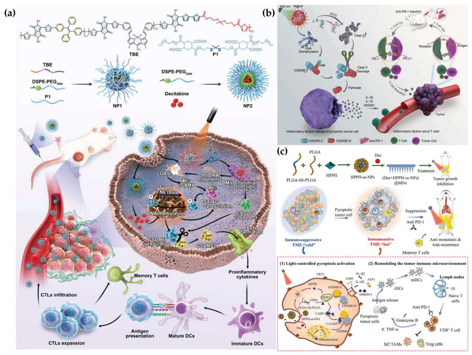
A study proposed an intelligent nanotheranostic system that integrates photodynamic therapy, epigenetic therapy, near-infrared fluorescence bioimaging, and TME modulation through simultaneous activation of pyroptosis and the cGAS-STING pathway for cancer treatment. This approach involves developing oxidation-sensitive nanoparticles (NP1) loaded with the photosensitizer TBE, alongside with decitabine loaded nanomicelles (NP2). NP2 restores STING and GSDME expression, while NP1-mediated PDT, upon 808 nm laser irradiation, exhibits exceptionally high photo-to-singlet oxygen (1O2) conversion efficiency, promoting the release of DNA fragments from damaged mitochondria, thereby enhancing the cGAS-STING pathway and facilitating caspase-3 activation. Activated caspase-3 subsequently cleaves upregulated GSDME into pore-forming GSDME-NT. Pro-inflammatory cytokines released concomitantly from pyroptosis and cGAS-STING pathway activation induce dendritic cell (DC) and natural killer (NK) cell maturation, eliciting cytotoxic T cell-mediated anti-tumor immunity and establishing long-term anti-tumor immune memory, thereby mounting robust and multifaceted anti-tumor immune responses. Collectively, this work presents an integrated strategy combining epigenetic therapy with photodynamic therapy. By simultaneously activating pyroptosis and the cGAS-STING pathway, this innovative approach holds promise for overcoming the limitations of existing treatments and offers valuable avenues for future clinical applications [240].
To enhance anti-tumor photodynamic therapy, Zheng et al. designed a nanosystem based on an iridium-based photosensitizer (R@IrP) that, upon light irradiation, induces pyroptosis through the caspase-3/GSDME pathway and combines with anti-PD-1 immunotherapy to remodel the tumor microenvironment, thereby enhancing therapeutic efficacy. Under specific green light irradiation (520 nm), IrP efficiently converts intracellular oxygen to singlet oxygen. RG108, a small-molecule DNMTi, functions primarily to upregulate GSDME protein expression. Results demonstrated distinct cellular morphological differences between apoptosis (shrinkage) and pyroptosis (swelling, bubble-like structures). Schematic illustrations of the caspase-3/GSDME pathway elucidated the transition from apoptosis to pyroptosis. Western blot analysis showed enhanced caspase-3 and GSDME expression in R@IrP treated cells under light irradiation. Furthermore, in B16 tumor-bearing mice, R@IrP combined with light irradiation and anti-PD-1 demonstrated superior control of tumor growth compared to anti-PD-1 alone. These findings indicate that pyroptosis-based photodynamic therapy effectively remodels the tumor microenvironment and activates the immune system for tumor eradication [241].
Photodynamic therapy in breast cancer still encounters significant obstacles, including insufficient immunogenicity and limited pyroptosis induction efficiency. To address these issues, researchers developed dissolvable microneedles (MNs) for the local co-delivery of decitabine and GSH-responsive photosensitizer nanoparticles (HPPH-ss-NPs), aimed at enhancing immunogenic pyroptosis in breast cancer cells. This microneedle platform achieves dissolution-dependent release of DAC and GSH-triggered activation of HPPH-ss-NPs, enabling spatiotemporal control that reduces systemic exposure while increasing drug concentration in tumor. Mechanistically, DAC restores expression of the pyroptosis execution factor GSDME, while HPPH-ss-NPs deplete intracellular GSH and generate ROS upon 660 nm laser irradiation, activating caspase-3. This synergistic effect triggers pyroptosis, releasing immunostimulatory DAMPs, thereby increasing the numbers of mature dendritic cells and tumor-infiltrating CD8+T cells. In an orthotopic model, (DAC+ HPPH-ss-NPs) @MNs suppressed primary tumor growth, while combination with anti-PD-1 antibody synergistically inhibited tumor recurrence and lung metastasis, establishing durable systemic immunity (Figure 6). In summary, this microneedle platform constructs a localized photodynamic-epigenetic interplay strategy, reshaping immunologically inert tumors into a pyroptosis-driven immunogenic microenvironment [242].

5.3. Epigenetic Enhancement of Immunotherapy

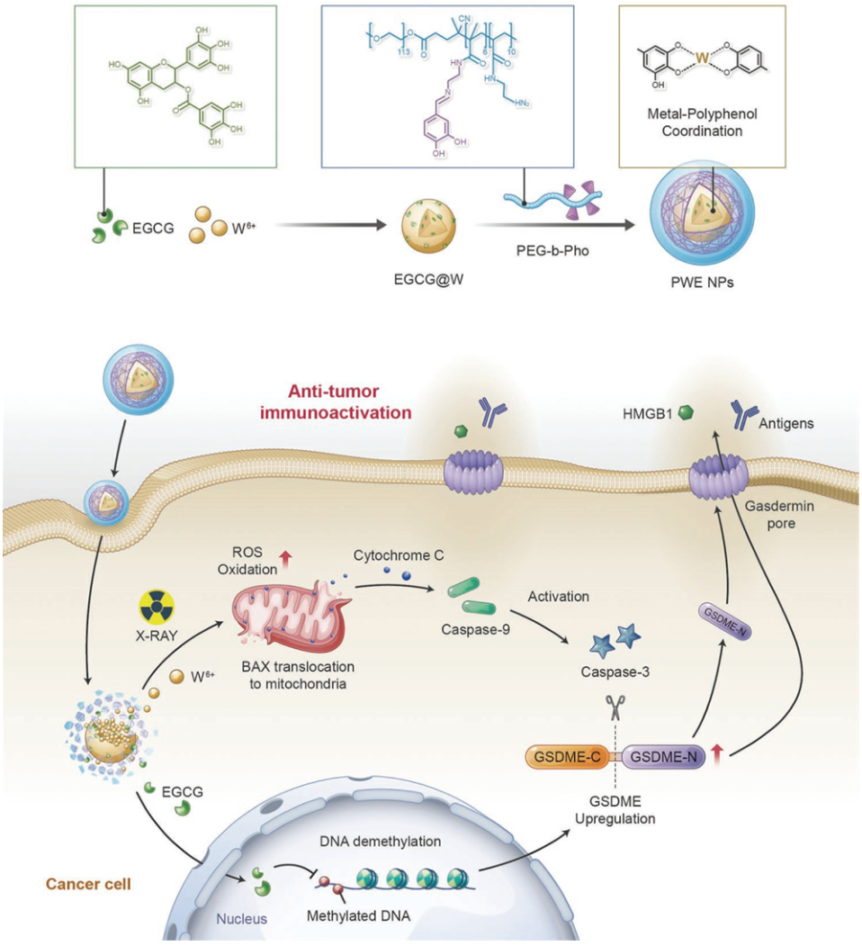
The success of tumor immunotherapy is highly dependent on the immune-permissive state of the tumor TME. However, the TME frequently exhibits an immunosuppressive phenotype, forming so-called “cold tumors”, characterized by impaired tumor antigen presentation and recognition, establishment of an immunosuppressive microenvironment, and functional exhaustion of effector T cells. Recent studies have revealed that the phenotypic and functional dysregulation of cells within the TME, including tumor cells, immune cells, and stromal cells, broadly influenced by reversible epigenetic regulation at the upstream level. The core mechanistic role of epigenetic therapy lies in its direct mediation of the dynamic switch between “cold” (immunosuppressive) and “hot” (immune-permissive) tumor phenotypes. Given the reversibility of epigenetic modifications, epigenetic drugs such as DNMTs are considered ideal tools for remodeling the TME. These agents not only directly induce antigen expression on tumor cells but also systematically reshape immune cell function. Consequently, combining epigenetic modulators with immune checkpoint inhibitors and other therapies constitutes a highly promising synergistic anti-tumor strategy. Pyroptosis plays a central role in immune surveillance, response initiation, and effector phases through its unique lytic death mechanism and capacity to provoke robust inflammatory responses. Integrating the synergistic effects of multiple mechanisms—epigenetic modulation, pyroptosis induction, and immune checkpoint blockade—into a systematic combination therapy strategy can not only enhance anti-tumor immune responses but also establish a critical scientific foundation for overcoming intrinsic drug resistance and adaptive immune evasion in tumors [253,254].
Tumor immunotherapy is often constrained by the immunosuppressive microenvironment, and GSDMD is expressed at low levels in most tumor cells, while small-molecule inhibitors of DNA methylation suffer from non-specificity or single-function deficiencies. To address these challenges, researchers constructed a dual-drug nano-delivery system (Nig-DAC) @HMA based on hexavalent histidine-metal coordination self-assembly (HMA), designed to remodel the immune microenvironment through synergistic induction of pyroptosis. This system co-delivers the DNA methyltransferase inhibitor DAC and the NLRP3 inflammasome activator nigericin (Nig). The mechanism involves two steps: DAC first upregulates the low-expressed pyroptosis execution protein GSDMD in bladder cancer tumor cells through demethylation; subsequently, Nig activates the NLRP3 inflammasome and caspase-1, cleaving the upregulated GSDMD and thereby efficiently triggering pyroptosis in cancer cells. The pyroptotic death releases substantial inflammatory factors, effectively reversing the local immunosuppressive state, eliciting robust systemic anti-tumor immune responses in vivo, and significantly inhibiting tumor growth. This study provides a novel strategy for temporally regulating epigenetic and inflammatory pathways through nanotechnology to enhance pyroptosis and anti-tumor immunity [243,255].
Regulating the immunosuppressive microenvironment and eliminating residual microscopic lesions is critical for inhibiting postoperative recurrence of triple-negative breast cancer [256]. Although immunotherapy holds potential, its anti-recurrence efficacy remains suboptimal due to multiple immunosuppression and insufficient apoptotic immunogenicity. To address this, researchers designed an injectable hydrogel encapsulating autocatalytic copper peroxide (CP@Gel) as a therapeutic platform, combined with the clinical-grade DNA methyltransferase inhibitor decitabine, aiming to overcome apoptosis resistance, enhance immunogenicity, and remodel the TIM through pyroptosis induction, thereby activating potent anti-tumor immune responses. DAC upregulates GSDME protein expression by inhibiting GSDMD; subsequently injected CP@Gel continuously releases copper peroxide, which autocatalytically generates ROS in the tumor microenvironment, thereby activating caspase-3. The synergistic action of these components strongly induces pyroptosis, promotes damage-associated molecular pattern release, enhances antigen presentation, and recruits cytotoxic T cells. In vivo experiments demonstrated that combination of DAC and CP@Gel reduced local tumor recurrence rates by 67%, showcasing the successful integration of sustained drug release, autocatalysis, and epigenetic modification [244]. These findings indicate that pyroptosis combined with injectable hydrogel-assisted strategies holds significant potential for preventing postoperative recurrence in triple-negative breast cancer.
Although gene therapy holds promise for treating genetic disorders, its application in cancer treatment faces numerous challenges due to the complex genetic heterogeneity of tumors and the immunosuppressive microenvironment. A research team developed a multimodal strategy therapeutic strategy that integrates alphavirus vector-based gene therapy, epigenetic regulation, and immune checkpoint blockade. Specifically, they designed a novel delivery system based on membrane fusion mechanisms—liposomes mimicking the penetrating properties of filamentous actin—capable of efficiently encapsulating and delivering Semliki Forest virus (pSFV)-based DNA vectors carrying the p53 tumor suppressor gene and the anti-PD-L1 single-chain antibody (scFv) gene. This system achieves cytosolic delivery of genetic contents directly through membrane fusion, thereby circumventing the vector degradation and low delivery efficiency associated with traditional endocytic pathways. To potentiate the immune activation effects of this combination therapy, they concurrently administered the DNA methyltransferase inhibitor decitabine. DAC upregulates GSDME expression in tumor cells through epigenetic demethylation, thereby converting p53-mediated apoptotic signals into GSDME-dependent pyroptosis. This switch in death modality not only effectively kills apoptosis-resistant tumor cells but also, through the characteristic release of inflammatory contents during pyroptosis, significantly promotes T cell infiltration and activation within the TIM, thereby enhancing anti-PD-L1 therapy and systemic anti-tumor immune responses (Figure 7). This multi-mechanism synergistic combination strategy offers a novel translationally promising approach for overcoming drug resistance and enhancing the efficacy of immune checkpoint therapy [245].
Although cancer immunotherapy has become an important treatment strategy for various malignancies, the TIM still severely constrains its clinical efficacy. To address this, a research team developed a dual-responsive DNMTi nanoprodrug (ACNPs) and combined it with oncolytic herpes simplex virus (oHSV) to synergistically regulate the TIM and enhance immune responses. The epigenetic drug 5-azacytidine (5-Aza) upregulates GSDME expression at the transcriptional level, while oHSV further enhances its protein stability by inhibiting the ubiquitin-proteasome degradation pathway of GSDME. The combination significantly potentiates GSDME-mediated tumor cell pyroptosis. In vivo models, the combination of ACNPs and oHSV not only effectively inhibited tumor growth but also significantly remodeled the TIM, manifested as increased immune cell infiltration and reduced inhibitory signals. Further investigation revealed that this combination strategy significantly enhances the anti-tumor efficacy of subsequent immune checkpoint blockade (ICB) therapy. Through the synergistic action of epigenetic drugs and oncolytic viruses, immunogenic pyroptosis was successfully induced and TME immunosuppression was reversed, providing a translationally promising combination strategy for overcoming resistance to current immunotherapies [246].

5.4. Epigenetic Priming for Radiotherapy-Induced Pyroptosis

Radiotherapy employs high-energy ionizing radiation to damage the genetic material of tumor cells, suppress their proliferative capacity, and induce cell death, so that it offers irreplaceable advantages in clinical oncology. However, its therapeutic effect is confined to the local irradiation field, with limited efficacy against established distant metastases. Although radiotherapy can elicit local immune responses, it often induces immune tolerance in most cases, primarily manifested as a significant increase in immunosuppressive cell populations—such as tumor-associated M2-type macrophages and regulatory T cells—within the irradiated microenvironment, accompanied by upregulation of anti-inflammatory cytokine signaling, thereby suppressing systemic anti-tumor immune responses [257,258,259,260].
To reverse the immunosuppressive state induced by radiotherapy and enhance therapeutic effects on distant metastases, a research team designed a multifunctional metal-phenolic network nanosystem (PWE) based on radiosensitizers, capable of inducing pyroptosis in 4T1 breast cancer cells during radiotherapy through epigenetic regulation strategies. This system is self-assembled from a polyphenolic DNMTi epigallocatechin gallate (EGCG), high atomic number radiation-sensitizing W6+ ions, and polyphenol-modified block copolymers. EGCG reverses the epigenetic silencing of the GSDME gene by inhibiting DNMT activity, thereby restoring GSDME protein expression in tumor cells. Subsequently, the PWE nanoplatform synergizes with radiotherapy to activate caspase-3, which cleaves the expressed GSDME protein to generate its N-terminal domain fragment. This fragment oligomerizes to form pores in the cell membrane, ultimately driving immunogenic pyroptosis in tumor cells (Figure 8). Experimental results demonstrated that this nanosystem not only enhanced dendritic cell maturation and increased CD8+ T cell infiltration, promoting secretion of pro-inflammatory cytokines such as TNF-α, IFN-γ, IL-6, and IL-12, but also inhibited immunosuppressive components including M2-type macrophages and Treg cells, ultimately achieving effective regression of primary tumors, distant metastases, and even systemic dissemination [247].
Inducing pyroptosis in tumor cells can elicit potent anti-tumor immune responses, offering a promising strategy for treating TNBC and preventing recurrence and metastasis. A research team developed an ultrasmall hafnium oxide nanoparticle composite system loaded with the DNMT inhibitor decitabine DAC. This nanosystem utilizes ultrasmall HfO2 nanoparticles to achieve deep penetration and prolonged retention in tumor tissues, enabling targeted delivery of DAC to tumor sites. Upon X-ray irradiation, HfO2 nanoparticles function as radiosensitizers to enhance local energy deposition and ROS generation and further activate the caspase-3 signaling pathway. Concurrently, DAC reverses the epigenetic silencing of the GSDME gene in TNBC cells by inhibiting DNA methyltransferase activity. Through the combined action of caspase-3 and functional GSDME, the cell death modality shifts from apoptosis to pyroptosis. This study not only overcomes the limitations of traditional pyroptosis inducers, including significant side effects and low efficiency, but also provides a synergistic strategy based on epigenetic remodeling and nano-radiosensitization for treating malignancies with low GSDME expression, further expanding the application prospects of pyroptosis-based therapy and radiotherapy in solid tumor treatment [248].

6. Conclusions

In summary, DNA methylation as a core mechanism of epigenetic regulation plays an indispensable role in all aspects of cancer initiation, progression, and therapeutic response. From global methylation pattern remodeling to the silencing of specific tumor suppressor genes such as CDKN2A, VHL, and BRCA1, and further to the aberrant activation of key signaling pathways, DNA methylation abnormalities profoundly shape the malignant phenotype of tumors. Of particular significance is the crosstalk between DNA methylation and pyroptosis, which provides a novel perspective for understanding mechanisms of drug resistance. Epigenetic silencing of pyroptosis execution proteins such as GSDME enables tumor cells to evade immunogenic cell death induced by chemotherapy and radiotherapy, constituting an important basis for acquired resistance. Therefore, targeting DNA methylation to restore tumor cell sensitivity to pyroptosis has emerged as a highly promising therapeutic strategy.
In recent years, the rapid development of nanomedicine has brought revolutionary breakthroughs to combination therapies that precisely intervene in DNA methylation and induce pyroptosis. Through the construction of diverse intelligent delivery platforms—including liposomes, polymeric nanoparticles, hydrogels, and metal-phenolic networks, researchers have successfully achieved spatiotemporally coordinated delivery of DNMT inhibitors (DAC, RG108, etc.) with chemotherapeutic agents (cisplatin, doxorubicin), photosensitizers, radiosensitizers, or immunomodulators. This “epigenetic priming-pyroptosis execution” combination strategy not only efficiently induces pyroptosis in tumor cells in vitro and in vivo but also effectively reverses the immunosuppressive microenvironment, activates systemic anti-tumor immune responses, and significantly enhances the efficacy of immune checkpoint blockade and other therapies [246,261,262]. These achievements conclusively demonstrate the tremendous potential of deeply integrating epigenetic regulation with nanotechnology for overcoming drug resistance and achieving long-term immune surveillance [263,264].

7. Future Perspectives

Despite the promising prospects, this field still faces numerous challenges. First, the synergistic mechanisms between DNA methylation and other epigenetic modifications—such as histone modifications and chromatin remodeling—in pyroptosis regulation require further elucidation. Second, the poor stability and short plasma half-life of DNA methyltransferase inhibitors, together with their ill-defined pharmacokinetics, hamper precise in vivo delivery; even with nanocarriers, systematic evaluations of drug release kinetics, metabolite toxicity, and long-term biodistribution remain lacking. Third, while GSDME-mediated pyroptosis efficiently kills tumor cells, it may trigger uncontrolled inflammatory cytokine storms (e.g., massive release of IL-1β and IL-18). This can lead to systemic inflammation, capillary leak syndrome, or cytokine release syndrome, representing a major safety concern for clinical translation. Fourth, precisely controlling the sequential release ratio of different drugs from nanocarriers to match the optimal window for “priming” and “execution” is a critical hurdle, as current platforms lack real-time feedback regulation. Given the substantial heterogeneity in GSDME methylation status and caspase-3 activity across patients and tumor regions, fixed spatiotemporal release profiles are inadequate for personalized therapy. Additionally, how pathogens such as viruses manipulate the host methylation system through “molecular mimicry” to evade pyroptosis introduces new complexities for anti-tumor therapy [265].
To address these translational barriers, future research should prioritize biomarker-guided dose scheduling and patient stratification based on tumor GSDME methylation status. Specifically, quantitative methylation-specific PCR (qMSP) or droplet digital PCR (ddPCR) could be employed to stratify patients into “high” versus “low” GSDME methylation groups, thereby identifying those most likely to benefit from DNMT inhibitor-based “epigenetic priming” before pyroptosis-inducing therapy. Furthermore, the optimal dosing window for decitabine (e.g., low-dose, short-course regimens to avoid off-target hypomethylation toxicity) should be systematically evaluated in relation to the kinetics of GSDME demethylation and re-expression. Real-time monitoring of GSDME expression using liquid biopsy-based circulating tumor DNA (ctDNA) methylation assays could enable adaptive dose adjustment, minimize the risk of uncontrolled inflammatory cytokine release while maximize therapeutic efficacy [266].
In parallel, integrating single-cell multi-omics technologies, organoid models, and more precise epigenetic editing tools such as CRISPR/dCas9-TET1/DNMT3A holds promise for in-depth dissection of the dynamic networks through which DNA methylation regulates pyroptosis in specific microenvironments. Concurrently, the development of intelligent nanomedicines with feedback-responsive capabilities—such as closed-loop delivery platforms that sense tumor microenvironmental cues (e.g., caspase-3 activity or GSDME expression) and programmable nanodevices integrating real-time biomarker monitoring—will be essential to achieve individualized “priming-execution” coordination. Furthermore, patient-derived organoids and micro physiological systems should be employed to systematically evaluate safety windows and pharmacokinetic-pharmacodynamic relationships before clinical translation. Collectively, these biomarker-driven, personalized strategies, together with advanced epigenetic and nanotechnological platforms, represent a concrete pathway toward safe and effective clinical application of the “epigenetic-pyroptosis” combination therapeutic strategy, bringing new hope to cancer patients, particularly those with refractory resistance to existing therapies.

Author Contributions

Writing—original draft preparation, S.W. and X.L.; software, H.L.; methodology, J.Z. and J.L.; writing—review and editing, X.J.; supervision, C.F. All authors have read and agreed to the published version of the manuscript.

Funding

This work was supported by Natural Science Foundation of Beijing Municipality (2232003 to C.F.), the Natural Science Foundation of China (21571133 to C.F.), and Beijing Natural Science Foundation Program and Scientific Research Key Program of Beijing Municipal Commission of Education (KZ201710025024 to C.F.).

Data Availability Statement

No new data were created or analyzed in this study. Data sharing is not applicable to this article.

Acknowledgments

During the preparation of this manuscript, the authors used Grammarly (v1.2.261.1889) for grammar checking and language refinement. The authors have reviewed and edited the output and take full responsibility for the content of this publication.

Conflicts of Interest

The authors declare no conflicts of interest.

References

  1. Johnstone, S.E.; Gladyshev, V.N.; Aryee, M.J.; Bernstein, B.E. Epigenetic clocks, aging, and cancer. Science 2022, 378, 1276–1277. [Google Scholar] [CrossRef] [Scilit]
  2. Hanahan, D. Hallmarks of Cancer: New Dimensions. Cancer Discov. 2022, 12, 31–46. [Google Scholar] [CrossRef] [Scilit] [PubMed]
  3. Sun, L.; Zhang, H.; Gao, P. Metabolic reprogramming and epigenetic modifications on the path to cancer. Protein Cell 2022, 13, 877–919. [Google Scholar] [CrossRef] [Scilit]
  4. Ge, T.; Gu, X.; Jia, R.; Ge, S.; Chai, P.; Zhuang, A.; Fan, X. Crosstalk between metabolic reprogramming and epigenetics in cancer: Updates on mechanisms and therapeutic opportunities. Cancer Commun. 2022, 42, 1049–1082. [Google Scholar] [CrossRef] [Scilit] [PubMed]
  5. Banerjee, R.; Smith, J.; Eccles, M.R.; Weeks, R.J.; Chatterjee, A. Epigenetic basis and targeting of cancer metastasis. Trends Cancer 2022, 8, 226–241. [Google Scholar] [CrossRef] [Scilit]
  6. Liu, H.; Li, P.; Wei, Z.; Zhang, C.; Xia, M.; Du, Q.; Chen, Y.; Liu, N.; Li, H.; Yang, X.P. Regulation of T cell differentiation and function by epigenetic modification enzymes. Semin. Immunopathol. 2019, 41, 315–326. [Google Scholar] [CrossRef] [Scilit]
  7. Yu, B.; Yu, X.; Xiong, J.; Ma, M. Methylation Modification, Alternative Splicing, and Noncoding RNA Play a Role in Cancer Metastasis through Epigenetic Regulation. BioMed Res. Int. 2021, 2021, 4061525. [Google Scholar] [CrossRef] [Scilit]
  8. Hartenian, E.; Broz, P. Pyroptosis: Palmitoylation regulates GSDMD activation and pore formation. Cell Res. 2024, 34, 675–676. [Google Scholar] [CrossRef] [Scilit]
  9. Yu, P.; Zhang, X.; Liu, N.; Tang, L.; Peng, C.; Chen, X. Pyroptosis: Mechanisms and diseases. Signal Transduct. Target. Ther. 2021, 6, 128. [Google Scholar] [CrossRef] [Scilit] [PubMed]
  10. Ouyang, X.; Zhou, J.; Lin, L.; Zhang, Z.; Luo, S.; Hu, D. Pyroptosis, inflammasome, and gasdermins in tumor immunity. Innate Immun. 2023, 29, 3–13. [Google Scholar] [CrossRef] [Scilit]
  11. Kahaer, G.; Pan, S.; Yang, C.; Xie, W.; Lu, Y. Dual function of Gasdermin E: Pyroptosis-mediated pan-cancer suppression versus HCC-specific oncogenic activity. Front. Immunol. 2025, 16, 1626311. [Google Scholar] [CrossRef] [Scilit] [PubMed]
  12. Yuan, Z.; Jiang, G.; Yuan, Y.; Liang, Q.; Hou, Y.; Zhang, W.; Tang, L.; Fan, K.; Feng, W. 5-FU@HFn combined with decitabine induces pyroptosis and enhances antitumor immunotherapy for chronic myeloid leukemia. J. Nanobiotechnol. 2025, 23, 252. [Google Scholar] [CrossRef] [Scilit] [PubMed]
  13. Issa, J.P.; Kantarjian, H.M. Targeting DNA methylation. Clin. Cancer Res. 2009, 15, 3938–3946. [Google Scholar] [CrossRef] [Scilit] [PubMed]
  14. Roberti, A.; Valdes, A.F.; Torrecillas, R.; Fraga, M.F.; Fernandez, A.F. Epigenetics in cancer therapy and nanomedicine. Clin. Epigenetics 2019, 11, 81. [Google Scholar] [CrossRef] [Scilit]
  15. Urbanova, M.; Cihova, M.; Buocikova, V.; Slopovsky, J.; Dubovan, P.; Pindak, D.; Tomas, M.; García-Bermejo, L.; Rodríguez-Garrote, M.; Earl, J.; et al. Nanomedicine and epigenetics: New alliances to increase the odds in pancreatic cancer survival. Biomed. Pharmacother. Biomed. Pharmacother. 2023, 165, 115179. [Google Scholar] [CrossRef] [Scilit]
  16. Zhang, Y.; Xu, D.; Hou, X.; Wang, X.; Zhao, S.; Jin, X. Perspectives on materials: Reality and potential of epigenetic drug nano-delivery. Chem. Eng. J. 2024, 502, 157746. [Google Scholar] [CrossRef] [Scilit]
  17. Wu, Y.L.; Lin, Z.J.; Li, C.C.; Lin, X.; Shan, S.K.; Guo, B.; Zheng, M.H.; Li, F.; Yuan, L.Q.; Li, Z.H. Epigenetic regulation in metabolic diseases: Mechanisms and advances in clinical study. Signal Transduct. Target. Ther. 2023, 8, 98. [Google Scholar] [CrossRef] [Scilit]
  18. Tammen, S.A.; Friso, S.; Choi, S.W. Epigenetics: The link between nature and nurture. Mol. Asp. Med. 2013, 34, 753–764. [Google Scholar] [CrossRef] [Scilit]
  19. Dawson, M.A.; Kouzarides, T. Cancer epigenetics: From mechanism to therapy. Cell 2012, 150, 12–27. [Google Scholar] [CrossRef] [Scilit]
  20. Chao, Y.L.; Pecot, C.V. Targeting Epigenetics in Lung Cancer. Cold Spring Harb. Perspect. Med. 2021, 11, a038000. [Google Scholar] [CrossRef] [Scilit]
  21. Nebbioso, A.; Tambaro, F.P.; Dell’Aversana, C.; Altucci, L. Cancer epigenetics: Moving forward. PLoS Genet. 2018, 14, e1007362. [Google Scholar] [CrossRef] [Scilit] [PubMed]
  22. Lin, J.; Rao, D.; Zhang, M.; Gao, Q. Metabolic reprogramming in the tumor microenvironment of liver cancer. J. Hematol. Oncol. 2024, 17, 6. [Google Scholar] [CrossRef] [Scilit]
  23. Tang, W.; Chen, Z.; Zhang, W.; Cheng, Y.; Zhang, B.; Wu, F.; Wang, Q.; Wang, S.; Rong, D.; Reiter, F.P.; et al. The mechanisms of sorafenib resistance in hepatocellular carcinoma: Theoretical basis and therapeutic aspects. Signal Transduct. Target. Ther. 2020, 5, 87. [Google Scholar] [CrossRef] [Scilit]
  24. Recillas-Targa, F. Cancer Epigenetics: An Overview. Arch. Med. Res. 2022, 53, 732–740. [Google Scholar] [CrossRef] [Scilit] [PubMed]
  25. D’Aversa, E.; Salvatori, F.; Vaccarezza, M.; Antonica, B.; Grisafi, M.; Singh, A.V.; Secchiero, P.; Zauli, G.; Tisato, V.; Gemmati, D. circRNAs as Epigenetic Regulators of Integrity in Blood–Brain Barrier Architecture: Mechanisms and Therapeutic Strategies in Multiple Sclerosis. Cells 2024, 13, 1316. [Google Scholar] [CrossRef] [Scilit] [PubMed]
  26. Yoo, C.B.; Jones, P.A. Epigenetic therapy of cancer: Past, present and future. Nat. Rev. Drug Discov. 2006, 5, 37–50. [Google Scholar] [CrossRef] [Scilit]
  27. Jarrold, J.; Davies, C.C. PRMTs and Arginine Methylation: Cancer’s Best-Kept Secret? Trends Mol. Med. 2019, 25, 993–1009. [Google Scholar] [CrossRef] [Scilit]
  28. Mattei, A.L.; Bailly, N.; Meissner, A. DNA methylation: A historical perspective. Trends Genet. 2022, 38, 676–707. [Google Scholar] [CrossRef] [Scilit]
  29. Wu, X.; Zhang, Y. TET-mediated active DNA demethylation: Mechanism, function and beyond. Nat. Rev. Genet. 2017, 18, 517–534. [Google Scholar] [CrossRef] [Scilit]
  30. Lamiable-Oulaidi, F.; Harijan, R.K.; Shaffer, K.J.; Crump, D.R.; Sun, Y.; Du, Q.; Gulab, S.A.; Khan, A.A.; Luxenburger, A.; Woolhouse, A.D.; et al. Synthesis and Characterization of Transition-State Analogue Inhibitors against Human DNA Methyltransferase 1. J. Med. Chem. 2022, 65, 5462–5494. [Google Scholar] [CrossRef] [Scilit]
  31. Gujar, H.; Weisenberger, D.J.; Liang, G. The Roles of Human DNA Methyltransferases and Their Isoforms in Shaping the Epigenome. Genes 2019, 10, 172. [Google Scholar] [CrossRef] [Scilit]
  32. Xu, Z.; Shi, J.; Chen, Q.; Yang, S.; Wang, Z.; Xiao, B.; Lai, Z.; Jing, Y.; Li, Y.; Li, X. Regulation of de novo and maintenance DNA methylation by DNA methyltransferases in postimplantation embryos. J. Biol. Chem. 2025, 301, 107990. [Google Scholar] [CrossRef] [Scilit] [PubMed]
  33. Ren, W.; Gao, L.; Song, J. Structural Basis of DNMT1 and DNMT3A-Mediated DNA Methylation. Genes 2018, 9, 620. [Google Scholar] [CrossRef] [Scilit] [PubMed]
  34. Wu, H.; Zhang, Y. Reversing DNA methylation: Mechanisms, genomics, and biological functions. Cell 2014, 156, 45–68. [Google Scholar] [CrossRef] [Scilit]
  35. Garcia-Martinez, L.; Zhang, Y.; Nakata, Y.; Chan, H.L.; Morey, L. Epigenetic mechanisms in breast cancer therapy and resistance. Nat. Commun. 2021, 12, 1786. [Google Scholar] [CrossRef] [Scilit] [PubMed]
  36. Tahiliani, M.; Koh, K.P.; Shen, Y.; Pastor, W.A.; Bandukwala, H.; Brudno, Y.; Agarwal, S.; Iyer, L.M.; Liu, D.R.; Aravind, L.; et al. Conversion of 5-methylcytosine to 5-hydroxymethylcytosine in mammalian DNA by MLL partner TET1. Science 2009, 324, 930–935. [Google Scholar] [CrossRef] [Scilit]
  37. Honer, M.A.; Ferman, B.I.; Gray, Z.H.; Bondarenko, E.A.; Whetstine, J.R. Epigenetic modulators provide a path to understanding disease and therapeutic opportunity. Genes Dev. 2024, 38, 473–503. [Google Scholar] [CrossRef] [Scilit]
  38. Ma, C.; Cheng, J.; Gu, J.; Wang, Q. Epigenetic drugs in cancer therapy: Mechanisms, immune modulation, and therapeutic applications. Mol. Biomed. 2025, 6, 132. [Google Scholar] [CrossRef] [Scilit]
  39. Wang, L.; Wu, Y.; Li, Z.; Lan, T.; Zhao, X.; Lv, W.; Shi, F.; Luo, X.; Rao, Y.; Cao, Y. Design and synthesis of water-soluble grifolin prodrugs for DNA methyltransferase 1 (DNMT1) down-regulation. RSC Adv. 2021, 11, 38907–38914. [Google Scholar] [CrossRef] [Scilit]
  40. Joshi, K.; Liu, S.; Breslin, S.J.P.; Zhang, J. Mechanisms that regulate the activities of TET proteins. Cell. Mol. Life Sci. 2022, 79, 363. [Google Scholar] [CrossRef] [Scilit] [PubMed]
  41. Lekesiz, R.T.; Koca, K.K.; Kugu, G.; Çalışkaner, Z.O. Versatile functions of methyl-CpG-binding domain 2 (MBD2) in cellular characteristics and differentiation. Mol. Biol. Rep. 2025, 52, 316. [Google Scholar] [CrossRef] [Scilit] [PubMed]
  42. Yang, A.Y.; Kim, H.; Li, W.; Kong, A.N. Natural compound-derived epigenetic regulators targeting epigenetic readers, writers and erasers. Curr. Top. Med. Chem. 2016, 16, 697–713. [Google Scholar] [CrossRef] [Scilit]
  43. Shruptha, P.; Poyya, J.; Vasudevan, T.G.; Satyamoorthy, K. Targeting TET enzymes in ovarian cancer: Epigenetic regulation, chemoresistance, and therapeutic opportunities. Epigenomics 2025, 17, 1551–1564. [Google Scholar] [CrossRef] [Scilit]
  44. Manna, S.; Mishra, J.; Baral, T.; Kirtana, R.; Nandi, P.; Roy, A.; Chakraborty, S.; Niharika; Patra, S.K. Epigenetic signaling and crosstalk in regulation of gene expression and disease progression. Epigenomics 2023, 15, 723–740. [Google Scholar] [CrossRef] [Scilit] [PubMed]
  45. Sarkar, D.; Leung, E.Y.; Baguley, B.C.; Finlay, G.J.; Askarian-Amiri, M.E. Epigenetic regulation in human melanoma: Past and future. Epigenetics 2015, 10, 103–121. [Google Scholar] [CrossRef] [Scilit]
  46. Biswas, S.; Rao, C.M. Epigenetic tools (The Writers, The Readers and The Erasers) and their implications in cancer therapy. Eur. J. Pharmacol. 2018, 837, 8–24. [Google Scholar] [CrossRef] [Scilit]
  47. Treviño, L.S.; Wang, Q.; Walker, C.L. Phosphorylation of epigenetic “readers, writers and erasers”: Implications for developmental reprogramming and the epigenetic basis for health and disease. Prog. Biophys. Mol. Biol. 2015, 118, 8–13. [Google Scholar] [CrossRef] [Scilit]
  48. Huang, Y.; Rao, A. Connections between TET proteins and aberrant DNA modification in cancer. Trends Genet. 2014, 30, 464–474. [Google Scholar] [CrossRef] [Scilit]
  49. Ito, S.; Shen, L.; Dai, Q.; Wu, S.C.; Collins, L.B.; Swenberg, J.A.; He, C.; Zhang, Y. Tet proteins can convert 5-methylcytosine to 5-formylcytosine and 5-carboxylcytosine. Science 2011, 333, 1300–1303. [Google Scholar] [CrossRef] [Scilit] [PubMed]
  50. Huang, F.; Wu, X.; Du, Q.; Lin, J.; Ma, W.; Liu, J. Systematic Characterization of DNA Methyltransferases Family in Tumor Progression and Antitumor Immunity. Technol. Cancer Res. Treat. 2024, 23, 15330338241260658. [Google Scholar] [CrossRef] [Scilit]
  51. Ding, J.; Shen, H.; Wang, D.; Kuang, W.; Wang, L.; Wang, X.; Yang, P. Pan-cancer analysis of DNA methyltransferase family with potential implications in prognosis and immunology in human cancer. Genes Dis. 2023, 10, 1206–1209. [Google Scholar] [CrossRef] [Scilit]
  52. Yan, X.; Qi, Y.; Yao, X.; Zhou, N.; Ye, X.; Chen, X. DNMT3L inhibits hepatocellular carcinoma progression through DNA methylation of CDO1: Insights from big data to basic research. J. Transl. Med. 2024, 22, 128. [Google Scholar] [CrossRef] [Scilit]
  53. Huang, G.; Cai, X.; Li, D. Significance of targeting DNMT3A mutations in AML. Ann. Hematol. 2025, 104, 1399–1414. [Google Scholar] [CrossRef] [Scilit]
  54. Zhang, X.; Bao, L.; Sun, M.; Chen, J. DNA Methyltransferases 1-Regulated Methylation of Protein Kinase C Zeta Influences Its Expression in Breast Cancer Cells. J. Breast Cancer 2025, 28, 72–85. [Google Scholar] [CrossRef] [Scilit]
  55. Wang, Q.; Liang, N.; Yang, T.; Li, Y.; Li, J.; Huang, Q.; Wu, C.; Sun, L.; Zhou, X.; Cheng, X.; et al. DNMT1-mediated methylation of BEX1 regulates stemness and tumorigenicity in liver cancer. J. Hepatol. 2021, 75, 1142–1153. [Google Scholar] [CrossRef] [Scilit]
  56. Cheng, T.; Zhou, C.; Bian, S.; Sobeck, K.; Liu, Y. Coordinated activation of DNMT3a and TET2 in cancer stem cell-like cells initiates and sustains drug resistance in hepatocellular carcinoma. Cancer Cell Int. 2024, 24, 110. [Google Scholar] [CrossRef] [Scilit]
  57. Liu, S.; Liu, Z.; Xie, Z.; Pang, J.; Yu, J.; Lehmann, E.; Huynh, L.; Vukosavljevic, T.; Takeki, M.; Klisovic, R.B.; et al. Bortezomib induces DNA hypomethylation and silenced gene transcription by interfering with Sp1/NF-kappaB-dependent DNA methyltransferase activity in acute myeloid leukemia. Blood 2008, 111, 2364–2373. [Google Scholar] [CrossRef] [Scilit]
  58. Lu, J.; Guo, Y.; Yin, J.; Chen, J.; Wang, Y.; Wang, G.G.; Song, J. Structure-guided functional suppression of AML-associated DNMT3A hotspot mutations. Nat. Commun. 2024, 15, 3111. [Google Scholar] [CrossRef] [Scilit]
  59. Sanaei, M.; Kavoosi, F.; Roustazadeh, A.; Golestan, F. Effect of Genistein in Comparison with Trichostatin A on Reactivation of DNMTs Genes in Hepatocellular Carcinoma. J. Clin. Transl. Hepatol. 2018, 6, 141–146. [Google Scholar] [CrossRef] [Scilit]
  60. Veeck, J.; Esteller, M. Breast cancer epigenetics: From DNA methylation to microRNAs. J. Mammary Gland Biol. Neoplasia 2010, 15, 5–17. [Google Scholar] [CrossRef] [Scilit]
  61. Ruan, B.; Dong, J.; Wei, F.; Huang, Z.; Yang, B.; Zhang, L.; Li, C.; Dong, H.; Cao, W.; Wang, H.; et al. DNMT aberration-incurred GPX4 suppression prompts osteoblast ferroptosis and osteoporosis. Bone Res. 2024, 12, 68. [Google Scholar] [CrossRef] [Scilit]
  62. Kuehner, J.N.; Bruggeman, E.C.; Wen, Z.; Yao, B. Epigenetic Regulations in Neuropsychiatric Disorders. Front. Genet. 2019, 10, 268. [Google Scholar] [CrossRef] [Scilit]
  63. Lin, R.K.; Wang, Y.C. Dysregulated transcriptional and post-translational control of DNA methyltransferases in cancer. Cell Biosci. 2014, 4, 46. [Google Scholar] [CrossRef] [Scilit] [PubMed]
  64. Selivanova, G. Wild type p53 reactivation: From lab bench to clinic. FEBS Lett. 2014, 588, 2628–2638. [Google Scholar] [CrossRef] [Scilit]
  65. Wang, A.; Xu, Q.; Sha, R.; Bao, T.; Xi, X.; Guo, G. MicroRNA-29a inhibits cell proliferation and arrests cell cycle by modulating p16 methylation in cervical cancer. Oncol. Lett. 2021, 21, 272. [Google Scholar] [CrossRef] [Scilit] [PubMed]
  66. Wu, H.; Zhang, W.; Wu, Z.; Liu, Y.; Shi, Y.; Gong, J.; Shen, W.; Liu, C. miR-29c-3p regulates DNMT3B and LATS1 methylation to inhibit tumor progression in hepatocellular carcinoma. Cell Death Dis. 2019, 10, 48. [Google Scholar] [CrossRef] [Scilit]
  67. Fabbri, M.; Garzon, R.; Cimmino, A.; Liu, Z.; Zanesi, N.; Callegari, E.; Liu, S.; Alder, H.; Costinean, S.; Fernandez-Cymering, C.; et al. MicroRNA-29 family reverts aberrant methylation in lung cancer by targeting DNA methyltransferases 3A and 3B. Proc. Natl. Acad. Sci. USA 2007, 104, 15805–15810. [Google Scholar] [CrossRef] [Scilit] [PubMed]
  68. Tan, M.; Wu, J.; Cai, Y. Suppression of Wnt signaling by the miR-29 family is mediated by demethylation of WIF-1 in non-small-cell lung cancer. Biochem. Biophys. Res. Commun. 2013, 438, 673–679. [Google Scholar] [CrossRef] [Scilit]
  69. Hong, Q.; Shao, Z.M. Ubiquitination/deubiquitination and acetylation/deacetylation: Making DNMT1 stability more coordinated. Acta Pharmacol. Sin. 2011, 32, 139–140. [Google Scholar] [CrossRef] [Scilit]
  70. Du, Z.; Song, J.; Wang, Y.; Zhao, Y.; Guda, K.; Yang, S.; Kao, H.Y.; Xu, Y.; Willis, J.; Markowitz, S.D.; et al. DNMT1 stability is regulated by proteins coordinating deubiquitination and acetylation-driven ubiquitination. Sci. Signal. 2010, 3, ra80. [Google Scholar] [CrossRef] [Scilit]
  71. Wong, K.K. DNMT1: A key drug target in triple-negative breast cancer. Semin. Cancer Biol. 2021, 72, 198–213. [Google Scholar] [CrossRef] [Scilit]
  72. Hegde, M.; Joshi, M.B. Comprehensive analysis of regulation of DNA methyltransferase isoforms in human breast tumors. J. Cancer Res. Clin. Oncol. 2021, 147, 937–971. [Google Scholar] [CrossRef] [Scilit]
  73. Chiba, S. Biomarkers predicting the efficacy of DNA hypomethylating agents in the treatment of myelodysplastic syndromes and acute myeloid leukemia: TET enzymes. Rinsho Ketsueki 2018, 59, 594–601. [Google Scholar] [CrossRef]
  74. Thienpont, B.; Steinbacher, J.; Zhao, H.; D’Anna, F.; Kuchnio, A.; Ploumakis, A.; Ghesquière, B.; Van Dyck, L.; Boeckx, B.; Schoonjans, L.; et al. Tumour hypoxia causes DNA hypermethylation by reducing TET activity. Nature 2016, 537, 63–68. [Google Scholar] [CrossRef] [Scilit]
  75. Cui, W.; Huang, Z.; Jin, S.G.; Johnson, J.; Lau, K.H.; Hostetter, G.; Pfeifer, G.P. Deficiency of the Polycomb Protein RYBP and TET Methylcytosine Oxidases Promotes Extensive CpG Island Hypermethylation and Malignant Transformation. Cancer Res. 2023, 83, 2480–2495. [Google Scholar] [CrossRef] [Scilit]
  76. Meisel, M.; Hinterleitner, R.; Pacis, A.; Chen, L.; Earley, Z.M.; Mayassi, T.; Pierre, J.F.; Ernest, J.D.; Galipeau, H.J.; Thuille, N.; et al. Microbial signals drive pre-leukaemic myeloproliferation in a Tet2-deficient host. Nature 2018, 557, 580–584. [Google Scholar] [CrossRef] [Scilit]
  77. Huang, F.; Sun, J.; Chen, W.; Zhang, L.; He, X.; Dong, H.; Wu, Y.; Wang, H.; Li, Z.; Ball, B.; et al. TET2 deficiency promotes MDS-associated leukemogenesis. Blood Cancer J. 2022, 12, 141. [Google Scholar] [CrossRef] [Scilit]
  78. Mercher, T.; Quivoron, C.; Couronné, L.; Bastard, C.; Vainchenker, W.; Bernard, O.A. TET2, a tumor suppressor in hematological disorders. Biochim. Biophys. Acta 2012, 1825, 173–177. [Google Scholar] [CrossRef] [Scilit]
  79. Ko, M.; Bandukwala, H.S.; An, J.; Lamperti, E.D.; Thompson, E.C.; Hastie, R.; Tsangaratou, A.; Rajewsky, K.; Koralov, S.B.; Rao, A. Ten-Eleven-Translocation 2 (TET2) negatively regulates homeostasis and differentiation of hematopoietic stem cells in mice. Proc. Natl. Acad. Sci. USA 2011, 108, 14566–14571. [Google Scholar] [CrossRef] [Scilit]
  80. Bick, A.G.; Weinstock, J.S.; Nandakumar, S.K.; Fulco, C.P.; Bao, E.L.; Zekavat, S.M.; Szeto, M.D.; Liao, X.; Leventhal, M.J.; Nasser, J.; et al. Inherited causes of clonal haematopoiesis in 97,691 whole genomes. Nature 2020, 586, 763–768. [Google Scholar] [CrossRef] [Scilit]
  81. Meng, L.; Shi, H.; Wang, Z.; Fan, M.; Pang, S.; Lin, R. The Gamma-glutamyltransferase gene of Helicobacter pylori can promote gastric carcinogenesis by activating Wnt signal pathway through up-regulating TET1. Life Sci. 2021, 267, 118921. [Google Scholar] [CrossRef] [Scilit]
  82. Zhao, X.; Cui, D.; Yan, F.; Yang, L.; Huang, B. Circ_0007919 exerts an anti-tumor role in colorectal cancer through targeting miR-942–5p/TET1 axis. Pathol.-Res. Pract. 2022, 229, 153704. [Google Scholar] [CrossRef] [Scilit]
  83. Alzahayqa, M.; Jamous, A.; Khatib, A.A.H.; Salah, Z. TET1 Isoforms Have Distinct Expression Pattern, Localization and Regulation in Breast Cancer. Front. Oncol. 2022, 12, 848544. [Google Scholar] [CrossRef] [Scilit]
  84. Wang, J.; Yu, Z.; Wang, J.; Shen, Y.; Qiu, J.; Zhuang, Z. LncRNA NUTM2A-AS1 positively modulates TET1 and HIF-1A to enhance gastric cancer tumorigenesis and drug resistance by sponging miR-376a. Cancer Med. 2020, 9, 9499–9510. [Google Scholar] [CrossRef] [Scilit]
  85. Hsu, C.H.; Peng, K.L.; Kang, M.L.; Chen, Y.R.; Yang, Y.C.; Tsai, C.H.; Chu, C.S.; Jeng, Y.M.; Chen, Y.T.; Lin, F.M.; et al. TET1 suppresses cancer invasion by activating the tissue inhibitors of metalloproteinases. Cell Rep. 2012, 2, 568–579. [Google Scholar] [CrossRef] [Scilit]
  86. Soubrier, F. TET2: A Bridge Between DNA Methylation and Vascular Inflammation. Circulation 2020, 141, 2001–2003. [Google Scholar] [CrossRef] [Scilit]
  87. Wang, K.; Chen, Z.; Shi, J.; Feng, Y.; Yu, M.; Sun, Y.; Zhuang, Q.; Liang, B.; Luo, G.; Xu, X.; et al. Resveratrol inhibits the tumor migration and invasion by upregulating TET1 and reducing TIMP2/3 methylation in prostate carcinoma cells. Prostate 2020, 80, 977–985. [Google Scholar] [CrossRef] [Scilit]
  88. He, J.; Peng, F.; Xu, Y.; Liu, Z.; Su, K.; Zhou, Y.; Jiang, Y.; Wang, M. The TET/5hmC mediated epigenetic landscape in glioma: From molecular mechanisms to therapeutic targeting and future perspectives. Pharmacol. Res. 2026, 224, 108095. [Google Scholar] [CrossRef] [Scilit]
  89. Intlekofer, A.M.; Dematteo, R.G.; Venneti, S.; Finley, L.W.; Lu, C.; Judkins, A.R.; Rustenburg, A.S.; Grinaway, P.B.; Chodera, J.D.; Cross, J.R.; et al. Hypoxia Induces Production of L-2-Hydroxyglutarate. Cell Metab. 2015, 22, 304–311. [Google Scholar] [CrossRef] [Scilit]
  90. Maifrede, S.; Le, B.V.; Nieborowska-Skorska, M.; Golovine, K.; Sullivan-Reed, K.; Dunuwille, W.M.B.; Nacson, J.; Hulse, M.; Keith, K.; Madzo, J.; et al. TET2 and DNMT3A Mutations Exert Divergent Effects on DNA Repair and Sensitivity of Leukemia Cells to PARP Inhibitors. Cancer Res. 2021, 81, 5089–5101. [Google Scholar] [CrossRef] [Scilit]
  91. Jing, C.B.; Fu, C.; Prutsch, N.; Wang, M.; He, S.; Look, A.T. Synthetic lethal targeting of TET2-mutant hematopoietic stem and progenitor cells (HSPCs) with TOP1-targeted drugs and PARP1 inhibitors. Leukemia 2020, 34, 2992–3006. [Google Scholar] [CrossRef] [Scilit]
  92. Laranjeira, A.B.A.; Nguyen, D.; Pelosof, L.C.; Doroshow, J.H.; Yang, S.X. Upregulation of TET2 and Resistance to DNA Methyltransferase (DNMT) Inhibitors in DNMT1-Deleted Cancer Cells. Diseases 2024, 12, 163. [Google Scholar] [CrossRef] [Scilit]
  93. Tricarico, R.; Madzo, J.; Scher, G.; Cohen, M.; Jelinek, J.; Maegawa, S.; Nagarathinam, R.; Scher, C.; Chang, W.C.; Nicolas, E.; et al. TET1 and TDG Suppress Inflammatory Response in Intestinal Tumorigenesis: Implications for Colorectal Tumors with the CpG Island Methylator Phenotype. Gastroenterology 2023, 164, 921–936.e1. [Google Scholar] [CrossRef] [Scilit]
  94. Thomson, J.P.; Ottaviano, R.; Unterberger, E.B.; Lempiäinen, H.; Muller, A.; Terranova, R.; Illingworth, R.S.; Webb, S.; Kerr, A.R.; Lyall, M.J.; et al. Loss of Tet1-Associated 5-Hydroxymethylcytosine Is Concomitant with Aberrant Promoter Hypermethylation in Liver Cancer. Cancer Res. 2016, 76, 3097–3108. [Google Scholar] [CrossRef] [Scilit]
  95. Counts, J.L.; Goodman, J.I. Hypomethylation of DNA: An epigenetic mechanism involved in tumor promotion. Mol. Carcinog. 1994, 11, 185–188. [Google Scholar] [CrossRef] [Scilit]
  96. Chandler, L.A.; Jones, P.A. Hypomethylation of DNA in the regulation of gene expression. Dev. Biol. 1988, 5, 335–349. [Google Scholar] [CrossRef] [Scilit]
  97. Jackson, K.; Yu, M.C.; Arakawa, K.; Fiala, E.; Youn, B.; Fiegl, H.; Müller-Holzner, E.; Widschwendter, M.; Ehrlich, M. DNA hypomethylation is prevalent even in low-grade breast cancers. Cancer Biol. Ther. 2004, 3, 1225–1231. [Google Scholar] [CrossRef] [Scilit]
  98. Guo, H.; Vuille, J.A.; Wittner, B.S.; Lachtara, E.M.; Hou, Y.; Lin, M.; Zhao, T.; Raman, A.T.; Russell, H.C.; Reeves, B.A.; et al. DNA hypomethylation silences anti-tumor immune genes in early prostate cancer and CTCs. Cell 2023, 186, 2765–2782.e28. [Google Scholar] [CrossRef] [Scilit]
  99. Anwar, S.L.; Wulaningsih, W.; Lehmann, U. Transposable Elements in Human Cancer: Causes and Consequences of Deregulation. Int. J. Mol. Sci. 2017, 18, 974. [Google Scholar] [CrossRef] [Scilit]
  100. Hong, Y.; Liu, N. Transposable elements in health and disease: Molecular basis and clinical implications. Chin. Med. J. 2025, 138, 2220–2233. [Google Scholar] [CrossRef] [Scilit]
  101. Beck, C.R.; Garcia-Perez, J.L.; Badge, R.M.; Moran, J.V. LINE-1 elements in structural variation and disease. Annu. Rev. Genom. Hum. Genet. 2011, 12, 187–215. [Google Scholar] [CrossRef] [Scilit]
  102. Murata, A.; Baba, Y.; Watanabe, M.; Shigaki, H.; Miyake, K.; Ishimoto, T.; Iwatsuki, M.; Iwagami, S.; Sakamoto, Y.; Miyamoto, Y.; et al. Methylation levels of LINE-1 in primary lesion and matched metastatic lesions of colorectal cancer. Br. J. Cancer 2013, 109, 408–415. [Google Scholar] [CrossRef] [Scilit]
  103. Mendez-Dorantes, C.; Burns, K.H. LINE-1 retrotransposition and its deregulation in cancers: Implications for therapeutic opportunities. Genes Dev. 2023, 37, 948–967. [Google Scholar] [CrossRef] [Scilit]
  104. Debernardi, C.; Libera, L.; Berrino, E.; Sahnane, N.; Chiaravalli, A.M.; Laudi, C.; Berselli, M.; Sapino, A.; Sessa, F.; Venesio, T.; et al. Evaluation of global and intragenic hypomethylation in colorectal adenomas improves patient stratification and colorectal cancer risk prediction. Clin. Epigenet. 2021, 13, 154. [Google Scholar] [CrossRef] [Scilit] [PubMed]
  105. Baba, Y.; Yasuda, N.; Bundo, M.; Nakachi, Y.; Ueda, J.; Ishimoto, T.; Iwatsuki, M.; Miyamoto, Y.; Yoshida, N.; Oshiumi, H.; et al. LINE-1 hypomethylation, increased retrotransposition and tumor-specific insertion in upper gastrointestinal cancer. Cancer Sci. 2024, 115, 247–256. [Google Scholar] [CrossRef] [Scilit]
  106. Yilmaz, F.; Kong, W.; Syed, S.A.; O’Neill, F.H.; Lombardi, J.T.; Ng, P.K.S.; Lau, C.C.; Lee, C. Satellite DNA fragility accompanies complex genome rearrangements and ecDNA oncogene amplification in canine osteosarcomas. bioRxiv 2025. The preprint server for biology. [Google Scholar] [CrossRef] [Scilit]
  107. Chen, D.; Zhang, X.R.; Zhang, Y.; Zhang, L.; Ma, J.L.; You, W.C.; Pan, K.F. Hypomethylation of repetitive elements in blood leukocyte DNA and risk of gastric lesions in a Chinese population. Cancer Epidemiol. 2016, 41, 122–128. [Google Scholar] [CrossRef] [Scilit]
  108. Nakaoka, T.; Saito, Y.; Saito, H. Aberrant DNA Methylation as a Biomarker and a Therapeutic Target of Cholangiocarcinoma. Int. J. Mol. Sci. 2017, 18, 1111. [Google Scholar] [CrossRef] [Scilit] [PubMed]
  109. Li, L.; Fan, Y.; Huang, X.; Luo, J.; Zhong, L.; Shu, X.S.; Lu, L.; Xiang, T.; Chan, A.T.C.; Yeo, W.; et al. Tumor Suppression of Ras GTPase-Activating Protein RASA5 through Antagonizing Ras Signaling Perturbation in Carcinomas. iScience 2019, 21, 1–18. [Google Scholar] [CrossRef] [Scilit] [PubMed]
  110. Chen, Z.Y.; Zhang, J.L.; Yao, H.X.; Wang, P.Y.; Zhu, J.; Wang, W.; Wang, X.; Wan, Y.L.; Chen, S.W.; Chen, G.W.; et al. Aberrant methylation of the SPARC gene promoter and its clinical implication in gastric cancer. Sci. Rep. 2014, 4, 7035. [Google Scholar] [CrossRef] [Scilit]
  111. Antequera, F. Structure, function and evolution of CpG island promoters. Cell. Mol. Life Sci. 2003, 60, 1647–1658. [Google Scholar] [CrossRef] [Scilit] [PubMed]
  112. Toyota, M.; Ahuja, N.; Ohe-Toyota, M.; Herman, J.G.; Baylin, S.B.; Issa, J.P. CpG island methylator phenotype in colorectal cancer. Proc. Natl. Acad. Sci. USA 1999, 96, 8681–8686. [Google Scholar] [CrossRef] [Scilit] [PubMed]
  113. Wodarz, D.; Boland, C.R.; Goel, A.; Komarova, N.L. Methylation kinetics and CpG-island methylator phenotype status in colorectal cancer cell lines. Biol. Direct 2013, 8, 14. [Google Scholar] [CrossRef] [Scilit]
  114. van Rijnsoever, M.; Grieu, F.; Elsaleh, H.; Joseph, D.; Iacopetta, B. Characterisation of colorectal cancers showing hypermethylation at multiple CpG islands. Gut 2002, 51, 797–802. [Google Scholar] [CrossRef] [Scilit]
  115. Ou, A.; Yung, W.K.A.; Majd, N. Molecular Mechanisms of Treatment Resistance in Glioblastoma. Int. J. Mol. Sci. 2020, 22, 351. [Google Scholar] [CrossRef] [Scilit]
  116. Wang, H.; Yan, W.; Zhang, S.; Gu, Y.; Wang, Y.; Wei, Y.; Liu, H.; Wang, F.; Wu, Q.; Zhang, Y. Survival differences of CIMP subtypes integrated with CNA information in human breast cancer. Oncotarget 2017, 8, 48807–48819. [Google Scholar] [CrossRef] [Scilit] [PubMed]
  117. Sproul, D.; Meehan, R.R. Genomic insights into cancer-associated aberrant CpG island hypermethylation. Brief. Funct. Genom. 2013, 12, 174–190. [Google Scholar] [CrossRef] [Scilit]
  118. Arya, A.K.; Bhadada, S.K.; Singh, P.; Sachdeva, N.; Saikia, U.N.; Dahiya, D.; Behera, A.; Bhansali, A.; Rao, S.D. Promoter hypermethylation inactivates CDKN2A, CDKN2B and RASSF1A genes in sporadic parathyroid adenomas. Sci. Rep. 2017, 7, 3123. [Google Scholar] [CrossRef] [Scilit]
  119. Qian, Z.R.; Sano, T.; Yoshimoto, K.; Asa, S.L.; Yamada, S.; Mizusawa, N.; Kudo, E. Tumor-specific downregulation and methylation of the CDH13 (H-cadherin) and CDH1 (E-cadherin) genes correlate with aggressiveness of human pituitary adenomas. Mod. Pathol. 2007, 20, 1269–1277. [Google Scholar] [CrossRef] [Scilit]
  120. Zhou, Y.; Wang, X.B.; Qiu, X.P.; Shuai, Z.; Wang, C.; Zheng, F. CDKN2A promoter methylation and hepatocellular carcinoma risk: A meta-analysis. Clin. Res. Hepatol. Gastroenterol. 2018, 42, 529–541. [Google Scholar] [CrossRef] [Scilit]
  121. Chan, S.H.; Chiang, J.; Ngeow, J. CDKN2A germline alterations and the relevance of genotype-phenotype associations in cancer predisposition. Hered. Cancer Clin. Pract. 2021, 19, 21. [Google Scholar] [CrossRef] [Scilit]
  122. Wu, T.; Wu, Y.; Jiang, D.; Sun, W.; Zou, M.; Vasamsetti, S.B.; Dutta, P.; Leers, S.A.; Di, W.; Li, G. SATB2, coordinated with CUX1, regulates IL-1β-induced senescence-like phenotype in endothelial cells by fine-tuning the atherosclerosis-associated p16(INK4a) expression. Aging Cell 2023, 22, e13765. [Google Scholar] [CrossRef] [Scilit]
  123. Chen, Y.; Li, Z.; Fang, Q.; Wang, H.; Li, C.; Gao, H.; Zhang, Y. CDKN2A (p16INK4A) affects the anti-tumor effect of CDK inhibitor in somatotroph adenomas. Int. J. Mol. Med. 2021, 47, 500–510. [Google Scholar] [CrossRef] [Scilit]
  124. Wasserman, J.S.; Fowle, H.; Hashmi, R.; Atar, D.; Patel, K.R.; Yarmahmoodi, A.; Macfarlane, A.W.; Tan, Y.; Cukierman, E.; Gligorijevic, B.; et al. Derivation of human primary prostate epithelial cell lines by differentially targeting the CDKN2A locus along with expression of hTERT. Sci. Rep. 2024, 14, 20409. [Google Scholar] [CrossRef] [Scilit]
  125. Tsujimoto, H.; Hagiwara, A.; Sugihara, H.; Hattori, T.; Yamagishi, H. Promoter methylations of p16INK4a and p14ARF genes in early and advanced gastric cancer. Correlations of the modes of their occurrence with histologic type. Pathol. Res. Pract. 2002, 198, 785–794. [Google Scholar] [CrossRef] [Scilit] [PubMed]
  126. Burri, N.; Shaw, P.; Bouzourene, H.; Sordat, I.; Sordat, B.; Gillet, M.; Schorderet, D.; Bosman, F.T.; Chaubert, P. Methylation silencing and mutations of the p14ARF and p16INK4a genes in colon cancer. Lab. Investig. 2001, 81, 217–229. [Google Scholar] [CrossRef] [Scilit]
  127. El Motiam, A.; Bouzaher, Y.H.; Chen, H.; Seoane, R.; Vidal, S.; Blanquer, M.; Tolosa, R.M.; Rodríguez-Lemus, B.; Herrera-Gavilán, J.A.; Vidal, A.; et al. SUMOylation of the lysine-less tumor suppressor p14ARF counters ubiquitylation-dependent degradation. Cell Death Dis. 2025, 16, 519. [Google Scholar] [CrossRef] [Scilit]
  128. Tam, K.W.; Zhang, W.; Soh, J.; Stastny, V.; Chen, M.; Sun, H.; Thu, K.; Rios, J.J.; Yang, C.; Marconett, C.N.; et al. CDKN2A/p16 inactivation mechanisms and their relationship to smoke exposure and molecular features in non-small-cell lung cancer. J. Thorac. Oncol. 2013, 8, 1378–1388. [Google Scholar] [CrossRef] [Scilit]
  129. Gopalakrishnan, S.; Pandi, A.; Arumugam, P.; Jayaseelan, V.P. MicroRNAs targeting CDKN2A gene as a potential prognostic marker in head and neck squamous cell carcinoma. Mol. Biol. Res. Commun. 2024, 13, 21–27. [Google Scholar] [CrossRef] [Scilit] [PubMed]
  130. Kimura, H.; Klein, A.P.; Hruban, R.H.; Roberts, N.J. The Role of Inherited Pathogenic CDKN2A Variants in Susceptibility to Pancreatic Cancer. Pancreas 2021, 50, 1123–1130. [Google Scholar] [CrossRef] [Scilit] [PubMed]
  131. Barrett, M.T.; Sanchez, C.A.; Prevo, L.J.; Wong, D.J.; Galipeau, P.C.; Paulson, T.G.; Rabinovitch, P.S.; Reid, B.J. Evolution of neoplastic cell lineages in Barrett oesophagus. Nat. Genet. 1999, 22, 106–109. [Google Scholar] [CrossRef] [Scilit]
  132. Duffy, M.J.; Synnott, N.C.; O’Grady, S.; Crown, J. Targeting p53 for the treatment of cancer. Semin. Cancer Biol. 2022, 79, 58–67. [Google Scholar] [CrossRef] [Scilit]
  133. Liu, Y.; Su, Z.; Tavana, O.; Gu, W. Understanding the complexity of p53 in a new era of tumor suppression. Cancer Cell 2024, 42, 946–967. [Google Scholar] [CrossRef] [Scilit]
  134. Zhang, H.; Xu, J.; Long, Y.; Maimaitijiang, A.; Su, Z.; Li, W.; Li, J. Unraveling the Guardian: P53’s Multifaceted Role in the DNA Damage Response and Tumor Treatment Strategies. Int. J. Mol. Sci. 2024, 25, 12928. [Google Scholar] [CrossRef] [Scilit]
  135. Tomicic, M.T.; Dawood, M.; Efferth, T. Epigenetic Alterations Upstream and Downstream of p53 Signaling in Colorectal Carcinoma. Cancers 2021, 13, 4072. [Google Scholar] [CrossRef] [Scilit]
  136. Xu, M.; Zhu, J.; Liu, S.; Wang, C.; Shi, Q.; Kuang, Y.; Fang, X.; Hu, X. FOXD3, frequently methylated in colorectal cancer, acts as a tumor suppressor and induces tumor cell apoptosis under ER stress via p53. Carcinogenesis 2020, 41, 1253–1262. [Google Scholar] [CrossRef] [Scilit]
  137. Benedetti, R.; Di Crosta, M.; D’Orazi, G.; Cirone, M. Post-Translational Modifications (PTMs) of mutp53 and Epigenetic Changes Induced by mutp53. Biology 2024, 13, 508. [Google Scholar] [CrossRef] [Scilit]
  138. Marouco, D.; Garabadgiu, A.V.; Melino, G.; Barlev, N.A. Lysine-specific modifications of p53: A matter of life and death? Oncotarget 2013, 4, 1556–1571. [Google Scholar] [CrossRef] [Scilit]
  139. Mahesh, A.; Khan, M.I.K.; Govindaraju, G.; Verma, M.; Awasthi, S.; Chavali, P.L.; Chavali, S.; Rajavelu, A.; Dhayalan, A. SET7/9 interacts and methylates the ribosomal protein, eL42 and regulates protein synthesis. Biochim. Biophys. Acta Mol. Cell Res. 2020, 1867, 118611. [Google Scholar] [CrossRef] [Scilit]
  140. Liu, Y.; Stockwell, B.R.; Jiang, X.; Gu, W. p53-regulated non-apoptotic cell death pathways and their relevance in cancer and other diseases. Nat. Rev. Mol. Cell Biol. 2025, 26, 600–614. [Google Scholar] [CrossRef] [Scilit]
  141. Zhan, M.; Zhao, B.; Chen, H.; Wu, J.; Shi, R.; Gao, F.; Zhao, L.; Zhu, J. Metabolic reprogramming in clear cell renal cell carcinoma: Core pathways and targeted therapeutic strategies. Front. Genet. 2025, 16, 1752384. [Google Scholar] [CrossRef] [Scilit]
  142. Shen, H.; Ojo, O.A.; Ding, H.; Mullen, L.J.; Xing, C.; Hossain, M.I.; Yassin, A.; Shi, V.Y.; Lewis, Z.; Podgorska, E.; et al. HIF1α-regulated glycolysis promotes activation-induced cell death and IFN-γ induction in hypoxic T cells. Nat. Commun. 2024, 15, 9394. [Google Scholar] [CrossRef] [Scilit]
  143. Zhang, Y.; Zhang, S.; Sun, H.; Xu, L. The pathogenesis and therapeutic implications of metabolic reprogramming in renal cell carcinoma. Cell Death Discov. 2025, 11, 186. [Google Scholar] [CrossRef] [Scilit]
  144. Miranda, M.; Ferreira, C.; Fernandes, M.; Lopes, F.; Ye, A.; Sousa, A.B.; Costa, L.; Palma Dos Reis, J.; Palmela Leitão, T. Hereditary renal cell carcinoma surveillance protocols: A review of the literature and proposed recommendations. Fam. Cancer 2026, 25, 10. [Google Scholar] [CrossRef] [Scilit]
  145. Carlo, M.I.; Hakimi, A.A.; Stewart, G.D.; Bratslavsky, G.; Brugarolas, J.; Chen, Y.B.; Linehan, W.M.; Maher, E.R.; Merino, M.J.; Offit, K.; et al. Familial Kidney Cancer: Implications of New Syndromes and Molecular Insights. Eur. Urol. 2019, 76, 754–764. [Google Scholar] [CrossRef] [Scilit]
  146. Nickerson, M.L.; Jaeger, E.; Shi, Y.; Durocher, J.A.; Mahurkar, S.; Zaridze, D.; Matveev, V.; Janout, V.; Kollarova, H.; Bencko, V.; et al. Improved identification of von Hippel-Lindau gene alterations in clear cell renal tumors. Clin. Cancer Res. 2008, 14, 4726–4734. [Google Scholar] [CrossRef] [Scilit]
  147. Clark, P.E. The role of VHL in clear-cell renal cell carcinoma and its relation to targeted therapy. Kidney Int. 2009, 76, 939–945. [Google Scholar] [CrossRef] [Scilit]
  148. Alleman, W.G.; Tabios, R.L.; Chandramouli, G.V.; Aprelikova, O.N.; Torres-Cabala, C.; Mendoza, A.; Rodgers, C.; Sopko, N.A.; Linehan, W.M.; Vasselli, J.R. The in vitro and in vivo effects of re-expressing methylated von Hippel-Lindau tumor suppressor gene in clear cell renal carcinoma with 5-aza-2′-deoxycytidine. Clin. Cancer Res. 2004, 10, 7011–7021. [Google Scholar] [CrossRef] [Scilit]
  149. Avissar-Whiting, M.; Koestler, D.C.; Houseman, E.A.; Christensen, B.C.; Kelsey, K.T.; Marsit, C.J. Polycomb group genes are targets of aberrant DNA methylation in renal cell carcinoma. Epigenetics 2011, 6, 703–709. [Google Scholar] [CrossRef] [Scilit]
  150. Valera, V.A.; Walter, B.A.; Linehan, W.M.; Merino, M.J. Regulatory Effects of microRNA-92 (miR-92) on VHL Gene Expression and the Hypoxic Activation of miR-210 in Clear Cell Renal Cell Carcinoma. J. Cancer 2011, 2, 515–526. [Google Scholar] [CrossRef] [Scilit]
  151. Kim, B.J.; Kim, J.H.; Kim, H.S.; Zang, D.Y. Prognostic and predictive value of VHL gene alteration in renal cell carcinoma: A meta-analysis and review. Oncotarget 2017, 8, 13979–13985. [Google Scholar] [CrossRef] [Scilit]
  152. Mir, S.M.; Landry, J.W.; Das, S.K.; Fisher, P.B. Epigenetic mechanisms and therapeutic advances in breast cancer: From molecular insights to clinical applications. Adv. Cancer Res. 2025, 168, 371–438. [Google Scholar] [CrossRef] [Scilit] [PubMed]
  153. Gao, D.; Herman, J.G.; Guo, M. The clinical value of aberrant epigenetic changes of DNA damage repair genes in human cancer. Oncotarget 2016, 7, 37331–37346. [Google Scholar] [CrossRef] [Scilit] [PubMed]
  154. Fink, J.L.; Jaradi, B.; Stone, N.; Sanker, B.; Zhang, F.; Dobrovic, A.; Kirschner, S.; Hadfield, J.; Kondrashova, O.; Waring, P.M. Validation and Performance of Quantitative BRCA1 and RAD51C Promoter Hypermethylation Testing in Breast and Ovarian Cancers. J. Mol. Diagn. 2025, 27, 139–153. [Google Scholar] [CrossRef] [Scilit]
  155. Jagtap, S.V.; Jagtap, S.S. Methylation of BRCA1 promoter in sporadic breast cancer. Indian J. Med. Res. 2023, 158, 85–87. [Google Scholar] [CrossRef] [Scilit]
  156. Patra, D.; Varghese, G.R.; Jaikumar, V.S.; Rajan, A.; Krishnan, N.; Kuppuswamy, K.; Thankappan, R.; Srinivas, P. Decoding BRCA1 promoter hypermethylation: A new frontier in understanding sporadic breast cancer. Cancer Gene Ther. 2025, 32, 1400–1413. [Google Scholar] [CrossRef] [Scilit]
  157. Ruscito, I.; Gasparri, M.L.; De Marco, M.P.; Costanzi, F.; Besharat, A.R.; Papadia, A.; Kuehn, T.; Gentilini, O.D.; Bellati, F.; Caserta, D. The Clinical and Pathological Profile of BRCA1 Gene Methylated Breast Cancer Women: A Meta-Analysis. Cancers 2021, 13, 1391. [Google Scholar] [CrossRef] [Scilit]
  158. Nindrea, R.D.; Harahap, W.A.; Aryandono, T.; Lazuardi, L. Association of BRCA1 Promoter Methylation with Breast Cancer in Asia: A Meta- Analysis. Asian Pac. J. Cancer Prev. 2018, 19, 885–889. [Google Scholar] [CrossRef] [Scilit]
  159. Tarapara, B.; Shah, F. BRCA1/2 methylation and expression dynamics in hereditary breast and ovarian cancer: Insights from gene, protein, and TCGA analysis. Clin. Transl. Oncol. 2025, 27, 3911–3923. [Google Scholar] [CrossRef] [Scilit]
  160. Fiegl, H.; Schnaiter, S.; Reimer, D.U.; Leitner, K.; Nardelli, P.; Tsibulak, I.; Wieser, V.; Wimmer, K.; Schamschula, E.; Marth, C.; et al. BRCA loss of function including BRCA1 DNA-methylation, but not BRCA-unrelated homologous recombination deficiency, is associated with platinum hypersensitivity in high-grade ovarian cancer. Clin. Epigenet. 2024, 16, 171. [Google Scholar] [CrossRef] [Scilit] [PubMed]
  161. Yamashita, N.; Tokunaga, E.; Kitao, H.; Hitchins, M.; Inoue, Y.; Tanaka, K.; Hisamatsu, Y.; Taketani, K.; Akiyoshi, S.; Okada, S.; et al. Epigenetic Inactivation of BRCA1 Through Promoter Hypermethylation and Its Clinical Importance in Triple-Negative Breast Cancer. Clin. Breast Cancer 2015, 15, 498–504. [Google Scholar] [CrossRef] [Scilit]
  162. Velazquez, C.; Orhan, E.; Tabet, I.; Fenou, L.; Orsetti, B.; Adélaïde, J.; Guille, A.; Thézénas, S.; Crapez, E.; Colombo, P.E.; et al. BRCA1-methylated triple negative breast cancers previously exposed to neoadjuvant chemotherapy form RAD51 foci and respond poorly to olaparib. Front. Oncol. 2023, 13, 1125021. [Google Scholar] [CrossRef] [Scilit]
  163. Ai, L.; Tao, Q.; Zhong, S.; Fields, C.R.; Kim, W.J.; Lee, M.W.; Cui, Y.; Brown, K.D.; Robertson, K.D. Inactivation of Wnt inhibitory factor-1 (WIF1) expression by epigenetic silencing is a common event in breast cancer. Carcinogenesis 2006, 27, 1341–1348. [Google Scholar] [CrossRef] [Scilit] [PubMed]
  164. Herbst, A.; Kolligs, F.T. Wnt signaling as a therapeutic target for cancer. Methods Mol. Biol. 2007, 361, 63–91. [Google Scholar] [CrossRef] [Scilit] [PubMed]
  165. Huth, L.; Rose, M.; Kloubert, V.; Winkens, W.; Schlensog, M.; Hartmann, A.; Knüchel, R.; Dahl, E. BDNF is associated with SFRP1 expression in luminal and basal-like breast cancer cell lines and primary breast cancer tissues: A novel role in tumor suppression? PLoS ONE 2014, 9, e102558. [Google Scholar] [CrossRef] [Scilit]
  166. Liu, X.; Fu, J.; Bi, H.; Ge, A.; Xia, T.; Liu, Y.; Sun, H.; Li, D.; Zhao, Y. DNA methylation of SFRP1, SFRP2, and WIF1 and prognosis of postoperative colorectal cancer patients. BMC Cancer 2019, 19, 1212. [Google Scholar] [CrossRef] [Scilit] [PubMed]
  167. Stewart, D.J. Wnt signaling pathway in non-small cell lung cancer. J. Natl. Cancer Inst. 2014, 106, djt356. [Google Scholar] [CrossRef] [Scilit]
  168. Ding, Z.; Qian, Y.B.; Zhu, L.X.; Xiong, Q.R. Promoter methylation and mRNA expression of DKK-3 and WIF-1 in hepatocellular carcinoma. World J. Gastroenterol. 2009, 15, 2595–2601. [Google Scholar] [CrossRef] [Scilit]
  169. Deng, Y.; Yu, B.; Cheng, Q.; Jin, J.; You, H.; Ke, R.; Tang, N.; Shen, Q.; Shu, H.; Yao, G.; et al. Epigenetic silencing of WIF-1 in hepatocellular carcinomas. J. Cancer Res. Clin. Oncol. 2010, 136, 1161–1167. [Google Scholar] [CrossRef] [Scilit]
  170. Debouki-Joudi, S.; Trifa, F.; Khabir, A.; Sellami-Boudawara, T.; Frikha, M.; Daoud, J.; Mokdad-Gargouri, R. CpG methylation of APC promoter 1A in sporadic and familial breast cancer patients. Cancer Biomark. 2017, 18, 133–141. [Google Scholar] [CrossRef] [Scilit]
  171. Xing, W.; Li, Y.; Chen, J.; Hu, Q.; Liu, P.; Ge, X.; Lv, J.; Wang, D. Association of APC Expression with Its Promoter Methylation Status and the Prognosis of Hepatocellular Carcinoma. Asian Pac. J. Cancer Prev. 2023, 24, 3851–3857. [Google Scholar] [CrossRef] [Scilit]
  172. Erdem, B.; Küçükyıldırım, S.; Sağlar, E.; Polat, Z.; Mergen, H. Promoter hypermethylation of p16 and APC in gastrointestinal cancer patients. Turk. J. Gastroenterol. 2014, 25, 512–517. [Google Scholar] [CrossRef] [Scilit]
  173. Ullah, R.; Yin, Q.; Snell, A.H.; Wan, L. RAF-MEK-ERK pathway in cancer evolution and treatment. Semin. Cancer Biol. 2022, 85, 123–154. [Google Scholar] [CrossRef] [Scilit]
  174. Xu, Y.; Du, W.; Xiao, Y.; Gao, K.; Li, J.; Li, S. A Number of the N-terminal RASSF Family: RASSF7. Anti-Cancer Agents Med. Chem. 2024, 24, 889–895. [Google Scholar] [CrossRef] [Scilit] [PubMed]
  175. Paschidis, K.; Zougros, A.; Chatziandreou, I.; Tsikalakis, S.; Korkolopoulou, P.; Kavantzas, N.; Saetta, A.A. Methylation analysis of APC, AXIN2, DACT1, RASSF1A and MGMT gene promoters in non-small cell lung cancer. Pathol. Res. Pract. 2022, 234, 153899. [Google Scholar] [CrossRef] [Scilit]
  176. Yadav, S.; Makker, A.; Agarwal, P.; Singh, U.; Nayak, S.; Goel, M.M. Phosphatase and Tensin Homolog Immunohistochemical Expression and Promoter Methylation Status in Endometrioid Endometrial Carcinoma and Its Precursor Lesions. Cureus 2022, 14, e30778. [Google Scholar] [CrossRef] [Scilit]
  177. Oceandy, D.; Amanda, B.; Ashari, F.Y.; Faizah, Z.; Azis, M.A.; Stafford, N. The Cross-Talk Between the TNF-α and RASSF-Hippo Signalling Pathways. Int. J. Mol. Sci. 2019, 20, 2346. [Google Scholar] [CrossRef] [Scilit]
  178. Vos, M.D.; Martinez, A.; Elam, C.; Dallol, A.; Taylor, B.J.; Latif, F.; Clark, G.J. A role for the RASSF1A tumor suppressor in the regulation of tubulin polymerization and genomic stability. Cancer Res. 2004, 64, 4244–4250. [Google Scholar] [CrossRef] [Scilit]
  179. Yu, L.; Wei, J.; Liu, P. Attacking the PI3K/Akt/mTOR signaling pathway for targeted therapeutic treatment in human cancer. Semin. Cancer Biol. 2022, 85, 69–94. [Google Scholar] [CrossRef] [Scilit] [PubMed]
  180. Alobid, S. Targeting the PI3K/AKT/mTOR signaling pathway in prostate cancer: Molecular dysregulation, therapeutic advances, and future directions. Saudi Pharm. J. 2026, 34, 2. [Google Scholar] [CrossRef] [Scilit] [PubMed]
  181. Qian, X.J.; Li, Y.T.; Yu, Y.; Yang, F.; Deng, R.; Ji, J.; Jiao, L.; Li, X.; Wu, R.Y.; Chen, W.D.; et al. Inhibition of DNA methyltransferase as a novel therapeutic strategy to overcome acquired resistance to dual PI3K/mTOR inhibitors. Oncotarget 2015, 6, 5134–5146. [Google Scholar] [CrossRef] [Scilit] [PubMed]
  182. Wang, M.; Jiang, S.; Zhang, Y.; Li, P.; Wang, K. The Multifaceted Roles of Pyroptotic Cell Death Pathways in Cancer. Cancers 2019, 11, 1313. [Google Scholar] [CrossRef] [Scilit]
  183. Wei, X.; Xie, F.; Zhou, X.; Wu, Y.; Yan, H.; Liu, T.; Huang, J.; Wang, F.; Zhou, F.; Zhang, L. Role of pyroptosis in inflammation and cancer. Cell. Mol. Immunol. 2022, 19, 971–992. [Google Scholar] [CrossRef] [Scilit] [PubMed]
  184. Wang, X.; Li, Q.; He, S.; Bai, J.; Ma, C.; Zhang, L.; Guan, X.; Yuan, H.; Li, Y.; Zhu, X.; et al. LncRNA FENDRR with m6A RNA methylation regulates hypoxia-induced pulmonary artery endothelial cell pyroptosis by mediating DRP1 DNA methylation. Mol. Med. 2022, 28, 126. [Google Scholar] [CrossRef] [Scilit]
  185. Shin, K.W.D.; Hamanaka, R.B. Tamping Down the Fire: Taming Pyroptosis through RNA Methylation in Acute Lung Injury. Am. J. Respir. Cell Mol. Biol. 2024, 70, 331–333. [Google Scholar] [CrossRef] [Scilit]
  186. Chen, W.; Ye, X.; Chen, Y.; Zhao, T.; Zhou, H. M6A methylation of FKFB3 reduced pyroptosis of gastric cancer by NLRP3. Anti-Cancer Drugs 2024, 35, 344–357. [Google Scholar] [CrossRef] [Scilit]
  187. Yang, F.; Bettadapura, S.N.; Smeltzer, M.S.; Zhu, H.; Wang, S. Pyroptosis and pyroptosis-inducing cancer drugs. Acta Pharmacol. Sin. 2022, 43, 2462–2473. [Google Scholar] [CrossRef] [Scilit]
  188. Rühl, S.; Broz, P. Regulation of Lytic and Non-Lytic Functions of Gasdermin Pores. J. Mol. Biol. 2022, 434, 167246. [Google Scholar] [CrossRef] [Scilit]
  189. Wang, C.; Ruan, J. Mechanistic Insights into Gasdermin Pore Formation and Regulation in Pyroptosis. J. Mol. Biol. 2022, 434, 167297. [Google Scholar] [CrossRef] [Scilit]
  190. Moretti, J.; Blander, J.M. Increasing complexity of NLRP3 inflammasome regulation. J. Leukoc. Biol. 2021, 109, 561–571. [Google Scholar] [CrossRef] [Scilit] [PubMed]
  191. Bhat, A.A.; Thapa, R.; Afzal, O.; Agrawal, N.; Almalki, W.H.; Kazmi, I.; Alzarea, S.I.; Altamimi, A.S.A.; Prasher, P.; Singh, S.K.; et al. The pyroptotic role of Caspase-3/GSDME signalling pathway among various cancer: A Review. Int. J. Biol. Macromol. 2023, 242, 124832. [Google Scholar] [CrossRef] [Scilit]
  192. De Schutter, E.; Croes, L.; Ibrahim, J.; Pauwels, P.; Op de Beeck, K.; Vandenabeele, P.; Van Camp, G. GSDME and its role in cancer: From behind the scenes to the front of the stage. Int. J. Cancer 2021, 148, 2872–2883. [Google Scholar] [CrossRef] [Scilit] [PubMed]
  193. Wei, S.; Feng, M.; Zhang, S. Molecular Characteristics of Cell Pyroptosis and Its Inhibitors: A Review of Activation, Regulation, and Inhibitors. Int. J. Mol. Sci. 2022, 23, 16115. [Google Scholar] [CrossRef] [Scilit] [PubMed]
  194. Liu, X.; Zhang, Z.; Ruan, J.; Pan, Y.; Magupalli, V.G.; Wu, H.; Lieberman, J. Inflammasome-activated gasdermin D causes pyroptosis by forming membrane pores. Nature 2016, 535, 153–158. [Google Scholar] [CrossRef] [Scilit] [PubMed]
  195. Wang, Y.; Gao, W.; Shi, X.; Ding, J.; Liu, W.; He, H.; Wang, K.; Shao, F. Chemotherapy drugs induce pyroptosis through caspase-3 cleavage of a gasdermin. Nature 2017, 547, 99–103. [Google Scholar] [CrossRef] [Scilit]
  196. Wang, C.; Dreyer, B.; Teran, E.; Ruan, J. From pores to rupture: Structural basis and regulation of lytic cell death by gasdermins and NINJ1. J. Biol. Chem. 2025, 301, 110698. [Google Scholar] [CrossRef] [Scilit]
  197. Ruan, J.; Wang, S.; Wang, J. Mechanism and regulation of pyroptosis-mediated in cancer cell death. Chem. Biol. Interact. 2020, 323, 109052. [Google Scholar] [CrossRef] [Scilit]
  198. Hou, J.; Hsu, J.M.; Hung, M.C. Molecular mechanisms and functions of pyroptosis in inflammation and antitumor immunity. Mol. Cell 2021, 81, 4579–4590. [Google Scholar] [CrossRef] [Scilit]
  199. Yuan, R.; Zhao, W.; Wang, Q.Q.; He, J.; Han, S.; Gao, H.; Feng, Y.; Yang, S. Cucurbitacin B inhibits non-small cell lung cancer in vivo and in vitro by triggering TLR4/NLRP3/GSDMD-dependent pyroptosis. Pharmacol. Res. 2021, 170, 105748. [Google Scholar] [CrossRef] [Scilit]
  200. Yan, H.; Luo, B.; Wu, X.; Guan, F.; Yu, X.; Zhao, L.; Ke, X.; Wu, J.; Yuan, J. Cisplatin Induces Pyroptosis via Activation of MEG3/NLRP3/caspase-1/GSDMD Pathway in Triple-Negative Breast Cancer. Int. J. Biol. Sci. 2021, 17, 2606–2621. [Google Scholar] [CrossRef] [Scilit]
  201. Tan, Y.; Sun, R.; Liu, L.; Yang, D.; Xiang, Q.; Li, L.; Tang, J.; Qiu, Z.; Peng, W.; Wang, Y.; et al. Tumor suppressor DRD2 facilitates M1 macrophages and restricts NF-κB signaling to trigger pyroptosis in breast cancer. Theranostics 2021, 11, 5214–5231. [Google Scholar] [CrossRef] [Scilit] [PubMed]
  202. Hou, J.; Zhao, R.; Xia, W.; Chang, C.W.; You, Y.; Hsu, J.M.; Nie, L.; Chen, Y.; Wang, Y.C.; Liu, C.; et al. PD-L1-mediated gasdermin C expression switches apoptosis to pyroptosis in cancer cells and facilitates tumour necrosis. Nat. Cell Biol. 2020, 22, 1264–1275. [Google Scholar] [CrossRef] [Scilit]
  203. Kuriakose, T.; Kanneganti, T.D. Pyroptosis in Antiviral Immunity. Curr. Top. Microbiol. Immunol. 2023, 442, 65–83. [Google Scholar] [CrossRef] [Scilit] [PubMed]
  204. Chen, C.; Wang, J.; Zhang, S.; Zhu, X.; Hu, J.; Liu, C.; Liu, L. Epigenetic regulation of diverse regulated cell death modalities in cardiovascular disease: Insights into necroptosis, pyroptosis, ferroptosis, and cuproptosis. Redox Biol. 2024, 76, 103321. [Google Scholar] [CrossRef] [Scilit]
  205. Gong, W.; Fang, P.; Leng, M.; Shi, Y. Promoting GSDME expression through DNA demethylation to increase chemosensitivity of breast cancer MCF-7/Taxol cells. PLoS ONE 2023, 18, e0282244. [Google Scholar] [CrossRef] [Scilit]
  206. Pan, Y.; Li, R.; Ma, H.; Hu, X.; Zhao, J.; Qiao, J.; Dou, X.; Wang, Y.; Zhang, Y.; Wang, X.; et al. 17 β-Estradiol inhibits GSDME-mediated pyroptosis in ERα-positive breast cancer cells by promoting GSDME promoter methylation. J. Steroid Biochem. Mol. Biol. 2026, 257, 106920. [Google Scholar] [CrossRef] [Scilit]
  207. Liu, Y.; He, J.; Chen, J.; Chen, T.; Li, W.; Yang, Z.; Zeng, F. Programmed cell death in triple-negative breast cancer. Cell. Mol. Biol. Lett. 2025, 30, 111. [Google Scholar] [CrossRef] [Scilit]
  208. Hu, Y.; Liu, Y.; Zong, L.; Zhang, W.; Liu, R.; Xing, Q.; Liu, Z.; Yan, Q.; Li, W.; Lei, H.; et al. The multifaceted roles of GSDME-mediated pyroptosis in cancer: Therapeutic strategies and persisting obstacles. Cell Death Dis. 2023, 14, 836. [Google Scholar] [CrossRef] [Scilit] [PubMed]
  209. Tian, A.; Wu, T.; Zhang, Y.; Chen, J.; Sha, J.; Xia, W. Triggering pyroptosis enhances the antitumor efficacy of PARP inhibitors in prostate cancer. Cell. Oncol. 2023, 46, 1855–1870. [Google Scholar] [CrossRef] [Scilit]
  210. Ibrahim, J.; Op de Beeck, K.; Fransen, E.; Croes, L.; Beyens, M.; Suls, A.; Vanden Berghe, W.; Peeters, M.; Van Camp, G. Methylation analysis of Gasdermin E shows great promise as a biomarker for colorectal cancer. Cancer Med. 2019, 8, 2133–2145. [Google Scholar] [CrossRef] [Scilit]
  211. Zhang, S.; Zhong, M.; Zhu, H.; You, Q.; Yuan, H.; Li, Y. Hypomethylation of DRD2 promotes breast cancer through the FLNA-ERK pathway. Cancer Genet. 2023, 278–279, 71–78. [Google Scholar] [CrossRef] [Scilit]
  212. Haldar, S.; Dru, C.; Mishra, R.; Tripathi, M.; Duong, F.; Angara, B.; Fernandez, A.; Arditi, M.; Bhowmick, N.A. Histone deacetylase inhibitors mediate DNA damage repair in ameliorating hemorrhagic cystitis. Sci. Rep. 2016, 6, 39257. [Google Scholar] [CrossRef] [Scilit] [PubMed]
  213. Huang, Y.H.; Lo, M.H.; Cai, X.Y.; Kuo, H.C. Epigenetic hypomethylation and upregulation of NLRC4 and NLRP12 in Kawasaki disease. Oncotarget 2018, 9, 18939–18948. [Google Scholar] [CrossRef] [Scilit]
  214. Zhong, W.; Li, B.; Xu, Y.; Yang, P.; Chen, R.; Wang, Z.; Shao, C.; Song, J.; Yan, J. Hypermethylation of the Micro-RNA 145 Promoter Is the Key Regulator for NLRP3 Inflammasome-Induced Activation and Plaque Formation. JACC. Basic Transl. Sci. 2018, 3, 604–624. [Google Scholar] [CrossRef] [Scilit]
  215. Sun, X.; Xiao, L.; Chen, J.; Chen, X.; Chen, X.; Yao, S.; Li, H.; Zhao, G.; Ma, J. DNA methylation is involved in the pathogenesis of osteoarthritis by regulating CtBP expression and CtBP-mediated signaling. Int. J. Biol. Sci. 2020, 16, 994–1009. [Google Scholar] [CrossRef] [Scilit] [PubMed]
  216. Guan, X.; Zhang, F.; Zhang, N.; Li, G.; Yin, F. Roles of METTL3 and NLRP3 in pyroptosis and prospects in SCIRI. Front. Immunol. 2025, 16, 1552704. [Google Scholar] [CrossRef] [Scilit] [PubMed]
  217. Zhang, R.N.; Jing, Z.Q.; Zhang, L.; Sun, Z.J. Epigenetic regulation of pyroptosis in cancer: Molecular pathogenesis and targeting strategies. Cancer Lett. 2023, 575, 216413. [Google Scholar] [CrossRef] [Scilit]
  218. Xie, X.; Fang, F. The METTL3/m6A Reader Protein YTHDF1 Regulates Endothelial Cell Pyroptosis by Enhancing NLRP3 Expression to Affect Soft Tissue Injury. J. Inflamm. Res. 2024, 17, 11331–11346. [Google Scholar] [CrossRef] [Scilit]
  219. Xu, G.; Wang, J.; Gu, N.; Yang, T. NSUN7-mediated m5C methylation of NLRP3 promotes pyroptosis in ovarian granulosa cells in polycystic ovary syndrome. Sci. Rep. 2025, 15, 35145. [Google Scholar] [CrossRef] [Scilit]
  220. Wen, R.; Yang, Y.H.; Zhang, T.N.; Liu, C.F.; Yang, N. Targeting epigenetic and post-translational modifications regulating pyroptosis for the treatment of inflammatory diseases. Pharmacol. Res. 2024, 203, 107182. [Google Scholar] [CrossRef] [Scilit]
  221. Seok, J.K.; Kang, H.C.; Cho, Y.Y.; Lee, H.S.; Lee, J.Y. Regulation of the NLRP3 Inflammasome by Post-Translational Modifications and Small Molecules. Front. Immunol. 2020, 11, 618231. [Google Scholar] [CrossRef] [Scilit]
  222. Poli, G.; Fabi, C.; Bellet, M.M.; Costantini, C.; Nunziangeli, L.; Romani, L.; Brancorsini, S. Epigenetic Mechanisms of Inflammasome Regulation. Int. J. Mol. Sci. 2020, 21, 5758. [Google Scholar] [CrossRef] [Scilit]
  223. Lee, M.H.; Lee, J.Y.; Kim, J.Y.; An, Y.R.; Lee, S.K. Epstein-Barr Virus Silences GSDME and Pyroptosis in Gastric Cancer. Microorganisms 2025, 13, 2704. [Google Scholar] [CrossRef] [Scilit]
  224. Nishikawa, J.; Iizasa, H.; Yoshiyama, H.; Nakamura, M.; Saito, M.; Sasaki, S.; Shimokuri, K.; Yanagihara, M.; Sakai, K.; Suehiro, Y.; et al. The Role of Epigenetic Regulation in Epstein-Barr Virus-Associated Gastric Cancer. Int. J. Mol. Sci. 2017, 18, 1606. [Google Scholar] [CrossRef] [Scilit] [PubMed]
  225. Wang, H.; Liang, M.; Zhang, J.; Tong, H.; Zhang, F.; Liu, Y.; Wang, P.; Chang, M.; Han, F.; Liu, S.; et al. The NSP5, ORF6 and NSP13 of SARS-CoV-2 Cooperate to Modulate Inflammatory Cell Death Activation. Adv. Sci. 2025, 12, e03977. [Google Scholar] [CrossRef] [Scilit]
  226. Zheng, M.; Kanneganti, T.D. The regulation of the ZBP1-NLRP3 inflammasome and its implications in pyroptosis, apoptosis, and necroptosis (PANoptosis). Immunol. Rev. 2020, 297, 26–38. [Google Scholar] [CrossRef] [Scilit]
  227. Zahoor, A.; Khazer, R.; Mehraj, I.; Gani, U.; Fayaz, F.; Khanday, F.A.; Bhat, S.S. Aberrant DNA methylation as a key modulator of cell death pathways: Insights into cancer progression and other diseases. Funct. Integr. Genom. 2025, 25, 50. [Google Scholar] [CrossRef] [Scilit] [PubMed]
  228. Zhou, S.; Liu, J.; Wan, A.; Zhang, Y.; Qi, X. Epigenetic regulation of diverse cell death modalities in cancer: A focus on pyroptosis, ferroptosis, cuproptosis, and disulfidptosis. J. Hematol. Oncol. 2024, 17, 22. [Google Scholar] [CrossRef] [Scilit] [PubMed]
  229. Tong, X.; Tang, R.; Xiao, M.; Xu, J.; Wang, W.; Zhang, B.; Liu, J.; Yu, X.; Shi, S. Targeting cell death pathways for cancer therapy: Recent developments in necroptosis, pyroptosis, ferroptosis, and cuproptosis research. J. Hematol. Oncol. 2022, 15, 174. [Google Scholar] [CrossRef] [Scilit]
  230. Damiescu, R.; Efferth, T.; Dawood, M. Dysregulation of different modes of programmed cell death by epigenetic modifications and their role in cancer. Cancer Lett. 2024, 584, 216623. [Google Scholar] [CrossRef] [Scilit]
  231. Churchill, M.J.; Mitchell, P.S.; Rauch, I. Epithelial Pyroptosis in Host Defense. J. Mol. Biol. 2022, 434, 167278. [Google Scholar] [CrossRef] [Scilit]
  232. Lamkanfi, M.; Dixit, V.M. Manipulation of host cell death pathways during microbial infections. Cell Host Microbe 2010, 8, 44–54. [Google Scholar] [CrossRef] [Scilit]
  233. Wanford, J.J.; Hachani, A.; Odendall, C. Reprogramming of Cell Death Pathways by Bacterial Effectors as a Widespread Virulence Strategy. Infect. Immun. 2022, 90, e0061421. [Google Scholar] [CrossRef] [Scilit]
  234. Wang, Y.; Ma, C.; Liu, X.; Cheng, J.; Zhu, D.; Liu, P.; Qi, P.; Li, X.; Gu, J.; Wang, Q. Epigenetic enzyme inhibitors targeting DNA, histone, and RNA methylation: Mechanisms and therapeutic applications in cancer. Eur. J. Med. Chem. 2026, 306, 118590. [Google Scholar] [CrossRef] [Scilit]
  235. Alghamian, Y.; Soukkarieh, C.; Abbady, A.Q.; Murad, H. Investigation of role of CpG methylation in some epithelial mesenchymal transition gene in a chemoresistant ovarian cancer cell line. Sci. Rep. 2022, 12, 7494. [Google Scholar] [CrossRef] [Scilit]
  236. Hogg, S.J.; Beavis, P.A.; Dawson, M.A.; Johnstone, R.W. Targeting the epigenetic regulation of antitumour immunity. Nat. Rev. Drug Discov. 2020, 19, 776–800. [Google Scholar] [CrossRef] [Scilit] [PubMed]
  237. Morel, D.; Jeffery, D.; Aspeslagh, S.; Almouzni, G.; Postel-Vinay, S. Combining epigenetic drugs with other therapies for solid tumours—Past lessons and future promise. Nat. Rev. Clin. Oncol. 2020, 17, 91–107. [Google Scholar] [CrossRef] [Scilit] [PubMed]
  238. Fan, J.X.; Deng, R.H.; Wang, H.; Liu, X.H.; Wang, X.N.; Qin, R.; Jin, X.; Lei, T.R.; Zheng, D.; Zhou, P.H.; et al. Epigenetics-Based Tumor Cells Pyroptosis for Enhancing the Immunological Effect of Chemotherapeutic Nanocarriers. Nano Lett. 2019, 19, 8049–8058. [Google Scholar] [CrossRef] [Scilit]
  239. Hou, X.; Xu, J.; Wang, Y.; Zhao, J.; Guan, Y.; Yang, X.; Xu, T.; Du, K.; He, S.; Shi, Y. Triggering Pyroptosis by Doxorubicin-Loaded Multifunctional Nanoparticles in Combination with Decitabine for Breast Cancer Chemoimmunotherapy. ACS Appl. Mater. Interfaces 2024, 16, 58392–58404. [Google Scholar] [CrossRef] [Scilit] [PubMed]
  240. Ding, F.; Liu, J.; Ai, K.; Xu, C.; Mao, X.; Liu, Z.; Xiao, H. Simultaneous Activation of Pyroptosis and cGAS-STING Pathway with Epigenetic/Photodynamic Nanotheranostic for Enhanced Tumor Photoimmunotherapy. Adv. Mater. 2024, 36, e2306419. [Google Scholar] [CrossRef] [Scilit]
  241. Zheng, L.L.; Fan, Y.; Wang, X.; Yang, Z.J.; Zhang, Y.L.; Liu, T.T.; Chen, M.Y.; Kang, S.F.; Guo, S.W.; Shi, Z.; et al. Nanoagonist-Mediated GSDME-Dependent Pyroptosis Remodels the Inflammatory Microenvironment for Tumor Photoimmunotherapy. Adv. Funct. Mater. 2023, 33, 2200811. [Google Scholar] [CrossRef] [Scilit]
  242. Yu, H.; Chen, Y.; Yin, J.; Yuan, Z.; Feng, S.; Duan, Y.; Yan, P.; Liu, S.; Zhu, W. Light-controlled pyroptosis via redox-responsive microneedles enhances photodynamic-epigenetic immunotherapy in breast cancer. Mater. Today Bio 2025, 34, 102158. [Google Scholar] [CrossRef] [Scilit] [PubMed]
  243. Niu, Q.; Liu, Y.; Zheng, Y.; Tang, Z.; Qian, Y.; Qi, R.; Shen, J.; Zhao, P. Co-delivery of nigericin and decitabine using hexahistidine-metal nanocarriers for pyroptosis-induced immunotherapeutics. Acta Pharm. Sin. B 2022, 12, 4458–4471. [Google Scholar] [CrossRef] [Scilit]
  244. Rao, Z.; Zhu, Y.; Chen, Z.; Luo, Y.; Yang, Z.; Liu, W.; Qiao, C.; Xia, Y.; Yang, P.; Ye, D.M.; et al. Injectable Autocatalytic Hydrogel Triggers Pyroptosis to Stimulate Anticancer Immune Response for Preventing Postoperative Tumor Recurrence. Adv. Sci. 2025, 12, e2408415. [Google Scholar] [CrossRef] [Scilit]
  245. Xia, Y.; Su, M.; Ye, Z.; Du, F.; Wang, X.; Guan, D.; Zhang, X.; Rao, Z.; Ning, P. An epigenetic regulator synergizes with alphavirus-mediated gene therapy via biomimetic delivery for enhanced cancer therapy. Trends Biotechnol. 2025, 43, 1196–1214. [Google Scholar] [CrossRef] [Scilit] [PubMed]
  246. Wang, Y.Y.; Wang, J.; Wang, S.; Yang, Q.C.; Song, A.; Zhang, M.J.; Wang, W.D.; Liu, Y.T.; Zhang, J.; Wang, W.M.; et al. Dual-Responsive Epigenetic Inhibitor Nanoprodrug Combined with Oncolytic Virus Synergistically Boost Cancer Immunotherapy by Igniting Gasdermin E-Mediated Pyroptosis. ACS Nano 2024, 18, 20167–20180. [Google Scholar] [CrossRef] [Scilit]
  247. Wang, G.H.; Li, B.; Tian, H.; Xie, L.S.; Yan, J.; Sang, W.; Li, J.; Zhang, Z.; Li, W.X.; Dai, Y.L. A Metal-Phenolic Nanocoordinator Launches Radiotherapeutic Cancer Pyroptosis Through an Epigenetic Mechanism. Adv. Funct. Mater. 2023, 33, 2213425. [Google Scholar] [CrossRef] [Scilit]
  248. Liu, R.; Wang, R.; Zhao, M.; Liu, Y.; Zhu, X.; Wu, X.; Du, S.; Gu, Z.; Du, J. Ultra-small radiosensitizers deliver epigenetic drugs to induce pyroptosis and boost triple-negative breast cancer radiotherapy. Nano Today 2023, 52, 101997. [Google Scholar] [CrossRef] [Scilit]
  249. Qin, S.Y.; Zhang, A.Q.; Cheng, S.X.; Rong, L.; Zhang, X.Z. Drug self-delivery systems for cancer therapy. Biomaterials 2017, 112, 234–247. [Google Scholar] [CrossRef] [Scilit] [PubMed]
  250. Weingart, S.N.; Zhang, L.; Sweeney, M.; Hassett, M. Chemotherapy medication errors. Lancet Oncol. 2018, 19, e191–e199. [Google Scholar] [CrossRef] [Scilit]
  251. Donohoe, C.; Senge, M.O.; Arnaut, L.G.; Gomes-da-Silva, L.C. Cell death in photodynamic therapy: From oxidative stress to anti-tumor immunity. Biochim. Biophys. Acta Rev. Cancer 2019, 1872, 188308. [Google Scholar] [CrossRef] [Scilit] [PubMed]
  252. Ji, B.; Wei, M.; Yang, B. Recent advances in nanomedicines for photodynamic therapy (PDT)-driven cancer immunotherapy. Theranostics 2022, 12, 434–458. [Google Scholar] [CrossRef] [Scilit] [PubMed]
  253. Pan, X.; Zheng, L. Epigenetics in modulating immune functions of stromal and immune cells in the tumor microenvironment. Cell. Mol. Immunol. 2020, 17, 940–953. [Google Scholar] [CrossRef] [Scilit]
  254. Topper, M.J.; Vaz, M.; Marrone, K.A.; Brahmer, J.R.; Baylin, S.B. The emerging role of epigenetic therapeutics in immuno-oncology. Nat. Rev. Clin. Oncol. 2020, 17, 75–90. [Google Scholar] [CrossRef] [Scilit]
  255. Xie, Q.; Ding, J.; Chen, Y. Role of CD8(+) T lymphocyte cells: Interplay with stromal cells in tumor microenvironment. Acta Pharm. Sin. B 2021, 11, 1365–1378. [Google Scholar] [CrossRef] [Scilit]
  256. Mohammad, S.; Chandrasekar, V.; Singh, A.V.; Aboumarzouk, O.M.; Al-Shamari, A.; Choudhary, S.; Gupta, N.; Dakua, S.P. Biomarker identification of triple negative breast cancer subtypes using machine learning. npj Syst. Biol. Appl. 2026. [Google Scholar] [CrossRef] [Scilit]
  257. Darragh, L.B.; Karam, S.D. Radiation as an immune modulator: Mechanisms and implications for combination with immunotherapy. Nat. Rev. Cancer 2026, 26, 270–284. [Google Scholar] [CrossRef] [Scilit]
  258. Bagley, A.F.; Ludmir, E.B.; Maitra, A.; Minsky, B.D.; Li Smith, G.; Das, P.; Koong, A.C.; Holliday, E.B.; Taniguchi, C.M.; Katz, M.H.G.; et al. NBTXR3, a first-in-class radioenhancer for pancreatic ductal adenocarcinoma: Report of first patient experience. Clin. Transl. Radiat. Oncol. 2022, 33, 66–69. [Google Scholar] [CrossRef] [Scilit]
  259. Zhou, X.; You, M.; Wang, F.; Wang, Z.; Gao, X.; Jing, C.; Liu, J.; Guo, M.; Li, J.; Luo, A.; et al. Multifunctional Graphdiyne-Cerium Oxide Nanozymes Facilitate MicroRNA Delivery and Attenuate Tumor Hypoxia for Highly Efficient Radiotherapy of Esophageal Cancer. Adv. Mater. 2021, 33, e2100556. [Google Scholar] [CrossRef] [Scilit]
  260. Pei, P.; Shen, W.; Zhang, Y.; Zhang, Y.; Qi, Z.; Zhou, H.; Liu, T.; Sun, L.; Yang, K. Radioactive nano-oxygen generator enhance anti-tumor radio-immunotherapy by regulating tumor microenvironment and reducing proliferation. Biomaterials 2022, 280, 121326. [Google Scholar] [CrossRef] [Scilit] [PubMed]
  261. Khandelwal, M.; Anand, V.; Appunni, S.; Seth, A.; Singh, P.; Mathur, S.; Sharma, A. Decitabine augments cytotoxicity of cisplatin and doxorubicin to bladder cancer cells by activating hippo pathway through RASSF1A. Mol. Cell. Biochem. 2018, 446, 105–114. [Google Scholar] [CrossRef] [Scilit] [PubMed]
  262. Hou, X.; Wang, Y.; Li, C.; Lu, Y.; Hao, J.; Zhao, J.; Hou, Y.; Yang, X.; He, S.; Shi, Y. Carboxymethyl cellulose-based injectable hydrogel as co-delivery platform for doxorubicin and decitabine: Inducing pyroptosis and enhancing immunotherapy in breast cancer. Colloids Surf. B Biointerfaces 2026, 260, 115391. [Google Scholar] [CrossRef] [Scilit]
  263. Xie, B.; Liu, T.; Chen, S.; Zhang, Y.; He, D.; Shao, Q.; Zhang, Z.; Wang, C. Combination of DNA demethylation and chemotherapy to trigger cell pyroptosis for inhalation treatment of lung cancer. Nanoscale 2021, 13, 18608–18615. [Google Scholar] [CrossRef] [Scilit]
  264. Wang, H.; Gao, Z.Y.; Jiao, D.; Zhang, Y.F.; Zhang, J.T.; Wang, T.J.; Huang, Y.H.; Zheng, D.H.; Hou, J.Q.; Ding, D.; et al. A Microenvironment Dual-Responsive Nano-Drug Equipped with PD-L1 Blocking Peptide Triggers Immunogenic Pyroptosis for Prostate Cancer Self-Synergistic Immunotherapy. Adv. Funct. Mater. 2023, 33, 2214499. [Google Scholar] [CrossRef] [Scilit]
  265. Chai, Q.; Yu, S.; Zhong, Y.; Lu, Z.; Qiu, C.; Yu, Y.; Zhang, X.; Zhang, Y.; Lei, Z.; Qiang, L.; et al. A bacterial phospholipid phosphatase inhibits host pyroptosis by hijacking ubiquitin. Science 2022, 378, eabq0132. [Google Scholar] [CrossRef] [Scilit] [PubMed]
  266. Ibrahim, J.; De Schutter, E.; Op de Beeck, K. GSDME: A Potential Ally in Cancer Detection and Treatment. Trends Cancer 2021, 7, 392–394. [Google Scholar] [CrossRef] [Scilit] [PubMed]
Figure 1. Overview of DNA methylation process in cancer. DNA methylation is dynamically regulated by “writer” (DNMTs), “eraser” (TETs), and “reader” (MBD-containing proteins) enzymes. DNMTs methylate cytosine to form 5mC, while TETs sequentially oxidize 5mC to 5hmC, 5fC, and 5caC, initiating active demethylation. DNMT inhibitors (5-azacytidine, 5-aza-2′-deoxycytidine) and oncometabolite D-2HG (produced by mutant IDH1/2) modulate this cycle via inhibiting DNMTs and TETs, respectively.
Jnt 07 00014 g001
Figure 2. Schematic overview of epigenetic modifier dysregulation which drives the acquisition of cancer hallmarks. The dysregulation of epigenetic modification systems including DNA methyltransferase abnormalities and TET demethylase inactivation leads to genome-wide reprogramming of methylation patterns. A core consequence is focal hypermethylation at CpG islands, which results in the transcriptional silencing of tumor suppressor genes such as p53, BRCA1, and VHL. These molecular alterations aberrantly activate key signaling pathways including Wnt, Ras/MAPK, and PI3K/AKT, ultimately conferring cancer cells with acquired functional hallmarks such as uncontrolled proliferation, drug resistance, invasion and metastasis, and evasion of apoptosis.
Jnt 07 00014 g002
Figure 3. Mechanisms of cellular pyroptosis. Various factors stimulate inflammasome assembly, leading to activation of the caspase family. Caspases cleave GSDMD and GSDME, triggering pyroptotic cell death and the release of the proinflammatory cytokines IL-18 and IL-1β. GSDMD is primarily involved in inflammasome-mediated pyroptosis (canonical pathway), while GSDME mediates chemotherapy-induced pyroptosis via the caspase-3 axis.
Jnt 07 00014 g003
Figure 4. Multifaceted regulation of pyroptosis by DNA methylation.
Jnt 07 00014 g004
Figure 5. (A) Epigenetics-based tumor cellular pyroptosis for enhancing the immunological effect of chemotherapeutic nanocarriers. (a) Illustrative diagram of tumor cellular pyroptosis triggered by DAC/chemotherapeutics. (b) Schematic illustration of demethylation for tumor cells by DAC. Therapeutic process of the immune activation triggered by DAC/chemotherapeutics through pyroptosis pathway. (c) The release of LDH after different treatments. (d) The release of ATP after different treatments. (e) Western blotting analysis of pyroptosis-related proteins expression (GSDME-FL, GSDME-N, Pro-CASP3, and Cleaved CASP3) in 4T-1 cells after different treatments. (f) Representative photographs of 4T-1 cells after different treatments. The white arrows pointed to pyroptosis cells. (g) Super-resolution confocal laser scanning microscopic images of 4T-1-EGFP cells after different treatments. Reprinted with permission from Ref. [238]. 2019, American Chemical Society. (B) Schematic illustration of the preparation of FA-modified and GSH/ROS-responsive FPSD NPs and the potential mechanism of DAC + DOX@FPSD NPs. In 4T1 tumor cells, the pretreatment with DAC improved the expression of GSDME, and caspase-3 activated by the DOX molecules released from DOX@FPSD NPs could cleave the GSDME protein to GSDME-N, further inducing the pyroptosis and antitumor immunity. Reprinted with permission from Ref. [239]. 2024, American Chemical Society.
Jnt 07 00014 g005
Figure 6. (a) Scheme illustration of the preparation of NP1 and NP2, and the activation of the innate immune system via simultaneous induction of pyroptosis and cGAS-STING signaling pathway for enhanced T cell-mediated antitumor immunity with the synergistic effects of NP1 and NP2. Reprinted with permission from Ref. [240]. 2023, Wiley-VCH GmbH. (b) Schematic diagram of light-induced GSDME-mediated pyroptosis with nanoagonist attenuating immune-cold tumors via inflammatory microenvironment remodeling. In this combined therapy strategy, photocatalytic-induced IrP activated caspase-3, and meanwhile, RG108 up-regulated GSDME expression. Pyroptotic cancer cells appeared and then released a substantial proportion of inflammatory factors, which recruited more T cells and activated T cell-mediated anti-tumor immunity. Furthermore, combined with anti-PD-1 that could activate T cells, this therapy could remodel the TME and alleviate the resistance of cold tumors to anti-PD-1 and enhance the immunotherapy effect. Reprinted with permission from Ref. [241]. 2022, Wiley-VCH GmbH. (c) The graphical abstract illustrates a dissolvable microneedle platform designed in this study for the co-delivery DAC and redox-responsive HPPH nanoparticles, enabling spatiotemporally controlled epigenetic-photodynamic combination therapy. Laser-triggered pyroptosis and immunogenic cell death reprogram the immunosuppressive tumor microenvironment and synergize with PD-1 blockade to effectively suppress primary and metastatic tumors while preventing tumor recurrence. Reprinted with permission from Ref. [242]. 2025, Elsevier.
Jnt 07 00014 g006
Figure 7. (a) Schematic illustration of DAC + CP@Gel for inhibiting the postoperative recurrence of TNBC. DAC + CP@Gel inhibits the postoperative recurrence of TNBC by inducing pyroptosis and anti-tumor immune response. Reprinted with permission from Ref. [244]. 2024, Wiley-VCH GmbH. (b) Schematic illustration of the dual-responsive epigenetic inhibitor nanoprodrug ACNPs combined with oHSV trigger cooperative immunological reactions against tumors by inducing GSDME-mediated pyroptosis. Reprinted with permission from Ref. [245]. 2024, American Chemical Society.
Jnt 07 00014 g007
Figure 8. PWE-relevant preparation and therapeutic mechanism. EGCG, W6+ and PEG-b-Pho were chosen to prepare PWE NPs via the metal–poly-phenol coordination. The mechanism of radiotherapeutic cell pyroptosis for tumor immunotherapy. EGCG upregulated GSDME expression. W6+ radiosensitized caspase-3 generation cleft GSDME, releasing GSDME N-terminal to form Gasdermin pores. Radiotherapeutic pyroptosis activated anti-tumor immunity efficiently. Reprinted with permission from Ref. [247]. 2023, Wiley-VCH GmbH.
Jnt 07 00014 g008
Table 2. Nanomedicine strategies targeting DNA methylation and pyroptosis in antitumor therapy.
Therapy TypeNanoplatformsPayload 1
(Epigenetic Modulator)
Payload 2TriggerTargetCancer TypeRef.
Epigenetic therapy/
chemotherapy
Lipo-DDPDecitabineCisplatin——TumorBreast cancer[238]
DAC + DOX@FPSD NPsDecitabineDOX——TumorBreast cancer[239]
Epigenetic therapy/photodynamic
therapy
Np1(TBE) + Np2(DAC) + LDecitabineTBELaser
irradiation
TumorBreast cancer[240]
R@IrPRG108IrPLaser
irradiation
TumorMelanoma[241]
DAC + HPPH-ss-NPs@MNsDecitabineHPPH-ss-NPsLaser
irradiation
TumorBreast cancer[242]
Epigenetic therapy/immunotherapy(Nig-DAC) @HMADecitabineNig——TumorBladder cancer[243]
DAC + CP@GelDecitabineCP@Gel——TumorTNBC[244]
DAC + CyBI7-IL CIL/pSFV-p53PsDecitabinepSFV-p53Ps——TumorBreast cancer[245]
ACNPs + oHSV5-AZAoHSV——TumorBreast cancer[246]
Epigenetic therapy/radiotherapyPWE NPsEGCGW6+X-ray
irradiation
TumorBreast cancer[247]
DAC@O-HONsDecitabineHfO2 NPsX-ray
irradiation
TumorTNBC[248]
Disclaimer/Publisher’s Note: The statements, opinions and data contained in all publications are solely those of the individual author(s) and contributor(s) and not of MDPI and/or the editor(s). MDPI and/or the editor(s) disclaim responsibility for any injury to people or property resulting from any ideas, methods, instructions or products referred to in the content.