Abstract
Dye-sensitized solar cells (DSSCs) have emerged as a promising technology for converting sunlight into electricity at a low cost; however, it is still necessary to find a photostable, low-cost, and efficient photosensitizer. In this sense, the natural product bixin (Dye 1) has previously been reported as a potential photosensitizer. Thus, the present work reports the full synthesis of diester and diacid hybrids (Dyes 2 and 3, respectively, with corresponding yields of 93% and 52%) using the natural product bixin as a starting material and 1,3,4-oxadiazole ring as a connected point. The hydrolysis step of Dye 2 aims to obtain Dye 3 with a structural capacity to anchor the titanium dioxide (TiO2) nanofilms via the carboxylic acid group. Both compounds (Dyes 1 and 3) can be adsorbed via pseudo-first order on the surface of TiO2 nanofilms, reaching saturation after 10 and 6 min of exposure in an organic solution (1 × 10⁻⁵ M), respectively, with adsorption kinetics of the semisynthetic compound almost twofold higher than the natural product. Contrary to expectations, Dye 3 had spectral behavior similar to Dye 1, but with better frontier molecular orbital (FMO) parameters, indicating that Dye 3 will probably behave very similarly or have slightly better photovoltaic performance than Dye 1 in future DSSC measurements.
1. Introduction
Sunlight remains one of the essential energy sources expected to provide electric energy for the future. However, harvesting solar light and converting it into electricity at low cost with high efficiency continues to present a significant challenge. In this regard, dye-sensitized solar cells (DSSCs) have emerged as a promising technology for this conversion, primarily due to their high light-to-electricity conversion efficiencies, ease of fabrication, low production costs, and the capacity to modulate physical and chemical properties by inserting specific chemical groups in the organic dye (photosensitizer) [1,2,3].
Humankind has widely used natural dyes to achieve different physicochemical properties in certain products, such as candles, cosmetics, textiles, and foods. Depending on the application, color is not the main target in choosing dye, but it is important to know its stability and toxicity. In this sense, research on the discovery of novel semisynthetic dyes has been increasing constantly to improve the general properties of natural dyes [4,5,6]. The seed of Bixa orellana plant (a shrub native to tropical American countries, also known as annatto or urucum) is one of the main natural sources used in food additives and in cosmetics to impart yellow, or orange-red color, mainly due to the presence of the diapocarotenoid bixin 9-cis-6,6′-diapo-ψ,ψ-carotenedioic acid, here denoted Dye 1 (Figure 1), a natural product previously assessed as a potential photosensitizer for DSSCs [7,8]. This compound has maximum light absorption close to 470 nm (molar absorptivity coefficient 6.1 × 10⁴ M−1 cm−1 in chloroform) and can interact with titanium oxide (TiO2) films. Despite Dye 1 having low open-circuit voltage (VOC) and photostability, the presence of 2,2′-bipyridine (bipy) in the electrolyte significantly increased the VOC value from 299 to 519 mV [9], indicating Dye 1 as an interesting scaffold for future optimization as a photosensitizer in DSSCs.
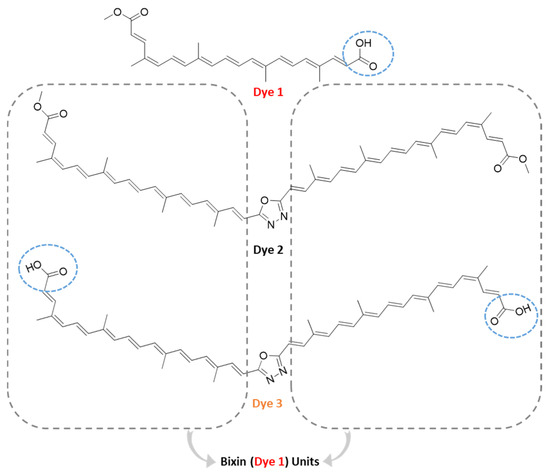
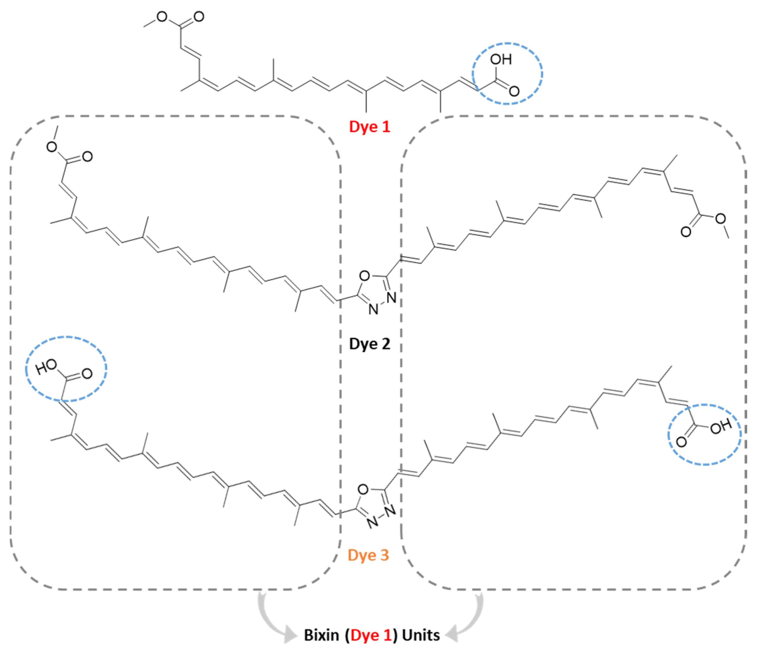
Figure 1.
Chemical structure of bixin (Dye 1), 1,3,4-oxadiazole bixin diacid hybrid (Dye 3), and its precursor 1,3,4-oxadiazole bixin diester hybrid (Dye 2). The blue circles indicate the groups anchoring to TiO2 nanofilms.
Since natural dyes have wide applications in different areas and can be used as scaffolds for the improvement of the desired physicochemical properties, the main purpose of this work is to report the complete synthesis of novel semisynthetic dyes: 1,3,4-oxadiazole bixin diester and diacid hybrids (Dyes 2 and 3, respectively, Figure 1) [10,11]. The 1,3,4-oxadiazole ring is a privileged heterocyclic scaffold widely explored in medicinal chemistry due to its unique physicochemical properties and a broad spectrum of biological activities. This five-membered heteroaromatic ring, containing two nitrogen and one oxygen atom, confers increased metabolic stability, great hydrogen bonding capacity, and favorable lipophilicity to drug candidates [11]. Moreover, 1,3,4-oxadiazoles often serve as bioisosteres for amide, ester, or carboxyl groups, enhancing pharmacokinetic profiles and improving membrane permeability when compared with their parent analogues [12]. Numerous studies have reported 1,3,4-oxadiazole derivatives exhibiting potent anti-inflammatory, antimicrobial, anticancer, anticonvulsant, and antitubercular activities. Their ability to interact with a variety of biological targets, including enzymes and receptors, underscores their versatility as key building blocks in rational drug design [13]. Consequently, the incorporation of 1,3,4-oxadiazole moieties has become a well-established strategy in the development of novel therapeutic agents with improved efficacy and safety profiles.
Beyond medicinal chemistry, the 1,3,4-oxadiazole ring plays a crucial role in the design of novel materials due to its unique electronic properties, chemical stability, and ability to participate in extended conjugated systems [14,15,16,17]. Its electron-withdrawing nature combined with aromatic stability makes it an excellent component for tuning the electronic characteristics of polymers, small molecules, and hybrid materials. The 1,3,4-oxadiazole derivatives are widely employed in the development of optoelectronic materials, such as organic light-emitting diodes (OLEDs), organic photovoltaics (OPVs), and organic field-effect transistors (OFETs), where they contribute to high electron mobility and thermal stability [14,16,17]. Additionally, their strong ability to participate in hydrogen bonding and π–π stacking interactions enables the formation of supramolecular architectures and advanced nanomaterials [15]. The incorporation of 1,3,4-oxadiazole units has also been explored in the design of sensors, corrosion inhibitors, and energetic materials, highlighting their versatility and growing relevance in materials science.
Overall, the planar structure of the 1,3,4-oxadiazole ring improves π-conjugation, charge transport, and luminescent properties. Its integration into small molecules and polymers enables precise tuning of molecular orbitals, resulting in improved device efficiency and longevity, as well as enhanced color purity with excellent thermal and oxidative stability, making it an effective electron-transporting and hole-blocking unit within OLED architectures [18]. Thus, the present work reports the synthesis of diester and diacid hybrids (Dyes 2 and 3, respectively, Figure 1) with a 1,3,4-oxadiazole core as a covalently connected point. Their physical properties were determined and compared with Dye 1 (Figure 1) through characterization by ultraviolet-visible (UV-vis) spectra, photostability, frontier molecular orbitals (FMOs) calculations, and electrochemistry for future DSSC measurements.
2. Results and Discussion
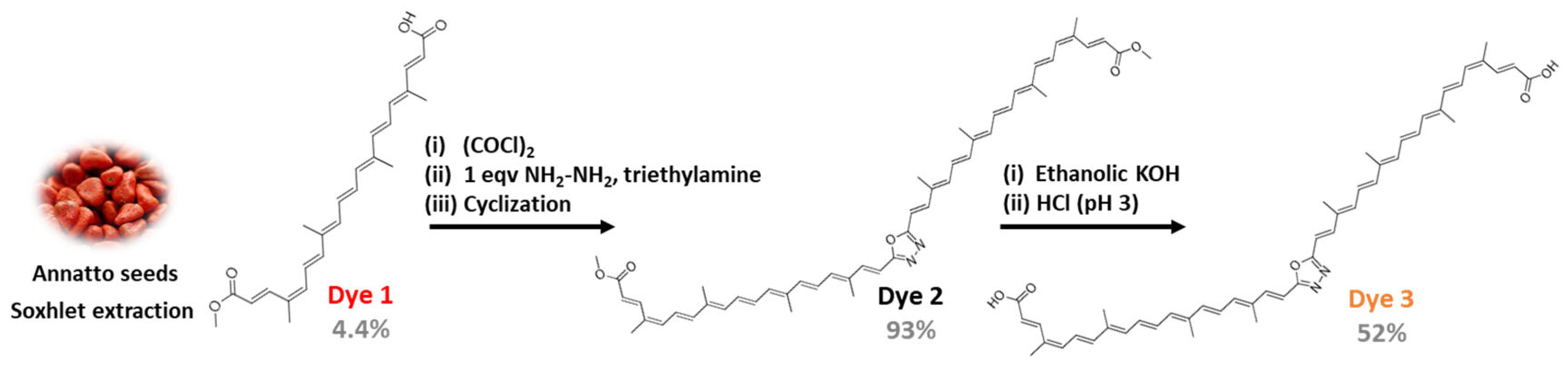
There are different methods for the extraction of bixin from annatto seeds [7,8]. In this work, the annatto seeds were used as the source to obtain the natural product Dye 1 after Soxhlet extraction with chloroform as solvent (Scheme 1) [19]. The total yield was 4.4%, and the chemical structure of Dye 1 was confirmed by melting point, Fourier transform infrared (FTIR) spectra, low-resolution mass spectrometry using electrospray ionization in the negative mode (LRMS (ESI-)), high-performance liquid chromatography (HPLC) coupled with a photodiode array (PDA) detector, and nuclear magnetic resonance (NMR) spectra (Section 3.2 and Figures S1–S5 in the Supplementary Material). The first step to synthesize Dye 2 was the synthesis of bixin acyl chloride through the reaction between Dye 1 and oxalyl chloride. The carboxyl group of bixin acyl chloride is more electrophilic than the carboxyl group of Dye 1. This reactivity improvement yielded the acyclic N-dimer after the reaction between 2 mol of bixin acyl chloride with 1 mol of hydrazine (NH2-NH2) in a dichloromethane medium with triethylamine [20]. The 1,3,4-oxadiazole bixin diester hybrid (Dye 2) was obtained after the cyclization step of a reaction between the acyclic N-dimer with tosyl chloride in triethylamine medium, yielding 93% [21,22]. The chemical structure of Dye 2 was confirmed by the same methods reported above (Section 3.3 and Figures S6–S11 in the Supplementary Material). It is important to highlight that Figure S10 in the Supplementary Materials shows the peak of molecular ion m/z 770.00 = [M+3H]+ and the peak m/z 738.00 = [[M+2H] − OCH3]+, which helps confirm the structure of Dye 2. Since the methyl ester groups in Dye 2 are not able to interact with TiO2 nanofilms, in this work, Dye 2 was converted into the corresponding bixin diacid hybrid through basic hydrolysis and then acidified to pH 3.0 using HCl, yielding 52% of the neutral Dye 3. The low yield may be due to the short hydrolysis time, which was carried out gently and without heating. It may also be due to the difficulty of filtering the solid, which is sticky before drying. The characterization of the chemical structure of Dye 3 was also conducted by melting point, FTIR, LRMS (ESI-), HPLC-PDA, and NMR spectroscopy (Figures S12–S16 in the Supplementary Material). In the IR spectrum, it is possible to observe the appearance of a broad band of 2000–3500 cm−1 that characterizes the appearance of the carboxylic acid function, in addition to the disappearance of the methyl signal in the 13C and 1H NMR spectra at 50.80 and 3.73 ppm, respectively, with the low-resolution mass spectrum with a peak at m/z 736, which refers to the [M-2H]− molecular ion peak. This peak can only be detected using the APCI detector, which requires very high ionization energy.
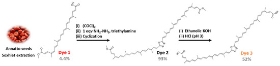
Scheme 1.
The main steps to synthesize the semisynthetic Dye 3.
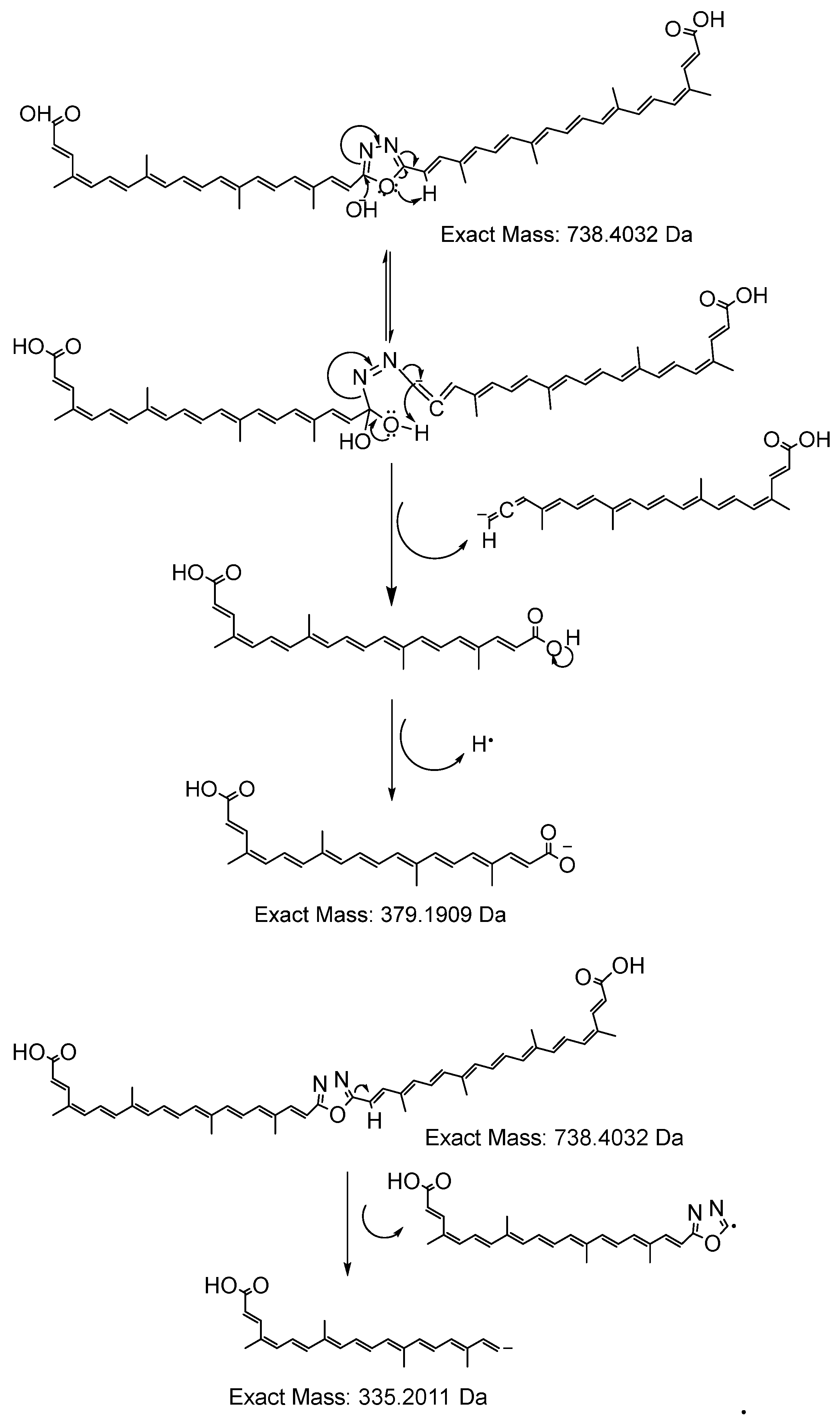
In addition to the above points confirming the structure of Dye 3, Figure 2 shows the fragmentation proposals to the highest-intensity ions observed in the high-resolution mass spectrometry (HRMS) with quadrupole time-of-flight (QTOF) analyzer and electrospray ionization (ESI-) data (Figure S17, in the Supplementary Material). Although the technique did not detect the molecular ion, possibly because of its large and complex structure, known molecule fragments were generated in high-resolution mode. The analysis of these two peaks helped confirm the structure by examining the masses of the fragments in the HRMS. For the highest-intensity peak, a calculated mass for C24H27O4− of m/z 379.1909 Da and m/z 379.1965 Da was found. For the second peak, a calculated mass for C23H27O2− of m/z 335.2011 Da and m/z 335.2061 Da was observed. Therefore, the structure of Dye 3 was unequivocally confirmed by the set of spectroscopic and spectrometric data discussed above and illustrated in Figure 2.

Figure 2.
Proposals for the fragmentation of the peaks in the HRMS spectra (see Figure S17 in the Supplementary Material) to Dye 3.
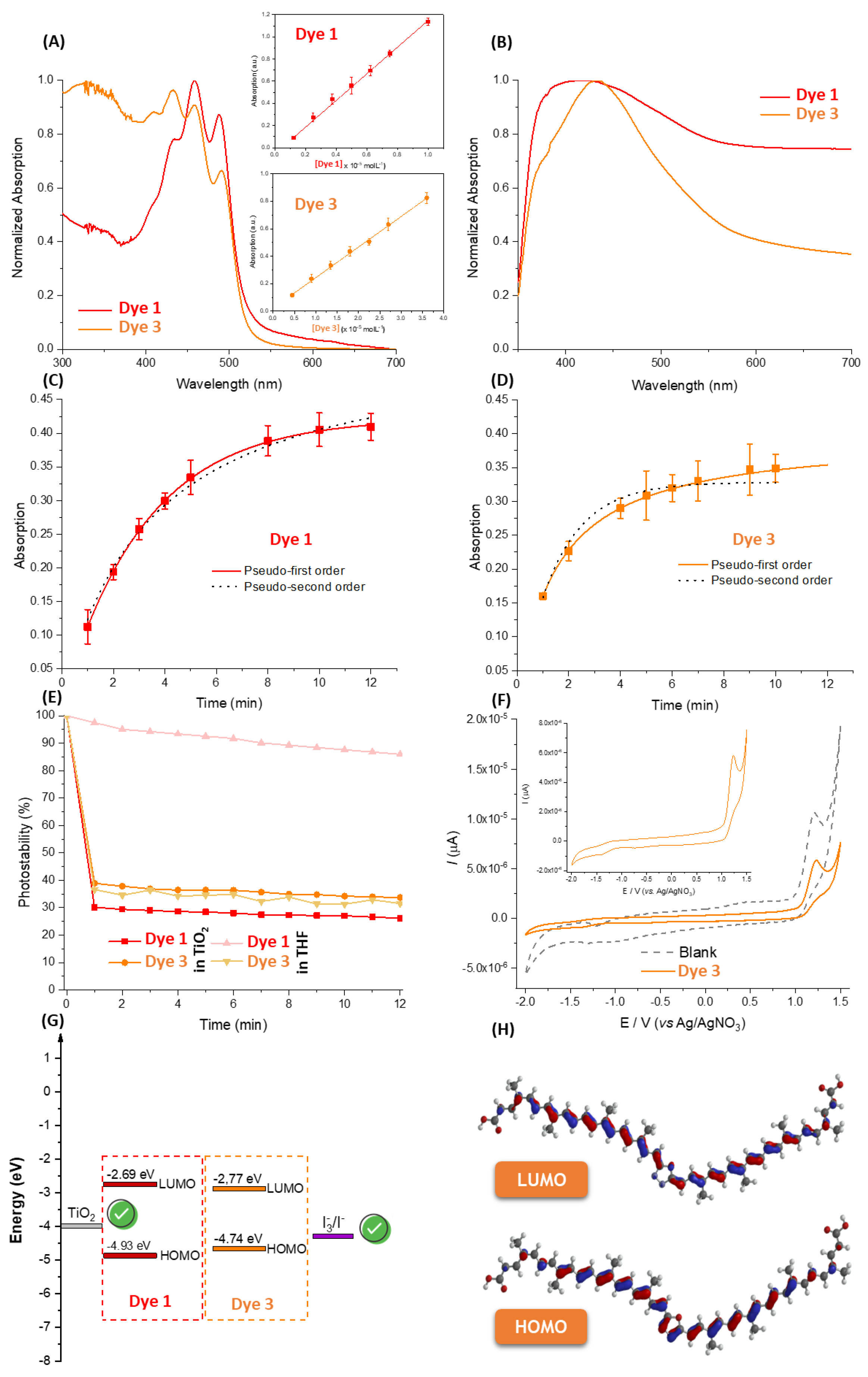
The normalized UV-vis spectra for Dyes 1 and 2 in tetrahydrofuran (THF) are shown in Figure 3A, exhibiting typical behavior for compounds belonging to the carotenoid family. The conjugated double-bond system in Dye 1, which constitutes the molecule’s chromophore, results in strong absorption in the 400–530 nm range with three distinct peaks (437, 462, and 493 nm), which arise from the lowest vibrational level of the electronic ground state to the lowest vibrational levels of the electronic excited states [10]. Despite Dye 1 showing an intense absorption band, 21Ag → 11Bu at 462 nm, the natural product under evaluation is the 9′-cis isomers, as evidenced by the weak absorption contribution at 355 nm, which corresponds to the transition to a higher-energy g state [23]. Interestingly, despite the coupling of two units of Dye 1 through a heterocycle, there was no shift in the absorption region, indicating that there was no increase in resonance states for the semisynthetic compound Dye 2. The hybrid Dye 2 showed lower photostability in THF than the natural product Dye 1 (Figure 3B), indicating that the dimer form connected by a central heterocycle did not provide greater stability.
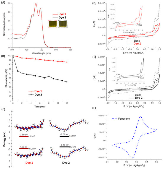
Figure 3.
(A) Normalized UV-vis spectra for Dyes 1 and 2 in THF. (B) Photostability plot for Dyes 1 and 2 in THF after different laboratory-simulated sunlight exposure periods with a potency of 0.1 W/cm2. (C) Theoretical estimation of HOMO-LUMO with DFT method B3LYP/6-31G* in THF. The CV for (D) Dye 1, (E) Dye 2, and (F) standard ferrocene in THF (0.5 mM).
The frontier molecular orbitals (FMOs) of Dyes 1 and 2 in THF were first assessed via computational calculations under DFT/B3LYP/6-31G* (Figure 3C). For Dye 1, the highest occupied molecular orbital (HOMO) and lowest unoccupied molecular orbital (LUMO) representations were in the conjugated double-bond system, with quantitative values of −4.93 and −2.69 eV, respectively, agreeing with the literature [23,24]. Following the experimental UV-vis trend (Figure 3A), Dye 2 showed practically the same theoretical FMO behavior as Dye 1 (Figure 3C). Qualitative analysis of the cyclic voltammograms (CV) in Figure 3D–F indicated that Dyes 1 and 2 do not present the self-regenerating shape of the ferrocene (positive control), but show slightly accentuated regions of oxidation and reduction, which can be explored to determine the experimental values of HOMO and LUMO, respectively. The experimental HOMO and LUMO values for both compounds were close to −4.50 and −2.30 eV, respectively, agreeing with the theoretical calculations.
Figure 4A,B depict the normalized UV-vis spectra for Dyes 1 and 3 in THF and supported in TiO2 nanofilms, respectively. Both compounds in organic solution exhibited absorption in the 400–530 nm range, with three peaks corresponding to the transitions from the lowest vibrational level of the electronic ground state to the lowest vibrational levels of the electronic excited states [10]. On the other hand, in TiO2 nanofilms, there was insufficient resolution to define the three absorption peaks clearly. However, an absorption maximum was observed at 450 nm. Despite Dyes 1 and 3 having similar absorption spectra behavior, surprisingly, the corresponding molar extinction coefficients (ε) were very different—120,401 ± 1907 and 22,273 ± 503 M−1 cm−1 (insets of Figure 4A)—indicating that the diacid dimer form of Dye 1 decreased the absorption capacity of the natural product.
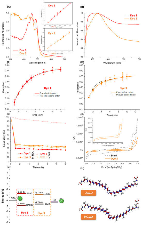
Figure 4.
(A) Normalized UV-vis spectra for Dyes 1 and 3 in (A) THF and (B) supported in TiO2 nanofilms. The insets in (A) are the Beer–Lambert plots used to calculate the ε values. Kinetic profile with the fit of pseudo-first and pseudo-second orders for the adsorption of (C) Dye 1 and (D) Dye 3 in TiO2 nanofilms. (E) Photostability plot for Dyes 1 and 3 in THF and supported in TiO2 nanofilms after different laboratory-simulated sunlight exposure (0.1 W/cm2) periods. (F) The CV for Dye 3 (0.5 mM) in THF. (G) Theoretical estimation of HOMO-LUMO for Dyes 1 and 3 under DFT/B3LYP/6-31G* in THF, with the corresponding (H) FMO representation for Dye 3.
Since the chemical structure of Dyes 1 and 3 presents anchoring groups (carboxylic acid) to interact with TiO2, the kinetic parameters of adsorption in nanofilms were determined (Figure 4C,D). The kinetic data are summarized in Table 1. Dyes 1 and 3 saturated the TiO2 surface after approximately 10 and 6 min of exposure in organic solution (1 × 10⁻⁵ M), respectively; therefore, the adsorption kinetics of the semisynthetic compound was almost twofold higher than the natural product, possibly due to the possibility of the two carboxylic groups of Dye 3 anchoring simultaneously to the TiO2 surface. This trend can be proven from the value of the pseudo-first-order rate constant (k1) for both dyes: kinetics are primarily controlled by external diffusion and independent of the concentration of the adsorbed dye [25].

Table 1.
Kinetic parameters for the adsorption of Dyes 1 or 3 in TiO2 nanofilms.
Figure 4E shows that after certain times of solar irradiation, both Dyes 1 and 3 adsorbed on the TiO2 surface photodegrade with similar kinetics—from 1 min of irradiation until 12 min of testing, the compounds reach constant values. However, Dye 3 is about 10% more photostable than Dye 1, probably due to the photostability generated by the central heterocycle. In organic solution, the natural product Dye 1 is more photostable in THF than on the TiO2 surface, probably due to the generation of more prominent radical species in the presence of the semiconductor, which is fully exposed to atmospheric air. Interestingly, the photodecomposition studies of Dye 2 in solution showed intermediate photostability between Dyes 1 and 3 in THF, indicating that the presence of free dicarboxylic acid groups in the dimer reduces its photostability, possibly due to its greater susceptibility to form reactive species.
The frontier molecular orbitals (FMOs) of Dye 3 in THF were firstly assessed via CV analysis (Figure 4F), with experimental highest occupied molecular orbital (HOMO) and lowest unoccupied molecular orbital (LUMO) values of −4.55 and −2.33 eV, respectively, compatible with the prediction under DFT/B3LYP/6-31G* (Figure 4G) with the corresponding FMO representations in the conjugated double-bond system (Figure 4H). Both experimental and theoretical energies at the LUMO level for Dyes 1 and 3 are higher than that of the conduction band of TiO2 [26], indicating that both dyes can inject electrons into the conduction band of TiO2, with Dye 3 presenting greater efficiency than Dye 1 in this electron injection. On the other hand, both experimental and theoretical HOMO values for Dyes 1 and 3 are lower than those found for the 3I−/I3− pair [27], indicating that the dyes can be quickly reduced after the injection of electrons into the conduction band of TiO2. Additionally, the shape of the CV for Dye 3 is not indicative of self-regeneration for future evaluation in a battery system [28].
Overall, the 1,3,4-oxadiazole bixin diester hybrid, Dye 2, was hydrolyzed in an acidic medium to obtain the diacid form (yield 52%). Dye 3 adsorbed via pseudo-first order on the surface of TiO2 nanofilms, following the same trend as Dye 2. Carvalho and collaborators [9] reported an open-circuit voltage (VOC) value of bixin (Dye 1) in the order of 299 mV, with short-circuit current (JSC), fill factor (FF), maximum power (Pmax), and efficiency (η) of the cells of 0.057 mA, 0.398, 6.76 µW, and 0.006%, respectively. Since this was different from what was expected, Dye 3 presented similar spectral behavior compared to the natural product Dye 1, but with better FMOs parameters, and it is expected that Dye 3 will show practically very similar or slightly better photovoltaic performance than Dye 1 on DSSC measurements. Thus, future determination of the parameters VOC, JSC, FF, Pmax, and η will be crucial to better understand the capacity of Dye 3 in DSSCs.
3. Material and Methods
3.1. General
All chemical reagents were purchased from Sigma–Aldrich (St. Louis, MO, USA). Solvents were treated with activated molecular sieves (3Å) before use. Reactions were monitored by thin-layer chromatography (TLC) on 0.25 mm Merck (Darmstadt, Germany) silica gel plates (60F-254) and visualized under a UV lamp (254 and 365 nm). All melting points (MPs) were measured using a an AAKER PFM-II and were uncorrected. The 1H-NMR (500 MHz) and 13C-NMR (125MHz) spectra were measured on a Bruker Ultrashield Plus spectrometer (Bruker BioSpin GmbH, Rheinstetten, Germany) at 25 °C and referenced to tetramethyl silane (TMS). Chemical shifts are reported in ppm (δ) using the residual solvent line as an internal standard. Splitting patterns were denoted s for singlet, d for doublet, t for triplet, m for multiplet, and brs for broad singlet. IR spectra were recorded on a Bruker-Vertex 70 FT-IR spectrophotometer using an ATR apparatus. Liquid chromatography–mass spectrometry (LC-MS) analyses were carried out on a Shimadzu LCMS 2020 (Shimadzu Inc., Kyoto, Japan) with the following analytical conditions: column of Kromasil C18, 150 mm × 4.6 mm × 5 µm (AkzoNobel, Amsterdam, the Netherlands); mobile phase of water with 0.02 M of AcONH4 (A), acetonitrile with 0.3% formic acid (B), 1.0 mL/min, linear gradient (indicated on trace); injection volume of 10 µL; detectors of PDA (200–800 nm), ESI+, and ESI- (low resolution). The UV-vis spectra were obtained with a PerkinElmer Lambda 25 spectrophotometer, while the cyclic voltammetric measurements were carried out in Autolab potentiostat/galvanostat PGSTAT204 running NOVA 2.1 software. The laboratory-simulated sunlight was obtained by a Newport Equipment model 67005 with a UV-vis lamp power of 50–500 W controlled with a power supply 69907. High-resolution mass spectrometry (HRMS) analyses were carried out on a QTOF Bruker Compact (Bruker-Daltonics, Bremen, Germany).
3.2. Bixin Extraction (Dye 1)
Annatto seeds (50 g) were subjected to Soxhlet extraction for a period of 4 h using chloroform as solvent. After the extraction period, the seeds were removed by filtration, and their volume was reduced by 50% using a rotary evaporator. After 24 h, bixin (Dye 1) precipitated in the form of dark-purple crystals, were filtered under vacuum and washed with ice-cold chloroform, generating 2.2 g of mass (4.4% yield). MP = 195–199 °C [29]. IR ν/cm−1 3405; 3178; 2946; 1712; 1606; 1562; 1284; 1157; 962. 1H-NMR (DMSO-d6) δ (ppm): δ 7.98 (dd, 1H); 7.5 (d, 1H); 6.9 (m, 1H); 6.72–6.57 (m, 5H); 6.57 (m, 4H); 5.9 (m, 2H); 3.82 (s, 3H); 2.0 (s, 12H). 13C-NMR DEPT-135 (DMSO-d6) δ (ppm): 168.28; 167.34; 148.67; 141.93; 141.07; 140.41; 139.53; 138.51; 137.18; 135.18; 135.02; 133.90; 131.78; 131.66; 125.27; 123.62; 118.12; 117.68; 51.86; 20.39; 13.14; 13.04; 12.94. LRMS (ESI-), m/z 393.0 [M-H]− (calculated for C25H29O4−, 393.0). All spectra are available in the Supplementary Material (Figures S1–S4). The purity of the final product was determined by HPLC-PDA (Figure S5).
3.3. Synthesis of 1,3,4-Oxadiazole Bixin Diester Hybrid (Dye 2)
In a flask equipped with a magnetic stirrer, under an inert atmosphere of nitrogen gas (N2), 1 (100.0 mg, 0.254 mmol) was added, and oxalyl chloride (20 mmol equivalent). The reaction proceeded for 30 min, and the evolution of HCl gas was observed. The end of the reaction was observed by TLC (dichloromethane–ethyl acetate 3%). Confirmation of the formation of acyl chloride was established by adding an aliquot of the reaction in methanol for the immediate formation of the bixin-derived methyl ester (Dye 2). At the end of the reaction, excess oxalyl chloride was removed in a rotary evaporator, and the solid remaining in the flask was solubilized in 2 mL of dry dichloromethane (under a molecular sieve activated in an oven for 120 h). In another flask, a solution of hydrazine hydrate (0.5 mmol equivalent) and triethylamine (10 mmol equivalent) in 1 mL of dry dichloromethane was prepared. The solution with bixin acyl chloride was added drop by drop to the hydrazine hydrate solution. The reaction was carried out for 2 h until the total formation of the orange-colored acyclic N-dimer was confirmed by TLC. Finally, tosyl chloride (0.75 mmol equivalent) and triethylamine (2 mmol equivalent) were added, and after 1 h of reaction, it was possible to observe a more nonpolar product with a reddish coloration by TLC in greater proportion. The contents of the flask were evaporated and suspended in an ethanol–water (3:7) solution, then vacuum-filtered and washed with distilled water, yielding dark-purple crystals of 90 mg (0.117 mmol, 93% yield). MP = 167–173 °C. IR ν/cm−1 2981; 2885; 1704; 1594; 1560; 1270; 1166; 964. 1H-NMR (Acetone-d6) δ (ppm): δ 8.0 (d, 1H); 7.56 (d, 1H); 6.98 (m, 1H); 6.67-6.91 (m, 4H); 6.38-6.64 (m, 4H); 5.9 (dd, 2H); 3.73 (s, 3H); 2.04 (s, 6H); 1.98 (s, 6H). 13C-NMR (Acetone-d6) δ (ppm): 167.03; 162.95; 152.14; 143.22; 141.93; 140.52; 139.94; 137.87; 137.20; 136.75; 135.75; 134.46; 133.39; 131.90; 131.54; 131.16; 131.05; 130.57; 128.25; 125.93; 124.47; 123.41; 117.68; 117.68; 114.72; 50.80; 19.39; 12.04; 11.89; 11.71. LRMS (ESI+), m/z 770.0 [M+3H]+ (calculated for C50H61N2O5+, 770.0) and peak m/z 738.00 = [[M+2H] − OCH3]+. All spectra obtained from this product are available in the Supplementary Material (Figures S6–S10). The purity of the final product was determined by HPLC-PDA (Figure S11).
3.4. Synthesis of 1,3,4-Oxadiazole Bixin Diacid Hybrid (Dye 3)
To synthesize the semisynthetic Dye 3, firstly 50 mg of Dye 2 was added to 5 mL of 10% ethanolic KOH solution in a flask and agitated with a magnetic stirrer. The reaction was carried out under stirring at room temperature for 4 h, followed by TLC (dichloromethane–3% ethyl acetate) until the total consumption of the reagents. Then, the reaction mixture was acidified to pH 3 using a concentrated HCl solution. The solid was vacuum-filtered and washed with ice-cold water, followed by ice-cold ethanol, yielding a dark-red solid of 26 mg (52% yield). MP > 250 °C. 1H-NMR (acetone-d6) δ (ppm): δ 7.99 (d, 1H); 7.36 (d, 1H); 6.96–7.06 (m, 2H); 6.75–6.88 (m, 4H); 6.61–6.69 (m, 3H); 6.46-6.58 (m, 4H); 5.87–5.96 (t, 3H); 2.03 (s, 6H); 2.00 (s, 6H). 13C-NMR (acetone-d6) δ (ppm): 167.24; 148.74; 141.66; 140.38; 140.00;139.22; 137.55; 136.86; 136.73; 134.82; 134.38; 133.48; 131.54; 131.36; 131.06; 124.56; 123.27; 118.25; 116.45; 19.41; 12.01; 11.89; 11.76. IR ν/cm−1 958, 1184, 1265, 1560, 1593, 1674, 2919. LRMS m/z 736 [M-2H]−. HRMS for the following peaks: calculated mass for C24H27O4− of m/z 379.1909 Da and m/z 379.1965 Da found; calculated mass for C23H27O2− of m/z 335.2011 Da and m/z 335.2061 Da found. All spectra obtained for the characterization of Dye 3 are available in the Supplementary Material (Figures S12–S17). The purity of the final product was determined by HPLC-PDA (Figure S16).
3.5. Adsorption
The TiO2 films were prepared by the doctor’s blade technique [30]. The TiO2 paste was deposited on a glass slide by screen-printing an area of 1 cm2. The resulting films were gradually heated with the following heating profile: 125 °C for 10 min, 325 °C for 5 min, 375 °C for 10 min, 450 °C for 10 min, and 500 °C for 15 min. The morphology and thickness of the TiO2 films (1.2 μm) were measured by scanning electron microscopy (SEM) as per previous work [31].
Dye 1 and 3 solutions (5 × 10⁻⁵ M) were prepared in THF. The TiO2 films were immersed in each solution, and absorption spectra were taken at selected time intervals from 1 to 12 min. Excess dye was removed by rinsing with solvent before measurement. The amount of adsorbed dye was monitored as a function of time by the absorbance of the dye-modified TiO2 films at the maximum dye absorption band in a PerkinElmer Lambda 25 spectrophotometer at room temperature (~20 °C). The kinetic profile for the adsorption of Dyes 1 and 3 on TiO2 nanofilms was determined following the pseudo-first (Equation (1)) and pseudo-second (Equation (2)) orders:
where t is the time (min), while k1 and k2 are the pseudo-first- and pseudo-second-order rate constants, respectively. The q1 and q2 are plateau regions based on pseudo-first and pseudo-second order, respectively.
3.6. Photostability
The solution of Dyes 1–3 (1 mM) was exposed to laboratory-simulated sunlight with a potency of 0.1 W/cm2 for periods of 0, 1, 2, 3, 4, 5, 6, 7, 8, 9, 10, 11, and 12 min at room temperature (~20 °C). After each exposure, the UV-vis spectrum was recorded with a PerkinElmer Lambda 25 spectrophotometer at room temperature (~20 °C). Additionally, the UV-vis spectra for Dyes 1 and 3 supported in TiO2 nanofilms (dyes in a saturated condition) before and after (0, 1, 2, 3, 4, 5, 6, 7, 8, 9, 10, 11, and 12 min) exposure to laboratory-simulated sunlight with a potency of 0.1 W/cm2 were also recorded.
3.7. Electrochemistry
The cyclic voltammetry measurements were carried out with an Autolab potentiostat/galvanostat PGSTAT204 running NOVA 2.1 software. Three-electrode systems were added to one electrochemical cell with a capacity of 10 mL. A glassy carbon electrode (GCE, d = 3.0 mm) was the working electrode, a glassy carbon wire (GC, d = 1.6 mm) was the counter electrode, and Ag/AgNO3 (0.01 M AgNO3 and 0.1 M tetrabutylammonium hexafluorophosphate in acetonitrile) was the reference electrode. Ferrocene (0.5 mM) was used as a reference. The reference and samples (Dyes 1–3, in 0.5 mM) were dissolved in THF. CV values were obtained at a scan rate of 50 mV/s. Before each electrochemical experiment, the GCE was polished with appropriate polishing pads, first using aluminum oxide with particle size of 0.3 μm and then aluminum oxide with particle size of 0.075 μm. After polishing, the electrode was rinsed thoroughly with Milli-Q water and then sonicated in a container filled with a 50:50 (v/v) mixture of Milli-Q water and ethanol for 5 min. In the electrochemical cell, nitrogen-saturated solutions were obtained by bubbling high-purity N2 for a minimum of 5 min in the solution and a continuous flow of pure gas over the solution during the voltammetric experiments. All the assays were conducted at room temperature (~20 °C). The HOMO and LUMO values were determined following equations reported in the literature [32,33].
3.8. Computational Procedure
The chemical structure for Dyes 1–3 was built with Spartan’14 software (Wavefunction, Inc., Irvine, USA). The same software was used to estimate the HOMO and LUMO parameters with the DFT method under Becke-3 Lee–Yang–Parr (B3LYP) 6-31G* in THF medium [34].
4. Conclusions
The natural compound bixin (Dye 1) was obtained from annatto seeds (yield 4.4%), and its diester and diacid hybrids (Dyes 2 and 3, respectively) were synthesized considering the 1,3,4-oxadiazole ring as the connected point with corresponding yields of 93% and 52%. Interestingly, despite the coupling of two units of Dye 1, there was no shift in the absorption capacity of the hybrids. However, Dye 3 was about 10% more photostable than Dye 1. Dyes 1 and 3 had anchoring groups (carboxylic acid) to interact with TiO2 via pseudo-first-order kinetics, with adsorption kinetics of the semisynthetic compound almost twofold higher than the natural product. The experimental and theoretical FMOs values indicated that the evaluated dyes had no self-regeneration for future evaluation in a battery system; however, the FMO representations in the conjugated double-bond system of the dyes presented parameters that were indicative that the compounds can be quickly reduced after the injection of electrons into the conduction band of TiO2 and that the dyes can inject electrons into the conduction band of TiO2, with Dye 3 presenting greater efficiency than Dye 1. Future determination of the parameters VOC, JSC, FF, Pmax, and η will be crucial to better understand the capacity of Dye 3 in DSSCs.
Supplementary Materials
The following supporting information can be downloaded at https://www.mdpi.com/article/10.3390/reactions6030039/s1. Figure S1: FTIR spectrum of Dye 1; Figure S2: 1H-NMR spectrum of Dye 1 in DMSO-d6 (followed by its expansion); Figure S3: 13C-NMR DEPT-135 spectrum of Dye 1 in DMSO-d6 (followed by its expansion); Figure S4: LRMS (ESI-) of Dye 1; Figure S5: HPLC-PDA of Dye 1; Figure S6: FTIR spectrum of Dye 2; Figure S7: 1H-NMR spectrum of Dye 2 in acetone-d6 (followed by its expansion); Figure S8: 13C-NMR DEPT-135 spectrum of Dye 2 in acetone-d6 (followed by its expansion); Figure S9: Overlay of part of the DEPT-135 13C-NMR spectra that proves the formation of the 1,3,4-oxadiazole moiety, where there is a clear change from 168.28 ppm (referring to the bixin carboxylic acid carbonyl) to 162.95 ppm (referring to the carbons of the 1,3,4-oxadiazole ring); Figure S10: LRMS (ESI+) of Dye 2, m/z 738.00 = [[M+2H] − OCH3]+. To obtain LRMS, the APCI detector was used, requiring very high ionization energy; Figure S11: HPLC-PDA of Dye 2; Figure S12: 1H-NMR spectrum of Dye 3 in acetone-d6 (followed by its expansion); Figure S13: 13C-NMR spectrum of Dye 3 in acetone-d6 (followed by its expansion); Figure S14: FTIR spectrum of Dye 3; Figure S15: LRMS ESI(-) of Dye 3; Figure S16: HPLC-PDA of Dye 3; Figure S17: HRMS ESI(-) of Dye 3.
Author Contributions
Conceptualization, O.A.C., C.S. and M.E.F.d.L.; methodology, A.S.M.M.V., O.A.C., D.P., C.S., R.N.C. and M.E.F.d.L.; software, O.A.C.; validation, C.S. and M.E.F.d.L.; formal analysis, A.S.M.M.V., O.A.C., D.P., R.N.C. and M.E.F.d.L.; investigation, A.S.M.M.V., O.A.C., D.P., C.S., R.N.C. and M.E.F.d.L.; resources, C.S. and M.E.F.d.L.; data curation, O.A.C. and M.E.F.d.L.; writing—original draft preparation, O.A.C.; writing—review and editing, A.S.M.M.V., O.A.C., D.P., C.S. and M.E.F.d.L.; visualization, O.A.C. and M.E.F.d.L.; supervision, C.S. and M.E.F.d.L.; project administration, O.A.C., C.S. and M.E.F.d.L.; funding acquisition, C.S. and M.E.F.d.L. All authors have read and agreed to the published version of the manuscript.
Funding
This project received funding from the Brazilian agencies Coordenação de Aperfeiçoamento de Pessoal de Nível Superior (CAPES, finance code 001), Conselho Nacional de Desenvolvimento Científico e Tecnológico (CNPq), and Fundação Carlos Chagas Filho de Amparo à Pesquisa do Estado do Rio de Janeiro (FAPERJ). The Coimbra Chemistry Centre is supported by the Fundação para a Ciência e a Tecnologia (FCT—Portuguese Agency for Scientific Research) through the projects UIDB/00313/2025 and UIDP/00313/2025.
Data Availability Statement
Data are contained within the article and Supplementary Materials.
Acknowledgments
O.A.C. also thanks FCT for his PhD fellowship 2020.07504.BD as well as the Programa de Pós-Graduação em Biologia Celular e Molecular from the Oswaldo Cruz Foundation (Rio de Janeiro, Brazil) and CAPES for grant PIPD (process SCBA 88887.082745/2024-00 with subproject 31010016). D.P. acknowledges Santander Universidades and UC for the award of the Projetos Semente de Investigação Científica Interdisciplinar Santander UC—Project INOCleanMat. The authors thank the Multiuser Analytical Center of IQ-UFRRJ for performing the NMR, FTIR, and LRMS analyses and the Analytical Center of Farmanguinhos (Fiocruz-RJ) for the HRMS of the final product.
Conflicts of Interest
The authors declare no conflicts of interest.
References
- Rodríguez-Morgade, M.S.; Pellejà, L.; Torres, T.; Palomares, E. Ti (IV) phthalocyanines for dye sensitized solar cells. J. Porphyr. Phthalocyanines 2013, 17, 814–820. [Google Scholar] [CrossRef]
- Fernandes, S.S.M.; Castro, M.C.R.; Pereira, A.I.; Mendes, A.; Serpa, C.; Pina, J.; Justino, L.L.G.; Burrows, H.D.; Raposo, M.M.M. Optical and Photovoltaic Properties of Thieno[3,2-b]thiophene-Based Push–Pull Organic Dyes with Different Anchoring Groups for Dye-Sensitized Solar Cells. ACS Omega 2017, 2, 9268–9279. [Google Scholar] [CrossRef] [PubMed]
- Grätzel, M. Recent Advances in Sensitized Mesoscopic Solar Cells. Acc. Chem. Res. 2009, 42, 1788–1798. [Google Scholar] [CrossRef] [PubMed]
- Che, J.; Yang, X. A recent (2009–2021) perspective on sustainable color and textile coloration using natural plant resources. Heliyon 2022, 8, e10979. [Google Scholar] [CrossRef] [PubMed]
- Hagan, E.; Poulin, J. Statistics of the early synthetic dye industry. Herit. Sci. 2021, 9, 33. [Google Scholar] [CrossRef]
- Pizzicato, B.; Pacifico, S.; Cayuela, D.; Mijas, G.; Riba-Moliner, M. Advancements in Sustainable Natural Dyes for Textile Applications: A Review. Molecules 2023, 28, 5954. [Google Scholar] [CrossRef]
- Huamán, A.A.; Celestino, M.R.; Quintana, M.E. Theoretical and experimental study of solar cells based on nanostructured films of TiO2 sensitized with natural dyes extracted from Zea mays and Bixa orellana. RSC Adv. 2021, 11, 9086–9097. [Google Scholar] [CrossRef]
- Rahmalia, W.; Silalahi, I.H.; Usman, T.; Fabre, J.-F.; Mouloungui, Z.; Zissis, G. Stability, reusability, and equivalent circuit of TiO2/treated metakaolinite-based dye-sensitized solar cell: Effect of illumination intensity on Voc and Isc values. Mater. Renew. Sustain. Energy 2021, 10, 10. [Google Scholar] [CrossRef]
- Carvalho, I.C.; Barbosa, M.L.; Costa, M.J.S.; Longo, E.; Cavalcante, L.S.; Viana, V.G.F.; Santos, R.S. TiO2-based dye-sensitized solar cells prepared with bixin and norbixin natural dyes: Effect of 2,2′-bipyridine additive on the current and voltage. Optik 2020, 218, 165236. [Google Scholar] [CrossRef]
- Rahmalia, W.; Fabre, J.-F.; Usman, T.; Mouloungui, Z. Aprotic solvents effect on the UV–visible absorption spectra of bixin. Spectrochim. Acta Part A Mol. Biomol. Spectrosc. 2014, 131, 455–460. [Google Scholar] [CrossRef]
- Pitasse-Santos, P.; Sueth-Santiago, V.; Lima, M. 1,2,4- and 1,3,4-Oxadiazoles as Scaffolds in the Development of Antiparasitic Agents. J. Braz. Chem. Soc. 2017, 29, 435–456. [Google Scholar] [CrossRef]
- Dick, A.; Cocklin, S. Bioisosteric Replacement as a Tool in Anti-HIV Drug Design. Pharmaceuticals 2020, 13, 36. [Google Scholar] [CrossRef]
- Kumar, D.; Aggarwal, N.; Deep, A.; Kumar, H.; Chopra, H.; Marwaha, R.K.; Cavalu, S. An Understanding of Mechanism-Based Approaches for 1,3,4-Oxadiazole Scaffolds as Cytotoxic Agents and Enzyme Inhibitors. Pharmaceuticals 2023, 16, 254. [Google Scholar] [CrossRef]
- Bolton, O.; Kim, J. Design principles to tune the optical properties of 1,3,4-oxadiazole-containing molecules. J. Mater. Chem. 2007, 17, 1981. [Google Scholar] [CrossRef]
- Najare, M.S.; Patil, M.K.; Nadaf, A.A.; Mantur, S.; Inamdar, S.R.; Khazi, I.A.M. Synthesis, characterization and photophysical properties of a new class of pyrene substituted 1,3,4-oxadiazole derivatives. Opt. Mater. 2019, 88, 256–265. [Google Scholar] [CrossRef]
- Maniyar, A.K.; Nadaf, Y.F.; Khasim, S.; Hamdallah, T.A.; Murugendrappa, M.V. Photophysical studies on donor-p-acceptor substituted 1,3,4-oxadiazole derivatives for optoelectronic application: Experimental and theoretical analysis. J. Opt. 2024. [Google Scholar] [CrossRef]
- Naik, L.; Khazi, I.A.M.; Malimath, G.H. Studies on photosensitization of TiO2 nanoparticles by novel 1,3,4-oxadiazoles derivatives. Optik 2019, 183, 732–741. [Google Scholar] [CrossRef]
- Paun, A.; Hadade, N.D.; Paraschivescu, C.C.; Matache, M. 1,3,4-Oxadiazoles as luminescent materials for organic light emitting diodes via cross-coupling reactions. J. Mater. Chem. C 2016, 4, 8596–8610. [Google Scholar] [CrossRef]
- Taham, T.; Cabral, F.A.; Barrozo, M.A.S. Extraction of bixin from annatto seeds using combined technologies. J. Supercrit. Fluids 2015, 100, 175–183. [Google Scholar] [CrossRef]
- Jandl, B.; Zheng, R.; Muttenthaler, M.; Baell, J. Novel Diacyl-hydrazide Compounds as Potential Therapeutics for Visceral Leishmaniasis. ACS Omega 2024, 9, 37170–37182. [Google Scholar] [CrossRef]
- Lamani, R.S.; Nagendra, G.; Sureshbabu, V.V. A facile synthesis of N-Z/Boc-protected 1,3,4-oxadiazole-based peptidomimetics employing peptidyl thiosemicarbazides. Tetrahedron Lett. 2010, 51, 4705–4709. [Google Scholar] [CrossRef]
- Dolman, S.J.; Gosselin, F.; O’Shea, P.D.; Davies, I.W. Superior Reactivity of Thiosemicarbazides in the Synthesis of 2-Amino-1,3,4-oxadiazoles. J. Org. Chem. 2006, 71, 9548–9551. [Google Scholar] [CrossRef]
- Scotter, M. The chemistry and analysis of annatto food colouring: A review. Food Addit. Contam. Part A 2009, 26, 1123–1145. [Google Scholar] [CrossRef]
- Lima, I.T.; da Silva Crispim, J.; de Sá Neto, O.P.; de Sousa Júnior, R.T.; Ribeiro Júnior, L.A.; da Silva Filho, D.A. Organic Electronics from Nature: Computational Investigation of the Electronic and Optical Properties of the Isomers of Bixin and Norbixin Present in the Achiote Seeds. Molecules 2022, 27, 2138. [Google Scholar] [CrossRef]
- Malik, P.K. Use of activated carbons prepared from sawdust and rice-husk for adsorption of acid dyes: A case study of Acid Yellow 36. Dyes Pigment. 2003, 56, 239–249. [Google Scholar] [CrossRef]
- Yu, H.; Irie, H.; Hashimoto, K. Conduction Band Energy Level Control of Titanium Dioxide: Toward an Efficient Visible-Light-Sensitive Photocatalyst. J. Am. Chem. Soc. 2010, 132, 6898–6899. [Google Scholar] [CrossRef]
- Hagfeldt, A.; Boschloo, G.; Sun, L.; Kloo, L.; Pettersson, H. Dye-Sensitized Solar Cells. Chem. Rev. 2010, 110, 6595–6663. [Google Scholar] [CrossRef]
- Bandodkar, A.J.; López, C.S.; Vinu Mohan, A.M.; Yin, L.; Kumar, R.; Wang, J. All-printed magnetically self-healing electrochemical devices. Sci. Adv. 2016, 2, e1601465. [Google Scholar] [CrossRef]
- REITH, J.F.; GIELEN, J.W. Properties of bixin and norbixin and the composition of annatto extracts. J. Food Sci. 1971, 36, 861–864. [Google Scholar] [CrossRef]
- Ramos-Hernández, R.; Calvo, F.D.; Pérez-Gutiérrez, E.; Percino, M.J. Large area small-molecule thin films deposited by the doctor blade technique implemented with computer numerical control machine. Thin Solid Film. 2023, 771, 139787. [Google Scholar] [CrossRef]
- Monteiro, C.J.P.; Jesus, P.; Davies, M.L.; Ferreira, D.; Arnaut, L.G.; Gallardo, I.; Pereira, M.M.; Serpa, C. Control of the distance between porphyrin sensitizers and the TiO2 surface in solar cells by designed anchoring groups. J. Mol. Struct. 2019, 1196, 444–454. [Google Scholar] [CrossRef]
- Fernandes, S.S.M.; Belsley, M.; Pereira, A.I.; Ivanou, D.; Mendes, A.; Justino, L.L.G.; Burrows, H.D.; Raposo, M.M.M. Push–Pull N,N-Diphenylhydrazones Bearing Bithiophene or Thienothiophene Spacers as Nonlinear Optical Second Harmonic Generators and as Photosensitizers for Nanocrystalline TiO2 Dye-Sensitized Solar Cells. ACS Omega 2018, 3, 12893–12904. [Google Scholar] [CrossRef]
- Rodrigues, A.C.B.; Eckert, A.; Pina, J.; Scherf, U.; Seixas de Melo, J.S. Polymeric near infrared emitters with bay-annulated indigo moieties. Mater. Adv. 2021, 2, 3736–3743. [Google Scholar] [CrossRef]
- Chaves, O.A.; Sueth-Santiago, V.; Pinto, D.C.d.A.; Netto-Ferreira, J.C.; Decote-Ricardo, D.; Lima, M.E.F. 2-Chloro-4,6-bis{(E)-3-methoxy-4-[(4-methoxybenzyl)oxy]styryl}pyrimidine: Synthesis, Spectroscopic and Computational Evaluation. Molbank 2021, 2021, M1276. [Google Scholar] [CrossRef]

Disclaimer/Publisher’s Note: The statements, opinions and data contained in all publications are solely those of the individual author(s) and contributor(s) and not of MDPI and/or the editor(s). MDPI and/or the editor(s) disclaim responsibility for any injury to people or property resulting from any ideas, methods, instructions or products referred to in the content. |
© 2025 by the authors. Licensee MDPI, Basel, Switzerland. This article is an open access article distributed under the terms and conditions of the Creative Commons Attribution (CC BY) license (https://creativecommons.org/licenses/by/4.0/).