Article

Biofilm Inhibition Against Staphylococcus aureus and Alizarin Red Dye-Removing Capability of Plant-Based Green Synthesis of Lanthanum Oxide (La2O3NPs) Nanoparticles

by
Krishnamoorthy Uma Sivakami
1,*,
Sundararajan Vaideeswaran
2,
Ambrose Rosevenis
3,
Rajender Boddula
4,*,
Kanagarajan Shenbagam
1,
Muniraj Balaganesh
5,
Usan Pathinathan Saleth Prabhakar
6,
Paramasivam Shanmugam
7,8,*,
Fatemah M. Barakat
9,
Supakorn Boonyuen
8 and
Ramyakrishna Pothu
10,*
1
Department of Chemistry, Cauvery College for Women (Autonomous), Affiliated to Bharathidasan University, Tiruchirappalli 620018, Tamil Nadu, India
2
Department of Science and Humanities, Amrita College of Engineering and Technology, Nagercoil 629901, Tamil Nadu, India
3
Department of Chemistry, St. Joseph’s College, Affiliated to Bharathidasan University, Tiruchirappalli 620002, Tamil Nadu, India
4
School of Sciences, Woxsen University, Hyderabad 502345, Telangana State, India
5
Department of Chemistry, BWDA Arts and Science College, Tindivanam 604304, Tamil Nadu, India
6
Department of Chemistry, Arul Anandar College, Madurai 625514, Tamil Nadu, India
7
Department of Biomaterials, Saveetha Dental College and Hospital, SIMATS, Saveetha University, Chennai 600077, Tamil Nadu, India
8
Department of Chemistry, Faculty of Science and Technology, Thammasat University, Pathum Thani 12120, Thailand
9
Department of Physics and Astronomy, College of Science, King Saud University, Riyadh 11451, Saudi Arabia
10
School of Physics and Electronics, College of Chemistry and Chemical Engineering, Hunan University, Changsha 410082, China
*
Authors to whom correspondence should be addressed.
Reactions 2025, 6(2), 32; https://doi.org/10.3390/reactions6020032
Submission received: 18 October 2024 / Revised: 20 January 2025 / Accepted: 30 January 2025 / Published: 2 May 2025

Abstract

:
The unique properties of lanthanum oxide nanoparticles (La2O3NPs) make them highly suitable for various environmental applications. This study explores the plant-based synthesis of La2O3NPs using Drypetes sepiaria as a reducing agent. The synthesized La2O3NPs were characterized through a range of spectroscopic and microscopic techniques. Scanning electron microscopy (SEM) revealed that the La2O3NPs have an uneven surface and a stony appearance. A morphological analysis indicated that the nanoparticles range in size from 20 to 50 nm. The appreciable band gap energy values were concluded as 5.5 eV. The crystal structure and elemental composition were determined using X-ray diffraction (XRD) and energy-dispersive spectroscopy (EDS). The results from the microplate assay method demonstrated enhanced anti-biofilm properties, and photocatalytic tests showed significant dye-degradation capabilities. The degradation efficiency and zone inhibition values of the La2O3NPs were found to be 90.12% and 39.18%, respectively.

Graphical Abstract

1. Introduction

Biomolecules existing in plant materials having capability of act as both reducing and stabilizing agents, serving as an alternate to chemical reducing agents for converting metal ions into their respective nanoparticles in a single-step process known as the green synthesis method [1,2]. The biogenic reduction methodology of a metal ion is typically rapid, even at room temperature, and quite effortless [3,4]. The eco-friendly reducing agents used include various water- and alcohol-soluble plant metabolites and co-enzymes. In addition to the extracts from a wide variety of species, live plants have also been successfully used in nanoparticle synthesis, resulting in nanoparticles with a reduced size range [5,6,7]. Nanoparticles have gained considerable technical attention, as they act as bridges between bulk materials and molecular structures [4,8,9]. In recent years, nanotechnology has developed with multifunctional applications across a variety of engineering fields [10,11]. The majority of the research has mainly focused on the synthetic aspect of nanoparticles, as they can be easily prepared and utilized [12,13]. Although various physical and chemical methods have been conventionally used for the nanoparticles synthesis, recent research has shifted toward the advancement of environmental friendly synthesis approaches [6,14,15,16]. Recently, phyto-nanotechnology has emerged as a novel approach for single-step synthesis, offering a non-toxic, straightforward, robust, and inexpensive method [17,18]. This technique utilizes plant-derived nanoparticles by employing readily available plant materials and capitalizing on the non-toxic nature of plants, making it well-suited to meet the high demand of nanoparticles in biomedical and environmental applications [18,19,20]. Notably, various parts of plants, beyond just leaves, have been employed in the synthesis of metal nanoparticles (MNPs). Plant extracts containing flavonoids, alkaloids, polyphenols, terpenoids, heterocyclic compounds, and polysaccharides play significant roles in the synthesis of MNPs [21,22]. These compounds act as reducing agents for the conversion of metal ions and also serve as stabilizing, impregnating, and fabricating agents during synthesis methods [23,24]. Nickolas et al. reported the synthesis of silver nanoparticles using origanum vulgare, where the NaOH concentration played a dominant role in the synthesis of AgNPs [25]. Lanthanum oxide nanoparticles (La2O3NPs) are among the most optically active oxides of all other rare earth oxides. The wide band gap energy is typically greater than 4 eV [26,27,28]. Bhusankar et al. reported rod-shaped lanthanum oxide nanoparticles as efficient bifunctional electrocatalysts for hydrogen and oxygen evolution reactions. As a result, rod-shaped lanthanum oxide exhibits good electrocatalytic activity [29]. La-based materials are receiving increasing attention for their magnetic, optical, electrical, and therapeutic properties [30,31].
La2O3NPs have been synthesized in various compositions, including hydroxides, fluoride, carbonate, phosphates, borate, and oxides [32,33,34]. Normally, hydrothermal, chemical precipitation methods have been used to prepare La2O3NPs. To minimize the interaction of harmful substances, eco-friendly green synthesis methods have been employed [6,35,36]. This article mainly focuses on the application part of bio fabricated nanomaterials as effective anti-biofilm agents and photocatalytic applications. Biofilms are accumulations of bacterial cells; they cause infections and are problematic for components and environments. These infectious cells, along with their extracellular polymeric substances, adhere to both living and non-living surfaces, posing serious problems.
Engineering, such as food [37], water [38,39], dairy, textiles, concrete [40] constructions, metals like steel, iron, etc. [41,42], water purification units, and marine production are susceptible to biofilm formation. Although numerous traditional methods have been implemented to control these biofilms, most provide only short-term solutions. This highlights the need for advanced research to discover anti-biofilm agents. Many studies have reported the degradation of toxic dyes using various metal particles, such as Au, Ag, Co, Cu, Pd, and Pt [43,44,45]. In this context, plant-based synthesized La2O3NPs, with their unique properties and high efficiency, were explored as catalysts for the degradation of Alizarine red dye. Typically, photo-catalytic processes involve the generation of free radicals under light irradiation. However, sunlight penetration is often insufficient in heavily colored wastewater, such as dye-laden industrial effluents, which limits the effectiveness of photo-catalytic methods [46,47]. Alizarin Red dye, also known as 3,4-dihydroxy-9,10-dioxo-9,10-dihydroanthracene-2-sulfonic acid, is a water-soluble sodium salt of Alizarin sulfonic acid. It belongs to the category of azo dyes and finds widespread use in the dyeing of wool and nylon. In this context, biosynthesized La2O3NPs were utilized to investigate degradation, offering a promising solution for treating wastewater from the textile industry.
This study introduces the new plant-based synthesis of La2O3NPs using Drypetes sepiaria as a reducing agent, providing an eco-friendly alternative to conventional synthetic methods. Furthermore, the research highlights the unique structural and functional properties of the synthesized La2O3NPs, including their distinct stony morphology, a well-defined energy band gap of 3.2 eV, and excellent photocatalytic and biofilm activity. These findings demonstrate the dual potential of La2O3NPs for environmental applications, offering an innovative approach to addressing biofilm-related challenges and dye pollution simultaneously.

2. Materials and Methods

2.1. Materials

Lanthanum chloride (99.5%), sodium hydroxide (98%), and ethanol (99%) were procured from SRL Chemicals, Mumbai, India. Sodium acetate (99.5%) and crystal violet were obtained from Nice Chemicals (P) Ltd., Ernakulam, India, while Alizarin Red (ARS) dye was sourced from Loba Chemie (P) Ltd., Mumbai, India.

2.2. Qualitative Preliminary Phytochemical Screening

The powdered leaves of Drypetes sepiaria (DS) were collected from Pachaimalai hills, located near Tiruchirappalli district at 119° N, 78°210 E of Tiruchirappalli, Tamil Nadu, India. Initially, the leaves were thoroughly washed several times with running tap water and then dried under shade. Approximately, 100 g of the powdered leaves was soaked in 500 mL of ethanol using the cold percolation method [48]. The extract was filtered at regular intervals, and the crude extract was collected through distillation. The extract was preserved in an amber bottle and refrigerated. This prepared extract was utilized for phytochemical screening.

2.3. Synthesis of Lanthanum Oxide Nanoparticles Using Eco-Friendly Leaf Extract as a Reducing Agent

La2O3NPs were synthesized using a green synthetic process with a Drypetes sepiaria (DS) leaf extract. A stoichiometric amount of analytical-grade lanthanum chloride (0.1 M, 100 mL) was added 100 mL of the plant extract, and the reaction mixture was stirred continuously for 1 h [49]. Subsequently, 0.1 M NaOH was added to the reaction mixture to adjust the pH to above 12.5. A magnetic stirrer was used to maintain the reaction mixture at 70 °C for 3 h. During the reaction, a color change was observed, indicating the formation NPs. After the completion of the reaction, the resulting La2O3NPs were dried in a hot-air oven at 60 °C for 24 h. After drying, the NPs were calcined at 600 °C for 4 h. The resulting La2O3NPs were characterized using various spectroscopic and microscopic techniques. Their biofilm-inhibition activity and dye-degradation efficiency for waste water were subsequently evaluated [50].

2.4. Microplate Assay for Biofilm Inhibition

To assess the effectiveness of the medication in preventing biofilm formation, we used 96-well flat-bottom polystyrene plates for the MTP assay. Each well was filled with 180 µL of BHI broth, followed by the addition of 10 µL of an overnight culture of pathogenic bacteria as an inoculant. Additionally, 10 µL of the TR-FEND sample, prepared from stock solutions with concentrations ranging from 500 to 31.25 µg/mL, was added. The mixture was then incubated for 24 h at 37 °C. After incubation, the well contents were aspirated and washed with 0.2 mL of phosphate-buffered saline (PBS, pH 7.2) to remove free-floating bacteria. Sessile bacteria were fixed using 2% sodium acetate and stained with 0.1% w/v crystal violet. Excess stain was rinsed off with deionized water. The percentage of biofilm inhibition was calculated using the below-mentioned formula (Equation (1)) [51].
% Biofilm inhibition = [(Control OD − Test OD/Control OD)] × 100

2.5. Photocatalytic Dye Degradation

The photocatalytic efficiency of the La2O3NPs was tested based on the degradation of Alizarin Red (ARS) under visible light irradiation. Initially, 50 mL of Alizarin Red (10 mg/L) solution was taken in a beaker, and 30 mg of La2O3NP photocatalysts was added. The degradation of Alizarin Red dye was studied, through the elimination of the Alizarin Red dye. The photocatalytic behavior of the synthetic sample was determined using the Alizarin Red dye under UV or solar radiation. A 150 W Xe (Xenon) lamp was used as the light source, with a 10 cm gap between the UV source and the photo-reaction vessel. The suspensions were magnetically agitated for 30 min in the dark before being exposed to radiation. Next, under typical ambient conditions, UV irradiation was applied to the photoreaction vessel. In the photo-removal experiment, the dye of choice was used with the sample; 3 mL of the suspension was extracted at regular intervals of time to remove the catalyst particles using a centrifugal process, and then, UV-Vis absorption spectrometer was used. Using the equation below, the photo-removal efficiency percentage was calculated. Following that, five consecutive cycles of recycling experiments (0, 60, 120, 180, 240, and 300 min) were conducted to assess the durability of the photo-catalytic sample. Prior to being used again in the subsequent trial, the NPs were dried, centrifuged, and cleaned in ethanol and deionized water. The degradation efficiency was calculated using the following formula (Equation (2)) [52].
% Photo-removal efficiency = C0 − C/C0 × 100
where C0 is the initial concentration of dye and C is the concentration of dye after photo-irradiation (final).

2.6. Characterization

To determine the absorbance, the synthesized La2O3NPs were exposed to a UV visible spectrometer (Perkin Elmer, Lambda 365). The morphological property was studied using SEM analysis, which clearly described the exact size of the synthesized La2O3NPs (JEOL Model JSM—6390LV). The functional group analysis was obtained from the FT-IR spectrometer (Thermo Nicolet, Avatar 370, Waltham, MA, USA). The crystalline structure of the sample was determined from the Bruker AXS Advance powder X-ray method. The elements present, along with their respective percentages, in the synthesized biofabricated La2O3NPs were explained using an elemental analysis (EDAX).

3. Results

The active phyto compounds present in the alcoholic leaf extract of Drypetes sepiaria (DS) are illustrated in Table 1.

3.1. Gas Chromatography-Mass Spectrum Study (GC-MS)
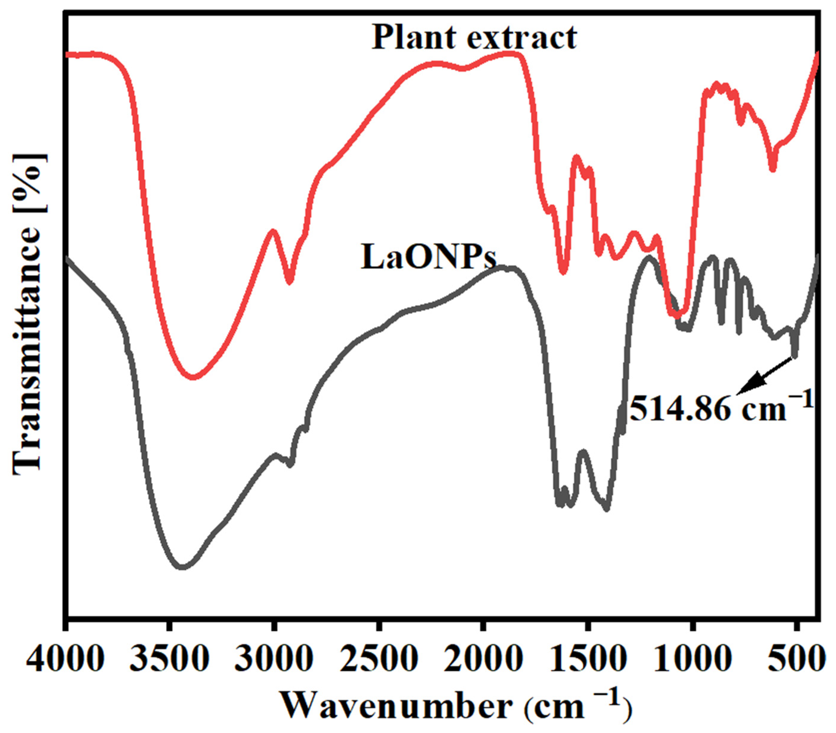
The mass spectrum was compared and identified with the database of the National Institute of Standard Technology (NIST). The results indicated twenty peaks, showing the presence of twenty chemical constituents with the retention time (RT), molecular formula, molecular weight (MW), and peak area (%) (Figure 1). The structures of the compounds were given (Figure 2). The prevailing compounds were 1,1,3-triethoxy-propane (35.88%), (2e, 6e)-1,1-dideutero-3,7,11-trimethyl-2,6,10-dodecatrien-1-ol (14.38%), alpha-tocopheryl acetate (10.72%), 1-hexadecanol (6.18%), and ethyl ester hexadecanoic acid (5.99%) (Table 2).
A particle size analysis using Dynamic Light Scattering (DLS) indicated that the synthesized particles were approximately 60 nm in size. The presence of La2O3NPs was confirmed via UV spectroscopy (Figure 3), which showed a symmetric single-band absorption peak at 230.9 nm (λ max) that steadily increased in intensity over time, with no observed shift.
The diffuse reflectance and energy spectra of bio-fabricated La2O3NPs synthesized using Drypetes sepiaria (DS) showed major peaks at 230 nm, as depicted in Figure 3a. Additionally, a few weak peaks were observed, possibly due to external factors, such as surface traps, shape defects, or impurities. The direct band gap energy values were concluded to be 5.5 eV for biosynthesized La2O3NPs and was given by Wood and Tauc’s relation [53]. The appreciable band gap energy value was clearly due to the existence of smaller-particle-size nanoparticles of biosynthesized La2O3NPs.
The SEM investigation (Figure 4) indicated that as the temperature increased, the grain size increased, leading to crystal-like grain growth with a grain size of 1 µm. The surface morphology was found to be a spherical shape with an uneven, porous surface, suggesting the possibility of the rapid release of byproducts during the progression of the reaction.

3.2. FT-IR

The FTIR spectra of the green synthesis of the plant extract (PE) and La2O3NPs are shown in Figure 5. In the FTIR spectrum of PE, the downshift of the O-H frequency from 3600 to 3434.12 cm−1 denoted hydrogen bonding. The peaks at 2928.19 cm−1, 2862.41 cm−1 represent the presence of –C-H- bonding. The peak observed at 1586.16 cm−1 indicates the presents of an NO2 stretch. The peaks at 1627.43 cm−1 and 1640.88 cm−1 indicate the presents of alkene bonding. The peak at 1414.90 cm−1 indicates the presents of a CH2 bend, and 1336.90 cm−1 indicates the presents of the -CH3 bend. The observed peaks at 1022.15 cm−1 and 1049.20 cm−1 indicate the presence of C-F bonds. Additionally, peaks at 864.62 cm−1, 713.06 cm−1, and 703 cm−1 indicate the presents of carbon and halogen bonding. The same characteristic peaks appeared in the plant extract stabilized La2O3NPs. Furthermore, an additional peak at 514.86 cm−1 appeared due to the metal–oxygen bond, thus confirming that the La2O3NPs were stabilized by the plant extract [54].

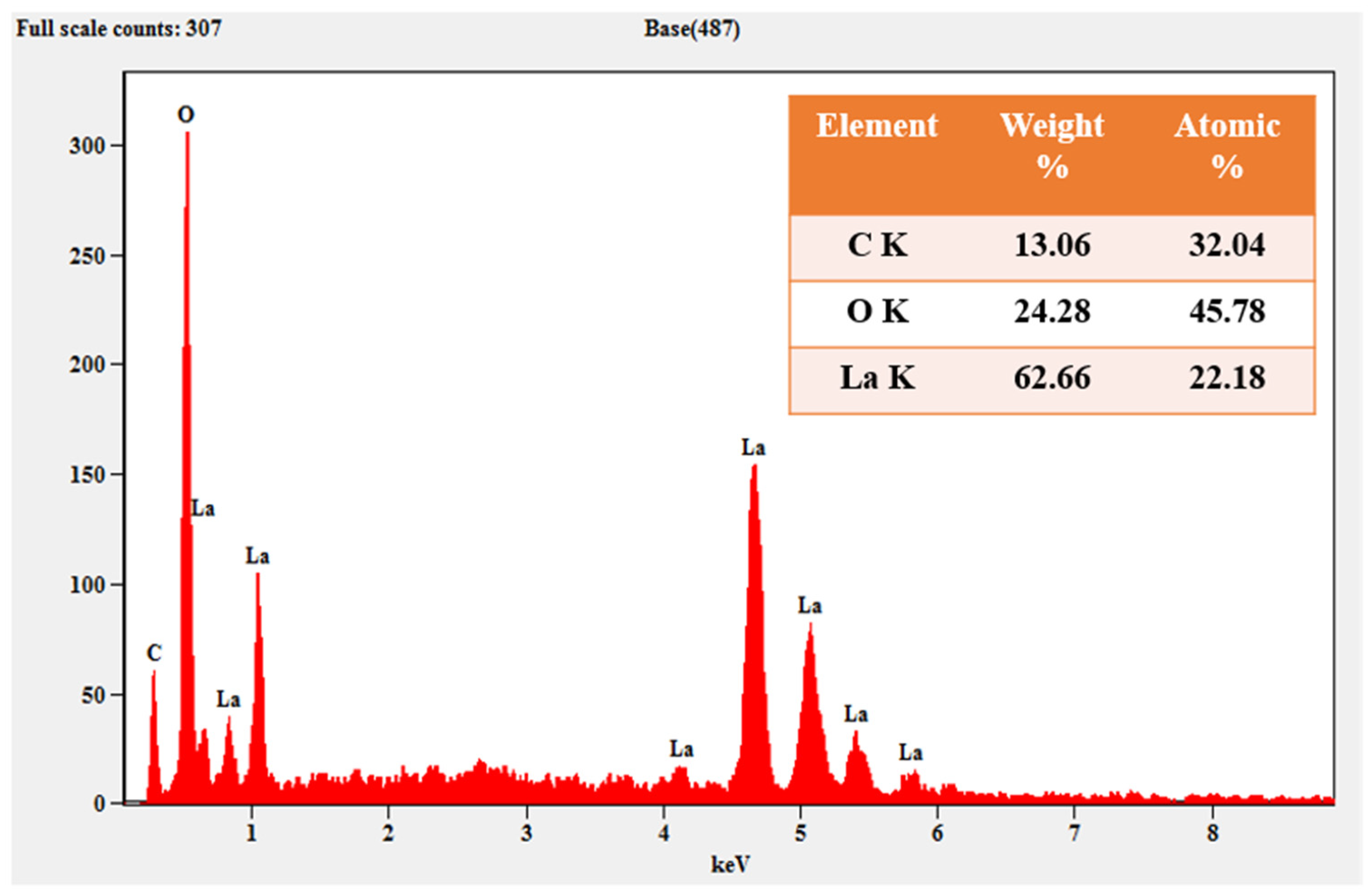
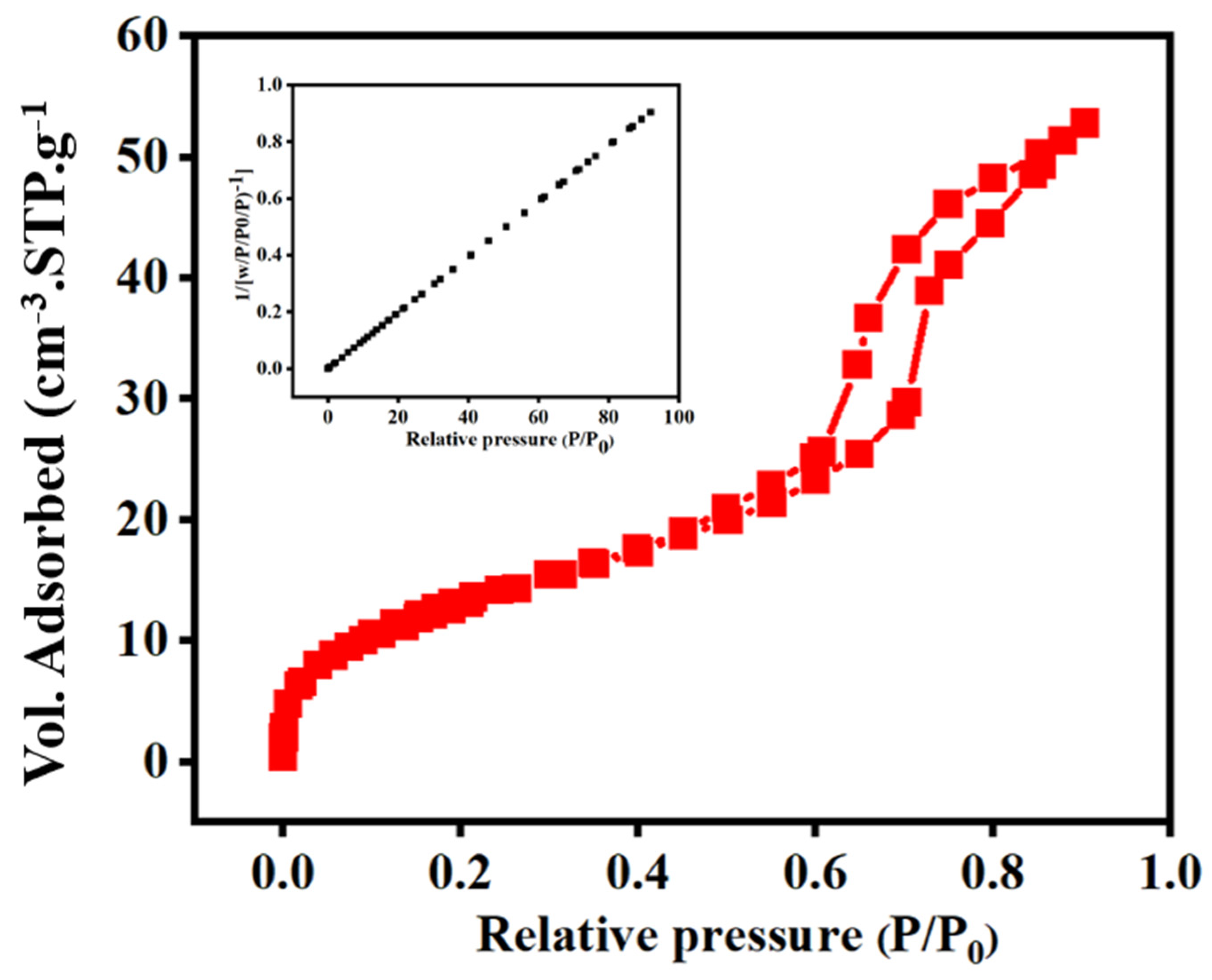
3.3. XRD Analysis and EDAX Analysis

XRD analysis plays a vital role in determining the microstructure property of different phases of materials (Figure 6). For La2O3NPs, the observed 2θ peaks were at 25°, 30°, 40°,44°, and 57°, which corresponded to plans of (100), (101), (102), (110), and (103), respectively. The peaks obtained were compared with Joint Committee for Powder Diffraction studies (JCPDS) card No: 05–0602, which shows that the synthesized La2O3NPs have a hexagonal structure.
Equations (1) and (2) represent the Debye–Scherer equation and Bragg’s law, which were used to calculate the particle size [55].
Debye-Scherer equation (D) = 0.9λ/β cosθ
Bragg’s Law (nλ) = 2dsinθ
In this context, λ denotes the wavelength of X-rays (0.154 nm), β represents angular or line broadening at full width at half maximum (FWHM), θ stands for the diffraction angle, D denotes the particle diameter in nanometers, and d refers to planar spacing. The elements and their respective percentages, as shown in the table indexed in Figure 7, are depicted in the EDAX analysis. The EDAX analysis reveals that the sample contains a reasonable number of C, O, and La atoms.
The HRTEM analysis was used to examine the surface of biosynthesized La2O3NPs at different magnifications (Figure 8). The HRTEM images provide detailed morphological and structural information regarding the La2O3NPs. Figure 8a shows a higher magnification image, where individual particle sizes are more distinguishable. The particles exhibit a relatively uniform size distribution, with dimensions ranging between 28 nm to 32 nm, as indicated by the measurements. However, the overall morphology suggests that the particles are slight aggregates of varying sizes. This is consistent with the expected size range obtained from the synthesis method, further confirming the nano-scale nature of the particles.
Figure 8b provides a closer look at the crystalline structure of the particles, where the lattice fringes are clearly visible. The interplanar spacing (d-spacing) of 0.248 nm, as marked in the image, corresponds to the characteristic crystallographic planes of the synthesized material, confirming the crystalline nature of the nanoparticles. Figure 8c shows the selected area electron diffraction (SAED) pattern, which consists of a series of concentric rings. This pattern indicates that the nanoparticles possess a polycrystalline structure. The well-defined diffraction spots further confirm the crystallinity observed in the high-resolution TEM image (Figure 8b), supporting the notion that the particles are crystalline in nature with some degree of an ordered structure. Further, they provide a comprehensive understanding of the particle size distribution, a particle size histogram was constructed, as shown in Figure 8d. The histogram indicates a relatively narrow size distribution, with most particles falling within the range of 30 ± 2 nm. This consistent size range suggests that the synthesis process using Drypetes sepiaria as a reducing agent facilitated controlled and uniform nanoparticle formation. The particle sizes obtained through the plant-based green synthesis method result in La2O3NPs with a uniform size distribution, crystalline structure, and favorable morphology, making them well-suited for photocatalytic and anti-biofilm applications.
The specific surface area of the La2O3NPs was obtained using the Brunauer–Emmett–Teller (BET) analysis, as shown in Figure 9, based on the N2 adsorption/desorption isotherm [56]. The observed shape of the isotherm suggests Type IV behavior according to the IUPAC classification, which is characteristic of mesoporous materials with well-defined pores. The specific surface area of the La2O3NPs was found to be 49.24 m2/g. Furthermore, the pore volume and pore diameter of the La2O3NPs were determined to be 0.0817 cm3/g and 6.64 nm, respectively. Additionally, the average pore size distribution was calculated using the BJH (Barrett–Joyner–Halenda) method, which is a procedure for determining pore size distribution based on the Kelvin equation and DH methods. The average pore size distribution of the La2O3NPs was found to be 4.10 nm. The obtained pore size distribution results reveal that the La2O3NPs exhibit a broad range of pore diameters, with a peak in the mesoporous range at around 4.10 nm. Moreover, the green-synthesized La2O3NPs provide more active sites for light absorption and catalysis, thereby enhancing the photocatalytic degradation of organic dyes, such as ARS.

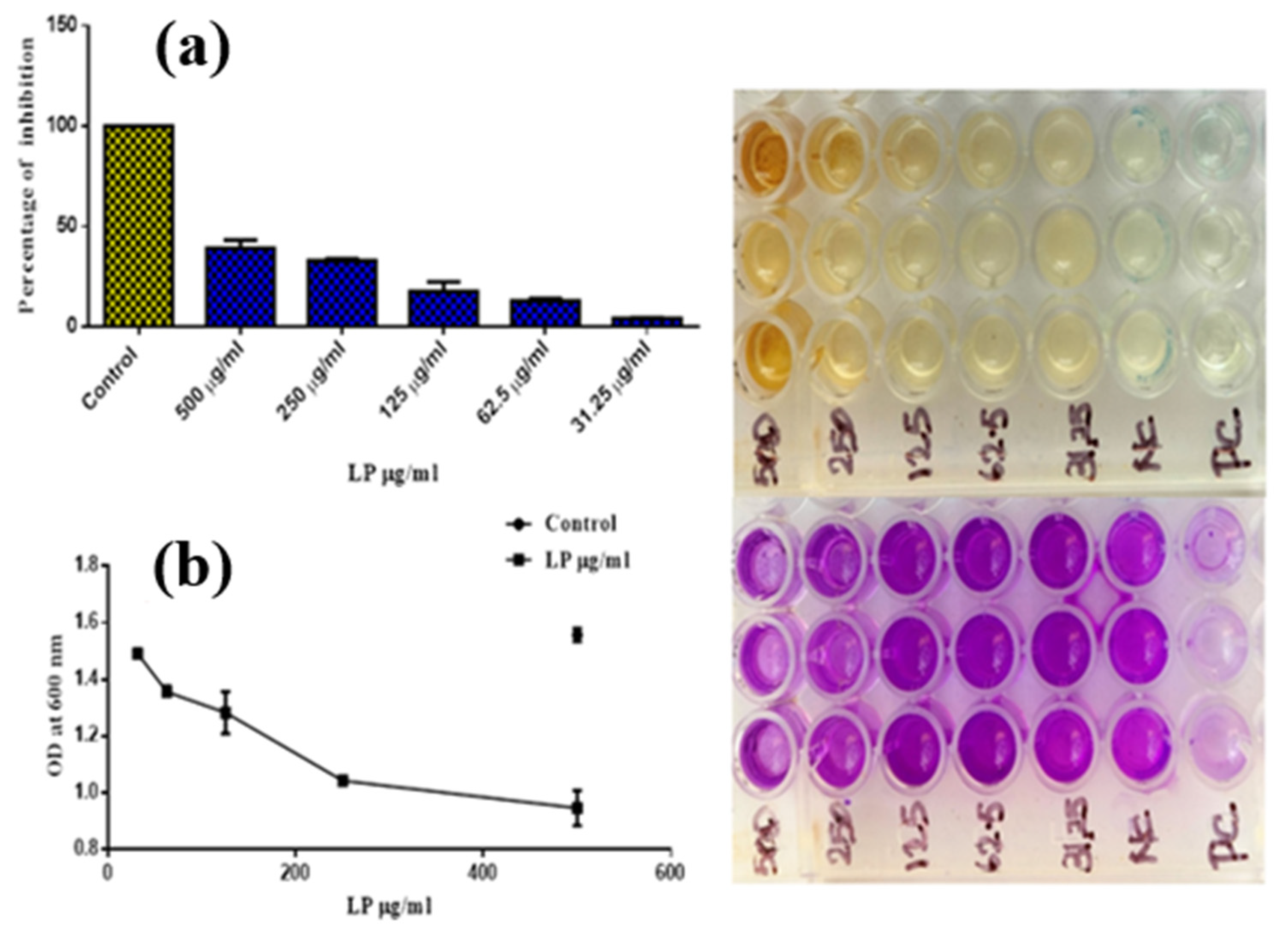
3.4. Biofilm-Inhibition Activities

The percentages of biofilm inhibition were calculated and tabulated. The OD value at 600 nm with various concentrations of synthesized bio-fabricated nanoparticles is represented in Table 3, which shows the inhibition of bio-film formation against Staphylococcus aureus, with a clear inhibition zone caused by the antibacterial properties of the synthesized La2O3NPs, as observed in the in vitro results (Figure 10a,b).
The optical density (OD) values were also observed, showing that the OD values increase with a decrease in the concentration of the samples [31]. The percentage of inhibition was higher at the higher concentration of 500 µg/mL and decreased with a lower concentration (Table 4). The synthesized bio-fabricated La2O3NPs demonstrated enhanced biofilm-inhibition capability.

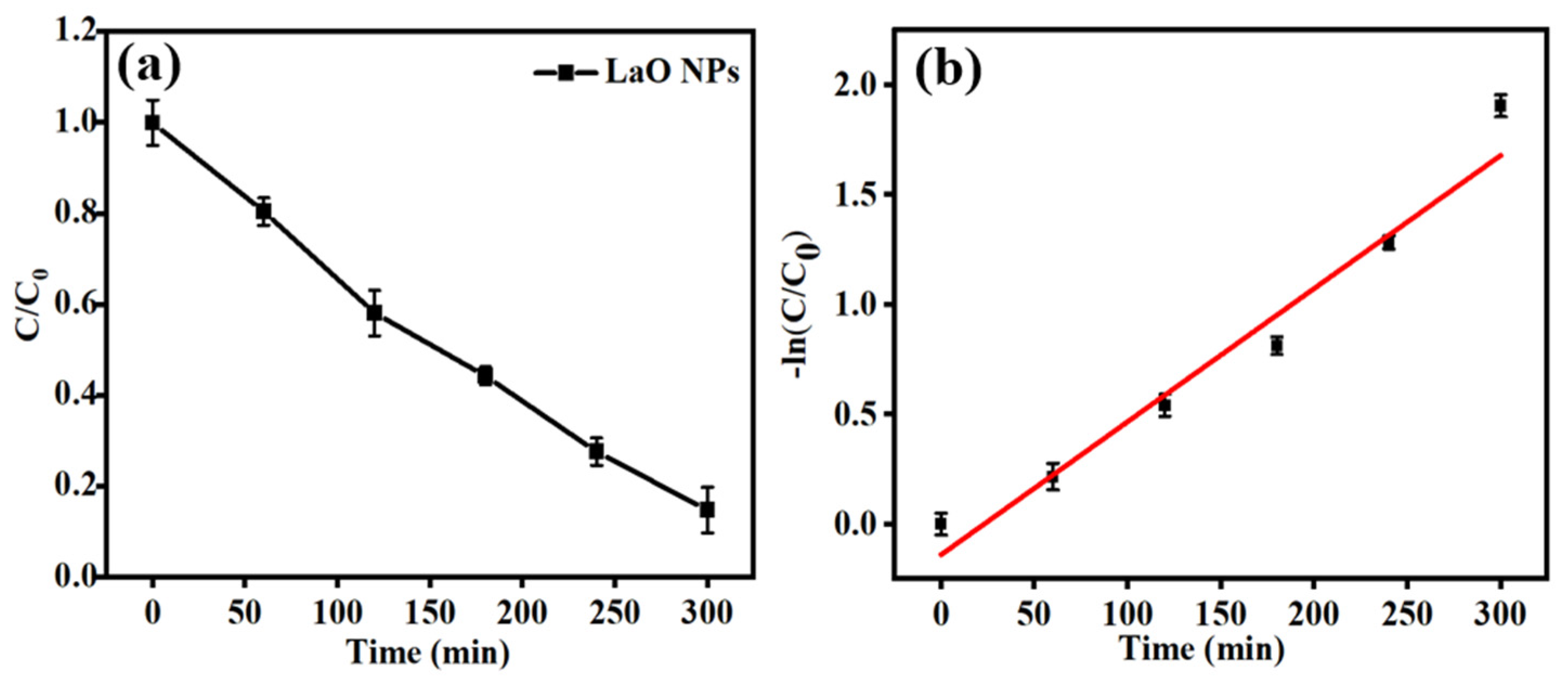
3.5. Photocatalytic Dye-Degradation Activities

During the photodegradation process, color changes in the dye solution were observed at different time intervals, as shown in Figure 11. Figure 12a illustrates the photodegradation performance of biosynthesized La2O3NPs for Alizarin Red (ARS) dye under visible light illustration. The concentration ration ratio C/C0 of ARS steadily decreased over time, indicating a progressive degradation process. The concentration ratio (C/C₀) of ARS steadily decreased over time, indicating a progressive degradation process. A degradation efficiency of 90.12% was achieved within 5 h, demonstrating the effective photocatalytic activity of the La2O3 NPs. Figure 12b further confirms the kinetics of the degradation process by plotting the −ln(C/C0) vs. as a function of time. The linearity of the data suggests that the degradation follows pseudo-first-order kinetics, as evidenced by the excellent fit, with an R2 value close to unity. Further, the rate constant values were found to be 6.19 × 10⁻², indicating a rapid reaction rate under the given experimental conditions. These results highlight the potential of La2O3NPs as effective photocatalysts for the degradation of organic pollutants like ARS, which is attributed to their enhanced light absorption and active surface properties. The linear kinetic trend and high-rate constant underline the nanoparticles’ promising role in environmental remediation applications.
The reusability of La2O3NPs was evaluated over five consecutive cycles for the photodegradation of Alizarin Red, as depicted in Figure 13. The results demonstrate that the degradation efficiency remained consistently high, with only a slight decrease observed with repeated use. In the first cycle, the degradation efficiency was 90.12%, and even after five cycles, it remained at 84.24%, indicating the excellent stability and reusability of the catalyst. The reusable property was undergoing the practical applicability of La2O3NPs as a cost-effective and sustainable photocatalyst for the treatment of organic dyes like Alizarin Red in wastewater treatment. These findings reveal that La2O3NPs possess significant potential for long-term environmental applications with minimal performance degradation over repeated cycles. Further, the photocatalytic activity of the La2O3NP photocatalyst was compared with various types of catalysts and provided in Table 5.
A photocatalytic mechanism of Alizarin Red (ARS) using La2O3NPs as a photocatalysts under sunlight irradiation is depicted in the Figure 14. As is known to all, the electrons are excited and transferred from the valence band (VB) to the conduction band (CB) with the illustration of light. The photogenerated charge carriers plays an crucial role in the photodegradation process. The electrons (e) in the CB combined with dissolved oxygen (O2) to generated superoxide radicals O2. The superoxide radical further converted into hydroxyl radicals (OH) in in the presence of protons (H+), which are highly effective oxidizing agents. Instantaneously, the holes (h+) in the VB react with water (H2O) to produce another hydroxyl radical. The combined form of reactive oxygen species helps to break down the toxic ARS molecule in non-toxic degraded products. This mechanism highlights the high photocatalytic efficiency of La2O3NPs, attributed to its ability to generate and separate charge carriers effectively under sunlight. The production of hydroxyl radicals as the primary oxidative species ensures the rapid degradation of ARS, showcasing the potential of La2O3NPs as a sustainable photocatalyst for environmental remediation applications.

4. Conclusions

This study demonstrates a novel green synthetic route for the synthesis of La2O3NPs using an alcoholic extract of Drypetes sepiaria (DS). The bio-synthesized La2O3NPs exhibit unique structural and functional properties, including a hexagonal crystalline structure, a significant band gap energy of 5.5 eV, and a porous spherical morphology with a stony-like structure. These characteristics, confirmed through UV, FT-IR, XRD, EDAX, and surface analysis, highlight the potential of Drypetes sepiaria as a sustainable resource for nanomaterial synthesis. The 90.12% of degradation efficiency was achieved within 5 h. The green synthesized La2O3NPs have good recycling efficiency, and the degradation efficiency remains constant up to the 5th cycles. Further, zone inhibition values (39.18%) demonstrate that the green synthesized La2O3 has good antibacterial efficiency against S. aureus. Furthermore, the nanoparticles show promising applications, evidenced by their efficient bio-scavenging activity and superior dye-absorption capabilities, making them a valuable candidate for environmental and biotechnological applications.

Author Contributions

Conceptualization, K.U.S.; methodology, S.V. and R.B.; software, M.B. and R.P.; validation, F.M.B.; formal analysis, S.B. and R.P.; investigation, K.U.S.; data curation, A.R. and U.P.S.P.; writing—original draft preparation, K.U.S.; writing—review and editing, R.B. and P.S.; visualization, K.S.; supervision, K.U.S.; project administration, K.U.S.; funding acquisition, K.U.S. All authors have read and agreed to the published version of the manuscript.

Funding

The authors acknowledge the funding source from UGC (4-4/2015-16-MRP/UGC-SERO) and the Tamil Nadu State Council for Science and Technology, TNSCST (PS020), for their financial support to carry out the research work.

Data Availability Statement

Data will be made available on request.

Conflicts of Interest

The authors declare no conflicts of interest.

References

  1. Bonatto, C.C.; Silva, L.P. Higher Temperatures Speed up the Growth and Control the Size and Optoelectrical Properties of Silver Nanoparticles Greenly Synthesized by Cashew Nutshells. Ind. Crops Prod. 2014, 58, 46–54. [Google Scholar] [CrossRef]
  2. Mishra, A.N.; Bhadauria, S.; Gaur, M.S.; Pasricha, R.; Kushwah, B.S. Synthesis of Gold Nanoparticles by Leaves of Zero-Calorie Sweetener Herb (Stevia rebaudiana) and Their Nanoscopic Characterization by Spectroscopy and Microscopy. Int. J. Green Nanotechnol. Phys. Chem. 2010, 1, P118–P124. [Google Scholar] [CrossRef]
  3. Yilmaz, M.; Turkdemir, H.; Kilic, M.A.; Bayram, E.; Cicek, A.; Mete, A.; Ulug, B. Biosynthesis of Silver Nanoparticles Using Leaves of Stevia rebaudiana. Mater. Chem. Phys. 2011, 130, 1195–1202. [Google Scholar] [CrossRef]
  4. Jagtap, U.B.; Bapat, V.A. Green Synthesis of Silver Nanoparticles Using Artocarpus Heterophyllus Lam. Seed Extract and Its Antibacterial Activity. Ind. Crops Prod. 2013, 46, 132–137. [Google Scholar] [CrossRef]
  5. Worakitjaroenphon, S.; Shanmugam, P.; Boonyuen, S.; Smith, S.M.; Chookamnerd, K. Green Synthesis of Silver and Gold Nanoparticles Using Oroxylum Indicum Plant Extract for Catalytic and Antimicrobial Activity. Biomass Conv. Bioref. 2023. [Google Scholar] [CrossRef]
  6. Shanmugam, P.; Boonyuen, S.; Tangjaideborisu, Y.; Na Nakorn, P.; Tantayanon, S.; Pothu, R.; Boddula, R. Anthocyanin Rich-Berry Extracts Coated Magnetic Fe3O4 Bionanocomposites and Their Antibacterial Activity. Inorg. Chem. Commun. 2023, 156, 111291. [Google Scholar] [CrossRef]
  7. Thakkar, K.N.; Mhatre, S.S.; Parikh, R.Y. Biological Synthesis of Metallic Nanoparticles. Nanomed. Nanotechnol. Biol. Med. 2010, 6, 257–262. [Google Scholar] [CrossRef]
  8. Fathima, J.B.; Pugazhendhi, A.; Venis, R. Synthesis and Characterization of ZrO2 Nanoparticles-Antimicrobial Activity and Their Prospective Role in Dental Care. Microb. Pathog. 2017, 110, 245–251. [Google Scholar] [CrossRef]
  9. Li, D.; Liu, Z.; Yuan, Y.; Liu, Y.; Niu, F. Green Synthesis of Gallic Acid-Coated Silver Nanoparticles with High Antimicrobial Activity and Low Cytotoxicity to Normal Cells. Process Biochem. 2015, 50, 357–366. [Google Scholar] [CrossRef]
  10. Khan, Z.U.H.; Khan, A.; Chen, Y.; Shah, N.S.; Muhammad, N.; Khan, A.U.; Tahir, K.; Khan, F.U.; Murtaza, B.; Hassan, S.U.; et al. Biomedical Applications of Green Synthesized Nobel Metal Nanoparticles. J. Photochem. Photobiol. B Biol. 2017, 173, 150–164. [Google Scholar] [CrossRef]
  11. Ramkumar, V.S.; Pugazhendhi, A.; Gopalakrishnan, K.; Sivagurunathan, P.; Saratale, G.D.; Dung, T.N.B.; Kannapiran, E. Biofabrication and Characterization of Silver Nanoparticles Using Aqueous Extract of Seaweed Enteromorpha compressa and Its Biomedical Properties. Biotechnol. Rep. 2017, 14, 1–7. [Google Scholar] [CrossRef]
  12. Sivakami, K.U.; Vaideeswaran, S.; Venis, A.R.; Juliet Helina, J.K.A.; Balaganesh, M. Impregnation, Silver Activating Capability and Biological Applications of Ionic Liquids. Mater. Lett. X 2022, 13, 100134. [Google Scholar] [CrossRef]
  13. Nnadozie, E.C.; Ajibade, P.A. Green Synthesis and Characterization of Magnetite (Fe3O4) Nanoparticles Using Chromolaena Odorata Root Extract for Smart Nanocomposite. Mater. Lett. 2020, 263, 127145. [Google Scholar] [CrossRef]
  14. Venkateswarlu, S.; Natesh Kumar, B.; Prasad, C.H.; Venkateswarlu, P.; Jyothi, N.V.V. Bio-Inspired Green Synthesis of Fe3O4 Spherical Magnetic Nanoparticles Using Syzygium cumini Seed Extract. Phys. B Condens. Matter 2014, 449, 67–71. [Google Scholar] [CrossRef]
  15. Agarwal, H.; Venkat Kumar, S.; Rajeshkumar, S. A Review on Green Synthesis of Zinc Oxide Nanoparticles—An Eco-Friendly Approach. Resour.-Effic. Technol. 2017, 3, 406–413. [Google Scholar] [CrossRef]
  16. Mokhtary, M.; Torabi, M. Nano Magnetite (Fe3O4), an Efficient and Robust Catalyst for the One-Pot Synthesis of 1-(Aryl(Piperidin-1-Yl)Methyl)Naphthalene-2-Ol and 1-(α-Amido Alkyl)-2-Naphthol under Ultrasound Irradiation. J. Saudi Chem. Soc. 2017, 21, S299–S304. [Google Scholar] [CrossRef]
  17. Vanathi, P.; Rajiv, P.; Narendhran, S.; Rajeshwari, S.; Rahman, P.K.S.M.; Venckatesh, R. Biosynthesis and Characterization of Phyto Mediated Zinc Oxide Nanoparticles: A Green Chemistry Approach. Mater. Lett. 2014, 134, 13–15. [Google Scholar] [CrossRef]
  18. Ebrahiminezhad, A.; Zare-Hoseinabadi, A.; Sarmah, A.K.; Taghizadeh, S.; Ghasemi, Y.; Berenjian, A. Plant-Mediated Synthesis and Applications of Iron Nanoparticles. Mol. Biotechnol. 2018, 60, 154–168. [Google Scholar] [CrossRef]
  19. Luo, F.; Yang, D.; Chen, Z.; Megharaj, M.; Naidu, R. One-Step Green Synthesis of Bimetallic Fe/Pd Nanoparticles Used to Degrade Orange II. J. Hazard. Mater. 2016, 303, 145–153. [Google Scholar] [CrossRef]
  20. Sareethammanuwat, M.; Boonyuen, S.; Arpornmaeklong, P. Effects of Beta-Tricalcium Phosphate Nanoparticles on the Properties of a Thermosensitive Chitosan/Collagen Hydrogel and Controlled Release of Quercetin. J. Biomed. Mater. Res. Part A 2021, 109, 1147–1159. [Google Scholar] [CrossRef]
  21. Salayová, A.; Bedlovičová, Z.; Daneu, N.; Baláž, M.; Lukáčová Bujňáková, Z.; Balážová, Ľ.; Tkáčiková, Ľ. Green Synthesis of Silver Nanoparticles with Antibacterial Activity Using Various Medicinal Plant Extracts: Morphology and Antibacterial Efficacy. Nanomaterials 2021, 11, 1005. [Google Scholar] [CrossRef] [PubMed]
  22. Zheng, A.L.T.; Boonyuen, S.; Ohno, T.; Andou, Y. Accessing Effects of Aliphatic Dicarboxylic Acid towards the Physical and Chemical Changes in Low Temperature Hydrothermally Reduced Graphene Hydrogel. J Porous Mater. 2021, 28, 1291–1300. [Google Scholar] [CrossRef]
  23. Aswini, R.; Hartati, S.; Jothimani, K.; Pothu, R.; Shanmugam, P.; Lee, Y.-Y.; Masimukku, S.; Boddula, R.; Selvaraj, M.; Al-Qahtani, N. Revolutionizing Microorganism Inactivation: Magnetic Nanomaterials in Sustainable Photocatalytic Disinfection. J. Environ. Manag. 2024, 370, 122738. [Google Scholar] [CrossRef]
  24. Jeevanandam, J.; Kiew, S.F.; Boakye-Ansah, S.; Lau, S.Y.; Barhoum, A.; Danquah, M.K.; Rodrigues, J. Green Approaches for the Synthesis of Metal and Metal Oxide Nanoparticles Using Microbial and Plant Extracts. Nanoscale 2022, 14, 2534–2571. [Google Scholar] [CrossRef]
  25. Rigopoulos, N.; Gkaliouri, C.M.; Sakavitsi, V.; Gournis, D. Full Factorial Design Synthesis of Silver Nanoparticles Using Origanum Vulgare. Reactions 2023, 4, 505–517. [Google Scholar] [CrossRef]
  26. Lin, J.; Huang, Y.; Zhang, J.; Ding, X.; Qi, S.; Tang, C. Preparation and Characterization of Lanthanum Borate Nanowires. Mater. Lett. 2007, 61, 1596–1600. [Google Scholar] [CrossRef]
  27. Bu, W.; Zhang, L.; Hua, Z.; Chen, H.; Shi, J. Synthesis and Characterization of Uniform Spindle-Shaped Microarchitectures Self-Assembled from Aligned Single-Crystalline Nanowires of Lanthanum Phosphates. Cryst. Growth Des. 2007, 7, 2305–2309. [Google Scholar] [CrossRef]
  28. Parameswaran, S.; Bakkiyaraj, R.; Shanmugam, P.; Boonyuen, S.; Venugopal, T. Investigation of Biological Efficacy Assessment of Cobalt-Doped Cerium Oxide Nanocomposites against Pathogenic Bacteria, Fungi, and Lung Cancer Cells. Mater. Chem. Phys. 2024, 321, 129496. [Google Scholar] [CrossRef]
  29. Talluri, B.; Yoo, K.; Kim, J. Lanthanum Oxide Rods as a Novel and Efficient Bifunctional Hydrogen and Oxygen Evolution Electrocatalyst for Overall Water Splitting. Ceram. Int. 2022, 48, 18645–18650. [Google Scholar] [CrossRef]
  30. Zhang, J.; Yang, Q.; Gao, C.; Wang, X.; Gao, C.; Liu, X. Copper Doped Lanthanum Hydroxide Nanorods as a Low Temperature Processable Hole Transport Material for Perovskite Solar Cells. J. Power Sources 2024, 590, 233797. [Google Scholar] [CrossRef]
  31. Aswini, R.; Jothimani, K.; Kannan, K.; Pothu, R.; Shanmugam, P.; Boddula, R.; Radwan, A.B.; Periyasami, G.; Karthikeyan, P.; Al-Qahtani, N. Carica Papaya Leaf-Infused Metal Oxide Nanocomposite: A Green Approach towards Water Treatment and Antibacterial Applications. Environ. Geochem. Health 2024, 46, 334. [Google Scholar] [CrossRef]
  32. Alphonsa Juliet Helina, J.K.; Aswin Kumar, I.; Viswanathan, N. Fabrication and Analyzing of Drypetes sepiaria Encapsulated Chitosan Hybrid Beads as Anticorrosion Agent. Mater. Today Proc. 2021, 47, 1929–1936. [Google Scholar] [CrossRef]
  33. Ismail, W.; Belal, A.; Abdo, W.; El-Shaer, A. Investigating the Physical and Electrical Properties of La2O3 via Annealing of La(OH)3. Sci. Rep. 2024, 14, 7716. [Google Scholar] [CrossRef]
  34. Xiao, Y.-F.; Zhang, Y.; Wang, D.-C.; Su, Y.-M.; Wu, J.; Liu, J.-Q.; Yang, L.-L.; Jin, Z. Hydrothermal Synthesis of Lanthanum Oxide Nanoparticles Modified Pumice: High Lanthanum Oxide Loading Ratio and Efficiency Phosphate Removal. J. Environ. Chem. Eng. 2024, 12, 111587. [Google Scholar] [CrossRef]
  35. Udomkun, P.; Boonupara, T.; Smith, S.M.; Kajitvichyanukul, P. Green Ag/AgCl as an Effective Plasmonic Photocatalyst for Degradation and Mineralization of Methylthioninium Chloride. Separations 2022, 9, 191. [Google Scholar] [CrossRef]
  36. Priyadharsan, A.; Ramar, K.; Handayani, M.; Kasilingam, T.; Gnanamoorthy, G.; Shaik, M.R.; Shaik, B.; Guru, A. Hydrothermal Green Synthesis of Aloe Vera Gel-Biotemplated Iron Oxide Nanoparticles for Robust Photocatalytic Degradation of Methylene Blue, Chromium (VI) Reduction, and Antibacterial Efficacy. Water Air Soil Pollut. 2024, 235, 309. [Google Scholar] [CrossRef]
  37. Awad, T.S.; Asker, D.; Hatton, B.D. Food-Safe Modification of Stainless Steel Food-Processing Surfaces to Reduce Bacterial Biofilms. ACS Appl. Mater. Interfaces 2018, 10, 22902–22912. [Google Scholar] [CrossRef]
  38. Erdei-Tombor, P.; Kiskó, G.; Taczman-Brückner, A. Biofilm Formation in Water Distribution Systems. Processes 2024, 12, 280. [Google Scholar] [CrossRef]
  39. Jiang, Y.; Liu, Y.; Zhang, X.; Gao, H.; Mou, L.; Wu, M.; Zhang, W.; Xin, F.; Jiang, M. Biofilm Application in the Microbial Biochemicals Production Process. Biotechnol. Adv. 2021, 48, 107724. [Google Scholar] [CrossRef]
  40. Zhang, X.; Fan, X.; Li, M.; Samia, A.; Yu, X. (Bill) Study on the Behaviors of Fungi-Concrete Surface Interactions and Theoretical Assessment of Its Potentials for Durable Concrete with Fungal-Mediated Self-Healing. J. Clean. Prod. 2021, 292, 125870. [Google Scholar] [CrossRef]
  41. Keerthana, A.K.; Ashraf, P.M. Carbon Nanodots Synthesized from Chitosan and Its Application as a Corrosion Inhibitor in Boat-Building Carbon Steel BIS2062. Appln Nanoscin 2020, 10, 1061–1071. [Google Scholar] [CrossRef]
  42. Uthaman, S.; Vishwakarma, V.; George, R.P.; Ramachandran, D.; Kumari, K.; Preetha, R.; Premila, M.; Rajaraman, R.; Mudali, U.K.; Amarendra, G. Enhancement of Strength and Durability of Fly Ash Concrete in Seawater Environments: Synergistic Effect of Nanoparticles. Constr. Build. Mater. 2018, 187, 448–459. [Google Scholar] [CrossRef]
  43. Wang, L.; Wang, J.; Tang, M.; Wang, C.; Gao, D.; Zhou, Y. Developing a Z-Scheme Ag2CO3/ZIF-8 Heterojunction for the Surface Decoration of Cotton Fabric toward Repeatable Photocatalytic Dye Degradation. Appl. Surf. Sci. 2023, 610, 155605. [Google Scholar] [CrossRef]
  44. Umoren, S.A.; Solomon, M.M. Protective Polymeric Films for Industrial Substrates: A Critical Review on Past and Recent Applications with Conducting Polymers and Polymer Composites/Nanocomposites. Prog. Mater. Sci. 2019, 104, 380–450. [Google Scholar] [CrossRef]
  45. Aruljothi, C.; Balaji, P.; Vaishnavi, E.; Pazhanivel, T.; Vasuki, T. Magnetic Recyclable CuFe2O4/rGO Nanocomposite for the Degradation of Tetracycline under Sunlight Irradiation. J. Chem. Technol. Biotechnol. 2023, 98, 1908–1917. [Google Scholar] [CrossRef]
  46. Gatou, M.-A.; Syrrakou, A.; Lagopati, N.; Pavlatou, E.A. Photocatalytic TiO2-Based Nanostructures as a Promising Material for Diverse Environmental Applications: A Review. Reactions 2024, 5, 135–194. [Google Scholar] [CrossRef]
  47. Fuziki, M.E.K.; Tusset, A.M.; dos Santos, O.A.A.; Lenzi, G.G. Chlorophyll Sensitization of TiO2: A Mini-Review. Reactions 2023, 4, 766–778. [Google Scholar] [CrossRef]
  48. Bitwell, C.; Indra, S.S.; Luke, C.; Kakoma, M.K. A Review of Modern and Conventional Extraction Techniques and Their Applications for Extracting Phytochemicals from Plants. Sci. Afr. 2023, 19, e01585. [Google Scholar] [CrossRef]
  49. Rashmi, B.N.; Harlapur, S.F.; Gurushantha, K.; Ravikumar, C.R.; Kumar, M.R.A.; Santosh, M.S.; Kumar, V.G.D.; Kumar, A.N.; Azad, A.K.; Ananda Murthy, H.C. Facile Green Synthesis of Lanthanum Oxide Nanoparticles Using Centella asiatica and Tridax Plants: Photocatalytic, Electrochemical Sensor and Antimicrobial Studies. Appl. Surf. Sci. Adv. 2022, 7, 100210. [Google Scholar] [CrossRef]
  50. Salem, S.S.; Fouda, A. Green Synthesis of Metallic Nanoparticles and Their Prospective Biotechnological Applications: An Overview. Biol. Trace Elem. Res. 2021, 199, 344–370. [Google Scholar] [CrossRef]
  51. Prabhakar, U.P.S.; Shanmugam, P.; Boonyuen, S.; Chandrasekar, L.P.; Pothu, R.; Boddula, R.; Radwan, A.B.; Al-Qahtani, N. Non-Covalent Functionalization of Surfactant-Assisted Graphene Oxide with Silver Nanocomposites for Highly Efficient Photocatalysis and Anti-Biofilm Applications. Mater. Sci. Energy Technol. 2024, 7, 205–215. [Google Scholar] [CrossRef]
  52. Boonyuen, S.; Smith, S.M.; Luengnaruemitchai, A.; Nakorn, P.N.; Tangjaideborisu, Y.; Shanmugam, P. Amplified Photocatalytic Decay of Organic Dyes and Antibiotic Pollutants Using Hexagonal Honeycomb Mesoporous Silica (SBA-15)/CeO2 Nanocomposites under Visible-Light Exposure. Inorg. Chem. Commun. 2025, 173, 113862. [Google Scholar] [CrossRef]
  53. Cheng, Z.; Meng, D.; Gao, S.; Wang, X.; Gao, D.; Guo, Q.; Hu, X.; Wang, L.; Song, J. In-Situ Synthesis of La2O3/Sepiolite Composite with Promoted Visible-Light Driven Photocatalytic Decomposition for Tetracycline. J. Water Process Eng. 2025, 69, 106709. [Google Scholar] [CrossRef]
  54. Parasuraman, B.; Shanmugam, P.; Govindasamy, P.; Nangan, S.; Gnanasekaran, L.; Thangavelu, P. Photocatalytic Degradation of Tetracycline Contaminated Wastewater over Bi2S3/BiWO6/rGO Ternary Nanocomposite under Visible Light Irradiation. J. Taiwan Inst. Chem. Eng. 2023, 166, 105249. [Google Scholar] [CrossRef]
  55. Priyadharsan, A.; Kamaraj, C.; Ranjith, R.; Sivakumar, S.; Dewiani, R.Y.; Muqoyyanah; Thammasak, R.; Periyasami, G.; Murni, H. Synthesis of Multifunctional rGO/g-C3N4/FeTiO3 Ternary Nanocomposites for Photocatalyst, Antibacterial, and Ecotoxicity Assessment of Zebrafish Embryo Model. J. Alloys Compd. 2024, 1007, 176256. [Google Scholar] [CrossRef]
  56. Bhagya, N.P.; Prashanth, P.A.; Raveendra, R.S.; Sathyanarayani, S.; Ananda, S.; Nagabhushana, B.M.; Nagabhushana, H. Adsorption of Hazardous Cationic Dye onto the Combustion Derived SrTiO3 Nanoparticles: Kinetic and Isotherm Studies. J. Asian Ceram. Soc. 2016, 4, 68–74. [Google Scholar] [CrossRef]
  57. Abumousa, R.A. MgO@ZrO2@g-C3N4 Composite for Efficient Photodegradation of Alizarin Red Dye. Inorg. Chem. Commun. 2023, 155, 111086. [Google Scholar] [CrossRef]
  58. Aaga, G.F.; Anshebo, S.T. Green Synthesis of Highly Efficient and Stable Copper Oxide Nanoparticles Using an Aqueous Seed Extract of Moringa Stenopetala for Sunlight-Assisted Catalytic Degradation of Congo Red and Alizarin Red s. Heliyon 2023, 9, e16067. [Google Scholar] [CrossRef]
  59. Padmaja, B.; Dhanapandian, S.; Suthakaran, S.; Ashokkumar, K.; Krishnakumar, N. Hydrothermally Developed SnO2 Nanoparticles and Its Photocatalytic Degradation of Alizarin Red S, Brilliant Green and Methyl Orange Dyes and Electrochemical Performances. Inorg. Chem. Commun. 2023, 149, 110363. [Google Scholar] [CrossRef]
  60. Rao, Y.; Zhang, Y.; Li, A.; Zhang, T.; Jiao, T. Photocatalytic Activity of G-TiO2@Fe3O4 with Persulfate for Degradation of Alizarin Red S under Visible Light. Chemosphere 2021, 266, 129236. [Google Scholar] [CrossRef]
  61. Akshatha, S.; Sreenivasa, S.; Parashuram, L.; Udaya Kumar, V.; Sharma, S.C.; Nagabhushana, H.; Kuar, S.; Maiyalagan, T. Synergistic effect of hybrid Ce3+/Ce4+ doped Bi2O3 nano-sphere photocatalyst for enhanced photocatalytic degradation of alizarin red S dye and its NUV excited photoluminescence studies. J. Environ. Chem. Eng. 2019, 7, 103053. [Google Scholar] [CrossRef]
  62. Begum, S.; Ahmaruzzaman, M. Geen Synthesis of SnO2 Nanoparticles loaded on Activated Carbon and its Application as Photocalayst in the Degradation of Alizarin Red S Dye. Mater. Today Proc. 2018, 5, 2314–2320. [Google Scholar] [CrossRef]
  63. Jabeen, U.; Shah, S.M.; Khan, S.U. Photo Catalytic Degradation of Alizarin Red S Using ZnS and Cadmium Doped ZnS Nanoparticles under Unfiltered Sunlight. Surf. Interfaces 2017, 6, 40–49. [Google Scholar] [CrossRef]
Figure 1. (A) Drypetes seplaria. (B) Profile of Drypetes seplaria. (C) GC-MS chromatogram of the ethanolic leaves extract of Drypetes seplaria.
Reactions 06 00032 g001
Figure 2. Structures of predominant chemical constituents present in Drypetes seplaria.
Reactions 06 00032 g002
Figure 3. (a) DRS UV spectrum, (b) Tauc plot, and (c) particle size analysis curve of bio-fabricated La2O3NPs.
Reactions 06 00032 g003
Figure 4. SEM images of bio-fabricated La2O3NPs.
Reactions 06 00032 g004
Figure 5. FTIR spectra of La2O3NPs and plant extract.
Reactions 06 00032 g005
Figure 6. XRD analysis of bio-fabricated La2O3NPs.
Reactions 06 00032 g006
Figure 7. EDAX analysis of bio-fabricated La2O3NPs.
Reactions 06 00032 g007
Figure 8. TEM images of La2O3NPs; (a) low magnification, (b) low magnification, (c) SAED pattern, and (d) particle size distribution curve.
Reactions 06 00032 g008
Figure 9. 4 N2 adsorption–desorption isotherms for La2O3 NPs.
Reactions 06 00032 g009
Figure 10. (a) Percentage of inhibition and (b) OD value at 600 nm inhibition for bio-fabricated La2O3NPs.
Reactions 06 00032 g010
Figure 11. Dye degradation for different concentration levels with regular time intervals.
Reactions 06 00032 g011
Figure 12. (a) OD value; (b) percentage of dye degradation for different concentrations of Alizarin Red dye for bio-fabricated La2O3NPs.
Reactions 06 00032 g012
Figure 13. Recycling efficiency of ARS over La2O3NPs.
Reactions 06 00032 g013
Figure 14. Proposed photocatalytic mechanism of photodegradation of ARS using La2O3NPs.
Reactions 06 00032 g014
Table 1. Qualitative preliminary phytochemical screening of ethanolic extract of Drypetes sepiaria (DS).
Phyto-ConstituentsInferencePhyto-ConstituentsInference
Carbohydrates+Anthraquinone Glycosides+
Reducing Sugar+Saponin Glycosides+
Hexose Sugar+Cyanogenic Glycosides
Non-Reducing Sugar (Starch)+Alkaloids+
ProteinsTannins+
Amino AcidsPhenolic Compounds
TyrosineFlavonoids
Steroids+Terpenoids+
Glycosides+Saponins+
+ Present, − absent.
Table 2. Chemical components identified in the ethanolic extract of the leaves of Drypetes sepiaria (DS) by GC-MS.
RTName of the CompoundMolecular FormulaMolecular WeightPeak Area %
5.8111,1-diethoxy-3-methyl- butaneC9H20O21601.06
5.8431,1-diethoxy- pentaneC9H20O21601.36
6.0253,3-diethoxy-2-butanoneC8H16O31601.00
7.8401,1,3-triethoxy- propaneC9H20O317635.82
16.5902,5-dimethyl-5-hexen-3-olC8H16O1281.22
18.7395-cyclohexyl- undecaneC17H342381.50
19.758diundecyl ester 1,2-benzenedicarboxylic acidC30H50O44741.32
19.889ethyl ester hexadecanoic acidC18H36O22845.99
20.8511-hexadecanolC16H34O2426.18
21.0751-cyclopentylethyl ester pentanoic acidC12H22O21981.01
21.1943,7-dimethyl-6-octen-1-olC10H20O1563.58
21.7781,10-decanediolC10H22O21741.73
23.164Hexyl(hexyloxy)methanethioateC13H26O2S2461.19
23.824cis-3-hexenyl-.alpha.-methylbutyrateC11H20O21841.65
25.867tetrahydro-2h-pyran-2-oneC8H14O21422.50
26.1582-(2-propenyl)-1,4-butanediolC7H14O21302.45
26.322alpha-tocopheryl acetateC31H52O347210.72
26.648(2e,6e)-1,1-dideutero-3,7,11-trimethyl-2,6,10-dodecatrien-1-olC15H24D2O22414.38
28.28410-methyl-6-trimethylsilyl-5(z),9-undecadien-1-otmsC18H38OSi23261.76
28.757dinonyl ester 1,2-benzenedicarboxylic acidC26H42O44183.57
Table 3. OD value at 600 nm; control mean OD value: 1.556.
Tested Sample Concentration (μg/mL)OD Value at 600 nm (in Triplicate)
Control1.5771.5321.561
500 µg/mL0.9780.8740.987
250 µg/mL1.0321.0361.060
125 µg/mL1.1981.3351.315
62.5 µg/mL1.3631.3721.335
31.25 µg/mL1.4951.4861.494
Table 4. Percentage of inhibition.
Tested Sample Concentration (μg/mL)Percentage of Inhibition (in Triplicate)Mean Value (%)
Control100100100100
500 µg/mL37.1443.8336.5639.18
250 µg/mL33.6733.4131.8732.99
125 µg/mL23.0014.2015.4817.56
62.5 µg/mL12.4011.8214.2012.81
31.25 µg/mL3.924.493.984.13
Table 5. Comparisons of photodegradation of ARS using various photocatalysts.
Samples Quantity of ARS (mg/L) Dosage of Photocatalyst (mg) Photocatalytic Duration Light Source Degradation Efficiency (%) References
MgO@
ZrO2@g-C3N4
255060OSRAM lamp 58 IM/W92%[57]
CuO NPs40100120Solar light95.4[58]
SnO23.55120Sun light39[59]
G-TiO2@Fe3O4/PS/Vis10025060Visible light100[60]
Ce3+/Ce4+/Bi2O3/Vis201000120Visible light78[61]
SnO2-NP-AC357560Sun light99[62]
Cd–ZnS4500200Sunlight96.7[63]
La2O31050300150 W Xe90.12Present study
Disclaimer/Publisher’s Note: The statements, opinions and data contained in all publications are solely those of the individual author(s) and contributor(s) and not of MDPI and/or the editor(s). MDPI and/or the editor(s) disclaim responsibility for any injury to people or property resulting from any ideas, methods, instructions or products referred to in the content.

