Systematic Review

Machine Learning Techniques for Nematode Microscopic Image Analysis: A Systematic Review

by
Jose Luis Jimenez
1,†,
Prem Gandhi
1,†,
Devadharshini Ayyappan
1,†,
Adrienne Gorny
2,
Weimin Ye
3 and
Edgar Lobaton
1,*
1
Department of Electrical and Computer Engineering, North Carolina State University, Engineering Building II, 890 Oval Dr, Raleigh, NC 27606, USA
2
Department of Entomology and Plant Pathology, North Carolina State University, Raleigh, NC 27606, USA
3
Agronomic Service Division, North Carolina Department of Agriculture & Consumer Services (NCDA& CS), Raleigh, NC 27607, USA
*
Author to whom correspondence should be addressed.
These authors contributed equally to this work.
AgriEngineering 2025, 7(11), 356; https://doi.org/10.3390/agriengineering7110356
Submission received: 15 August 2025 / Revised: 6 October 2025 / Accepted: 9 October 2025 / Published: 22 October 2025

Abstract

Farmers rely on nematode analysis for critical crop management decisions, yet traditional detection and classification methods remain subjective, labor-intensive, and time-consuming. Advances in Machine Learning (ML) and Deep Learning (DL) offer scalable solutions for automating microscopy-based nematode analyses. This systematic literature review (SLR) analyzed 44 articles published between 2018 and 2024 on ML/DL-based nematode image analysis, selected from 1460 records screened across Web of Science, IEEE Xplore, Agricola, and supplemental Google scholar searches. The quality of reporting was examined by considering dataset documentation and code availability. The results were synthesized narratively, as diversity in datasets, tasks, and metrics precluded a meta-analysis. Performance was primarily reported using accuracy, precision, recall, F1-score, Dice coefficient, Intersection over Union (IoU), and average precision (AP). CNNs were the most commonly used architectures, with models such as YOLO providing the best detection performance. Transformer-based models excelled in dense segmentation and counting. Despite strong performance, challenges include limited training data, occlusion, and inconsistent metric reporting across tasks. Although ML/DL models hold promise for scalable nematode analysis, future research should prioritize real-world validation, diverse nematode datasets, and standardized benchmark datasets to enable fair and reproducible model comparison.

1. Introduction

Plant-parasitic nematodes are microscopic roundworms, typically between 300 and 1000 μ m long. They are differentiated from non-parasitic nematodes by the presence of a stylet—a hollow needle-like structure used by the nematode to pierce and divert nutrients from the plant. More than 4100 plant-parasitic nematode species have been described, grouped into genera that share similar life cycles, feeding habits, and morphology [1]. Plant-parasitic nematodes have negative impacts in agriculture, causing crop losses and plant diseases [2]. Therefore, being able to detect and treat plants affected by nematodes is critical, especially as concerns regarding food insecurity, environmental protection, and invasive species around the world rise [3]. A crucial step in the detection and diagnosis of plant-parasitic nematodes is evaluation of specimens under the microscope by a nematologist or diagnostician, with the objective of classifying the genera and/or species of the nematodes (based on the observed morphology of the individual nematodes) and quantifying the population count within a sample (to determine whether the population rises above risk thresholds). Morphology-based identification is conducted by observing characteristics unique to a nematode genus, such as overall body length, shape of the head and tail sections, length and shape of the stylet, cuticle adornments such as annulations, and arrangement of internal organs including the esophagus, intestine, and reproductive structures. Conducting these evaluations manually (i.e., by eye) under the microscope requires extensive technical training, and the slow, laborious process limits the total number of samples that can be evaluated by each technician. Furthermore, repetitive laboratory tasks like this can lead to operator fatigue and errors [4]. Taken together, this may limit the quantity and quality of data provided to farmers and plant protection specialists to make nematode management decisions, implement management tactics, and set regulatory policies.
New developments in image processing and computer vision have enabled the automation of various image analysis tasks through the use of artificial intelligence (AI) techniques. Machine learning (ML) and deep learning (DL) models, as specific tools in ML/DL research, have been developed and deployed to automate the process of microscopic nematode image analysis. Prior to the development of ML/DL techniques, nematode image analysis was limited to image processing algorithms that require high tuning and expert-level operation [5]; for example, tuning methods via ImageJ version 1.48 have been employed to automate the nematode assay counting process, albeit with minimal occlusion and limited results [5]. The same was also true for the generation of segmentation masks [6]. There has been a significant increase in the development and use of ML/DL models for automating nematode image analysis over the past decade. The tools and the knowledge necessary to construct these models have become ubiquitous with the increasing popularity of ML—and, in particular, DL—across various scientific applications. Furthermore, modifying a classical computer vision algorithm for image segmentation is often more technically challenging and time-consuming than training and using an ML/DL model, which can be trained with more data or modified for certain applications. One example that illustrates the impact of ML/DL in the field is model performance for species classification, which has improved dramatically from an accuracy of 88.3% for 3 classes in 2020 [7] to 96.9% for 11 classes in 2023 [8] through the use of more sophisticated deep learning techniques, more data, and data augmentation strategies.
ML/DL models have become more accessible for image analysis due to the proliferation of hardware for their acceleration and their democratization through cloud platforms. For example, multiple research articles screened as part of this study used the Google Colab platform [9,10,11,12,13]. The use of these cloud platforms provides a low-cost way to train and evaluate models. Additionally, the availability of various public datasets has enabled easier comparison of models [9,14,15,16,17,18]. However, there is little uniformity among the papers for performance evaluation on public datasets.
In this work, we distinguish between the computational methods used for image analysis tasks and biological inference tasks. Automated microscopic image analysis can be performed using ML/DL models to extract quantitative data from microscopic images. As described by Xing and Yang [19], the main goals are to process large volumes of images and improve consistency by removing the potential for human error and subjectivity. The foundational image analysis tasks covered in this review include image classification (labeling a nematode), object detection (locating a nematode), segmentation (outlining a nematode’s exact shape), and tracking (following a nematode’s movement). Object detection builds on the classification task. In its simplest form, image segmentation can be considered a binary classification problem (e.g., detecting whether a pixel in an image is a nematode or not). However, the most relevant version of this task provides different labels for species as well, particularly in cases of nematode overlap. Finally, tracking is often coupled with detection or segmentation to capture the location (configuration) of a moving object in a video. Segmentation and tracking were observed to be the most common combination in the articles surveyed.
On the other hand, biological inference tasks are those driven by biological targets. As seen in this literary review, researchers have tried to automate main tasks such as species classification, counting specimens, behavior tracking, and lifespan tracking. Figure 1 illustrates the relationships between image analysis and biological inference tasks. Species classification is relevant because plant-parasitic nematodes have a negative impact on agriculture, causing crop damage and plant diseases [1,2]. Counting of specimens is used for ecosystem evaluation, as nematodes serve as good bio-indicators; i.e., they play important roles in the ecosystem [20]. Behavior tracking refers to tracking of the mobility of the nematodes and derived parameters such as speed and aggregation. This is relevant because the nervous and locomotion systems nematode are simple enough to be modeled and tracking behavior informs research on how organisms respond to various stimuli [21]. Finally, lifespan tracking mainly focuses on the detection of live versus dead nematodes; however, researchers have also expanded these tools to track parameters such as speed during development, egg-laying rates, and behavioral decline in aging nematodes. This is relevant for modeling aging and neuro-degenerative diseases and screening drugs, based on their short lifespan and simple nervous system.
In this article, we are interested in addressed the following research questions:
  • What challenges are addressed by AI-based methods?
  • What are the most effective ML/DL models? What are their strengths and limitations?
  • What novel AI-driven techniques have been proposed to improve nematode inference?
  • What trends and opportunities exist for improving AI-based nematode inference?
The rest of this article is structured as follows: Section 2 outlines the systematic review methodology, including the literature search, selection criteria, data extraction procedures, and the background on performance metrics and ML models. Section 3 presents the core findings including in the articles reviewed, and performance results organized by biological inference tasks. Section 4 synthesizes and compares model performance across architectures, highlighting trends, limitations, and research gaps based on the results. Finally, Section 5 concludes the review and offers recommendations to guide future research focused on ML-based nematode image analysis.

2. Methodology

The methodological design for this Systematic Literature Review (SLR) was performed according to the Preferred Reporting Items for Systematic Reviews and Meta-Analyses (PRISMA) 2020 guidelines [22], ensuring a structured and transparent review process (see Supplementary Materials, Document S3). This comprehensive SLR was conducted based upon predefined research questions, a rigorous literature selection process, and strict eligibility criteria to maintain a clear focus on ML/DL techniques for nematode microscopy image analysis (MIA). The following subsections outline the systematic approach followed in this review, centered around a set of research questions and detailed strategies that ensure the reproducibility and validity of the findings.

2.1. Review Process

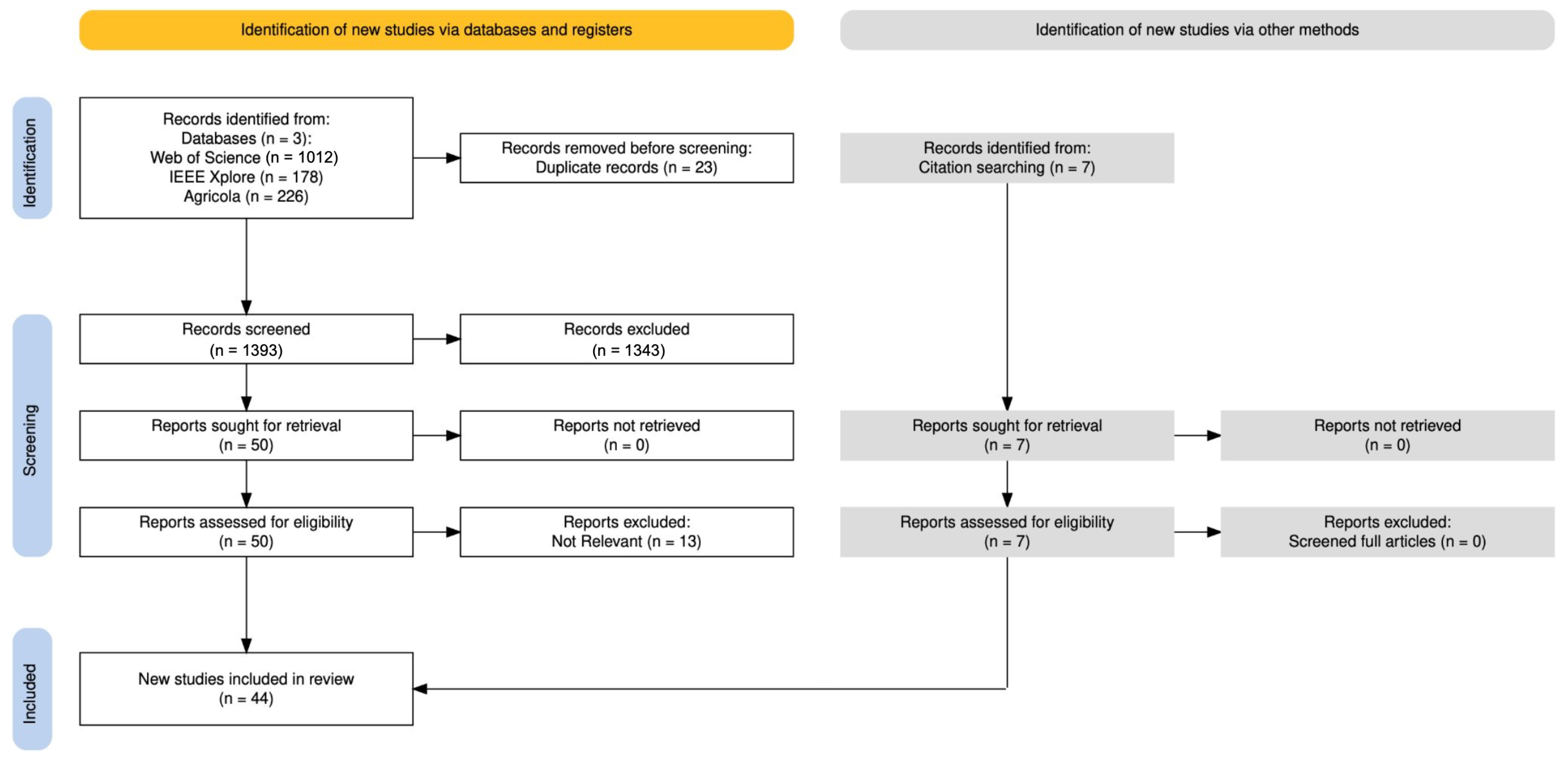
Three main phases were executed: Planning, conducting, and reporting the review process [23]. The planning phase defines the research questions, developing a review protocol, and designing a search strategy with relevant keywords. Three databases were selected for the search: Web of Science [24], IEEE Xplore [25], and Agricola [26]. In the conducting stage, a literature search was implemented using structured search queries for each specific database. Each source was last consulted on 16 May 2025. A total of 1460 articles were retrieved. A multiple-layer study selection process was implemented. The final stage encompassed synthesis and analysis of the collected data, centered around answering the research questions. This process integrated both qualitative and quantitative methods. A preliminary search was conducted across each database to identify the most relevant sources for ML-based Nematode MIA. Google Scholar was initially explored to compile a list of highly relevant journals and keywords; however, it was not included as a primary search database due to its limited export capabilities and lack of structured metadata retrieval (e.g., abstracts, authors). The primary database for this review was Web of Science, as it provided the most comprehensive collection of relevant articles. To ensure broader coverage and comparison of our topic in the AI and agricultural space, IEEE Xplore and Agricola were also included. Figure 2 shows the PRISMA flowchart for the overall process.

2.2. Search Strategy

The search strategy for this systematic review focused on retrieving relevant studies from the three major digital scientific databases mentioned in Table 1. Initially, exploratory search strings were tested across multiple databases, including Google Scholar, to refine keyword selection. The search process began by focusing on “organism-related” and “computational methods” terms in article titles; for instance, (Nematode OR Worm) AND (“Machine Learning” AND “Artificial Intelligence”). To ensure full coverage of the literature related to nematode inference, the search was expanded to include detection, segmentation, and classification inference tasks: (Nematode OR Worm) AND (“Machine Learning” AND “Artificial Intelligence”) AND (“Detection” AND “Classification” AND “Segmentation”). However, using the expanded search query limited the number of retrieved studies, yielding 189 using Google Scholar, 69 in Web of Science, and 14 in Agricola. The strategy was broadened by including only “organisms” and “inference tasks” terms, allowing for more inclusive search results. The term “worm” was excluded as it frequently returned irrelevant studies on malware worms, email worms, and AI cybersecurity applications. Instead, “C. elegans” was explicitly incorporated, as it is the most studied nematode species in microscopy image analysis research. Additionally, seven studies from Google Scholar were manually included as they were directly relevant to the research questions but did not appear in the primary databases used in this review. The final search keywords were structured using the search query detailed in Table 1.
This review restricted the publication years to 2018–2024 and included only English-language articles. The database search was conducted on 25 December 2024, across all three databases. The Web of Science and Agricola searches in Table 1 were restricted to Title-only (TI) queries to ensure high specificity in the studies. IEEE Xplore was intentionally queried without restrictions (e.g., Title-only), in order to broaden the scope and capture all the relevant ML research related to our topic. After executing the search queries, a total of 1416 articles were returned and screened based on predefined inclusion and exclusion criteria to ensure their relevance to the research objectives.

2.3. Selection Criteria

The selection criteria helped to identify articles that answered the research questions. These criteria were based on the SLR objectives and centered around the research questions. All selected studies were saved in a repository and recorded in a spreadsheet. An article was selected if it included the following criteria: (1) it is related to nematode analysis, (2) it makes use of ML/DL techniques in nematode image analysis research, and (3) it was published within the last 7 years. The exclusion criteria include: (1) the publication is a survey or review paper, (2) the publication is not available in English, and (3) the publication is not focused on nematodes and is not related to detection, classification, segmentation, morphological analysis, and so on using ML/DL techniques.

2.4. Selection Process

The chosen queries produced a total of 1416 articles, and 7 further articles were selected from Google Scholar as they were also considered relevant for inclusion in this study. The study selection process followed the PRISMA framework to ensure a rigorous and transparent methodology. Duplicate records were manually removed and irrelevant studies were excluded, including those focusing on Tree Nematode ML/DL models, Worm Malware, Worm Gear Neural Network, and Convolutional Neural Network (CNN) models trained on Nematode Connectome data. Two reviewers independently screened each article at the title, abstract, results, and/or full-text levels, with disagreements resolved through discussion and consensus or a third reviewer. This process ensured alignment with research objectives. Only studies explicitly focused on ML-driven nematode MIA tasks for detection, segmentation, and classification were included. Metadata were systematically recorded for each of the included studies.

2.5. Data Extraction Process

To systematically address the research questions, a structured data extraction framework was implemented. Each selected study was thoroughly reviewed, with key methodological and performance-related details recorded. A question-based Google Form was designed to standardize data collection, ensuring consistency across all studies. Data extraction was performed independently by the three main authors, with each reviewer responsible for a subset of the 44 included studies (16, 15, and 14 articles, respectively). To ensure accuracy, the data extracted for each study were cross-checked by one of the other two reviewers who had not originally collected data for that article. This framework captured essential metadata, methodological details, inference tasks, model architectures, and performance metrics. Each study was evaluated for dataset and code availability, as well as reported challenges, allowing for the synthesis of trends and research gaps in ML-based nematode microscopy image analysis. No study investigators were contacted to obtain or confirm additional data. Table 2 summarizes some of the collected data.
In accordance with the PRISMA 2020 guidelines, the primary outcomes of interest were the performance metrics. These metrics were used to evaluate ML/DL models for nematode microscopy image analysis (e.g., accuracy, precision, recall, F1-score, Intersection over Union (IoU), Dice coefficient, and average precision). When multiple metrics were reported, all were included in this study. Accuracy and recall were the most considered performance metrics, as they were frequently reported. To provide context for these outcomes, additional variables such as metadata, model type and architecture, inference tasks, data information, and code availability were extracted, as summarized in Table 2. Categorizing the inference tasks in each paper helped to map the range of MIA objectives targeted across studies. Model architectures were categorized to identify top-performing frameworks, offering insight into architectural preferences among researchers. Dataset information and code availability were collected to assess dataset reproducibility and accessibility for replication. It was also recorded whether authors reported challenges, limitations, or future research directions. When information was missing or unclear (e.g., dataset split, code availability), it was recorded as not reported rather than inferred.

2.6. Other Methods

The reviewed studies were not subjected to a standardized risk of bias tool (e.g., QUADAS-2 or RoB-2); instead, three reviewers qualitatively evaluated potential biases in dataset design, code/data availability, and reporting clarity, resolving internal disagreements through discussion. Randomness in model training and the lack of cross-study replication further complicated efforts to apply formal bias or certainty assessment between studies. The primary effect measures extracted were model performance metrics that are commonly used in the context of image analysis (see Section 2.7 Performance Metrics). These measures were synthesized narratively rather than pooled meta-analytically, as differences in datasets, task definitions, and reported metrics precluded quantitative aggregation. This narrative method involves the textual summary and interpretation of findings from the included articles, which were organized thematically to identify the key patterns, data trends, and research gaps.
Studies were included in each synthesis if they reported at least one performance metric relevant to nematode detection, segmentation, tracking, or classification. To prepare the data for synthesis, missing or unclear details (e.g., dataset splits, code availability) were recorded as “not reported” without restraint. The results were summarized through structured tables and figures (e.g., task distributions, model architectures, geographic trends), in order to facilitate comparison across studies. No meta-analysis, subgroup analysis, or sensitivity analysis was conducted, as variations across models and outcomes were substantial. Heterogeneity was noted by comparing results across ML and DL model families, dataset types, and reported inference tasks.
Reporting bias was assessed indirectly by noting the absence of dataset availability or selective reporting of performance metrics, which limited reproducibility in several studies. Certainty in the body of evidence was therefore judged qualitatively: while CNN- and YOLO-based models consistently showed strong results across multiple tasks, the overall confidence in generalizability remains moderate due to dataset limitations and inconsistent reporting standards.

2.7. Performance Metrics

The reviewed studies employed a variety of performance metrics, depending on the image analysis task. To compare ML models, it is necessary to understand their respective evaluation metrics. For nematode detection, researchers applied well-known performance metrics such as accuracy, precision, recall, and F1-score. For segmentation, researchers commonly used Intersection over Union (IoU), Jaccard Index, Dice Coefficient, average precision (AP), and F1-Score as well. In this section, some of the most common metrics reports are briefly described.

2.7.1. Standard Classification Metrics

The metrics presented in this section are derived from the true positives (TP), true negatives (TN), false positives (FP), and false negatives (FN). The corresponding formulas can be found below:
Accuracy = T P + T N T P + T N + F P + F N , Precision = T P T P + F P , Recall = T P T P + F N , F 1 - Score = 2 · Precision · Recall Precision + Recall .
All of these values range between 0 and 1, and a higher value indicates better performance. Accuracy measures how often a model correctly classifies samples. However, it can be a misleading measure when class imbalance is present in a dataset [28]. Precision quantifies the proportion of true positives (TP) among all predicted positives, making it especially valuable in imbalanced datasets where false positives (FP) must be minimized. Recall measures a model’s ability to correctly identify all positive classes, making it essential for evaluating binary segmentation and classification performances [29]. F1-Score is one of the most commonly used performance metrics in machine learning for binary and multi-class segmentation and classification. It is best-known as the harmonic mean of precision and recall, balancing both metrics [30].

2.7.2. Additional Metrics

Intersection over Union (IoU), also known as the Jaccard Index, is widely used in object detection and segmentation to measure the overlap between predicted and ground truth regions. It is defined as the ratio of the intersection to the union of the predicted and actual segments, making it scale-invariant and well-suited for microscopic inference tasks such as nematode detection [31]. Its formula is given by:
IoU = | A B | | A B | = T P T P + F P + F N
where A represents the prediction region, B the ground truth, and | · | represents the area of the region.
The Dice Coefficient (DC) is a commonly used evaluation metric in image segmentation to quantify the overlap between the predicted shape and ground truth regions. It is defined as the ratio of twice the intersection over the total number of pixels in both sets:
DC = 2 · | A B | | A | + | B | 2 · T P 2 · T P + F P + F N .
A dice score of 1 indicates perfect overlap, while 0 indicates no overlap. It is especially useful for assessing quality of pixel-level segmentation in nematode inference tasks [32].
Average precision (AP) measures overall model performance by computing the area under the precision–recall curve [33]. It captures the trade-off between precision and recall across varying confidence thresholds [34,35]. AP is typically approximated by the discrete summation:
AP = n ( R n R n 1 ) P n ,
where R n and P n are the recall and precision at the n-th threshold [34]. The PASCAL VOC benchmark introduced AP@0.5 using interpolated precision, which smooths the curve by selecting the highest precision at or above each recall level [35]. In contrast, the COCO benchmark extended this by averaging APs over IoU thresholds AP@[0.5–0.95] [36]. Both metrics are commonly used in the context of nematode image analysis.
For all these metrics, the values range between 0 and 1, with higher values indicating better performance.

2.8. Machine Learning Models

As mentioned in the Introduction, the recent decades have seen exponential growth in the use of deep learning models for image analysis tasks. Before these developments, classical machine learning methods would make use of hand-crafted features extracted from images, defined manually by experts, as inputs to a model that would turn vectors into a classification output. Classification models such as Support Vector Machine (SVM), Random Forest (RF), and artificial neural networks (ANNs) [37] fall in this category. However, thanks in part by the progress in computer hardware and the creation of large datasets enabled by the Internet, deep neural networks (DNNs) that can operate directly on the image domain became popular. These DNNs were the starting point of the deep learning revolution, and are capable of learning features directly from a dataset without the need for hand-crafted features. At present, there exist a large number of pre-trained models with different architectures that can be fine-tuned for many applications. Some of the standard architectures include Convolutional Neural Networks (CNNs), AutoEncoders (AEs), and Transformers [29]. Going over the details of all these architectures is beyond the scope of this article, but we provide some highlights on common architectures found in our survey in Appendix A.

3. Results

The database searches yielded a total of 1423 unique records, of which 1416 were screened based on their titles and abstracts. Following full-text assessment for eligibility, 44 studies were included in the final review (see the PRISMA flow diagram in Figure 2). Some papers that initially appeared to meet the inclusion criteria were excluded because one was a systematic review, another a Master’s thesis, and the rest either did not use appropriate imaging techniques or were not related to AI-based techniques [5,6,38,39,40,41,42,43].
Substantial heterogeneity was observed across the included studies in terms of study designs, dataset composition, and reported outcome metrics. Consequently, quantitative meta-analyses were not performed, and pooled summary estimates, precision intervals, or measures of statistical heterogeneity could not be generated. Similarly, subgroup analyses, formal investigations of heterogeneity among study results, and sensitivity analyses were not conducted due to incomplete and non-standardized reporting across studies. No formal assessment of reporting bias was made, as comparable effect sizes were rarely available. Nevertheless, the prevalence of high-performance results, coupled with limited dataset accessibility and small sample sizes, suggests that selective reporting cannot be excluded. A formal certainty-of-evidence framework, such as GRADE, was also not applied. Overall, confidence in the synthesized evidence was considered to be low to moderate, reflecting heterogeneous methods, small sample sizes, and incomplete reporting across studies.
This section covers the various tasks performed in the selected papers and provides a summarized view of recent research performed for the analysis and processing of nematode assays. Section 3.1 highlights the trend in the publications, Section 3.2 compares performances across the different image analysis tasks, and Section 3.3 provides an overview of trends on the biological inference tasks.

3.1. Publication Trends

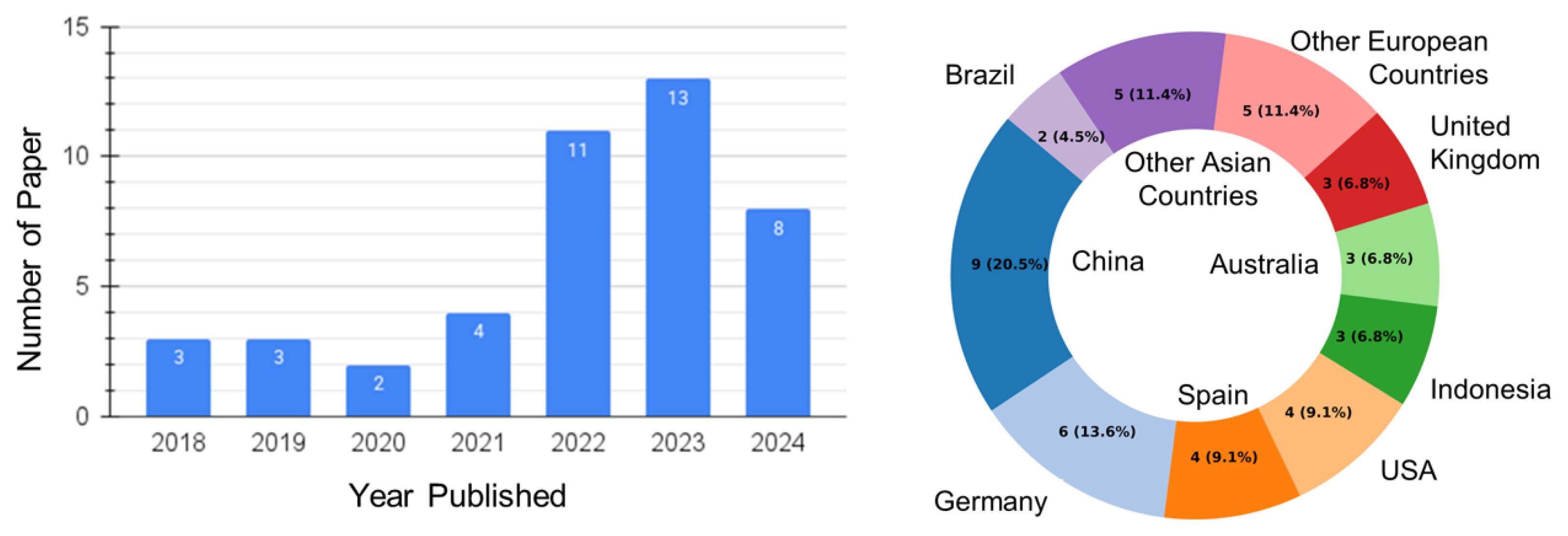
This SLR adopted a narrative synthesis approach instead of a meta-analysis due to the substantial differences in model architectures, dataset structure, performance metrics, which made it difficult to mathematically combine the performance results. Instead, however, this study employs structured tables and visualizations to display trends in model usage, research objectives, publication patterns, and performance outcomes. The number of publications has increased significantly since 2021 (see Figure 3 (left)), reflecting a growing interest in ML/DL models for nematode microscopy analysis. A geographic breakdown showed that China, Germany, Spain, the United States, Australia, and Indonesia were the most active contributors by country (see Figure 3 (right)), pointing to regional hubs of innovation and investment in agriculture ML applications. China and Germany lead the field in publication output.
As can be observed from Figure 4 (left), publication venues ranged from AI-focused to general science venues, with a total of 13 conference publications and 31 journal articles. Publication patterns revealed a wide array of venues, yet recurring appearances of Scientific Reports, Sensors, and IEEE-affiliated conferences highlight a multidisciplinary interest in the application of AI-based biological image analysis for nematodes. The citation distribution (Figure 4 (right)) shows that most studies received fewer than 10 citations; however, 9 studies stood out with 20 or more citation, and one particularly influential study reached 124 citations [44], indicating varying degrees of influence within the field over the past 7 years.
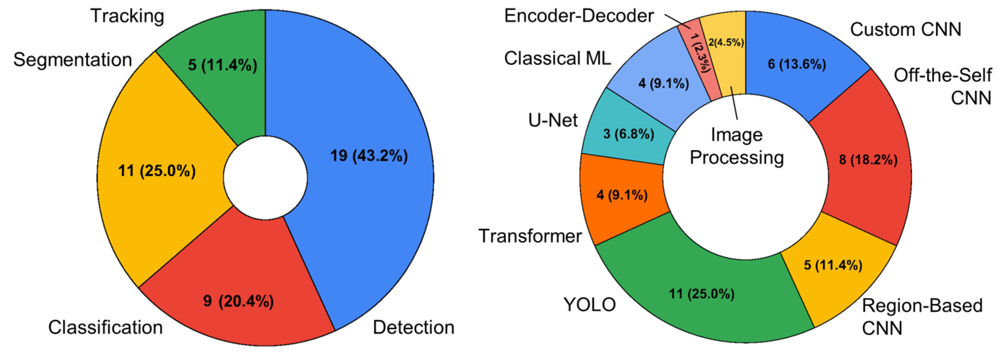
The trends in image analysis tasks are shown in Figure 5. The left plot illustrates the distribution of tasks across the different publications, from which it can be observed that detection was the most common task. The right plot illustrates the set of ML models used across all these tasks, indicating that the YOLO architecture for object detection was the most common.

3.2. Image Analysis Trends

This section summarizes the general trends found regarding the main three image analysis tasks studied; i.e., classification, detection, and segmentation. The papers associated with tracking are discussed in the biological inference task section, as there are fewer papers reporting metrics that can be compared across the board and they are more specific to the biological task at hand.
Table 3, Table 4 and Table 5 summarize the performances and datasets associated with these tasks. The “Year” column in the tables illustrates the chronological evolution of the models, helping readers to understand how model usage has progressed over time. Table 3 highlights the main classification results. Most of these results are associated with species classification from images, while one paper focused on videos [45] and one paper predicted lifespan [46]. It can be observed that the papers did not use consistent dataset sizes, with the smallest dataset including around 200 samples, largest dataset including around 50k samples, and a median of less than 1000 samples. The number of classes ranged from 2 to 19. Some papers specified the training and testing split used for evaluation, while most did not. The most common metrics are included in the table, with accuracy and F1 score having the most overlap across all the papers. Accuracy ranged from 0.69 to 0.99, and the paper including 19 classes [47] reported an accuracy of 0.94. These are relatively high values; however, due to the lack of accessibility to the datasets, it was not possible to reproduce the results for most of the models. The F1-score values ranged from 0.53 to 0.98.
Table 4 highlights the main detection results. In this case, all articles used their own custom dataset with sizes ranging from around 200 to 20 k (excluding the one obtained with data augmentation), with the only exception being [55], which used the public dataset BBBC010/11 [56]. The studies used accuracy, precision, recall, and F1-score more consistently as metrics for evaluation. The accuracy ranged from 0.57 to 0.99, precision from 0.59 to 1.00, recall from 0.68 to 0.99, and F1-score from 0.47 to 0.99. As in the previous case, relatively high scores reported were reported in several cases; however, it was not possible to reproduce or directly compare them due to the lack of accessible datasets. Over time, there is a clear trend showing that YOLO-based architectures have become increasingly dominant in recent years, reflecting the shift toward faster and more efficient object detection models.
Table 4. Detection Results.
Ref Detection
and Year Model Used Dataset: Size Train/Test Acc Prec Rec F1
[44] 2018Conv. Selective AECust: 30442435/6090.950.940.940.94
[57] 2018CNNCust: 11,82010,047/17730.93
[58] 2019HoG+SVMCust: 1200996/204 0.87
[45] 2019FCN 0.99
 [59] 2021CNN+LSTMCust: 23,58417,923/56610.91
[60] 2022YOLOv5sCust.: 19011520/3800.93
Faster R-CNN 0.94
[61] 2022YOLOv4-1 Class 0.910.790.85
YOLOv4-MulticlassCust.: 20,000 (augmented images)19,000/1000 0.930.930.92
[62] 2022Image processingN/A 0.980.980.98
[55] 2023YOLOv5sBBBC010/11 [56]: 17,21314,385/2828 0.880.76
[63] 2023YOLOv7-640Cust: 415332/830.990.990.990.99
[64] 2023YOLOv5Cust:13k (augmented)10,400/2600 1.000.990.99
[65] 2023Faster R-CNNCust.: 182163/190.860.940.830.88
YOLOv4-CSP 0.770.910.800.85
[66] 2023YOLOv7Cust.: 39313537/3940.990.970.960.97
Faster R-CNN 0.980.740.990.85
[67] 2023YOLOv5xCust: 3503- 0.850.75
[68] 2023Faster R-CNNCust: 16251413/2120.870.610.68
[69] 2024CNN and contour methodsCust: 10,468 0.99
[70] 2024YOLOv8Cust: 19,92915,677/42520.570.590.940.47
[71] 2024YOLOv5Cust: 894788/106 0.780.78
[72] 2024YOLOv5Cust: 6000 0.970.940.95
[73] 2024HoG+SVMCust: 20,647 0.910.920.970.95
Table 5 highlights the main segmentation results. In contrast to the detection studies, most segmentation works relied on publicly available datasets (Table 6), with several articles utilizing common datasets (D1–D5), thus enabling more consistent benchmarking across studies. The most frequently used models included U-Net, Mask R-CNN, and transformer-based architectures, with the Dice coefficient (DC) and AP50 being the most commonly reported evaluation metrics. Some studies also reported Precision and Recall values. Custom datasets (C1–C5) generally resulted in lower performance, particularly with conventional architectures such as U-Net. The highest results were consistently achieved by transformer-based models, reaching up to 0.99 on CSB-1 and 0.98 on the Mating Dataset. Despite these strong performances, not all studies provided clear details about the number of dataset images or train/test splits, which limits reproducibility and direct comparisons. Two segmentation papers [46,74] were not included in Table 5, as their reported evaluation metrics were not comparable with those adopted in our segmentation results table.
Table 5. Segmentation Results.
Ref Segmentation
Data and Year Model Used DC AP50 Prec/Rec @ 0.5
C4[75] 2019Image proc.0.76
C6[13] 2020U-Net 0.90/0.87
D1[76] 2021Mask R-CNN 0.9
C1[77] 2022U-Net0.46
C2 0.47
C3[78] 2022Mask R-CNN 0.89
D2[18] 2023Transformer-WormSwin 0.98
D3[33] 2024Mask2Former 0.62
D4[13] 2020U-Net 0.96/0.97
[18] 2023Transformer-WormSwin 0.95
[33] 2024Mask2Former 0.92
[79] 2024Mask2Former 0.71
D5[18] 2023Transformer-WormSwin 0.99
[79] 2024Mask2former 0.87
D3[33] 2024Multiple Instance Learning 0.65
D4 0.92
Table 6. Segmentation Datasets.
IDName and RefNo. of ImagesTrain/Test SplitAvail.
D1C. elegans [80]19081711/197Yes
D2Mating Dataset [18]450 patch images0/450Yes
D3Haemonchus Contortus [17]9576/19Yes
D4BBBC010 [56]100N/AYes
D5CSB-1 [18]4631 (10 videos)N/AYes
C1Custom Nematode Dataset [77]39673367/667No
C2Custom Cyst Dataset [77]487387/100No
C3Custom Nematode Dataset [78]16k14,880/1120No
C4Custom Cyst Dataset [75]132N/ANo
C5Potato Cyst Nematode [13]19731606/367No
Some papers addressed multiple tasks explicitly and, therefore, appear in more than one category. For example, ref. [46] used separate models for classification and segmentation. Thus, while it is shown under segmentation in Figure 5, it is also listed in Table 3. Likewise, ref. [45] is categorized under detection in Figure 5 but is also included in Table 3. Similarly, ref. [65] is grouped under tracking in Figure 5, yet it is also listed in Table 4.
CNNs were the most frequently used architectures across the 44 selected studies, appearing in 14 implementations and showing high adaptability across tasks such as classification, detection, and segmentation. YOLO-based models followed in adoption (12 studies), with YOLOv7 achieving the best performance for real-time species classification (99.56% mAP@0.5), with other studies also performing tasks in tracking and counting. Transformer-based models (4 studies) demonstrated exceptional accuracy for instance segmentation and counting when data were synthetically augmented. For instance, WormSwin reached 99% AP@0.5. R-CNN variants (5 studies) showed strong spatial localization but incurred higher computational cost. U-Net and encoder–decoder architectures (4 studies) performed moderately in segmentation tasks, while the Convolutional Selective Autoencoder (CSAE) achieved over 94% F1-Score for nematode egg counting. Finally, classical ML models (4 studies), including HOG+SVM pipelines, proved highly effective for nematode counting tasks (up to 95% F1-Score) but underperformed in segmentation applications.
Across the models, performance generally correlated with task complexity. YOLOv7 delivered the highest results, achieving 99.56% mAP@0.5 and 52.5 FPS, outperforming Faster R-CNN in both speed and accuracy for species classification. Transformer-based models such as WormSwin excelled in dense segmentation and counting tasks using synthetic data, while classical ML pipelines such as the HOG+SVM ML model remained effective for simple detection tasks. However, U-Net models showed limited segmentation accuracy and R-CNN variants incurred slower inference times, exceeding 43 ms per prediction.
Given the considerable heterogeneity in study design and inconsistent reporting, a formal risk-of-bias tool was not utilized. Many studies lacked essential details regarding dataset composition, annotation protocols, and train/test splits, which hindered a structured evaluation. Thus, the risk of bias across studies is deemed unclear to high, and the results should be interpreted with caution.

Datasets

The nematode public dataset shown in Table 3 focuses primarily on behavioral tracking, genetic diversity, lifespan analysis, and species recognition. The behavior dataset presented by Javer et al. [49] is among the most extensive resources, comprising nearly 15,000 single-worm tracking experiments across hundreds of genotypes, including over 2700 videos from the L4 larval stage to death for aging studies. The CeNDR dataset [50], in contrast, is not an image repository but a critical community resource that provides genetically diverse wild C. elegans strains, allowing for population-level genetics and strain-specific imaging experiments. A complementary microscopy dataset available on the BioImages Archive [52] offers wide-field time-lapse images of C. elegans annotated for lifespan, movement, and body part segmentation. Finally, the I-Nema dataset [16] extends beyond C. elegans, providing an open access dataset benchmark for species-level recognition with more than 2700 images representing 19 different species collected in both natural and laboratory environments.
Complementing the previously described datasets, the public datasets of nematodes in Table 6 differ considerably in species focus, imaging conditions, annotation strategies, and experimental design, which directly affects model performance and reproducibility. For example, the DIY microscope dataset by Fudickar et al. [80] provides high-resolution (3280 × 2464 px) Petri dish images of C. elegans acquired with a Raspberry Pi-based system, enabling affordable large-scale data collection but with variability in illumination. In contrast, the dataset used in WormSwim [18] includes three complementary sets: the CSB-1 dataset, which contains video-derived frames of C. elegans with pixel-level binary masks; a Synthetic dataset generated by compositing segmented worms onto background images to expand training diversity; and the Mating Dataset (MD), consisting of 450 dense, overlapping grayscale patches designed to challenge instances in segmentation under crowding conditions. Beyond C. elegans, Zofka et al. [17] introduced a dataset of Haemonchus Contortus (an animal-parasitic nematode) motility assays, pairing manually annotated microscopic images with motility videos across live–dead ratios. Lastly, the BBBC010 benchmark set [56] provides curated microscopy images of C. elegans subjected to pathogen and antibiotic treatments, annotated with foreground–background segmentation masks and biological phenotype (dead/alive) labels. Together, these datasets highlight key differences: controlled versus low-cost imaging setups, real versus synthetic data generation, single versus multiple nematode species, and annotations ranging from binary masks to biological treatment outcomes. The BBBC10 benchmark was also used in Table 4, together with the BBBC11 benchmark found in [56]. The difference in datasets here underscores the importance of standardized benchmarks such as CSB-1 and BBBC010 for fair model evaluation and reproducibility.

3.3. Biological Inference Trends

Table 7 shows the distribution of the biological inference tasks across all the publications. The number of papers do not add up to 44, as some papers addressed multiple biological tasks. It can be observed that the majority of the articles focused on species classification and counting tasks, while lifespan tracking was present in a relatively smaller percentage of articles. The rest of this section highlights some of the trends observed regarding the biological inference tasks.

3.3.1. Species Classification

Species classification is one of the most researched tasks for nematode microscopic image inference, as determined by this systematic review. The first major approach to species classification using DL was presented by Uhlemann et al. [7], who used CNNs to identify nematodes based on the variations present in the dataset of images to train where nematodes are classified.
Researchers have made use of high-magnification and high-resolution images to focus on specific areas of interest on the nematode’s body, such as the head, tail, and genitalia [8,11,15,47]. This approach focuses on the fact that nematode species tend to express most species-level differences around these areas, especially in adults. The use of close-up images led most researchers to focus on using “off-the-shelf” CNN architectures for training models, while sidestepping challenges associated with nematode detection in complex visual conditions such as low-lighting, high debris, occlusion, and overlapping worms.
The current state-of-the-art (SOTA) species classification model, developed by Qayyum et al. [53], uses close-up images and train a majority voting Vision Transformer with test time augmentation—a method that augments the test data during evaluation to get average predictions of the variations of the test data—to perform species classification across 19 classes with overall accuracy of 83%, precision of 82.%, and recall of 81%. This method still requires further validation with more complex datasets; however, it is a promising approach based on the improved performance of vision transformers over CNNs [81,82,83]. Abade et al. [15] also developed a SOTA model using NemaNet—a custom CNN based on a combination of DenseNet121 and InceptionV3—and close-up images obtained using traditional imaging techniques. NemaNet was trained to classify 5 different plant-parasitic nematodes with an accuracy of 98.82% and precision of 98% [15]. A similar approach was evaluated by Verma et al., who used InceptionV3 to classify two nematode species: Acrobeles and Acrobeloides [54].
A variety of modifications have been made to the problem to reduce the complexity of inference, such as including pose, nematode length, and focal length. Javer et al. [45] used the pose to classify nematode species by postural dynamics alone using a model inspired by a Dilated ResNet architecture, and made their dataset public [49]. They found that using the worm angle is the most effective feature for identifying species based on postural dynamics. Another approach is to focus on nematodes in the infective juvenile (IJ) stage, as they can be more easily classified by their body length [7,63,70,71]. Finally, some researchers have combined custom computer vision algorithms with DL models to produce effective results. One such example is that of Thevenoux et al. [51], who introduced an algorithm that isolates the nematode head from the image background using contour detection, mathematical morphology, and a landmark-based EB-Net architecture CNN model to automate nematode classification. These kinds of algorithms may be difficult to scale to additional species due to the parameters and design of the custom computer vision algorithms, which may require redesign. Lui et al. [48] captured images of the same nematode at multiple focal lengths. Passing the image stack through a CNN, they then performed high-dimensional data analysis to classify species; however, the used techniques impose a heavy data collection burden, reducing their possibility for use in automated approaches to nematode species classification.

3.3.2. Counting

Nematode counting is a common task in handling nematode assays and samples, and having a high-performance model ultimately enables many of the more complex tasks that follow to be accurate. The models most commonly employed for nematode counting are YOLO models; however, other models have been implemented as well (see Table 4). The main challenges in nematode counting are noise handling, occlusion, and overlapping nematodes. The limitations of using non-DL techniques is that they require specific conditions for the nematodes to be detected, such as the entire nematode body being visible [64]. Many researchers have tried to create robust models to handle these challenges using YOLO models, Faster R-CNN, and Histogram of Gradients (HOG) with Support Vector Machine (SVM). Rico-Guardiola et al. [60] showed that both YOLOv5 and Faster R-CNN models performed similarly in terms of accuracy; however, the smaller version of the YOLO model, YOLOv5s, obtained better results when considering computational costs. This indicates that YOLO models may be more suited for high-speed nematode inference applications when considering the one-stage detector architecture, which is faster than the two-stage detector architecture used in other CNN models. The most accurate model was given by Pun et al. [64], who trained various YOLO models and found that the YOLOv5s model performed almost perfectly on the validation set with a precision of 100% and mAP of 99.5% under conditions such as debris and overlapped nematodes. They implemented a mosaic augmentation technique to improve the model robustness and found that it slightly improves the model performance as it models the use of varied lenses in nematode counting. Similarly, Rico-Guardiola et al. [60] obtained an accuracy of 93.2%. Mori et al. [61] addressed the problem of counting overlapping worms by making an object classification for two, three, or four overlapping worms with a comparable performance of AP@.5 at 90%, following the approach cited by Pun et al. [64]. Another technique implemented by researchers to improve model robustness when dealing with noise, occlusion, and overlapping worms is to use dataset augmentation techniques such as Style transfer, Pix2Pix, and Supervised self-labeling [55]. The Pix2Pix method allows researchers to generate synthetic images of a dataset which are capable of matching a given real appearance.
Finally, there is a small, growing body of nematode detection literature focused on counting eggs to estimate the number of nematodes—specifically, root-knot nematodes (RKNs)—within a collected sample. The studies below highlight the use of CNN-based models to estimate egg counts within microscopic images of a sample. Akintayo et al. [44] have been cited frequently in the literature, as they used a Convolutional Sparse Auto-Encoder (CSAE) model which has served as a baseline in other works; however, other approaches are emerging. The best performance reported to date is with the YOLO model, which provides an accuracy of 99.1% with images that contain debris and occlusions at an inference speed of up to 1.3 FPS for eggs and 4 FPS for nematodes [63]. Saikai et al. [70] used two YOLOv8 models to detect RKN eggs and second-stage juvenile (J2) nematodes, respectively. This paper differs in that it considers the inclusion of free-living nematodes (FLN) within the microscopic image, thereby introducing complexities to the inference task. Another method by Fraher, Watson et al. [69] is a hybrid approach employing a contour-based method and a CNN to count RKN eggs. Methods employing ML techniques yield higher performance than techniques utilizing traditional image processing techniques, such as the novel extreme point method proposed by Pun et al. [62], which achieved a detection coefficient of determination ( R 2 ) of 90.5%. The benefit of methods employing traditional image processing techniques is that they are not black-box models, such that the algorithms can be modified to account for new factors.

3.3.3. Behavioral Tracking

Another area of research has developed, which involves detecting and tracking nematodes in order to measure and evaluate their responses to various controlled stimuli. Research focused on behavioral tracking of nematodes is quite new, with all research having been published starting from 2023. A major consideration in behavior tracking research is the inference speed, which should be performant due to the requirements of real-time tracking. These algorithms are further derived for applications such as lifespan tracking, where behavior is often an important parameter [9,59].
One of the most relevant results comes from Deep-worm [10], using YOLOv5 and Strong SORT tracking backbones to detect nematodes and track their movement and predicted trajectories in real-time. They use the Strong SORT algorithm to make the model more robust to overlapping and occluded worms, and they have made the dataset publicly available for use [10]. The Deep-worm nematode tracking pipeline obtained an impressive precision of 98%, recall of 94%, and mAP.5 of 98% with a 9 min training time [10]. Ribeiro et al. [65] used a different approach, involving Faster R-CNN and ResNet18 for nematode tracking by implementing tracking algorithms (e.g., IoU Tracker, SORT, Centroid-Kalman) directly onto the raw holographic patterns. Additionally, Ribeiro et al. extracted biomechanical parameters such as nematode speed and movement patterns [65].
Some other uses of behavioral tracking include pose prediction and analysis. Castro et al. [84] developed a novel method for predicting C. elegans poses in low-resolution images of multiple worms in full 55 mm Petri dishes. The predicted poses are images of worm skeletons, and are the result of a neural network trained to predict aggregation behaviors between worms. Zhang and Chen [85] simplified the problem by implementing microfluidic chips; in particular, they proposed an algorithm for automatic counting and analysis of the body-bending behavior of nematodes for microfluidic chips.

3.3.4. Lifespan Tracking

Nematode lifespan tracking and prediction is a newer topic of research, with most of the papers published after 2021 and the majority being in 2024. Nematodes have become model organisms for understanding neuro-degenerative diseases and aging, based on their short lifespan and simple nervous system.
Dead or alive nematode classification has served as a model problem for researchers. For example, Garvi et al. [59] presented an automated system for classifying dead or live C. elegans using a ResNet18 with a unidirectional long short-term memory (LSTM) model. Additionally, they evaluated the system using an original, simulated, and mixed dataset, and showed that the mixed dataset improved the hit ratio by 3.20%, resulting in an accuracy of 83.66% for dead C. elegans and 98.56% for live C. elegans. Galimov and Yakimovich [46] used WormNet and HydraNet to show that the posterior part of the C. Elegans is most useful for classifying long-lived C. elegans, which was achieved by predicting the lifespan class of C. elegans nematodes based on young adult images (1-3-day-old adults), using HydraNet to classify age and WormNet to segment the worms into anterior, mid-body, and posterior parts. Radvansky et al. [73] are the only researchers to use ML techniques, who proposed using a HoG and SVM and tracking the nematodes over time to determine whether they are dead or alive. Finally, Bates et al. [9] demonstrated the variety of inferences possible through the use of a Faster R-CNN to detect and identify nematodes; another one to detect the stage of life they are in; and another to determine whether something is an egg, nematode, or neither. They utilized 3 different Faster R-CNN models to track nematode speeds during development, measure egg-laying rates, map spatial distribution in reproductive adults, and show behavioral decline in aging nematode populations [9].
Researchers have used custom and off-the-shelf CNN architectures for lifespan tracking, with YOLO being the most commonly used architecture in the last year. For example, Phuyued et al. [71] used a YOLOv5 model with contrast-limited adaptive Histogram Equalization (CLAHE) to improve image clarity and detect nematodes in the infective juvenile stage.

4. Discussion

4.1. Advantages

This review highlights the increasing application of deep learning methods in nematode image analysis tasks such as classification, detection, segmentation, and tracking, with a focus on the first three tasks. Biological inference tasks included species classification, counting, behavior monitoring, and lifespan tracking. We observed that YOLO-based architectures consistently performed well in detection and counting tasks, offering fast inference and high accuracy, even in visually complex environments with overlapping worms or background noise. For classification, CNN-based models and transformer architectures achieved strong results, particularly when applied to high-resolution or close-up images of key morphological regions. Transformer-based models also demonstrated superior performance in segmentation tasks, especially when applied to well-annotated public datasets such as BBBC010 and CSB-1. These findings indicate that data-driven approaches are well positioned to improve diagnostic accuracy, reproducibility, and efficiency compared with manual microscopy.

4.2. Challenges and Proposed Solutions

Despite these encouraging outcomes, there are notable limitations in the current body of research. Many studies do not provide detailed information about the dataset composition, train/test splits, or annotation protocols, which undermines their reproducibility and limits the ability to perform fair comparisons. The utilized evaluation metrics also vary across studies, with some using accuracy and F1-score, while others use Dice coefficient, AP, or mAP, further complicating benchmarking efforts. In addition, three major challenges identified in this review are detailed below.
  • Limited training data—Custom datasets are often small or inconsistently annotated. Another barrier identified in this review is the lack of standardized benchmark (SB) datasets for nematodes. In the context of ML, an SB is a common, objective framework that is used to evaluate and compare the performance of different models and algorithms. It refers to a curated and publicly available dataset with clearly defined tasks, consistent annotation standards, and well-established evaluation metrics. Solution: Wider use of data augmentation (e.g., rotation, scaling, and synthetic image generation), transfer learning, and collaborative dataset sharing can improve the generalizability of models. Many studies relied on private datasets and inconsistent metrics, making direct evaluation difficult. A prime example of the SB approach is the I-Nema dataset presented by Lu et al. [16], which provides images of 19 nematode species with a rigorous, repeatable experimental protocol. Adopting such benchmarks would directly address the challenges of reproducibility and inconsistent reporting.
  • Occlusion and overlapping objects—Worms and eggs often overlap, complicating detection and counting. Solution: Advanced models such as transformer-based detectors and instance segmentation approaches can help to resolve occluded objects more reliably.
  • Inconsistent metric reporting—Variation in reported evaluation metrics makes cross-study comparisons difficult. Solution: Adoption of standardized evaluation frameworks and transparent reporting of datasets and splits will facilitate reproducibility and fair benchmarking.
A practical consideration for applying the models reviewed in this study is their handling of large-scale, high-resolution images. CNN- and YOLO-based models are trained at fixed input resolutions, meaning that high-resolution images must be either rescaled—potentially losing critical details—or divided into smaller tiles for analysis. In practice, patch-based processing with overlapping tiles and stitched outputs is the standard approach for dealing with these large images. This allows high-resolution images to be analyzed accurately, though at the cost of additional computational effort.
This review itself is also subject to certain limitations. It focused primarily on three major databases and English-language articles, which may have led to the omission of relevant studies published elsewhere. In addition, no formal risk of bias assessment or meta-analysis was conducted; therefore, the synthesis presented here is qualitative in nature and may be influenced by reporting variations across studies.

4.3. Future Perspectives

These and future advancements in deep learning methods for nematode image analysis stand to improve plant-parasitic nematode detection and diagnostics, thus allowing nematologists and diagnosticians to improve the speed and ease with which samples are processed, while maintaining highly accurate specimen classification and counting. This, in turn, will directly support farmers and plant protection specialists in making timely nematode management decisions and avoiding yield losses. Additionally, future cloud-based imaging processing may support farmers in under-resourced regions of the world, where a nematologist or diagnostician may not be physically present to conduct manual microscopic evaluations. Expanding and sharing plant-parasitic nematode datasets and the application of image analysis programs in plant inspection facilities can assist in preventing the introduction of invasive nematode species to new areas and support regulatory policies.

5. Conclusions

Based on this Systematic Literature Review (SLR), machine learning (ML) and deep learning (DL) models offer a scalable solution for automating microscopic nematode image analysis, which has traditionally been labor-intensive and slow. A total of 44 articles were reviewed in detail. It was observed that Convolutional Neural Networks (CNNs) were the most frequently used models, with specific architectures such as YOLO excelling in detection, while Transformer-based models were more effective for dense segmentation and counting. Despite these advancements and strong performance metrics (with accuracy values reported as high as 0.99 for classification, detection, and segmentation), challenges persist—including a limited number of diverse nematode datasets for training, issues relating to occlusion, and inconsistent reporting of performance metrics across different studies. It is recommended that future work should focus on addressing these gaps.

Supplementary Materials

The following supporting information can be downloaded at: https://www.mdpi.com/article/10.3390/agriengineering7110356/s1, Document S1: Example Review Data Collection Form; Document S2: PRISMA_2020_Abstract Checklist; Document S3: PRISMA_2020_Checklist [86].

Author Contributions

J.L.J. was responsible for the Abstract, Methodology, the publication trends and dataset section in the Results, and contributed to the Introduction, Discussion, and Appendix A. P.G. was responsible for the biological inference trends in the Results section, and contributed to the Introduction and Appendix A.7. D.A. contributed to the image analysis trends discussed in the Results and Discussion, and also assisted with manuscript revisions. E.L. helped to revise and edit the manuscript and contributed to data curation. A.G. and W.Y. contributed additional content to the Introduction and Discussion sections. All authors have read and agreed to the published version of the manuscript.

Funding

This work was funded by a multi-commodity coalition, including the North Carolina Soybean Producers Association, North Carolina Sweetpotato Commission, North Carolina Cotton Producers Association, North Carolina Peanut Growers Association, Tobacco Growers Association of North Carolina, North Carolina Agricultural Consultants Association, and North Carolina Department of Agriculture and Consumer Services (NCDA&CS). Funding was provided through the NCDA&CS Program, Innovations to Advance the Agricultural Economy. Additional funding was provided by the N.C. Plant Sciences Initiative (N.C. PSI) at NC State university.

Data Availability Statement

The data collection form and PRISMA checklists used in this study are provided in the Supplementary Materials. Further inquiries can be directed to the corresponding author.

Acknowledgments

We thank the North Carolina Soybean Producers Association, North Carolina Sweetpotato Commission, North Carolina Cotton Producers Association, North Carolina Peanut Growers Association, Tobacco Growers Association of North Carolina, North Carolina Agricultural Consultants Association, NCDA&CS, N.C. PSI, and NC Farm Bureau. Their stewardship and support were instrumental in connecting NC State faculty with growers and commodity leaders to develop research priorities that are directly informed by the agricultural community. We also acknowledge Rachel Vann, N.C. PSI Platform Director for Extension Outreach and Engagement; and Lauren Maynard, Interdisciplinary Project Launch Director, for their guidance. Finally, we thank Lauren Maynard for her valuable technical assistance. During the preparation of this manuscript, the authors used AI-based tools to assist in reading and organizing of the relevant literature. However, the authors independently performed all summarization and interpretation of the information. The authors have reviewed and edited all content and take full responsibility for the accuracy and integrity of this publication.

Conflicts of Interest

The authors declare no competing interests.

Abbreviations

The following abbreviations are used in this manuscript:
AIArtificial Intelligence
MLMachine Learning
DLDeep Learning
APAverage Precision
SLRSystematic Literature Review
PRISMAPref. Reporting Items for Syst. Reviews and Meta-Analyses
C. elegansCaenorhabditis elegans
MIAMicroscopy Image Analysis
CNNConvolutional Neural Network
IoUIntersection over Union
TPTrue Positives
FPFalse Positives
APAverage Precision
mAPmean Average Precision
SVMSupport Vector Machine
RFRandom Forest
ANNArtificial Neural Network
DNNDeep Neural Network
AEAutoencoder
R-CNNRegion-Based Convolutional Neural Network
YOLOYou Only Look Once
HoGHistogram of Gradients
SVMSupport Vector Machine
RKNRoot-knot nematode
CSAEConvolutional Sparse Auto-Encoder
FLNFree-living nematode
R 2 coefficient of determination
RPNRegion Proposal Network
IJInfective Juvenile
LSTMLong short-term memory
RoIRegion of Interest
SotAState-of-the-Art

Appendix A. Top-Performing DL and ML Models

This appendix highlights the architectural trends observed for the best-performing models. Results are grouped by the type of models encountered.

Appendix A.1. Classical Machine Learning Models

Most classical machine learning (ML) models apply statistical principles to estimate complex functions computationally, often relying on manually engineered features such as texture, intensity, and shape, which are then fed into classifiers like SVMs, decision trees, or k-nearest neighbors [29,87,88]. In this review, four studies employed ML models for nematode image analysis: two focused on segmentation mask generation [74,75] and two on object counting and detection [58,73]. Segmentation-focused models achieved below 70% accuracy, while counting/detection approaches achieved stronger results, with one study reporting an F1-score of 0.88 [58] and another exceeding 92% across accuracy, precision, recall, and F1-score [73]. This indicates the relative strength of ML approaches in quantification tasks over pixel-level segmentation.
In [73], the authors combined Histogram of Oriented Gradients (HOG) feature extraction with a Support Vector Machine (SVM) classifier to detect and count live nematodes in Petri dish images. The model was trained using over 40,000 annotated image patches and validated through 10-fold cross-validation. It achieved over 92% accuracy, 92% precision, 98% recall, and a 95% F1-score, demonstrating strong generalization and offering a rapid solution for automating nematode quantification in soil assays.

Appendix A.2. Convolutional Neural Network (CNN) Models

CNNs are deep learning models designed for grid-structured data such as images, which are well-suited for object detection, segmentation, and classification due to their ability to extract multi-scale features [29]. In this review, 14 of the 44 included studies employed CNN-based architectures, making them the most frequently used approach for nematode detection and classification. The implementations varied and included off-the-shelf CNNs (e.g., ResNet101, EfficientNet), as well as custom designed CNNs (e.g., HydraNet, NemaNet).
One of the most influential papers in this category introduced a customized CNN architecture named NemaNet [15]. This model combines components from two well-established architectures—InceptionV3 and DenseNet121—for enhanced deep feature extraction. The authors incorporated a DenseNet-style structure along with multiple Inception blocks to create a hybrid network. The Inception modules enable the use of multiple filter sizes within the same layer, allowing the model to capture features at different spatial scales. These blocks also allow for control over the depth, size, and number of parameters. By leveraging this dual-architecture approach, NemaNet enhanced both flexibility and performance in nematode detection tasks.

Appendix A.3. Region-Based CNN Models

Region-Based Convolutional Neural Networks (R-CNNs) enhance standard CNNs by combining region proposal methods with deep feature extraction and classification, enabling both object identification and precise localization [89]. In this work, 5 out of 44 studies employed R-CNN-based architectures, including three using Mask R-CNN and two using Faster R-CNN. These models were applied to tasks such as segmentation, binary classification, counting, and object tracking.
In [60], the authors proposed the use of Faster R-CNN to detect C. elegans in low-resolution images captured using a Raspberry Pi-based imaging system. The model employed a ResNet-50-FPN backbone pre-trained on the COCO dataset, which was further trained over 150 epochs using Stochastic Gradient Descent (SGD) optimization and standard data augmentation techniques (e.g., vertical flip, hue-saturation-value (HSV) adjustment, scaling, and translation). Faster R-CNN achieved a high mean Average Precision (mAP@0.5) of 94.4%, but showed higher computational cost (43.25 ms per prediction) compared to YOLOv5s (6.03 ms). Despite this, the method proved highly reliable for detection tasks requiring spatial precision. This application of Faster R-CNN demonstrates its effectiveness for nematode detection and counting, offering great performance in identifying and localizing nematodes under variable imaging conditions.

Appendix A.4. YOLO Object Detection Models

The You Only Look Once (YOLO) series reframed object detection as a single regression task, with YOLOv1 predicting bounding boxes and class probabilities in one forward pass [90]. Newer versions, such as YOLOv7, have improved precision while maintaining efficiency, making them effective for high-throughput nematode detection in microscopy images [91]. In this review, 11 of 44 studies utilized a YOLO-based architecture; specifically, versions 4 through 8. These models demonstrated strong performance in a range of microscopy-based tasks for nematode analysis, including species classification, object tracking, detection, and egg/nematode counting.
In [66], the authors showed how YOLOv7 can outperform YOLOv5 and Faster R-CNN for C. elegans species classification acquired from microfluidic chips, achieving an mAP@0.5 of 99.56%. This improvement was attributed to several architecture enhancements, including multi-branch stacking modules, dense residual layers, and a spatial pyramid pooling component (SPPCSPC) which collectively enhanced multiscale feature extraction. Additional components such as transition blocks and RepConv layers contributed to improve scalability and inference speed. YOLOv7 also generalized well across external datasets, achieving 94.21% mAP and 52.5 frames per second (FPS), outperforming YOLOv5 and Faster R-CNN, which demonstrated reduced accuracy and slower speeds. These results indicate YOLOv7’s strong potential for real-time nematode detection in microscopy-based image analysis for agricultural applications.

Appendix A.5. U-Net and Encoder–Decoder Models

U-Net, introduced by Ronneberger et al. [92], is widely used in biomedical segmentation due to its ability to preserve spatial resolution, enabling precise pixel-wise segmentation even with limited training data. Encoder–decoder models map inputs to outputs by compressing and then reconstructing data, enabling tasks such as denoising or translation [29]. Unlike basic encoder–decoders, which often suffer from resolution loss, U-Net’s architecture preserves fine-grained localization through its design.
In this review, we identified three studies using U-Net models for segmentation and tracking tasks, and one study using an encoder–decoder architecture. Of these, two studies [13,77] applied U-Net for the generation of segmentation masks and achieved a dice coefficient greater than 59% and precision, recall, and AP@0.5 greater than 84%, respectively. The next U-Net study, [84], combined tracking and segmentation, reporting more than 74.5% in precision and recall and an IoU of 63%. Finally, an encoder–decoder model—namely, Convolutional Selective Autoencoder (CSAE)—was used for egg counting [44]. This model demonstrated strong performance in all the metrics evaluated, with accuracy, precision, recall, and F1-score values exceeding 94%. The U-Net models studied in these works showed limited effectiveness for segmentation tasks, with only moderate performance observed. However, they showed relatively stronger results in detection-related use-cases.
In [44], the authors proposed a Deep Convolutional Selective Autoencoder (CSAE) architecture, which was designed to detect and count soybean cyst nematodes (SCN) in complex, debris-filled microscopy images. Structured as an encoder–decoder architecture, the model’s encoder extracts salient features from image patches, while the decoder selectively reconstructs only SCN egg regions, masking background noise in the process, a mechanism termed “selectivity.” This is achieved through patch-wise training on expert-annotated egg-containing images, enabling the CSAE to distinguish between nematode eggs and visually similar debris. When tested on 24,000 images, the model achieved an accuracy of 95% and an F1-score of 94%, outperforming conventional image processing methods that required extensive manual tuning and exhibited high false-positive rates. The CSAE achieved detection performance comparable to human experts across both high- and low-occlusion scenes, supporting its use in high-throughput nematode detection. Nonetheless, the authors acknowledged a trade-off due to overlapping patch analysis, especially in highly cluttered environments.

Appendix A.6. Transformer Models

Transformer-based models use self-attention mechanisms to capture global dependencies without recurrence or convolution, enabling faster and more scalable training. Originally developed for Natural Language Processing, they have been adapted for vision tasks such as image segmentation and object detection [93]. This review identified four studies employing transformer-based architecture [18,33,53,79]. Three of these focused primarily on segmentation mask generation, with one of them also incorporating object counting and tracking tasks [18]. The remaining study [53] applied a transformer model exclusively for species classification. Among these, ref. [18] reported high performance across tasks, achieving 99% AP@0.5 overall and 96% AP@0.5 on the benchmark dataset BBC010, positioning it as one of the top-performing studies in terms of both segmentation and detection. Similarly, ref. [33] evaluated their model on the same dataset, reporting 92% AP@0.5. In contrast, ref. [79] obtained moderate results, with an AP@0.5 of 71% on BBC010. The classification-focused transformer model in [53] achieved moderate performance, with accuracy, precision, recall, and F1-score ranging from 81% to 83%. These findings indicate that while transformer-based models show strong potential for segmentation and detection, their effectiveness varies across tasks and datasets.
In [18], the authors presented WormSwin—a hybrid model combining the Hybrid Task Cascade (HTC) architecture with a Swin Transformer backbone for instance segmentation of C. elegans. Designed to extract single-worm postures from multi-worm microscopy recordings, the model incorporates Group Normalization with Weight Standardization, replaces Shared2FC bounding box heads with Shared4Conv1FC heads, and uses Soft Non-Maximum Suppression to better detect overlapping worms. The Swin Transformer’s shifted window attention and hierarchical feature maps enable detailed segmentation of high-resolution images. Trained on synthetic frames from the CSB-1 dataset, WormSwin achieved 0.99% AP@0.5 for both bounding boxes and segmentation masks, with strong generalization to the BBBC010 and MD benchmarks, making it well-suited for scalable, accurate nematode detection in the context of agricultural imaging.

Appendix A.7. Segmentation Models

Nematode segmentation is another area of active research within nematode MIA, due to the rich information provided by the segmentation mask. Much of the motivation with regard to automated nematode segmentation is focused on developing better tools for genetic studies, drug testing, ethology, nematode behavioral tracking, and nematode processing [10,18,33].
Researchers have utilized various techniques to generate segmentation masks, such as creating image segmentation and nematode skeletonization algorithms, implementing HoG with SVM techniques, and developing and training DL methods such as U-Net or transformer models.
The use of non-DL techniques is quite sparse in the literature; however, there are a few examples. Chen et al. used a custom algorithm called Local Maximum of Boundary Intensity, which works by taking a region of interest (RoI) and expanding it to cover the entire area of an object in an image, then classified the object using an SVM [75]. Lehner et al. segmented nematodes using image processing techniques and ML [74]. However, implementing fluorescence processing on nematodes takes additional time and resources. Finally, compared to DL models, non-DL techniques are difficult to modify, not robust, and tedious to tune compared to DL techniques, which tend to be more robust and are easier to develop.
Two-shot segmentation CNN models provide another avenue for automating nematode segmentation, which often achieve higher precision at the cost of increased computation [18]. One approach proposed by Chen et al. [13] involves using a U-Net model to automate nematode segmentation. In order to solve the issues of detecting nematodes with overlapping and occluded poses, the U-Net model was modified by adding two additional branches for predicting worm skeleton and body endpoints along with detecting worm-shaped objects in microscopic images. Chen et al. showed, in a separate article [77], that modification to the U-Net model with Global Weighted Pooling and superpixel voting resulted in finer segmentation of the nematodes. Another two-shot model approach involves the Mask R-CNN model, which some researchers have assigned as future work [9,60,63] and others have already implemented [76,78,94]. One application of the Mask R-CNN model is to develop a cheaper way for obtaining nematode masks with a DIY microscope [76]—a major improvement over using the SVM for counting nematodes [58]. Some improvements in Mask R-CNN performance have been obtained by modifying the model to use a Pr Rol Pooling layer instead of the RoI align layer, and implementing an improved NMS method to reduce missed and false detections of nematodes [78].
Vision Transformer models are the newest approach for performing nematode segmentation. All of the papers involving transformer models have been published in the last two years, with the majority published in the last year. One noticeable trend detected in this SLR is that transformer models have only been used for nematode segmentation tasks. One reason for the shift in the model type used may be due to the fact that it has recently been demonstrated that Transformer models perform better for object detection [81] and segmentation [82,83] than CNN-based models. WormSwin—a state-of-the-art vision transformer model for nematode segmentation—was the best-performing model, with an 99% AP@.5 for bounding boxes and segmentation masks on CSB-1 (a popular nematode dataset) and similar performance on BBBC010 (another popular nematode dataset) [18]. This model uses the Swin Transformer architecture combined with Hybrid Task Cascade for segmentation and to accurately handle occlusions and overlapping worms. Another transformer model is Mask2former, utilized by Cheng et al., which incorporate a semantic guidance structure to address the problem of overlapping nematodes with an associated loss calculation module to segment nematodes [79]. Zhang, Xu, Zhang, et al. [33] proposed to address the occlusion problem in nematode segmentation by integrating mask prediction and skeleton points prediction. This approach enhances the contextual information, enabling more precise separation of overlapping nematodes and outperforming existing segmentation methods on benchmark datasets [33].
Finally, a wide variety of data annotation pipelines have been proposed in nematode segmentation research due to the significant data requirements of more complex models such as vision transformers. Ding et al. [94] showed that pixel-level annotations of an image containing an average of 2.8 objects requires 4 minutes, whereas bounding box annotations can be performed in less than a minute. These new approaches significantly improve annotation workflows, thereby reducing the time necessary to train a model for performing a nematode inference task. The time necessary to annotate data within nematode segmentation tasks has been significantly decreased through the use of annotation methods like line annotations [13] or image-level annotations [77]. In order to solve the data challenges associated with training large DL models, weakly supervised approaches have been developed to make data annotation more efficient. One specific implementation of weakly supervised learning is to use multiple instance learning, which classifies a collection of images as containing a label and then determines the common features that all images with the label have. Ding et al. [94] constructed and deployed a weakly supervised segmentation model using multiple instance learning, which allows for weakly supervised bounding box-level annotation, offering time savings by an order of magnitude. The segmentation branch takes the bounding box from the detection branch and feeds it into a model similar to Mask R-CNN. The segmentation branch uses multiple instance learning to group instances and then classify them [94]. The segmentation output of the model is not very reliable; however, the detection branch obtained an accuracy of 91.68 and a Dice coefficient of 70.36%.

References

  1. Jones, J.T.; Haegeman, A.; Danchin, E.G.J.; Gaur, H.S.; Helder, J.; Jones, M.G.K.; Kikuchi, T.; Manzanilla-López, R.; Palomares-Rius, J.E.; Wesemael, W.M.L.; et al. Top 10 plant-parasitic nematodes in molecular plant pathology. Mol. Plant Pathol. 2013, 14, 946–961. [Google Scholar] [CrossRef]
  2. Savary, S.; Willocquet, L.; Pethybridge, S.J.; Esker, P.; McRoberts, N.; Nelson, A. The global burden of pathogens and pests on major food crops. Nat. Ecol. Evol. 2019, 3, 430–439. [Google Scholar] [CrossRef] [PubMed]
  3. Food and Agriculture Organization of the United Nations (FAO). The State of the World’s Land and Water Resources for Food and Agriculture 2021 (SOLAW 2021): Systems at Breaking Point. 2021. Available online: https://openknowledge.fao.org/items/55def12b-2a81-41e5-91dc-ac6c42f1cd0f (accessed on 21 July 2025).
  4. Min, Y.Y.; Toyota, K.; Sato, E. A novel nematode diagnostic method using the direct quantification of major plant-parasitic nematodes in soil by real-time PCR. Nematology 2012, 14, 265–276. [Google Scholar] [CrossRef]
  5. Holladay, B.H.; Willett, D.S.; Stelinski, L.L. High throughput nematode counting with automated image processing. BioControl 2016, 61, 177–183. [Google Scholar] [CrossRef]
  6. Abdelsamea, M.M. A semi-automated system based on level sets and invariant spatial interrelation shape features for Caenorhabditis elegans phenotypes. J. Vis. Commun. Image Represent. 2016, 41, 314–323. [Google Scholar] [CrossRef]
  7. Uhlemann, J.; Cawley, O.; Kakouli-Duarte, T. Nematode Identification using Artificial Neural Networks. In Proceedings of the 1st International Conference on Deep Learning Theory and Applications—DeLTA. INSTICC, Virtual Event, 8–10 July 2020; SciTePress: Setubal, Portugal, 2020; pp. 13–22. [Google Scholar] [CrossRef]
  8. Shabrina, N.H.; Lika, R.A.; Indarti, S. The Effect of Data Augmentation and Optimization Technique on the Performance of EfficientNetV2 for Plant-Parasitic Nematode Identification. In Proceedings of the 2023 IEEE International Conference on Industry 4.0, Artificial Intelligence, and Communications Technology (IAICT), Bali, Indonesia, 13–15 July 2023; pp. 190–195. [Google Scholar] [CrossRef]
  9. Bates, K.; Le, K.N.; Lu, H. Deep learning for robust and flexible tracking in behavioral studies for C. elegans. PLoS Comput. Biol. 2022, 18, 1–24. [Google Scholar] [CrossRef]
  10. Banerjee, S.C.; Khan, K.A.; Sharma, R. Deep-worm-tracker: Deep learning methods for accurate detection and tracking for behavioral studies in C. elegans. Appl. Anim. Behav. Sci. 2023, 266, 106024. [Google Scholar] [CrossRef]
  11. Shabrina, N.H.; Lika, R.A.; Indarti, S. Deep learning models for automatic identification of plant-parasitic nematode. Artif. Intell. Agric. 2023, 7, 1–12. [Google Scholar] [CrossRef]
  12. Shabrina, N.H.; Indarti, S.; Lika, R.A.; Maharani, R. A comparative analysis of convolutional neural networks approaches for phytoparasitic nematode identification. Commun. Math. Biol. Neurosci. 2023, 2023, 65. [Google Scholar] [CrossRef]
  13. Chen, L.; Strauch, M.; Daub, M.; Jiang, X.; Jansen, M.; Luigs, H.G.; Schultz-Kuhlmann, S.; Krüssel, S.; Merhof, D. A CNN Framework Based on Line Annotations for Detecting Nematodes in Microscopic Images. In Proceedings of the 2020 IEEE 17th International Symposium on Biomedical Imaging (ISBI), Iowa City, IA, USA, 3–7 April 2020; pp. 508–512. [Google Scholar] [CrossRef]
  14. Wählby, C.; Kamentsky, L.; Liu, Z.H.; Riklin-Raviv, T.; Conery, A.L.; O’Rourke, E.J.; Sokolnicki, K.L.; Visvikis, O.; Ljosa, V.; Irazoqui, J.E.; et al. An image analysis toolbox for high-throughput C. elegans Assays. Nat. Methods 2012, 9, 714–716. [Google Scholar] [CrossRef]
  15. Abade, A.; Porto, L.F.; Ferreira, P.A.; de Barros Vidal, F. NemaNet: A convolutional neural network model for identification of soybean nematodes. Biosyst. Eng. 2022, 213, 39–62. [Google Scholar] [CrossRef]
  16. Lu, X.; Wang, Y.; Fung, S.; Qing, X. I-Nema: A Biological Image Dataset for Nematode Recognition. arXiv 2021, arXiv:2103.08335. [Google Scholar] [CrossRef]
  17. Žofka, M.; Thuy Nguyen, L.; Mašátová, E.; Matoušková, P. Image recognition based on deep learning in Haemonchus contortus motility assays. Comput. Struct. Biotechnol. J. 2022, 20, 2372–2380. [Google Scholar] [CrossRef] [PubMed]
  18. Deserno, M.; Bozek, K. WormSwin: Instance segmentation of C. elegans using vision transformer. Sci. Rep. 2023, 13, 11021. [Google Scholar] [CrossRef] [PubMed]
  19. Xing, F.; Yang, L. Chapter 4-Machine Learning and Its Application in Microscopic Image Analysis. In Machine Learning and Medical Imaging; Wu, G., Shen, D., Sabuncu, M.R., Eds.; The Elsevier and MICCAI Society Book Series; Academic Press: Washington, DC, USA, 2016; pp. 97–127. [Google Scholar] [CrossRef]
  20. Wilson, M.; Kakouli-Duarte, T. Nematodes as Environmental Indicators; CABI: Wallingford, UK, 2009; pp. 1–326. [Google Scholar] [CrossRef]
  21. Walter, T.; Couzin, I.D. TRex, a fast multi-animal tracking system with markerless identification, and 2D estimation of posture and visual fields. eLife 2021, 10, e64000. [Google Scholar] [CrossRef] [PubMed]
  22. Moher, D.; Shamseer, L.; Clarke, M.; Ghersi, D.; Liberati, A.; Petticrew, M.; Shekelle, P.; Stewart, L.A.; PRISMA-P Group. Preferred reporting items for systematic review and meta-analysis protocols (PRISMA-P) 2015 statement. Syst. Rev. 2015, 4, 1. [Google Scholar] [CrossRef]
  23. Kitchenham, B.A.; Charters, S. Guidelines for Performing Systematic Literature Reviews in Software Engineering; EBSE Technical Report EBSE-2007-001; Keele University: Keele, UK; Durham University: Durham, UK, 2007. [Google Scholar]
  24. Web of Science. Web of Science Digital Library. Available online: https://clarivate.com/webofsciencegroup/solutions/web-of-science (accessed on 12 August 2025).
  25. IEEE Xplore. IEEE Xplore Digital Library. Available online: https://ieeexplore.ieee.org (accessed on 12 August 2025).
  26. AGRICOLA. AGRICOLA Digital Library. Available online: https://www.nal.usda.gov/agricola (accessed on 12 August 2025).
  27. Haddaway, N.R.; Page, M.J.; Pritchard, C.C.; McGuinness, L.A. PRISMA2020: An R package and Shiny app for producing PRISMA 2020-compliant flow diagrams, with interactivity for optimised digital transparency and Open Synthesis. Campbell Syst. Rev. 2022, 18, e1230. [Google Scholar] [CrossRef]
  28. Gupta, M.; Mishra, A. A systematic review of deep learning based image segmentation to detect polyp. Artif. Intell. Rev. 2024, 57, 7. [Google Scholar] [CrossRef]
  29. Goodfellow, I.; Bengio, Y.; Courville, A. Deep Learning; MIT Press: Cambridge, MA, USA, 2016; Available online: http://www.deeplearningbook.org (accessed on 15 August 2025).
  30. Chicco, D.; Jurman, G. The advantages of the Matthews correlation coefficient (MCC) over F1 score and accuracy in binary classification evaluation. BMC Genom. 2020, 21, 6. [Google Scholar] [CrossRef]
  31. Rezatofighi, H.; Tsoi, N.; Gwak, J.; Sadeghian, A.; Reid, I.; Savarese, S. Generalized Intersection Over Union: A Metric and a Loss for Bounding Box Regression. In Proceedings of the 2019 IEEE/CVF Conference on Computer Vision and Pattern Recognition (CVPR), Long Beach, CA, USA, 15–20 June 2019; pp. 658–666. [Google Scholar] [CrossRef]
  32. Shamir, R.R.; Duchin, Y.; Kim, J.; Sapiro, G.; Harel, N. Continuous Dice Coefficient: A Method for Evaluating Probabilistic Segmentations. bioRxiv 2018. Available online: https://www.biorxiv.org/content/early/2018/04/25/306977.full.pdf (accessed on 15 August 2025). [CrossRef]
  33. Ma, T.; Zhang, K.; Xu, J.; Xu, L.; Zhang, C.; Guo, L.; Li, Y. Instance Segmentation of Nematode Image Based on Transformer with Skeleton Points. In Proceedings of the 2024 7th International Conference on Advanced Algorithms and Control Engineering (ICAACE), Shanghai, China, 1–3 March 2024; pp. 442–448. [Google Scholar] [CrossRef]
  34. Davis, J.; Goadrich, M. The relationship between Precision-Recall and ROC curves. In Proceedings of the 23rd International Conference on Machine Learning, ICML ’06, Pittsburgh, PA, USA, 25–29 June 2006; pp. 233–240. [Google Scholar] [CrossRef]
  35. Everingham, M.; Van Gool, L.; Williams, C.K.I.; Winn, J.; Zisserman, A. The Pascal Visual Object Classes (VOC) Challenge. Int. J. Comput. Vis. 2010, 88, 303–338. [Google Scholar] [CrossRef]
  36. Lin, T.Y.; Maire, M.; Belongie, S.; Hays, J.; Perona, P.; Ramanan, D.; Dollár, P.; Zitnick, C.L. Microsoft COCO: Common Objects in Context. In Proceedings of the Computer Vision—ECCV 2014, Zurich, Switzerland, 6–12 September 2014; Fleet, D., Pajdla, T., Schiele, B., Tuytelaars, T., Eds.; Springer: Cham, Switzerland, 2014; pp. 740–755. [Google Scholar]
  37. Hastie, T.; Tibshirani, R.; Friedman, J.H.; Friedman, J.H. The Elements of Statistical Learning: Data Mining, Inference, and Prediction; Springer: Berlin/Heidelberg, Germany, 2009; Volume 2. [Google Scholar]
  38. Jiménez-Chavarría, J. SegNema: Nematode Segmentation strategy in Digital Microscopy Images Using Deep Learning and Shape Models. 2019. Available online: https://api.semanticscholar.org/CorpusID:204827619 (accessed on 15 August 2025).
  39. Haque, S.; Lobaton, E.; Nelson, N.; Yencho, G.C.; Pecota, K.V.; Mierop, R.; Kudenov, M.W.; Boyette, M.; Williams, C.M. Computer vision approach to characterize size and shape phenotypes of horticultural crops using high-throughput imagery. Comput. Electron. Agric. 2021, 182, 106011. [Google Scholar] [CrossRef]
  40. Ropelewska, E.; Skwiercz, A.; Sobczak, M. Distinguishing Cyst Nematode Species Using Image Textures and Artificial Neural Networks. Agronomy 2023, 13, 2277. [Google Scholar] [CrossRef]
  41. Yuan, Z.; Musa, N.; Dybal, K.; Back, M.; Leybourne, D.; Yang, P. Quantifying Nematodes through Images: Datasets, Models, and Baselines of Deep Learning. arXiv 2024, arXiv:2404.19748. [Google Scholar] [CrossRef]
  42. Chou, Y.; Dah-Jye, L.; Zhang, D. Edge Detection Using Convolutional Neural Networks for Nematode Development and Adaptation Analysis. In Computer Vision Systems. ICVS 2017; Springer: Cham, Switzerland, 2017; pp. 228–238. [Google Scholar] [CrossRef]
  43. Zhan, M.; Crane, M.M.; Entchev, E.V.; Caballero, A.; Fernandes de Abreu, D.A.; Ch’ng, Q.; Lu, H. Automated processing of imaging data through multi-tiered classification of biological structures illustrated using Caenorhabditis elegans. PLoS Comput. Biol. 2015, 11, e1004194. [Google Scholar] [CrossRef]
  44. Akintayo, A.; Tylka, G.L.; Singh, A.K.; Ganapathysubramanian, B.; Singh, A.; Sarkar, S. A deep learning framework to discern and count microscopic nematode eggs. Sci. Rep. 2018, 8, 9145. [Google Scholar] [CrossRef]
  45. Javer, A.; Brown, A.E.X.; Kokkinos, I.; Rittscher, J. Identification of C. elegans Strains Using a Fully Convolutional Neural Network on Behavioural Dynamics. In Proceedings of the Computer Vision—ECCV 2018 Workshops, Munich, Germany, 8–14 September 2018; Leal-Taixé, L., Roth, S., Eds.; Springer: Cham, Switzerland, 2019; pp. 455–464. [Google Scholar]
  46. Galimov, E.; Yakimovich, A. A tandem segmentation-classification approach for the localization of morphological predictors of C. elegans Lifesp. Motility. Aging 2022, 14, 1665–1677. [Google Scholar] [CrossRef]
  47. Qing, X.; Wang, Y.; Lu, X.; Li, H.; Wang, X.; Li, H.; Xie, X. NemaRec: A deep learning-based web application for nematode image identification and ecological indices calculation. Eur. J. Soil Biol. 2022, 110, 103408. [Google Scholar] [CrossRef]
  48. Liu, M.; Wang, X.; Zhang, H. Taxonomy of multi-focal nematode image stacks by a CNN based image fusion approach. Comput. Methods Programs Biomed. 2018, 156, 209–215. [Google Scholar] [CrossRef]
  49. Javer, A.; Currie, M.; Lee, C.W.; Hokanson, J.; Li, K.; Martineau, C.N.; Yemini, E.; Grundy, L.J.; Li, C.; Ch’ng, Q.; et al. An open source platform for analyzing and sharing worm behavior data. bioRxiv 2018. Available online: https://www.biorxiv.org/content/early/2018/07/26/377960.full.pdf (accessed on 15 August 2025). [CrossRef]
  50. Cook, D.E.; Zdraljevic, S.; Roberts, J.P.; Andersen, E.C. CeNDR, the Caenorhabditis elegans natural diversity resource. Nucleic Acids Res. 2017, 45, D650–D657. [Google Scholar] [CrossRef]
  51. Thevenoux, R.; LE, V.L.; Villessèche, H.; Buisson, A.; Beurton-Aimar, M.; Grenier, E.; Folcher, L.; Parisey, N. Image based species identification of Globodera quarantine nematodes using computer vision and deep learning. Comput. Electron. Agric. 2021, 186, 106058. [Google Scholar] [CrossRef]
  52. EMBL-EBI BioImage Archive. Raw images of Caenorhabditis elegans with Lifespan, Movement, and Segmentation Annotations (Accession: S-BIAD300). 2022. Available online: https://www.ebi.ac.uk/biostudies/bioimages/studies/S-BIAD300 (accessed on 15 August 2025).
  53. Qayyum, A.; Benzinou, A.; Nasreddine, K.; Foulon, V.; Martinel, A.; Borremans, C.; Hagemann, R.; Zeppilli, D. A Deep Learning Vision Transformer using Majority Voting Ensemble for Nematode Classification with Test Time Augmentation Technique. In Proceedings of the 4rd International Conference on Advances in Signal Processing and Artificial Intelligence (ASPAI’ 2022), Corfu, Greece, 19–21 October 2022. [Google Scholar]
  54. Verma, M.; Manhas, J.; Parihar, R.D.; Sharma, V. A Framework for Classification of Nematodes Species Using Deep Learning. In Emerging Trends in Expert Applications and Security; Rathore, V.S., Piuri, V., Babo, R., Ferreira, M.C., Eds.; Springer: Singapore, 2023; pp. 71–79. [Google Scholar]
  55. Escobar-Benavides, S.; García-Garví, A.; Layana-Castro, P.E.; Sánchez-Salmerón, A.J. Towards generalization for Caenorhabditis elegans detection. Comput. Struct. Biotechnol. J. 2023, 21, 4914–4922. [Google Scholar] [CrossRef] [PubMed]
  56. Ljosa, V.; Sokolnicki, K.L.; Carpenter, A.E. Annotated high-throughput microscopy image sets for validation. Nat. Methods 2012, 9, 637. [Google Scholar] [CrossRef] [PubMed]
  57. Hakim, A.; Mor, Y.; Toker, I.A.; Levine, A.; Neuhof, M.; Markovitz, Y.; Rechavi, O. WorMachine: Machine learning-based phenotypic analysis tool for worms. BMC Biol. 2018, 16, 8. [Google Scholar] [CrossRef] [PubMed]
  58. Bornhorst, J.; Nustede, E.J.; Fudickar, S. Mass Surveilance of C.elegans—Smartphone-Based DIY Microsc. Mach.-Learn. Approach Worm Detect. Sensors 2019, 19, 1468. [Google Scholar] [CrossRef]
  59. García Garví, A.; Puchalt, J.C.; Layana Castro, P.E.; Navarro Moya, F.; Sánchez-Salmerón, A.J. Towards Lifespan Automation for Caenorhabditis elegans Based on Deep Learning: Analysing Convolutional and Recurrent Neural Networks for Dead or Live Classification. Sensors 2021, 21, 4943. [Google Scholar] [CrossRef]
  60. Rico-Guardiola, E.J.; Layana-Castro, P.E.; García-Garví, A.; Sánchez-Salmerón, A.J. Caenorhabditis Elegans Detection Using YOLOv5 and Faster R-CNN Networks. In Optimization, Learning Algorithms and Applications; Pereira, A.I., Košir, A., Fernandes, F.P., Pacheco, M.F., Teixeira, J.P., Lopes, R.P., Eds.; Springer: Cham, Switzerland, 2022; pp. 776–787. [Google Scholar]
  61. Mori, S.; Tachibana, Y.; Suzuki, M.; Harada, Y. Automatic worm detection to solve overlapping problems using a convolutional neural network. Sci. Rep. 2022, 12, 8521. [Google Scholar] [CrossRef]
  62. Pun, T.B.; Neupane, A.; Koech, R.; Owen, K.J. Detection and Quantification of Root-Knot Nematode (Meloidogyne Spp.) Eggs from Tomato Plants Using Image Analysis. IEEE Access 2022, 10, 123190–123204. [Google Scholar] [CrossRef]
  63. Pun, T.B.; Neupane, A.; Koech, R. A Deep Learning-Based Decision Support Tool for Plant-Parasitic Nematode Management. J. Imaging 2023, 9, 240. [Google Scholar] [CrossRef]
  64. Pun, T.B.; Neupane, A.; Koech, R.; Walsh, K. Detection and counting of root-knot nematodes using YOLO models with mosaic augmentation. Biosens. Bioelectron. X 2023, 15, 100407. [Google Scholar] [CrossRef]
  65. Ribeiro, P.; Sobieranski, A.C.; Gonçalves, E.C.; Dutra, R.C.; von Wangenheim, A. Automated classification and tracking of microscopic holographic patterns of nematodes using machine learning methods. Nematology 2023, 26, 183–201. [Google Scholar] [CrossRef]
  66. Zhang, J.; Liu, S.; Yuan, H.; Yong, R.; Duan, S.; Li, Y.; Spencer, J.; Lim, E.G.; Yu, L.; Song, P. Deep Learning for Microfluidic-Assisted Caenorhabditis elegans Multi-Parameter Identification Using YOLOv7. Micromachines 2023, 14, 1339. [Google Scholar] [CrossRef] [PubMed]
  67. Agarwal, S.; Curran, Z.C.; Yu, G.; Mishra, S.; Baniya, A.; Bogale, M.; Hughes, K.; Salichs, O.; Zare, A.; Jiang, Z.; et al. Plant Parasitic Nematode Identification in Complex Samples with Deep Learning. J. Nematol. 2023, 55, 20230045. [Google Scholar] [CrossRef] [PubMed]
  68. Angeline, N.; Husna Shabrina, N.; Indarti, S. Faster region-based convolutional neural network for plant-parasitic and non-parasitic nematode detection. Indones. J. Electr. Eng. Comput. Sci. 2023, 30, 316. [Google Scholar] [CrossRef]
  69. Fraher, S.P.; Watson, M.; Nguyen, H.; Moore, S.; Lewis, R.S.; Kudenov, M.; Yencho, G.C.; Gorny, A.M. A Comparison of Three Automated Root-Knot Nematode Egg Counting Approaches Using Machine Learning, Image Analysis, and a Hybrid Model. Plant Dis. 2024, 108, 2625–2629. [Google Scholar] [CrossRef] [PubMed]
  70. Saikai, K.K.; Bresilla, T.; Kool, J.; de Ruijter, N.C.A.; van Schaik, C.; Teklu, M.G. Counting nematodes made easy: Leveraging AI-powered automation for enhanced efficiency and precision. Front. Plant Sci. 2024, 15, 1349209. [Google Scholar] [CrossRef]
  71. Phuyued, U.; Kangkachit, T.; Jitkongchuen, D. Detection of Infective Juvenile Stage of Entomopathogenic Nematodes Using Deep Learning. In Proceedings of the 2024 5th International Conference on Big Data Analytics and Practices (IBDAP), Bangkok, Thailand, 23–25 August 2024; pp. 50–55. [Google Scholar] [CrossRef]
  72. Weng, J.; Lin, Q.; Chen, S.; Chen, W. Method for Live Determination of Caenorhabditis Elegans Based on Deep Learning and Image Processing. In Proceedings of the 2024 IEEE International Conference on Advanced Information, Mechanical Engineering, Robotics and Automation (AIMERA), Wulumuqi, China, 18–19 May 2024; pp. 62–68. [Google Scholar] [CrossRef]
  73. Radvansky, M.; Kadlecova, A.; Kudelka, M. Utilizing Image Recognition Methods for the Identification and Counting of Live Nematodes in Petri Dishes. In Proceedings of the 2024 25th International Carpathian Control Conference (ICCC), Krynica Zdrój, Poland, 22–24 May 2024; pp. 1–6. [Google Scholar] [CrossRef]
  74. Lehner, T.; Pum, D.; Rollinger, J.M.; Kirchweger, B. Workflow for Segmentation of Caenorhabditis elegans from Fluorescence Images for the Quantitation of Lipids. Appl. Sci. 2021, 11, 1420. [Google Scholar] [CrossRef]
  75. Chen, L.; Strauch, M.; Daub, M.; Jansen, M.; Luigs, H.G.; Merhof, D. Instance Segmentation of Nematode Cysts in Microscopic Images of Soil Samples. In Proceedings of the 2019 41st Annual International Conference of the IEEE Engineering in Medicine and Biology Society (EMBC), Berlin, Germany, 23–27 July 2019; pp. 5932–5936. [Google Scholar] [CrossRef]
  76. Fudickar, S.; Nustede, E.J.; Dreyer, E.; Bornhorst, J. Mask R-CNN Based C. Elegans Detection with a DIY Microscope. Biosensors 2021, 11, 257. [Google Scholar] [CrossRef]
  77. Chen, L.; Strauch, M.; Daub, M.; Luigs, H.G.; Jansen, M.; Merhof, D. Learning to Segment Fine Structures Under Image-Level Supervision With an Application to Nematode Segmentation. In Proceedings of the 2022 44th Annual International Conference of the IEEE Engineering in Medicine & Biology Society (EMBC), Glasgow, UK, 11–15 July 2022; pp. 2128–2131. [Google Scholar] [CrossRef]
  78. Liu, T.; Liu, Y.; Long, R.; Song, Y. Case Segmentation of Nematode Microscopic Image Based on Improved Mask R-CNN. In Proceedings of the CAIBDA 2022: 2nd International Conference on Artificial Intelligence, Big Data and Algorithms, Nanjing, China, 17–19 June 2022; pp. 1–5. [Google Scholar]
  79. Cheng, Y.; Xu, J.; Xu, L.; Guo, L.; Li, Y. Semantic-guided Nematode Instance Segmentation. In Proceedings of the 2024 5th International Conference on Artificial Intelligence and Electromechanical Automation (AIEA), Shenzhen, China, 14–16 June 2024; pp. 282–286. [Google Scholar] [CrossRef]
  80. Fudickar, S.; Nustede, E.J.; Dreyer, E.; Bornhorst, J. C. elegans Detection with DIY Microscope Dataset; Zenodo. 2021. Available online: https://doi.org/10.5281/zenodo.5144802 (accessed on 15 August 2025).
  81. Zong, Z.; Song, G.; Liu, Y. DETRs with Collaborative Hybrid Assignments Training. arXiv 2023, arXiv:2211.12860. [Google Scholar] [CrossRef]
  82. Cheng, B.; Misra, I.; Schwing, A.G.; Kirillov, A.; Girdhar, R. Masked-attention Mask Transformer for Universal Image Segmentation. arXiv 2022, arXiv:2112.01527. [Google Scholar] [CrossRef]
  83. Xie, E.; Wang, W.; Yu, Z.; Anandkumar, A.; Alvarez, J.M.; Luo, P. SegFormer: Simple and Efficient Design for Semantic Segmentation with Transformers. arXiv 2021, arXiv:2105.15203. [Google Scholar] [CrossRef]
  84. Layana Castro, P.E.; Garví, A.G.; Sánchez-Salmerón, A.J. Automatic segmentation of Caenorhabditis elegans skeletons in worm aggregations using improved U-Net in low-resolution image sequences. Heliyon 2023, 9, e14715. [Google Scholar] [CrossRef] [PubMed]
  85. Zhang, H.; Chen, W. Automated recognition and analysis of body bending behavior in C. elegans. BMC Bioinform. 2023, 24, 175. [Google Scholar] [CrossRef] [PubMed]
  86. Page, M.J.; McKenzie, J.E.; Bossuyt, P.M.; Boutron, I.; Hoffmann, T.C.; Mulrow, C.D.; Moher, D. The PRISMA 2020 statement: An updated guideline for reporting systematic reviews. BMJ 2021, 372, n71. [Google Scholar] [CrossRef] [PubMed]
  87. Haralick, R.M.; Shanmugam, K.; Dinstein, I. Textural Features for Image Classification. IEEE Trans. Syst. Man Cybern. 1973, SMC-3, 610–621. [Google Scholar] [CrossRef]
  88. Cortes, C.; Vapnik, V. Support-vector networks. Mach. Learn. 1995, 20, 273–297. [Google Scholar] [CrossRef]
  89. Girshick, R.; Donahue, J.; Darrell, T.; Malik, J. Rich Feature Hierarchies for Accurate Object Detection and Semantic Segmentation. In Proceedings of the 2014 IEEE Conference on Computer Vision and Pattern Recognition, Columbus, OH, USA, 23–28 June 2014; pp. 580–587. [Google Scholar] [CrossRef]
  90. Redmon, J.; Divvala, S.K.; Girshick, R.B.; Farhadi, A. You Only Look Once: Unified, Real-Time Object Detection. arXiv 2015, arXiv:1506.02640. [Google Scholar]
  91. Terven, J.; Córdova-Esparza, D.M.; Romero-González, J.A. A Comprehensive Review of YOLO Architectures in Computer Vision: From YOLOv1 to YOLOv8 and YOLO-NAS. Mach. Learn. Knowl. Extr. 2023, 5, 1680–1716. [Google Scholar] [CrossRef]
  92. Ronneberger, O.; Fischer, P.; Brox, T. U-Net: Convolutional Networks for Biomedical Image Segmentation. In Proceedings of the Medical Image Computing and Computer-Assisted Intervention—MICCAI 2015, Munich, Germany, 5–9 October 2015; Navab, N., Hornegger, J., Wells, W.M., Frangi, A.F., Eds.; Springer: Cham, Switzerland, 2015; pp. 234–241. [Google Scholar]
  93. Vaswani, A.; Shazeer, N.; Parmar, N.; Uszkoreit, J.; Jones, L.; Gomez, A.N.; Kaiser, L.; Polosukhin, I. Attention Is All You Need. arXiv 2017, arXiv:1706.03762. [Google Scholar]
  94. Ding, M.; Liu, J.; Zhao, Z.; Luo, Y.; Tang, J. WSM-MIL: A weakly supervised segmentation method with multiple instance learning for C elegans image. Phys. Scr. 2024, 99, 065043. [Google Scholar] [CrossRef]
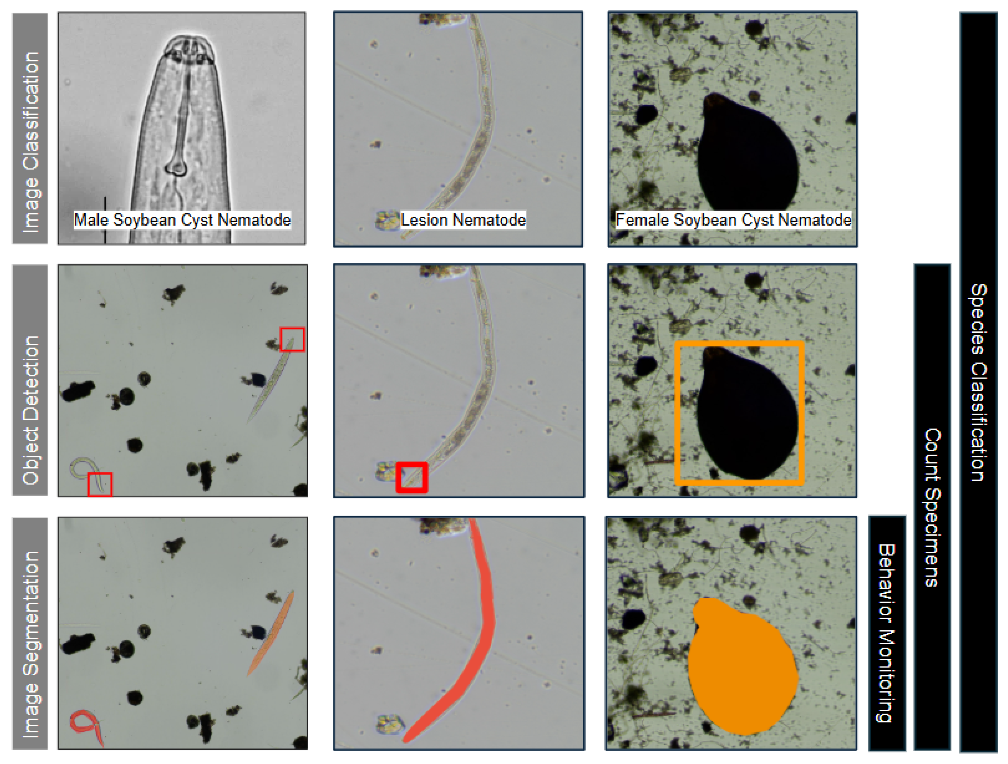
Figure 1. Illustration of the main image analysis tasks identified (left) and corresponding biological inference tasks (right). Object detection includes classification as part of its task. Similarly, image segmentation in this context also includes classification. Tracking is often combined with image segmentation in the use-cases observed. Image generated using pictures captured by our group.
Agriengineering 07 00356 g001
Figure 2. PRISMA flowchart for paper selection. Image generated using the tool of Haddaway et al. [27].
Agriengineering 07 00356 g002
Figure 3. Yearly distribution of publications (left). Geographic distribution of articles (right). Countries with three or more publications are shown individually, including China (9), Germany (6), Spain (4), USA (3), Indonesia (3), and Australia (3). Countries with fewer than three publications are grouped by region: United Kingdom (UK, Ireland), Other Europe Countries (France, Netherlands, Austria, Czech Republic), and Other Asian Countries (Japan, India, Israel, Thailand). Brazil is shown individually as the only South American country represented.
Agriengineering 07 00356 g003
Figure 4. (Left) Distribution of selected publications categorized by venue type (Conference vs. Journal) and thematic focus. This thematic grouping provides a clearer breakdown of the types of publications across the included studies. (Right) Histogram showing the distribution of citation counts among the selected articles (bin size = 5) summarizing publication impact.
Agriengineering 07 00356 g004
Figure 5. (Left) Distribution of image analysis tasks. Four primary image analysis tasks identified across the selected studies: object detection, image classification, image segmentation, and tracking. (Right) Distribution of model architectures across the selected studies. This chart illustrates the diversity of DL and ML model types applied in nematode image analysis tasks.
Agriengineering 07 00356 g005
Table 1. Search Strings and Results from Databases.
Database NameSearch StringResults
Web of ScienceTI = (Nematode OR Nematodes OR elegans OR C. elegans) AND TI = (detecting OR detect OR detection OR tracking OR track OR identifying OR identify OR identification OR classifying OR classify OR classification OR segmentation OR segment OR recognizing OR recognize OR recognition OR automating OR automate OR automation OR automated OR sorting OR sort OR counting OR count OR phenotypic OR phenotype OR phenotypes OR deciding OR decide OR decision)1012
Agricola178
IEEE Xplore226
Table 2. Data Collected in Research Papers.
CategoryData Collected
MetadataTitle, Authors, Year, Citations, Publication Type, Location (Country), Institution/University, Journal/Publisher
MethodsML/DL Model Type, Architecture, Computational and Pre-processing Techniques
Inference TasksCounting, Lifespan, Tracking, Morphological Analysis, Segmentation, Classification
Performance MetricsAccuracy, Precision, Recall, F1-Score, mAP, AP, mAP50, AP@0.5, Avg Mask, IoU, Dice Coefficient
Dataset InformationDataset Size, Availability, Splits (Train, Validation, Test)
Nematode SpeciesSpecies studied in each paper
Code AvailabilityPublicly available source code
ChallengesIdentified gaps, limitations, and proposed improvements
Table 3. Classification Results. (S) under Class indicates that the task was species-related, while (L) indicates lifespan-related.
Ref Classification
and Year Model Used Dataset: Size Train/Test Class Acc Prec Rec F1
[48] 2018Cust. SiameseCust.: 50 k-5 (S)0.93 0.94
Neural Net
[45] 2019Cust. Fully Conv. Nets (FCN)Open Worm Mov. [49] (Single): 10,476 (V)-5 (S)0.81 0.53
Open Worm Mov. [50] (Multiple): 308 (V)95 / 512 (S)0.99 0.98
[51] 2021Custom CNN ModelCust.: 390330/6020.83
[7] 2020XceptionCust.: 188-3 (S)0.88
Cust.: 234-3 (S)0.69
 [46] 2022Cust. CNN-WormNetBioImage Archive (EMBL) [52]: 734-2 (L)0.72
[47] 2022ResNet101Cust.: 257190 / 6719 (S)0.94
[11] 2022EfficientNetCust.: 957862 / 9511 (S)0.970.980.970.97
[15] 2022Cust. CNN-NemaNetCust.: 3063-5 (S)0.980.980.980.98
[53] 2022Vision TransformerCust.: 27692215 / 55419 (S)0.830.820.810.81
[54] 2023Inception V3I-Nema Dataset [16]: 277227 / 502 (S)0.9
[8] 2023EfficientNetCust.: 957-11 (S)0.960.970.960.96
Table 7. Proportion of Papers Addressing Biological Inference Tasks.
Biological Inference TaskProportion of Papers
Species Classification 14 / 44 = 0.32
Counting Specimens 16 / 44 = 0.36
Behavior Monitoring 11 / 44 = 0.25
Lifespan Tracking 4 / 44 = 0.09
Disclaimer/Publisher’s Note: The statements, opinions and data contained in all publications are solely those of the individual author(s) and contributor(s) and not of MDPI and/or the editor(s). MDPI and/or the editor(s) disclaim responsibility for any injury to people or property resulting from any ideas, methods, instructions or products referred to in the content.