Article

Biodegradation and Microbial Contamination of Limestone Surfaces: An Experimental Study from Batalha Monastery, Portugal

by
Yufan Ding
1,2,
Catia Sofia Clemente Salvador
1,
Ana Teresa Caldeira
1,3,
Emma Angelini
2 and
Nick Schiavon
1,*
1
Hercules Laboratory, University of Évora, 7000-812 Évora, Portugal
2
Department of Applied Science and Technology, Polytechnic of Turin, 10138 Torino, Italy
3
Department of Chemistry, University of Évora, 7000-812 Évora, Portugal
*
Author to whom correspondence should be addressed.
Corros. Mater. Degrad. 2021, 2(1), 31-45; https://doi.org/10.3390/cmd2010002
Submission received: 30 November 2020 / Revised: 4 January 2021 / Accepted: 8 January 2021 / Published: 13 January 2021
(This article belongs to the Special Issue Cultural Heritage Materials Degradation and Its Prevention)

Abstract

:
An experimental study was conducted to assess the nature and extent of the biodeterioration of the limestone in the Batalha Monastery in Portugal. Stone fragments covered with microbial biofilms and lichenous crusts were investigated using Optical Microscopy (OM), Low Vacuum Scanning Electron Microscopy with Energy Dispersive Spectroscopy (LV-SEM + EDS), and X-ray micro-Diffractometry (μ-XRD). Microbial samples were collected from the stone surface, cultured, and analyzed with NGS metagenomic DNA test to classify the bacterial communities associated with the formation of the biofilms. Particulate air pollutants collected on Pall GN-6 paper filters using a cascade impactor were characterized by SEM-EDS + NGS. The results showed that lichens play a major role in biodeterioration by promoting both physical and chemical attack on the limestone substrate via hyphae mechanical penetration along calcite inter-crystalline spaces, the dissolution/leaching of calcite minerals, and the precipitation of secondary minerals such as Ca-oxalates within the stone porosity framework. DNA analyses identified the bacterial communities within the biofilms and their relative abundances. Air quality monitoring results suggest that the microbial population colonizing the monastery limestone could at least partially be derived from the dry and wet deposition of airborne biological particles on the stone surfaces and that S, N, and P-rich air pollutants may have provided nutrients and energy for the bacteria communities, thus indirectly facilitating biofilm formation, the growth of a lichenous crusts, and limestone biodeterioration effects.

1. Introduction

The microbial contamination of stone surfaces in buildings and monuments leads to the formation of biofilms which are in turn responsible for physical and chemical bio-deterioration processes that may severely affect the preservation of Cultural Heritage assets in both urban and rural environments. Several types of autotrophic and heterotrophic microorganisms may colonize stone surfaces such as bacteria, fungi, algae, and lichens. Lichens may be described as an association between a fungus, usually an ascomycete (MYCOBIONT) and one or more photosynthetic partners, generally green algae or cyanobacteria (PHOTOBYONT) with the fungus forming a thallus or lichenized stroma that may penetrate deeply within the stone substrate through the action of fungal hyphae [1,2]. In recent studies, with the help of molecular assays, it was revealed that besides the photobiont algae and cyanobacteria, a high diversity of bacteria such as Proteobacteria, Actinobacteria, Acidobacteria, Bacteroidetes and Firmicutes are also associated with the lichen symbiotic system. These bacteria play multiple roles: nutrient supply (especially nitrogen, phosphorus, and sulfur), resistance against biotic and abiotic stress factors, detoxification of metabolites, provision of vitamin B12 for photosynthesis, production of hormones to support fungal and algal growth, and degradation of older parts of the lichen thallus [3]. Lichens are active agents in the biodegradation of minerals in building stone and natural stone. In a research of fungal diversity and distribution on stone surfaces in the old cathedral of Coimbra, both traditional cultivation and modern NGS (Next Generation Sequencing) techniques were used to establish a relationship between the identified fungal populations and different biodeterioration processes active at the surface of the cathedral [4]. A review paper described the microbial agents identified as responsible for stone biodeterioration processes on several famous Portuguese cultural heritage monuments in Lisbon, Coimbra, and Tomar [5]. For instance, the St. Cruz Church in Coimbra was constructed with Ançã limestone and presented various species of algae and phylum cyanobacterium (Gloeocapsa novacekii Komarek & Anagnostidis and kuetzingiana Nageli, Myxosarcina sp., Nostoc sp., Phormidium sp., Scytonema sp., Tolypothrix sp.) [6]. In the Cathedral of Guarda, which was also built with Ançã limestone, several species of fungi (Botrytis cinerea, Cladosporium sp., Engyodontium album, Penicillium sp., Trichoderma viride) were identified [7]. In the Belem Tower and Jeronimos Monastery located in Lisbon and built with Lioz limestone, Phylum Cyanobacteria and several lichens species (Aspicilia sp., Caloplaca aurantia, Lecanora sp., Squamarina crassa, Thyrea, Verrucaria and Xanthoria parietina) were identified [8,9]. The limestone of the Convent of Christ in Tomar was unspecified; in this site, algae, bacteria, fungi, and lichens were confirmed, but the species were not determined [10]. Methods used for the identification of microbial communities included Optical Microscopy, Scanning Electron Microscopy combined with Energy-Dispersive X-ray Spectroscopy, Transmission Electron Microscopy, X-ray Diffraction Spectroscopy, the identification of bacteria, and molecular biology protocols.
It has been shown that different atmospheric air pollutants present in urban environments may interact with the microbial community in the weathering of building stones: for instance, pioneering bacteria may use hydrocarbon from air pollution as a source of nutrients [11]; a lichenous cover may facilitate the deposition of particulate airborne pollutants on the stone surface [12]; sulfate or non-sulfate soiling layers from urban air pollution may also act as localized sites of intense desegregation of the stone underneath [12]. Previous studies also revealed that air pollution can affect the nature and distribution of bacterial communities in biofilms growing on building stone [13].
The Batalha Monastery (Figure 1) is listed as a UNESCO World Heritage Site and is located in central Portugal. The monastery was built using a Middle–Late Jurassic oolitic limestone that was extracted from several ancient quarries located in sites not far from the monument itself: Pidiogo, Valinho do Rei, Carvalhos, Reguengo do Fetal, Cabeço do Roxo, and Outeiro de Sebastião [14]. The outer surfaces of the oolitic limestone in the walls of the monastery show extensive coating by biofilms and lichenous crusts leading to intense biodeterioration and aesthetical disfigurement effects. The aim of the current study was threefold: (a) assess whether microbial colonization and the formation of lichenous crusts is responsible for the decay of the limestone used in the Batalha Monastery: (b) identify the microflora colonizing the limestone surface of the Batalha Monastery, in order to provide a microbial database that could help restorers in the selection of the correct cleaning procedure to be adopted for the conservation of the Monastery; and (c) assess the relative importance of biodeterioration versus anthropogenic air pollution in the limestone weathering in Batalha.

2. Materials and Methods

2.1. Location and Sampling

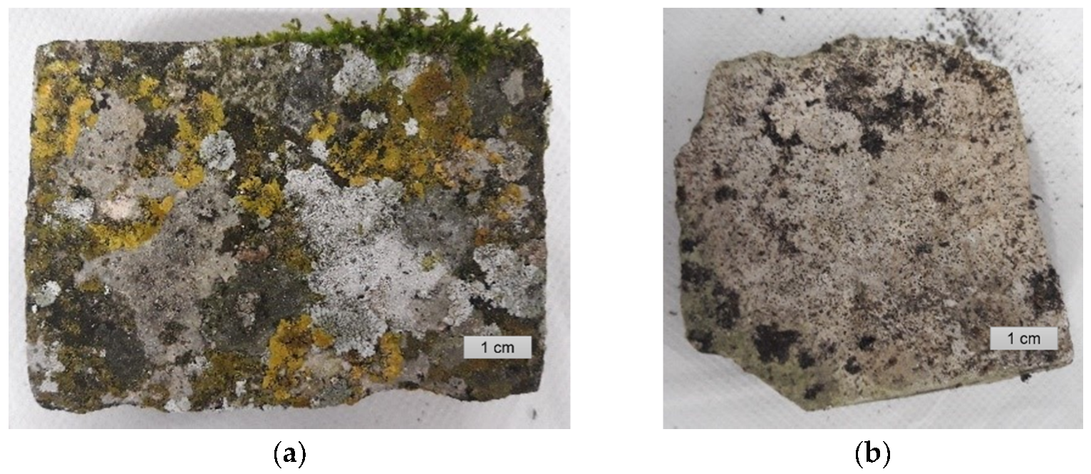
Samples (Figure 2) were collected from stone façades and roof areas of the Batalha Monastery (GPS latitude 39°39′32″ N, longitude 8°49′30″ W). The temperature at the site often reaches 30 °C (rarely 40 °C) in summer and −3 °C in winter with an average daily humidity of 81%. The maximum solar radiation in summer can reach 30,000 kJ/m2, while in winter, it averages around 5000 kJ/m2 (meteorological data from the Instituto Português do Mar e da Atmosfera, monitored in the meterological station in Leiria, Portugal, 13 km from Batalha).
The microbial sampling was carried out on representative areas of the stone materials, including areas showing evidence of weathering and stone decay such as flaking and granular desegregation. Sterile microinvasive (chisel and scalpels) and noninvasive (swabs) tools were used during the sampling under semi-aseptic conditions. Sterile swabs were placed in a suspension of transport MRD medium (Maximum Recovery Diluent) and preserved at 4 °C until utilization.

2.2. Air Monitoring

A cascade impactor (Optical Particle Sizer Spectrometer Model 3330, TSI, Minneapolis, MN, USA) was located outdoor at the roof level of the Batalha Monastery to sample particulate air pollutants. Particle collection campaign was carried out continuously for 1 week starting January 20th and ending January 27th 2020. Sterilized paper filters with pore size 0.45 µm (GN-6 Membrane, 47 mm, White, Gridded, Gamma Irradiated, Pall Coorperation) were used. Then, collected filters were stored in a sterile box for microbial characterization and Scanning Electron Microscopy with Energy Dispersive Spectroscopy (SEM + EDS) observations.

2.3. Molecular Analysis of Bacterial Communities

High-throughput sequencing (HTS) based on 16S rRNA gene analysis was conducted for the identification of bacterial communities present within biofilms on the limestone surface of the Batalha Monastery and deposited on the cascade impactor filter.
Microsamples were collected from limestone facades of the monastery, and the metagenomic DNA was extracted using the QIAmp DNA Stool Mini Kit (Qiagen, Limburg, Netherlands) with slight modifications from the manufacturer’s instructions [15]. The bacterial communities were characterized by the 16S rRNA V3-V4 region using the Illumina Sequencing platform. The analytical procedure was the same as thoroughly described by Dias et al. and Rosado el al. [16,17].

2.4. Morphology Observation, Mineralogical, and Chemical Characterization of the Bio-Deteriorated Stone

2.4.1. Optical Microscopy (OM)

Samples were observed under LEICA M205C Stereo Microscope (Leica, Wetzlar, Germany), photos were taken by a uEyeUI 149xSE-C industrial camera (IDS, Obsesulm, Germany).

2.4.2. X-Ray Micro-Diffractometry (µ-XRD)

The mineralogical characterization of the stone surface was carried out by μ-X-ray diffraction using a commercial Bruker AXS D8 Discovery diffractometer (Bruker, Karlsruhe, Germany) with the DaVinci design and Cu Ka radiation, Gadds detector, interval 3–708 2μ and step of 0.028/s. The EVA code and Highscore Plus software were used for the identification of the peaks using the PDF-2 International Centre for Diffraction Data (ICDD) mineralogical database.

2.4.3. Low-Vacuum Scanning Electron Microscopy Coupled with Energy-Dispersive Spectrometry (LV-SEM + EDS)

Scanning electron microscopy coupled with energy-dispersive X-ray spectrometry was carried out using a Hitachi S3700N (Tokyo, Japan) SEM coupled to a Bruker (Karlsruhe, Germany) XFlash 5010 SDD Detector system. Operating conditions were as follows: back-scattered imaging detector (BSEM); low-vacuum mode (pre-set pressure of 40 Pa) allowing the observation of non-coated biological samples; accelerating voltage—20 kV; current: 120 mA. For surface observation, a tablet (2 cm × 3 cm × 1 cm) with biofilm was cut from the stone sample and put into the chamber directly. For cross-section investigation, stone tablets were consolidated with resin, cut, and polished before observation.

3. Results

3.1. Optical Microscope Observation

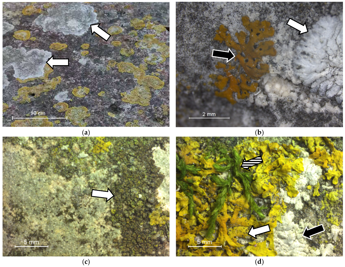
Field photographs from the monastery sampling sites are shown in Figure 3. Macroscopical visual examination allowed the identification of the following lichens species: Aspicilia sp., Caloplaca sp., Lecanora sp., Verrucaria, Dirina massiliensis, and Xanthoria [18]. These lichens are also found at other sites in Portugal and in Mediterranean countries such as Italy and Spain [19].

3.2. XRD Results

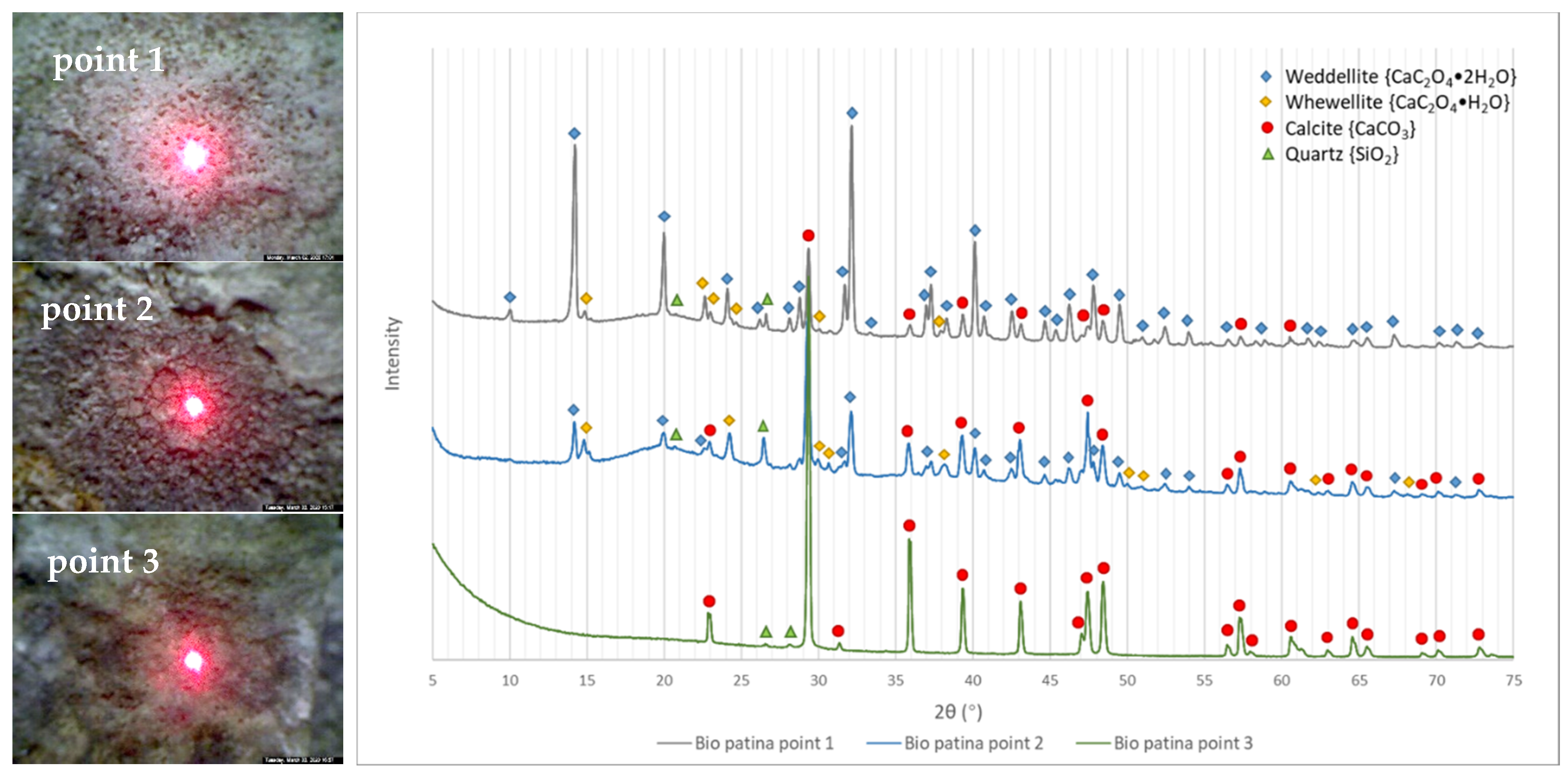
Point micro-XRD analyses (Figure 4) were performed on three areas characterized macroscopically by different colors, i.e., respectively white, black, and greenish (the latter possibly suggesting the presence of cyanobacteria, i.e., blue–green algae). Diffractograms corresponding to white and black areas show the presence of Ca-oxalate mineral peaks of weddellite and whewellite, while in green areas, only calcite and quartz of the limestone substrate are detected. The building stone used in the construction of the Batalha Monastery was in fact an oolitic limestone with a mineralogical composition characterized by more than 97% calcite with minor amounts of quartz [14]. It is well known how fungi are able to excrete different organic compounds such as oxalic acid as a result of their metabolic activity, which may react with CaCO3-rich materials such as limestones, leading to the crystallization of secondary Ca minerals in limestones [20,21]. In this case, the whewellite and weddellite could be a product from the reaction between the limestone substrate and the microflora metabolic products.

3.3. SEM-EDS Results

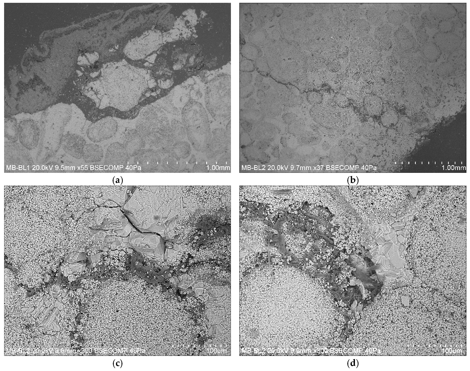
In Figure 5, SEM imaging of cross-sections of bio-deteriorated stone fragments shows (a) and (b) lichen hyphae mechanically penetrating inside the stone fabric up to a depth of 3 mm; (c) and (d) a close-up of hyphae progression preferentially along limestone oolite grain boundaries leading to the complete detachment and incorporation of stone substrate fragments within the advancing lichenous weathering crust.
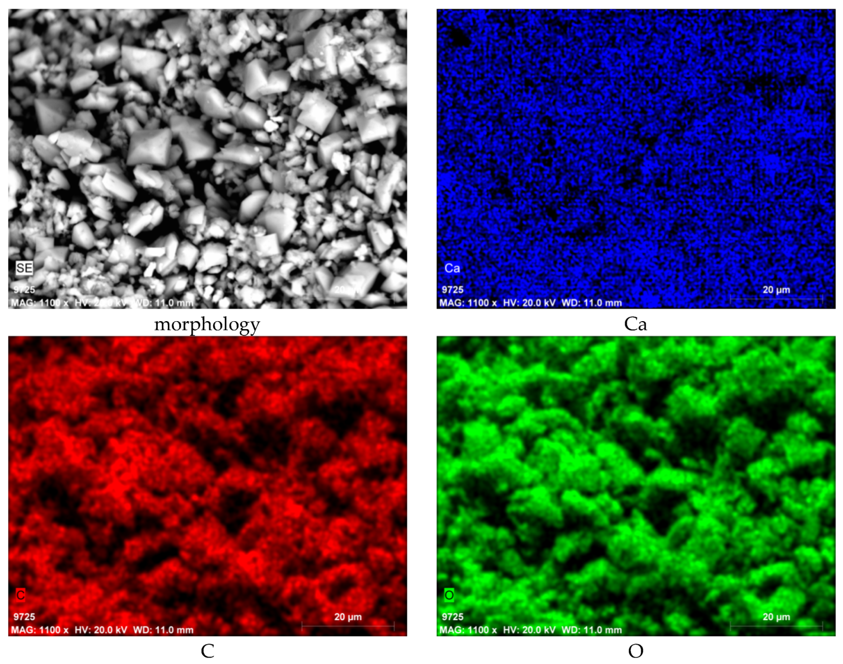
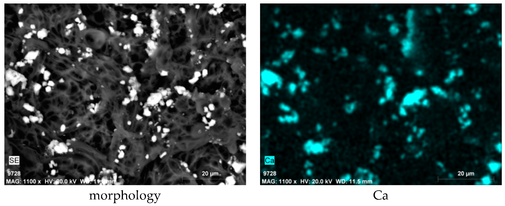
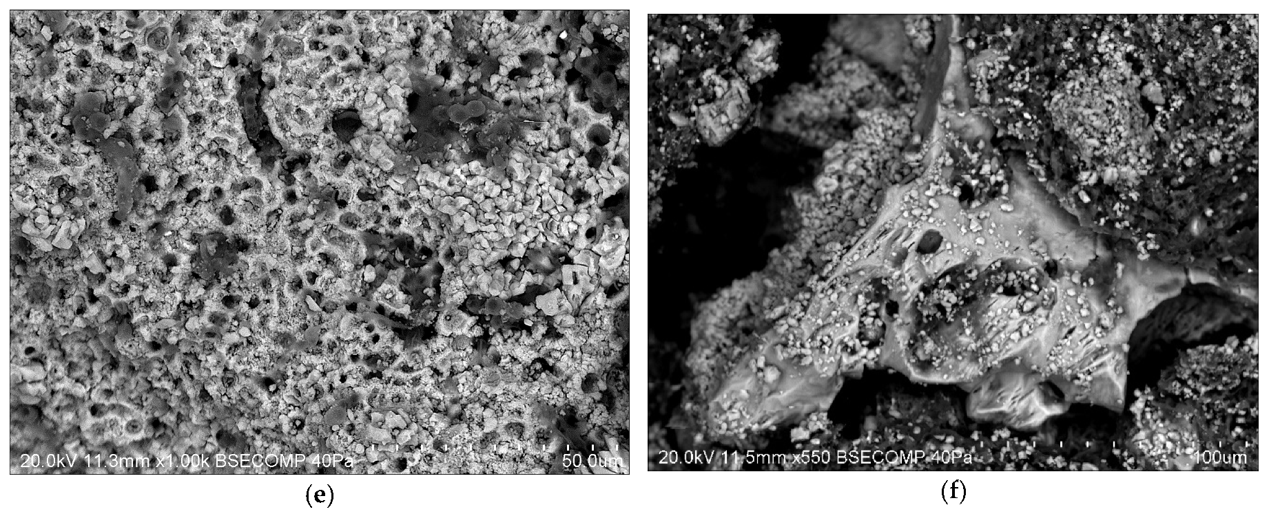
Figure 6a shows an intermediate stage of stone fragmentation with evidence of chemical etching patterns on mineral faces by fungal hyphae exploiting intracrystalline cleavage planes of calcite. Some hyphae were able to desegregate the stone fabric into fragments with grainsize 1–10 μm (black arrow) with granular size 1–10 µm. Figure 6b shows a close-up view of fungal hyphae and spores particles disrupting the inner stone microfabric. Figure 6c shows an area where the limestone substrate was completely desegregated into micron-size fragments. Several crystals (with dimension around 10 µm) can be seen displaying a tetragonal crystalline habit typical of Ca-oxalates, the interpretation of which is confirmed by EDS elemental analysis (see Figure A1). Figure 6d shows the re-precipitation of microcrystalline calcite crystals as confirmed by the EDS spectrum showing Ca peaks (Figure A2). Figure 6e shows again the presence of etching patterns (pit size 2–5 µm) on the stone surface. Hyphae here are septate and have a spherical shape. Figure 6f is an epitome of the above-mentioned degradation forms: dissolution cavities, pitting, penetrated hyphae, and oxalate precipitation can all be seen on this part of the stone. The increase in surface roughness and porosity favors adhesion and colonization by microorganisms, which in turn make the stone more prone to mechanical weathering [22].
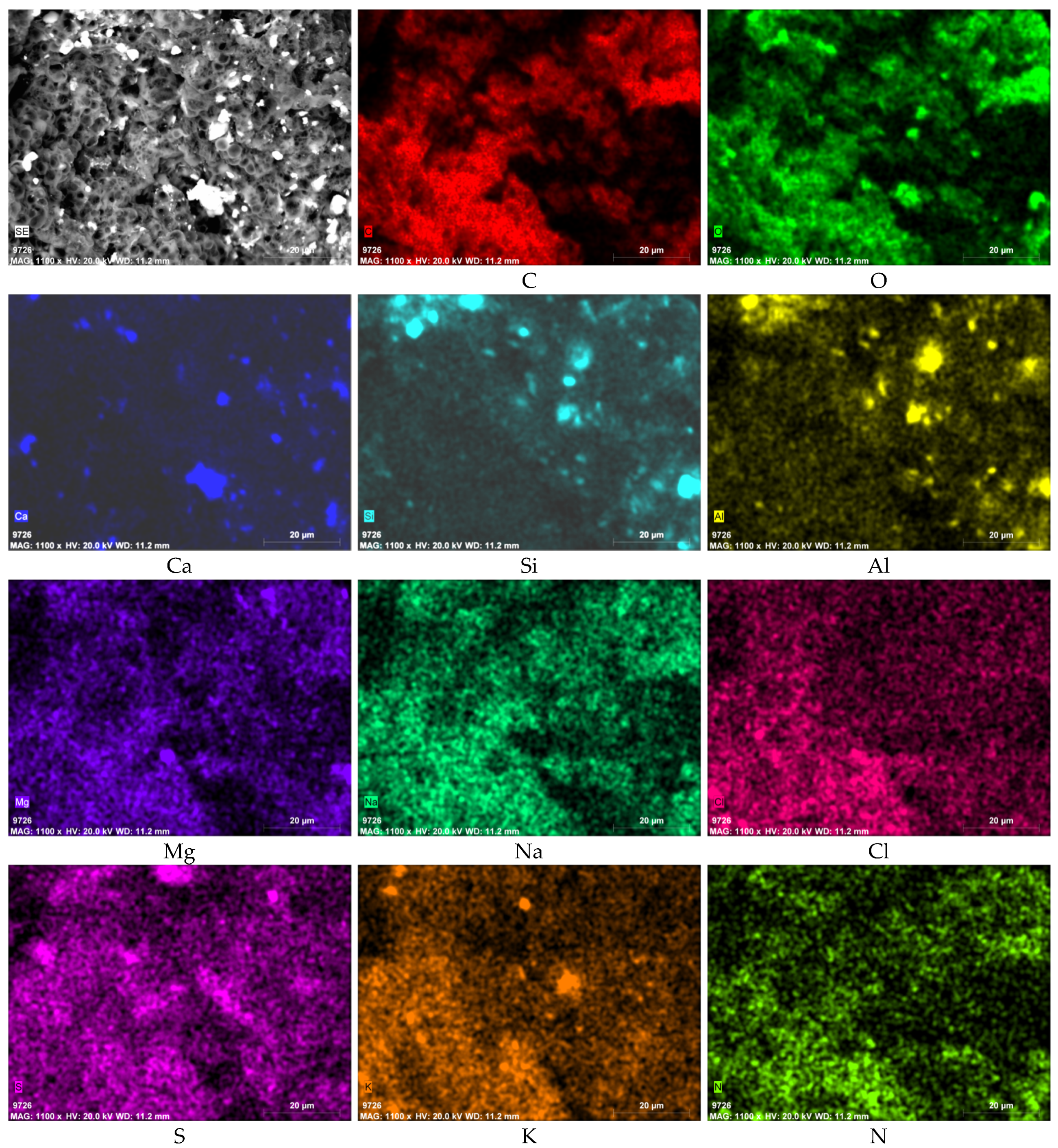
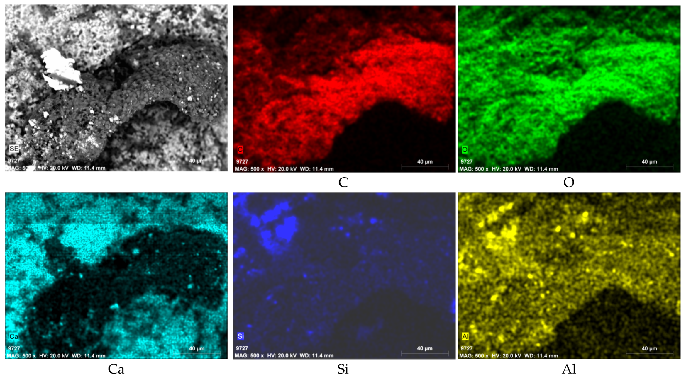
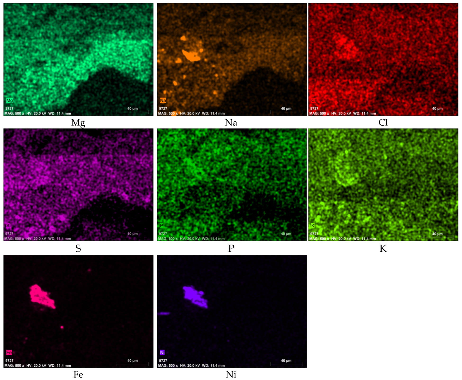
Element mapping of a lichen-covered area is shown in Figure 7. The strong signal of C and O correlating with the lichen represents the organism, while Ca is almost undetectable at the substrate covered by lichen except for some scattered grains on the lichen surface. Al, Mg, Na, and Si showed positive correlation with the lichen colonization, suggesting a silicate soil dust source. Cl and K are distributed evenly on the investigated area possibly from a fertilizer source. There are also a few Fe-Ni particles in this mapping, which are likely generated by vehicular traffic or industrial activity.
There is a noteworthy presence of P and S peaks associated with the presence of lichenous crusts. In other areas (Figure A3), the element N also shows a distribution highly correlated with the presence of fungal hyphae. It is evident that the presence of a lichenous cover on the stone surface facilitates the deposition of soil dust and other airborne particles while at the same time increasing the retention of water and the wet deposition of gaseous and particulate pollutants containing N, P, and S derived from anthropogenic sources such as agricultural fertilizers (N and P) and combustion-related (S) processes [23].
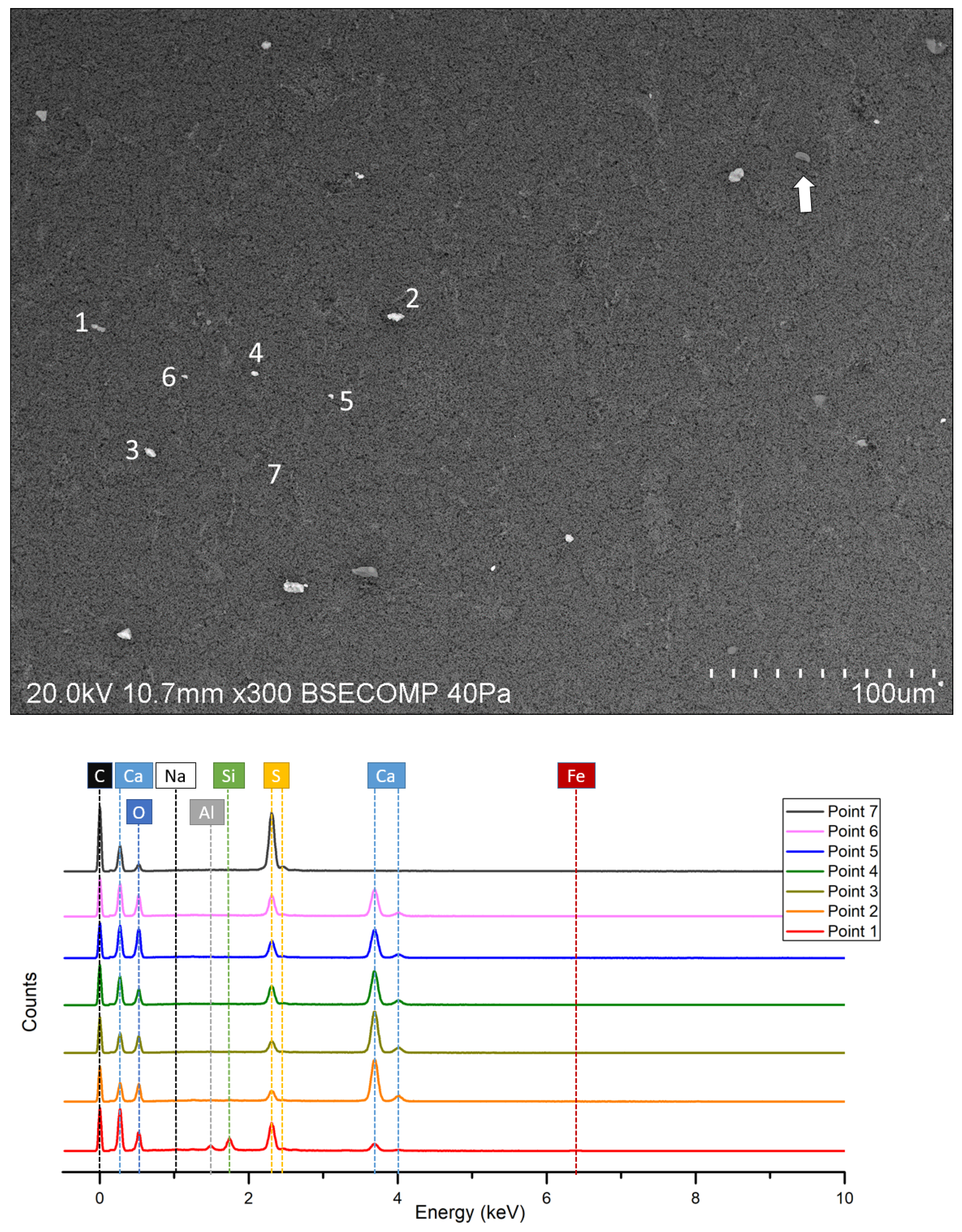
SEM investigation on the filter papers from the cascade impactor and the multi-point element analysis are shown in Figure 8. Particles are mostly of sizes less than 20 µm, and spores can also be seen (white arrow). Multi-point EDS analysis shows that the particles (point 1–6) are mainly Ca-C-O-S rich, suggesting the presence of CaCO3, CaC2O4, and CaSO4. Some particles show the presence of Na, Si, and Al, which are likely to be associated with deposited windborne silicate soil dust. The weak signal of Fe might come from oxidized iron products. A notable result is that on the filter paper where no particles are present (point 7), there is a significant peak of S, suggesting sulfur compounds are still affecting today’s air quality in the outdoor environment surrounding the Batalha Monastery. The spores present on the filter papers proved that they have been deposited on the stone surface by dry and wet deposition as airborne atmospheric particles.

3.4. High-Throughput Sequencing

The identification of bacterial communities is shown in Figure 9. The dominant airborne bacteria from the surrounding atmospheric environment on the filter paper belong to phyla Proteobacteria (61.86%) and Firmicutes (25.46%); aside from that, Bacteroidetes (4.17%), Actinobacteria (2.53%), and Acidobacteria (1.67%) are also present. For the sample MB-R3, the dominant bacteria phyla are Proteobacteria (44.23%), Actinobacteria (14.59%), Cyanobacteria (11.52%), Bacteroidetes (7.68%), Acidobacteria (5.82%), Firmicutes (5.48%), Verrucomicrobia (3.10%), and Planctomycetes (2.85%). Sample P2-5 is characterized by analogous microbial communities but with different relative abundances: Proteobacteria (52.40%), Cyanobacteria (9.39%), Bacteroidetes (8.85%), Actinobacteria (6.92%), Acidobacteria (3.24%), and a small quantity of Deinococcus (1.44%) and Planctomycetes (1.23%).
It is noteworthy that Proteobacteria phylum is by far the most represented in all samples, with Gammaproteobacteria and Alphaproteobacteria as the two predominant classes. The percentage of Actinobacteria and Acidobacteria has increased from filter paper to sample P2-5 to MB-R3, while Firmicutes bacterial abundance reduces following this sequence. Considering the degradation level of the two stone specimens, MB-R3 has a massive lichen layer on the surface, while P2-5 has not yet formed an evident lichen crust. It can be speculated that the lichen flora has changed the microenvironment, which selectively benefits the growth and reproduction of certain bacterial species, thus rearranging the relative abundance of bacteria. In fact, a previous study illustrated that the metabolite production and phenoloxidase activity of lichens can affect the bacterial community structure [24].
Another significant discovery is that Cyanobacteria, while abundant on the stone samples, seem not to be present on the filter papers. In fact, cyanobacteria could be found present at a few millimeters of depth from the stone–atmosphere interface as they seek shelter from desiccation and intense UV radiation [6]. Therefore, the Cyanobacteria found on the stone samples could have already been present in the stone materials from the original quarries.

4. Discussion

Proteobacteria and Bacteroidetes are believed to provide nutrients, mobilize iron and phosphate, and fix nitrogen for the lichen symbiotic growth [25]. Firmicutes is also well known for symbiotic nitrogen fixation and their beneficial and endophytic interaction with plants resulting in plant growth promotion [26]. Actinobacteria play the leading role in cellulose degradation, promoting the decomposition of plant remains [27]. Cyanobacterial are the typical photolithoautotrophic organisms that synthesize carbohydrate. Planctomycetes reproduction has been reported to be associated with the enrichment of N and P [27]. Hence, the increase of Planctomycetes ratio from membrane filter to sample P2-5 to MB-R3 may be due to the growth of a lichenous cover which in turn facilitates the fixation of nitrogen and a phosphorus-rich compound, resulting in the abundance increase of Planctomycetes bacteria; the EDS analysis of the biofilm (Figure 7 and Figure A3) is also in agreement with that.
The Nitrosomonas spp., Nitrobacter spp., and Thiobacillus spp. from Alphaproteobacteria and Betaproteobacteria class are chemolithoautotrophic bacteria that obtain energy from the oxidation of inorganic compounds (ammonia, nitrites, hydrogen sulfide, thiosulfates, or elementary sulfur), fixing CO2 from the atmosphere and releasing nitrous acids, nitric acid, or sulfuric acid [3]. The above-mentioned bacteria detected on the stone samples microbiomes in this research, combined with the significant presence of S-rich compounds on the filter samples, may suggest that air pollution leads to a richness of nutrients for the chemolithoautotrophic bacteria and indirectly promotes lichen growth.
The presence of bacterial community in lichen microbiomes was also reported in other studies. For instance, Paramo lichen harbored microbiomes composed mainly of the phyla Proteobacteria, Acidobacteria, Verrucomicrobia, Bacteroidetes, and Actinobacteria [28]. In other research, four lichen species from SW-Norway were analyzed with the bacteria detected in the lichen–rock samples, which were affiliated with the major lineages Acidobacteria, Actinobacteria, Proteobacteria (Alpha-, Beta-, Gamma-), Bacteriodetes, Chloroflexi, Deinococcus, Firmicutes, Planctomycetes, Tenericutes, and Cyanobacteria [29]. Even in the Antarctic and Arctic, the lichens-associated bacteria are Actinobacteria, Bacteroidetes, Deinococcus-Thermus, Firmicutes, and Proteobacteria (Alpha-, Beta-, and Gamma-) [30]. Regardless of the lichen species, there is some consistency in the bacterial phyla associated with lichens around the world, although the community structure can differ according to geographical location.

5. Conclusions

The biodegradation processes occurring on Batalha Monastery limestone constistute a complex phenomenon involving different steps. At the beginning, photolithoautotrophic bacteria (e.g., cyanobacteria) present in the stone started to grow using sunlight as an energy source for photosynthesis. Airborne bacterial spores from the surrounding atmospheric environment were also deposited to the stone surface, some of which are chemolithoautotrophic; they grow by utilizing the sulfates, nitrates, phosphate, and metallic particles from the air as nutrients, and being responsible for the production of acidic substances attacking the stone substrate, the use of fertilizers and combustion of fossil fuel can promote this process. These primary colonizers accumulate biomass, forming a biofilm enriched with organic and inorganic substances and growth factors, providing an excellent nutrient base for the subsequent heterotrophic microflora. As a result of metabolic activity, a series of acid compounds and expansion of hyphae damage the stone substrate by making fissures and boreholes as well as pitting and detached fragments, which in turn increase the capillary adsorption and accelerate degradation from the environment and microbial activity [31]. The development of a lichen cover promotes the fixation of phosphorous and nitrogen, which is selectively beneficial to some bacterial species, and the secondary metabolites of lichens can affect the bacterial community structure. In this complicated process, contamination in the air plays an important role to exacerbate limestone deterioration in the Monastery of Batalha. Therefore, an effective conservation strategy to preserve the Batalha Monastery would be twofold: i.e., to reduce S, N, and P-based air pollution in the atmosphere surrounding the monument while at the same time eliminating (using the appropriate cleaning procedure) the microbial colonization on the stone.

Author Contributions

Conceptualization, Y.D., N.S.; methodology, Y.D., A.T.C., N.S.; formal analysis, Y.D., C.S.C.S., A.T.C.; investigation, Y.D.; data curation, Y.D.; writing—original draft preparation, Y.D.; writing—review and editing, N.S., A.T.C., Y.D.; supervision, N.S., E.A.; project administration, N.S. All authors have read and agreed to the published version of the manuscript.

Funding

This research has received funding from the European Union’s Horizon 2020 research and innovation programme under the Marie Skłodowska-Curie grant agreement No 766311.

Informed Consent Statement

Not applicable.

Data Availability Statement

Data is contained within the article.

Acknowledgments

Samples collection in this research was by special permission of the Direção-Geral do Patrimonio Cultural and the Mosteiro da Batalha authorities. The research presented in this paper was carried out mainly using data collected at Universidade de Évora, Politecnico di Torino and Direcao-Geral do Patrimonio Cultural, as part of H2020-MSCA-ITN-2017, ED-ARCHMAT (ESR1).

Conflicts of Interest

The authors declare no conflict of interest.

Appendix A

Figure A1. SEM-EDS picture of tetragonal crystals in the biofilm.
Cmd 02 00002 g0a1
Figure A2. SEM-EDS picture of hyphae cohesive grains.
Cmd 02 00002 g0a2
Figure A3. SEM-EDS element mapping of a lichen covered area on sample MB-R3.
Cmd 02 00002 g0a3

References

  1. Ahmadjian, V. Resynthesis of lichens. In The Lichens; Elsevier Inc.: Cambridge, MA, USA; Academic Press: Cambridge, MA, USA, 1973; pp. 565–579. [Google Scholar] [CrossRef]
  2. Warscheid, T.; Braams, J. Biodeterioration of stone: A review. Int. Biodeterior. Biodegrad. 2000, 46, 343–368. [Google Scholar] [CrossRef]
  3. Grube, M.; Cernava, T.; Soh, J.; Fuchs, S.; Aschenbrenner, I.; Lassek, C.; Wegner, U.; Becher, D.; Riedel, K.; Sensen, C.W.; et al. Exploring functional contexts of symbiotic sustain within lichen-associated bacteria by comparative omics. ISME J. 2015, 9, 412–424. [Google Scholar] [CrossRef] [PubMed] [Green Version]
  4. Trovão, J.; Portugal, A.; Soares, F.; Paiva, D.S.; Mesquita, N.; Coelho, C.; Pinheiro, A.C.; Catarino, L.; Gil, F.; Tiago, I. Fungal diversity and distribution across distinct biodeterioration phenomena in limestone walls of the old cathedral of Coimbra, UNESCO World Heritage Site. Int. Biodeterior. Biodegrad. 2019, 142, 91–102. [Google Scholar] [CrossRef]
  5. Pinheiro, A.C.; Mesquita, N.; Trovão, J.; Soares, F.; Tiago, I.; Coelho, C.; Carvalho, H.P.; Gil, F.; Catarino, L.; Piñar, G.; et al. Limestone biodeterioration: A review on the Portuguese cultural heritage scenario. J. Cult. Herit. 2019, 36, 275–285. [Google Scholar] [CrossRef]
  6. Macedo, M.F.; Miller, A.Z.; Dionísio, A.; Saiz-Jimenez, C. Biodiversity of cyanobacteria and green algae on monuments in the Mediterranean Basin: An overview. Microbiology 2009, 155, 3476–3490. [Google Scholar] [CrossRef] [PubMed] [Green Version]
  7. Prada, J.L.; Pugés, M.; Rocabayera, R. Estudio de las patologias del Retablo del Altar Mayor de la Sé da Guarda. In Património Estudos; Instituto Português do Património Arquitectónico: Lisbon, Portugal, 2002; p. 70. [Google Scholar]
  8. Ascaso, C.; Wierzchos, J.; Delgado Rodrigues, J.; Aires-Barros, L.; Henriques, F.; Charola, A.E. Endolithic microorganisms in the biodeterioration of the tower of Belem. Int. Z. FürBauinstandsetz. 1998, 4, 627–640. [Google Scholar]
  9. Ascaso, C.; Wierzchos, J.; Souza-Egipsy, V.; De los Rıos, A.; Rodrigues, J.D. In situ evaluation of the biodeteriorating action of microorganisms and the effects of biocides on carbonate rock of the Jeronimos Monastery (Lisbon). Int. Biodeterior. Biodegrad. 2002, 49, 1–12. [Google Scholar] [CrossRef] [Green Version]
  10. Mateus, D.M.; Silva, R.B.; Costa, F.M.; Coroado, J.P. Diversidade microbiológica do edifício da Sacristia Incompleta do Convento de Cristo, em Tomar, e avaliação do seu controlo por biocidas. Conserv. Património 2013, 17, 11–17. [Google Scholar] [CrossRef]
  11. Schiavon, N.; Chiavari, G.; Schiavon, G.; Fabbri, D. Nature and decay effects of urban soiling on granitic building stones. Sci. Total Environ. 1995, 167, 87–101. [Google Scholar] [CrossRef]
  12. Schiavon, N. Biodeterioration of calcareous and granitic building stones in urban environments. Geol. Soc. Lond. Spec. Publ. 2002, 205, 195–205. [Google Scholar] [CrossRef]
  13. Ortega-Morales, O.; Montero-Muñoz, J.L.; Neto, J.A.B.; Beech, I.B.; Sunner, J.; Gaylarde, C. Deterioration and microbial colonization of cultural heritage stone buildings in polluted and unpolluted tropical and subtropical climates: A meta-analysis. Int. Biodeterior. Biodegrad. 2019, 143, 104734. [Google Scholar] [CrossRef]
  14. Ding, Y.; Mirão, J.; Redol, P.; Dias, L.; Moita, P.; Angelini, E.; Grassini, S.; Schiavon, N. A combined petrographic and geochemical metrological approach to assess the provenance of the building limestone used in the Batalha Monastery (Portugal). In Metrology for Archaeology and Cultural Heritage; Battisti, V., Gallo, V., Eds.; IMEKO: Florence, Italy, 2019; p. 338. [Google Scholar]
  15. Dias, L.; Rosado, T.; Coelho, A.; Barrulas, P.; Lopes, L.; Moita, P.; Candeias, A.; Mirão, J.; Caldeira, A.T. Natural limestone discolouration triggered by microbial activity—A contribution. AIMS Microbiol. 2018, 4, 594. [Google Scholar] [CrossRef] [PubMed]
  16. Rosado, T.; Dias, L.; Lança, M.; Nogueira, C.; Santos, R.; Martins, M.R.; Candeias, A.; Mirão, J.; Caldeira, A.T. Assessment of microbiota present on a Portuguese historical stone convent using high-throughput sequencing approaches. MicrobiologyOpen 2020, 9, 1067–1084. [Google Scholar] [CrossRef] [PubMed]
  17. Dias, L.; Rosado, T.; Candeias, A.; Mirão, J.; Caldeira, A.T. A change in composition, a change in colour: The case of limestone sculptures from the Portuguese National Museum of Ancient Art. J. Cult. Herit. 2020, 42, 255–262. [Google Scholar] [CrossRef]
  18. Mifsud, S.; Lanfranco, E.; Fiorentino, J.; Mifsud, S.D. An Updated Flora of Selmunett (St. Paul’s Island) including Mosses and Lichens. XJENZA 2016, 4, 142–159. [Google Scholar]
  19. Lisci, M.; Monte, M.; Pacini, E. Lichens and higher plants on stone: A review. Int. Biodeterior. Biodegrad. 2003, 51, 1–17. [Google Scholar] [CrossRef]
  20. Sayer, J.A.; Kierans, M.; Gadd, G.M. Solubilisation of some naturally occurring metal-bearing minerals, limescale and lead phosphate by Aspergillus niger. FEMS Microbiol. Lett. 1997, 154, 29–35. [Google Scholar] [CrossRef]
  21. Gharieb, M.M.; Sayer, J.A.; Gadd, G.M. Solubilization of natural gypsum (CaSO4.2H2O) and the formation of calcium oxalate by Aspergillus niger and Serpula himantioides. Mycol. Res. 1998, 102, 825–830. [Google Scholar] [CrossRef]
  22. Scheerer, S.; Ortega-Morales, O.; Gaylarde, C. Microbial deterioration of stone monuments—an updated overview. Adv. Appl. Microbiol. 2009, 66, 97–139. [Google Scholar] [CrossRef]
  23. Shi, J.; Wang, N.; Gao, H.; Baker, A.; Yao, X.; Zhang, D. Phosphorus solubility in aerosol particles related to particle sources and atmospheric acidification in Asian continental outflow. Atmos. Chem. Phys. 2019, 19, 847–860. [Google Scholar] [CrossRef] [Green Version]
  24. Leiva, D.; Clavero-León, C.; Carú, M.; Orlando, J. Intrinsic factors of Peltigera lichens influence the structure of the associated soil bacterial microbiota. FEMS Microbiol. Ecol. 2016, 92, fiw178. [Google Scholar] [CrossRef] [PubMed] [Green Version]
  25. Ryan, R.P.; Germaine, K.; Franks, A.; Ryan, D.J.; Dowling, D.N. Bacterial endophytes: Recent developments and applications. FEMS Microbiol. Lett. 2008, 278, 1–9. [Google Scholar] [CrossRef] [PubMed] [Green Version]
  26. Bates, S.T.; Cropsey, G.W.; Caporaso, J.G.; Knight, R.; Fierer, N. Bacterial communities associated with the lichen symbiosis. Appl. Environ. Microbiol. 2011, 77, 1309–1314. [Google Scholar] [CrossRef] [PubMed] [Green Version]
  27. Pankratov, T.A.; Dedysh, S.N.; Zavarzin, G.A. The leading role of actinobacteria in aerobic cellulose degradation in Sphagnum peat bogs. Dokl. Biol. Sci. 2006, 410, 428–430. [Google Scholar] [CrossRef] [PubMed]
  28. Sierra, M.A.; Danko, D.C.; Sandoval, T.A.; Pishchany, G.; Moncada, B.; Kolter, R.; Mason, C.E.; Zambrano, M.M. The microbiomes of seven lichen genera reveal host specificity, a reduced core community and potential as source of antimicrobials. Front. Microbiol. 2020, 11, 398. [Google Scholar] [CrossRef] [Green Version]
  29. Bjelland, T.; Grube, M.; Hoem, S.; Jorgensen, S.L.; Daae, F.L.; Thorseth, I.H.; Øvreås, L. Microbial metacommunities in the lichen–rock habitat. Environ. Microbiol. Rep. 2011, 3, 434–442. [Google Scholar] [CrossRef]
  30. Lee, Y.M.; Kim, E.H.; Lee, H.K.; Hong, S.G. Biodiversity and physiological characteristics of Antarctic and Arctic lichens-associated bacteria. World J. Microbiol. Biotechnol. 2014, 30, 2711–2721. [Google Scholar] [CrossRef]
  31. Özvan, A.; Dinçer, İ.; Akın, M.; Oyan, V.; Tapan, M. Experimental studies on ignimbrite and the effect of lichens and capillarity on the deterioration of Seljuk Gravestones. Eng. Geol. 2015, 185, 81–95. [Google Scholar] [CrossRef]
Figure 1. (a) The external wall of Batalha Monastery, covered by dark brown/gray biofilms and crusts; (b) Balustrade on the roof of the monastery showing thick biofilms and lichenous crusts.
Cmd 02 00002 g001
Figure 2. Microbial colonized stone samples from the Batalha Monastery: (a) Sample MB-R3; (b) Sample P2-5.
Cmd 02 00002 g002
Figure 3. Batalha Monastery, lichenous crusts on limestone: (a) Dirina massiliensis (white arrow); (b) Crustose Caloplaca sp. (black arrow) and Lecanora sp. (white arrow); (c) Aspicilia sp. (white arrow); (d) Foliose Xanthoria (white arrow), Lecanora (black arrow) and higher plant (stripe arrow).
Cmd 02 00002 g003
Figure 4. XRD result of the bio-colonized stone surface.
Cmd 02 00002 g004
Figure 5. Back-scattered imaging detector (BSEM) micrographs of biodeteriorated Batalha oolitic limestone: (a) Detached stone fragments incorporated within the growing lichenous weathering crust; (bd) Lichen hyphae penetrating deeply inside the limestone fabric using pre-existing intracrystalline cracks and/or porosity within the sparitic cement and leading to the detachment of the oolitic grains.
Cmd 02 00002 g005
Figure 6. BSEM photos of limestone specimen from Batalha Monastery, surface: (a) Stone full of rifts, hyphae penetrating (black arrow) and expanding across the calcite cleavage planes (white arrow); (b) Stone surface with fragmented granules and microbial spores (white arrow); (c) Calcium oxalate crystals (weddellite) showing typical tetragonal habit; (d) Microorganism tissues and cohesive calcium composite; (e) Pitting corrosion associated with hyphae penetration and Ca-oxalate precipitation; (f) A stone showing multiple degradation forms.
Cmd 02 00002 g006aCmd 02 00002 g006b
Figure 7. Scanning Electron Microscopy with Energy Dispersive Spectroscopy (SEM-EDS) element mapping of a lichen-covered area on the sample MB-R3.
Cmd 02 00002 g007aCmd 02 00002 g007b
Figure 8. SEM-EDS analysis of filter paper. A spore particle is shown (white arrow).
Cmd 02 00002 g008
Figure 9. Relative abundance of the dominant bacterial classes in the stone samples and filter paper with corresponding phyla (on the right side).
Cmd 02 00002 g009
Publisher’s Note: MDPI stays neutral with regard to jurisdictional claims in published maps and institutional affiliations.