Review

Surface Effects in Irradiation Damage: A Review of Underlying Multi-Scale Mechanisms and Cross-System Behaviors

1
School of Mechano-Electronic Engineering, Xidian University, Xi’an 710071, China
2
Amazon AWS Finance, Amazon re:Invent, 2121 8th Ave., Seattle, WA 98121, USA
3
Chengdu Development Center of Science and Technology, China Academy of Engineering Physics, Chengdu 610200, China
4
College of Engineering and Computer Science, Arkansas State University, Jonesboro, AR 72467, USA
5
School of Mechanical Engineering, University of Birmingham, Birmingham B15 2TT, UK
6
Yangtze·Delta Region Institute of Tsinghua University, Jiaxing 314006, China
7
Science and Technology on Vacuum Technology and Physics Laboratory, Lanzhou 730000, China
8
CityU-Xidian Joint Laboratory of Micro/Nano Manufacturing, Shenzhen 518057, China
*
Authors to whom correspondence should be addressed.
Surfaces 2026, 9(2), 40; https://doi.org/10.3390/surfaces9020040
Submission received: 28 February 2026 / Revised: 15 April 2026 / Accepted: 22 April 2026 / Published: 28 April 2026
(This article belongs to the Collection Featured Articles for Surfaces)

Abstract

Structural materials in nuclear energy, aerospace, and electronics face long-term irradiation by high-energy particles, triggering microscopic defect evolution and macroscopic performance degradation that limits service safety. This review provides a systematic overview of irradiation damage mechanisms, with particular emphasis on the role of surfaces. The discussion traces the evolution from initial defect generation through energy deposition and displacement cascades to the migration and aggregation of defects toward surfaces, culminating in their interactions with near-surface microstructures. A comparative analysis of damage behaviors in metals, ceramics, silicon-based materials, and polymers is presented, elucidating how distinct mechanisms arise from fundamental differences in crystal structure and chemical bonding. The integration of multiscale simulation techniques with advanced in situ characterization is highlighted as a critical approach for deciphering the cross-scale processes. Current strategies for enhancing radiation resistance including composition optimization, microstructure regulation, and interface design are summarized. Finally, the review outlines key challenges such as multi-field coupling damage characterization and long-term predictive modeling. Future research directions are foreseen to emphasize closer simulation–experiment integration and the design of smart, self-adapting materials, thereby providing comprehensive theoretical and technical support for the development of next-generation radiation-tolerant materials.

1. Introduction

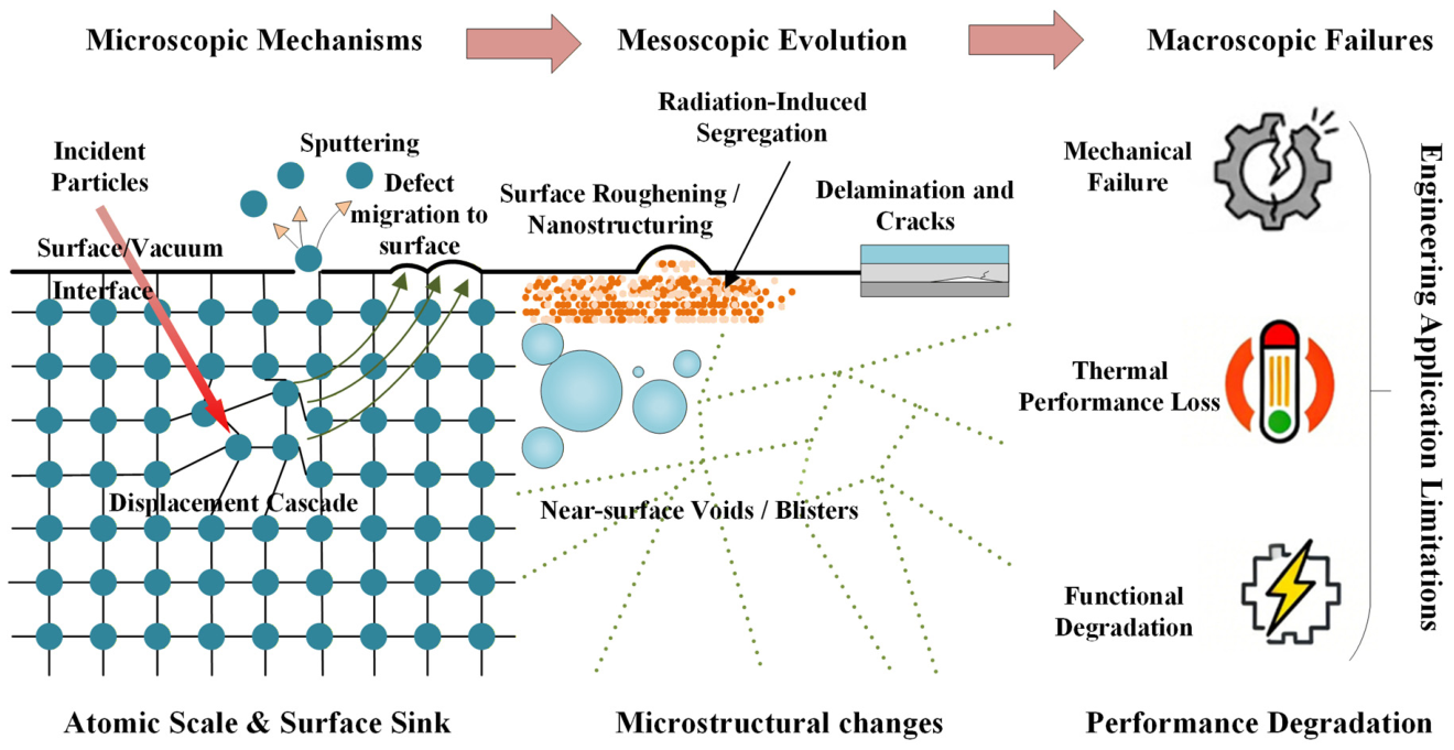
Long-term operation in extreme radiation environments poses a fundamental challenge for critical materials. This includes structural alloys for fission/fusion reactors, core electronic components in spacecraft, and nuclear waste forms, all of which are subjected to continuous fluxes of high-energy particles [1,2,3]. Under such conditions, irradiation induces continuous evolution of microscopic defects. Critically, the proximity of these defects to the material surface or internal interfaces governs their migration and annihilation behaviors, leading to distinct surface-mediated lattice distortions and morphological evolution. This cascade process, extending from near-surface microstructural changes to macroscopic property degradation as illustrated in Figure 1, has become a key scientific bottleneck restricting the long-term service life and safety of these materials [4,5].
The collision of high-energy particles with the material lattice directly disrupts the bonding equilibrium between atoms, generating primary defects such as vacancies, interstitials, and Frenkel pairs [6,7]. Driven by thermodynamics, these primary defects further migrate and aggregate, and interact with intrinsic microstructural features including grain boundaries, phase boundaries, and dislocations, thereby inducing secondary structural evolutions such as lattice distortion, phase transformation, interface damage, and void swelling [8,9]. Such microstructural degradation directly propagates to the macroscopic scale, manifesting as service performance deterioration behaviors including thermal conductivity attenuation, mechanical embrittlement, reduced corrosion resistance, and increased tritium retention, and ultimately leads to material failure and potential safety hazards [10]. Therefore, revealing the mechanism of the structure–property correlation between microstructural evolution and macroscopic performance degradation of materials under irradiation environments is a core prerequisite for breaking through the bottlenecks in the design and application of materials for extreme environments.
Building on prior research, this work analyzes the fundamental theories and core mechanisms that govern irradiation damage, from defect nucleation and migration to their agglomeration and interactions with interfaces. The review provides a systematic analysis of irradiation-induced microstructural evolution and property degradation, highlighting the intrinsic correlation between surface properties and collective material degradation across various systems, including metals, ceramics, and composites, to reveal how surface states dictate distinct damage resistance and cross-scale behaviors. Concurrently, this study delineates the prevailing challenges in the field of irradiation damage research, which include the refined characterization of damage mechanisms under multi-field coupling, the establishment of predictive models for long-term service-induced damage, and the exploitation of high-efficiency shielding technologies tailored for extreme environments. Furthermore, an outlook is presented on the prospective development trends, such as the integration of multi-scale simulations with experimental validations, the rational design of smart responsive radiation-resistant materials, and the advancement of in situ real-time monitoring techniques. Ultimately, the findings presented herein are intended to furnish theoretical underpinnings and technical guidelines for the development and engineering deployment of radiation-tolerant materials.

2. The Basic Mechanism of Irradiation Damage

2.1. Primary Irradiation Damage

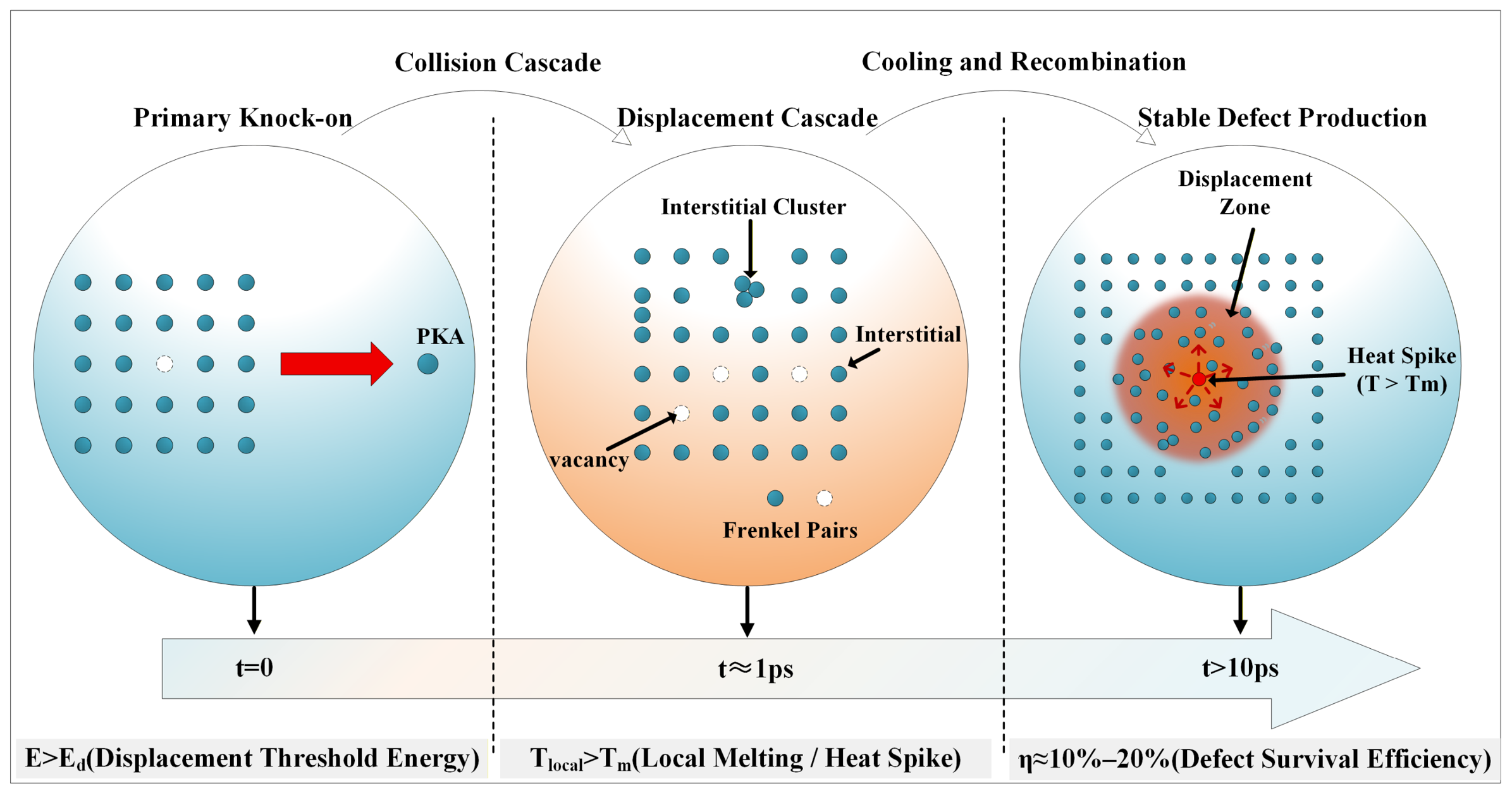
Primary radiation damage refers to the initial stage where high-energy particles interact with lattice atoms of materials, inducing energy transfer, atomic displacement, and the nucleation of microscopic defects [11]. In regions proximate to a material boundary, this process is fundamentally reshaped by surface-induced broken symmetry, distinguishing its underlying physics from isotropic bulk displacements. As shown in Figure 2, this process highly condenses three core stages within the picosecond scale: energy transfer and atomic displacement [12], displacement cascade formation [13], and thermal spike and quenching effects [14]. Unlike the infinite lattice in the bulk, the near-surface region imposes a geometric truncation on the collision sequence. The presence of a free surface serves as a dominant sink for mobile defects and a potent driver for near-surface cascade collapse or atomic sputtering [15], thereby significantly altering the surviving defect population. These surface-governed initial responses dictate the long-term microstructural evolution and the eventual degradation of macroscopic properties in irradiated materials.
Primary radiation damage, fundamentally characterized by the production of Frenkel pairs, exhibits profound sensitivity to both the incident particle species and the geometric constraints imposed by the surface [16,17]. Unlike neutron irradiation, which typically facilitates volumetric swelling in the bulk, ion and electron interactions are inherently governed by near-surface energy deposition and ballistic sputtering [18]. These effects are particularly dominant in silicon-based materials and low-dimensional systems, where the high surface-to-volume ratio dictates the overall radiation tolerance [19]. Consequently, these surface-mediated mechanisms trigger localized morphological instability and non-uniform defect clustering, which collectively determine the material’s structural integrity and its capacity for self-healing under radiation environments.
The subsequent displacement cascades and thermal spikes further determine the final defect configuration [18,19]. Crucially, when these cascades occur near a free surface or interface, the inherent symmetry of defect production is broken. The surface acts as a natural boundary that facilitates the rapid recombination or escape of point defects, significantly altering the defect survival rate compared to the interior of the material. This localized behavior, coupled with transient thermal energy relaxation, governs the specific microstructural features of the near-surface region. The dynamic process of defects moving toward and interacting with the surface provides the necessary physical foundation for the detailed mechanisms discussed in the following subsection.

2.2. Microscopic Evolution of Defects

The microstructural evolution of irradiation-induced defects, from point defect migration to the formation of complex clusters, is a dynamic process driven by thermodynamic forces and internal lattice stresses. Driven by their respective migration energy barriers, interstitials and vacancies undergo recombination or agglomerate into stable configurations such as vacancy disks, voids, and dislocation loops [10]. In the vicinity of a free surface, this process is fundamentally altered as the surface acts as a non-equilibrium sink that leads to surface-induced defect-depleted zones. The presence of the surface induces a biased diffusion flux of point defects, causing a preferential loss of mobile species to the boundary and thereby spatially redefining the localized defect density. Consequently, these surface-proximity effects redefine the stability of defect clusters and the subsequent degradation of the material’s mechanical integrity.
The interaction between migrating defects and the material’s boundary is bidirectional. On one hand, surfaces and interfaces (such as grain and phase boundaries) serve as effective sites for defect annihilation, potentially suppressing the nucleation and coarsening of voids and loops [20,21]. On the other hand, the persistent accumulation of defects at these boundaries can destabilize the local microstructure, leading to phenomena such as boundary decohesion, grain boundary migration, and localized stress concentration [21,22]. This localized evolution at the near-surface region is a critical factor in determining the overall radiation resistance of low-dimensional and coated materials.

2.3. Macroscopic Performance Degradation

The accumulation and evolution of irradiation-induced defects—such as vacancy clusters, voids, and dislocation loops—lead to a significant degradation of macroscopic material properties, ranging from mechanical embrittlement to physical and environmental instability. In metallic systems, the pinning effect of these defects on dislocation motion causes irradiation hardening and a transition from ductile to brittle fracture modes [23,24]. Similarly, in functional materials like semiconductors and ceramics, lattice distortion and defect scattering centers severely impair thermal conductivity and electrical performance [25,26,27]. As summarized in Table 1, these cross-scale degradations directly dictate the service life of materials in extreme irradiation environments.
Significantly, these performance losses are often initiated or exacerbated at the material’s boundaries. Surfaces and interfaces provide rapid diffusion channels for corrosive media, where irradiation-induced defect aggregation can drastically reduce pitting resistance and accelerate environmental erosion [23,28]. Conversely, tailored microstructural features, such as those in high-entropy ceramics or low-dimensional composites, can mitigate these synergistic effects by trapping gas atoms or promoting defect recombination [26,29]. This interplay between localized surface damage and global property attenuation underlines the necessity of focusing on surface-mediated strategies to enhance radiation tolerance.
In summary, the degradation of macroscopic performance represents the ultimate manifestation of multi-scale defect evolution, where the underlying mechanisms are intrinsically interrelated and synergistic. For surface-sensitive properties and low-dimensional systems, the magnitude of degradation and the specific evolutionary paths are fundamentally regulated by surface-mediated defect kinetics in conjunction with irradiation parameters and service environments. Consequently, the role of surfaces and interfaces as primary modulators of radiation tolerance constitutes a core consideration for the strategic design and life-prediction of advanced radiation-resistant materials.

3. Irradiation-Driven Surface Effects and Material Responses Across Different Systems

3.1. Irradiation-Induced Evolution of Surface Morphology and Performance Degradation

In extreme radiation environments, material surfaces often exhibit more pronounced damage than the bulk matrix, with surface effects becoming a decisive factor in the operational lifespan of spacecraft and nuclear facilities [30,31]. Irradiation-driven morphological evolution is primarily characterized by sputtering-induced roughening and the formation of functional nanostructures. These surface changes directly lead to the degradation of various macroscopic properties of materials, which originates from the coupled effects of point defects, dislocations, phase transformations and morphological coarsening in the near-surface region. This section reviews the general degradation mechanisms from six dimensions.
Regarding mechanical properties, irradiation introduces high-density point defects and dislocation loops near the surface, causing hardening and embrittlement by pinning dislocation slip, whereas defect recombination and grain growth may induce softening under high dose or high temperature. Surface roughening aggravates stress concentration, reducing ductility and fracture strength, and irradiation-induced microcracks and interfacial debonding directly weaken the load-bearing capacity, resulting in significant degradation of tensile and bending strength. For instance, Zr alloy is notably hardened by irradiation-induced dislocation loops [32], while Inconel 625 softens due to surface blistering under high-dose He ion irradiation [33], and for carbon fiber/epoxy composites, UV aging causes matrix degradation and interfacial debonding, leading to substantial deterioration of tensile strength [34]. These mechanical alterations further define the tribological behavior, where increased hardness and roughness often trigger a transition from adhesive wear to more destructive abrasive or delamination wear.
Surface morphology and wettability changes are most intuitive. Irradiation initially removes surface atoms layer by layer through physical sputtering, resulting in atomic-scale roughness. With increasing dose, bubbles such as He or H bubbles form and grow in the near-surface region, causing surface protrusion, blistering, and even spallation. Meanwhile, cascade collisions promote atomic migration and surface reconstruction, leading to the formation of nanoscale peaks and valleys or villus-like structures. These morphological changes, together with the modification of surface chemical groups induced by oxidation, jointly govern the wettability transition. For instance, heavy-ion irradiation produces nanopeaks and valleys on Ni-based alloy surfaces [21]. Conversely, advanced surface treatments like Ar plasma can drive atomic-scale reconstruction to repair machining-induced defects, restoring MgO surfaces to atomically flat terraces with Å-scale roughness [35]. Laser treatment also enables a controllable switch between hydrophilicity and superhydrophobicity on metallic surfaces via the synergistic effect of micro/nanostructures and oxide layers [36].
Degradation of thermal, electrical and optical properties originates from the obstruction of energy carriers (phonons, electrons) or photon transport by irradiation-induced near-surface defects. In terms of thermal properties, point defects, dislocations, grain boundaries and phase interfaces act as strong scattering centers for phonon propagation, significantly shortening the phonon mean free path and leading to a substantial decrease in surface thermal conductivity and thermal diffusivity; for example, the accumulation of point defects induced by proton irradiation in nuclear-grade graphite causes a remarkable reduction in thermal conductivity [37], and heavy-ion irradiation triggers an order-disorder phase transition in MgO-Nd2Zr2O7 ceramics, which also results in the degradation of thermal conductivity [28].
For electrical properties, deep-level defects introduced by irradiation serve as carrier recombination centers in semiconductors, reducing carrier mobility and concentration, while the accumulation of charge traps in dielectric materials leads to decreased surface resistivity and increased leakage current; for instance, proton irradiation induces significant degradation of electrical parameters in β-Ga2O3 diodes [38], and increases the interface state density of diamond Schottky diodes, resulting in a sharp drop in forward current [39]. Notably, in certain nanoscale systems, irradiation can also optimize electrical contacts by removing surface adsorbates [40], reflecting the dual role of surfaces in modifying electrical properties.
In optical performance, color centers induced by irradiation reduce transmittance mainly by enhancing extrinsic absorption; meanwhile, surface roughening caused by morphological evolution increases optical scattering loss, and near-surface microdefects may induce local electric field enhancement, thereby significantly lowering the laser-induced damage threshold; for example, the optical transmittance of SiO2 sol–gel antireflective coatings on KDP crystal surfaces decreases sharply after UV irradiation [41], and sputtered deposits generated from aluminum alloys under stray laser irradiation adhere to optical lens surfaces, leading to a significant reduction in mirror reflectivity and a maximum 75% decrease in the laser damage threshold [42].
Corrosion resistance generally deteriorates significantly under irradiation, which mainly stems from the fact that irradiation-induced point defects and dislocations provide rapid diffusion pathways for corrosive media. Meanwhile, grain boundary chromium depletion caused by radiation-induced segregation (RIS), together with surface microcracking and spallation, destroys the integrity and continuity of the passive film. However, in specific coating systems, the effect of irradiation on corrosion behavior exhibits remarkable duality. For instance, Au ion irradiation induces blistering and microcracking on the surface of Al/Al2O3 coatings and disrupts the interfacial continuity of the oxide layer, leading to a remarkable increase in deuterium permeability and degraded permeation resistance [43]. In contrast, some nanolayered coatings show irradiation-enhanced performance. For example, He/Au ion irradiation causes local amorphization in CrN/TiSiN multilayer coatings, which effectively blocks preferential diffusion pathways such as columnar grain boundaries, resulting in a positive shift in the free corrosion potential, a decrease in corrosion current density, and thus improved corrosion resistance [44].

3.2. Comparison and Summary of Irradiation Damage Characteristics in Different Material Systems

Although high-energy irradiation induces a cascading process from point defects to macroscopic performance degradation in all materials, different material systems exhibit distinct dominant damage mechanisms and evolution pathways due to intrinsic differences in crystal structure, chemical bonding, microstructure and thermodynamic stability. A systematic comparison and summary of these aspects are presented in Table 2.
The disparate irradiation responses across these material systems fundamentally stem from how their respective surface and interfacial states modulate atomic displacement and defect recovery. In metals, the damage evolution is primarily dictated by the kinetics of defect migration toward surface sinks, which compete with bulk aggregation. For ceramics, radiation tolerance is constrained by the stability of the lattice-surface coupling and chemical bond reorganization at boundaries. The performance of silicon-based materials (including monocrystalline silicon and silicide) hinges on how surface states and near-surface defects perturb electronic structures. In polymer/carbon-based systems, damage is essentially the irradiation-induced evolution of chemical bonds at the exposed surfaces or interfaces. Such surface-mediated characteristics not only determine distinct degradation pathways but also provide the fundamental principles for the targeted design of radiation-tolerant structures, such as high-entropy interfaces and surface-modified wide-bandgap semiconductors. Understanding these surface-driven commonalities is crucial for developing robust materials for extreme environments, ranging from nuclear cladding to space electronic devices.

3.3. Metals and Alloys

The behavior of metallic materials under irradiation is highly dependent on their crystal structures, defect migration energy barriers, and microstructural characteristics. From the perspective of crystal structure, this section evaluates how the presence of surfaces and interfaces creates unique behaviors and dominant damage modes that differ from isotropic bulk mechanisms.
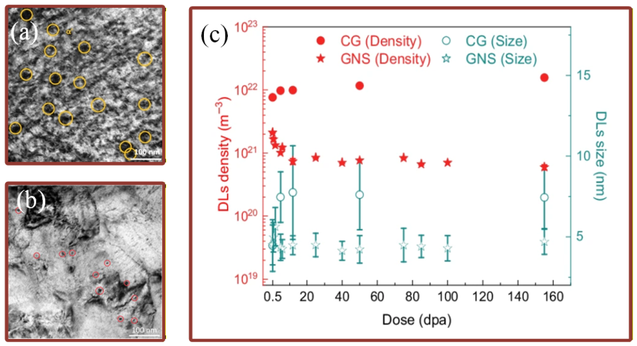
In Face-Centered Cubic (FCC) metals, the high atomic packing density and low vacancy migration energy inherently promote long-range defect diffusion. As illustrated in Figure 3, austenitic stainless steels exhibit a complex evolution of dislocation networks (DNs) and stacking faults (SFs) under irradiation. In gradient nanostructured (GNS) samples, the density of dislocation loops is reduced and SFs form a more distributed network, facilitating γ-to-α′ martensitic transformation near SFs. This highlights the role of nanoscale defect structures and interfaces in enhancing irradiation tolerance [45]. Recent studies highlight that gradient nanostructured surfaces can fundamentally enhance radiation tolerance by activating an adaptive martensitic transformation mechanism near the boundary [45]. This surface-induced transition effectively suppresses localized damage by absorbing primary defects. Furthermore, radiation-induced segregation (RIS) at these sites is highly sensitive to thermal environments and surface proximity. Such temperature-dependent chemical redistribution, driven by enhanced vacancy mobility, underscores that near surface stability is primarily dictated by surface mediated defect kinetics [46].
In contrast, Body-Centered Cubic (BCC) metals, such as tungsten (W), exhibit distinct recovery stages involving self-interstitial atoms, monovacancies, and multivacancy-hydrogen complexes, which are interpreted through specific migration barriers [47]. Due to the high migration resistance of vacancies in BCC lattices, these materials are highly sensitive to interfacial engineering. Studies on W-based composites and nanolayered films have confirmed that interfaces act as dominant sinks that regulate defect fluxes and suppress helium bubble nucleation [48,49,50]. By guiding defects toward surfaces or heterojunctions, these interfaces mitigate stress concentration and enhance the structural integrity of the near surface region.
In Hexagonal Close-Packed (HCP) systems, radiation damage is uniquely governed by inherent crystallographic anisotropy. Unlike Cubic systems, defect migration in HCP metals exhibits a strong preference for basal or prismatic planes. The presence of a free surface further breaks the symmetry of these migration paths, leading to the asymmetric growth of dislocation loops. Molecular dynamics studies indicate that vacancies and interstitials in HCP structures interact differently with the surface strain field, creating distinct ‘denuded zones that are orientation dependent. This surface driven anisotropic evolution is a precursor to macroscopic dimensional instability, such as irradiation-induced growth (IIG) [9]. In these systems, the surface state, encompassing grain orientation and the protective ZrO2 oxide layer, collaborates with anisotropic defect transport to dictate the long-term dimensional stability of the material and its resistance to irradiation-induced failure. For instance, recent studies on zirconium alloys reveal that Fe addition modifies irradiation-induced dislocation loop evolution, thereby influencing surface hardness and radiation response [32].

3.4. Ceramics and Oxides

Ceramics and oxides, including structural, high-entropy ceramics, and functional oxide systems, offer immense potential for extreme environments due to their exceptional thermal and chemical stability. Unlike metals, the radiation response of these materials is dictated by dense crystal packing and high bonding energies. In these materials, the free surface acts as a critical symmetry-breaking boundary that modulates displacement cascades. The high surface-to-volume ratio in advanced ceramic architectures ensures that surface-mediated defect annihilation and phase transformations play a dominant role in determining the radiation tolerance of the materials.
In oxide systems, radiation tolerance is increasingly linked to in situ defect recombination and surface morphology control. Wang et al. reported that heavy-ion irradiation of MgO-Nd2Zr2O7 composite ceramics induces surface roughening, swelling, and a pyrochlore-to-fluorite phase transition, directly linking surface topography evolution to structural degradation [28].
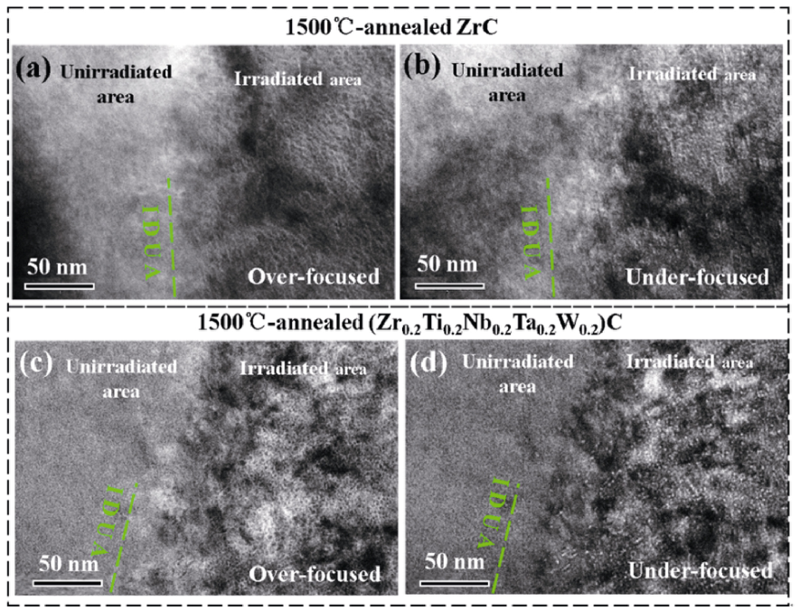
High-entropy ceramics (HECs) demonstrate exceptional structural stability due to severe local lattice distortions that suppress defect aggregation. As shown in Figure 4, Xin et al. [29] demonstrated that after annealing at 1500 °C, single-phase ZrC exhibited severe coarsening of helium bubbles near the surface damage layer, with an average size reaching 4.3 nm. In contrast, the (Zr0.2Ti0.2Nb0.2Ta0.2W0.2)C high-entropy ceramic displayed superior resistance to surface damage. The high-entropy structure effectively suppressed the migration and sequestration of helium near the surface, significantly reducing the average bubble size to 1.9 nm, thereby mitigating the degradation of surface microstructure induced by helium accumulation. Zhu et al. [51] identified the formation of defect-depleted zones near the surface of (WTiVNbTa)C5 at 650 K, demonstrating that surface proximity enhances defect recovery and prevents amorphization under high-fluence self-ion irradiation.
The performance of nuclear fuel cladding (typically Zircaloy-4) is critically dependent on the evolution of the surface ZrO2 layer formed during service. Giniyatova et al. [52] revealed that heavy-ion irradiation of CeO2 ceramics (a surrogate for nuclear fuels) induces surface cracking and spallation at high doses due to displacement damage and electronic excitation, threatening the mechanical integrity of the cladding surface.

3.5. Semiconductor Materials

Radiation damage in semiconductor materials is fundamentally governed by the generation and interaction of displacement defects. High-energy particles induce displacement cascades, creating recombination centers within the bandgap that reduce carrier mobility and lifetime while altering doping concentrations.
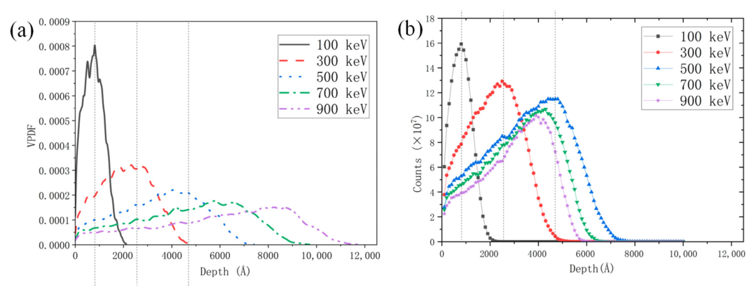
Beyond metallic systems, semiconductor materials—including bulk silicon devices and two-dimensional layered structures (e.g., MoS2)—exhibit extreme sensitivity to radiation due to their narrow bandgaps and high surface-to-volume ratios. For two-dimensional semiconductors, the damage mechanism shifts towards a surface-dominated interplay between elastic and inelastic scattering. Speckmann et al. [53] demonstrated that the sulfur displacement cross-section in monolayer MoS2 rises significantly above 80 keV, where surface-bound knock-on processes distinguish its behavior from the purely elastic mechanisms seen in graphene. Furthermore, the depth-dependent nature of surface damage is highlighted in silicon-based substrates under Ar+ irradiation (100–900 keV); low-energy ions concentrate high vacancy densities (up to ~0.0008 Å−3) at the immediate surface vacuum–solid interface, whereas higher energies shift the maximum defect density into the near-surface region as shown in Figure 5 [54]. Wide-bandgap (WBG) semiconductors, such as SiC and β-Ga2O3, exhibit superior radiation hardness due to their high bond energies and critical electric fields. However, they remain susceptible to displacement damage that traps primary carriers near critical interfaces. Under proton irradiation, β-Ga2O3 Schottky barrier diodes (SBDs) experience significant reductions in forward current and carrier concentration, primarily driven by Ga vacancies acting as deep-level traps near the metal–semiconductor interface [38]. In contrast, SiC-SBDs demonstrate higher stability with less pronounced current degradation and a notable increase in breakdown voltage. These divergent responses stem from the high defect formation energies in WBG materials, which suppress vacancy migration and agglomeration toward the surface. Such surface-mediated transport properties allow WBG electronics to maintain better functional integrity than conventional silicon-based devices under extreme irradiation [38].

3.6. Polymers and Carbon-Based Materials

The irradiation response of polymers and carbon-based materials is intrinsically linked to their molecular architecture, bonding energy, and the specific nature of energy deposition at surfaces and interfaces. Damage mechanisms in these systems primarily involve molecular chain modifications, defect-induced phase transformations, and surface-driven performance regulation. Due to their high surface-to-volume ratios, these materials often exhibit surface degradation that precedes bulk failure, making surface effects the dominant factor in determining their radiation tolerance.
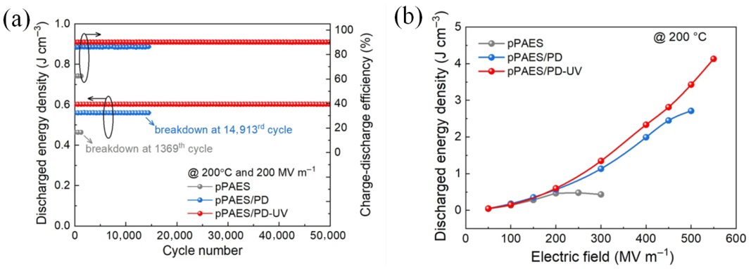
For polymers, irradiation primarily triggers chain scission, crosslinking, and functional group evolution at the surface. Furthermore, as shown in Figure 6, surface-driven modifications such as the introduction of deep-level charge traps via ultraviolet or gamma-ray irradiation effectively suppress conduction loss. For instance, polymer dots (PDs) in organic composites create traps that enhance high-temperature energy density [55]. Qu et al. [56] showed that UV irradiation of polyimide films induces surface roughening and a significant decrease in nanohardness and Young’s modulus, with the damaged layer progressively deepening with exposure time. Similarly, Shi et al. [34] reported that UV-aged carbon fiber/epoxy composites develop surface microcracks and fiber exposure due to matrix degradation, leading to substantial losses in tensile and flexural strength. Xu et al. [57] further demonstrated that UV irradiation of polyimide films causes surface oxygen enrichment, increased hydrophilicity, and chain scission, with the degradation morphology evolving into a honeycomb-like structure at high doses.
In contrast, certain irradiation conditions can improve surface properties. Naikwadi et al. [58] reviewed that gamma irradiation of polymers can induce surface crosslinking, enhancing hardness, wear resistance, and chemical stability while increasing hydrophilicity due to polar group formation. Wu et al. [59] found that electron-irradiated transparent polyimide films retain excellent optical transparency and thermal stability, with only slight surface roughening and increased hydrophilicity, demonstrating the importance of molecular design (bulky side groups, fluorine incorporation) in radiation tolerance. Zaharescu et al. [60] highlighted that controlled irradiation of polymer composites can tailor surface wettability, adhesion, and barrier properties by balancing crosslinking and chain scission.
In carbon-based materials, the surface plays a dual role as both a defect source and a sink. Arregui et al. [61] investigated neutron-irradiated glassy carbon and observed surface-connected nanopore closure and a temperature-dependent sp2-to-sp3 bonding transition, leading to significant densification and volume shrinkage. This demonstrates that the near-surface region is the primary site for irradiation-induced structural reorganization.
In nanocomposites, interfaces act as critical defect traps. Zong et al. [62] performed molecular dynamics simulations on NiFe-graphene nanocomposites and found that the graphene interface effectively absorbs point defects and promotes recombination, suppressing void formation in the metal matrix. This surface-mediated effect is amplified by the chemical complexity of the metal matrix.
For carbon nanotubes (CNTs), surface defects such as vacancies and C-C bond breakages follow the Griffith criterion, where the characteristic length of surface defects dictates the ultimate mechanical strength reduction. Xu et al. [63] further reported that heavy-ion irradiation of carbon foils and graphene films causes surface sputtering and thinning, with multilayer graphene exhibiting superior structural stability compared to amorphous carbon or graphene oxide due to interlayer energy dissipation.
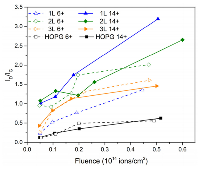
Furthermore, the damage morphology is highly sensitive to the incident ion’s electronic properties at the extreme surface. Figure 7 demonstrates the significant ion charge state dependence of irradiation damage in monolayer graphene. When the surface is struck by ions in a high equilibrium charge state (Q = 14+), the localized deposition of intense potential energy triggers severe structural degradation. In contrast, multi-layered graphene and graphite show no such charge state effect [64]. This indicates that subsurface layers effectively act as a functional sink for surface-excited electrons, shielding the material from potential energy-driven surface damage. Finally, for carbon nanotubes (CNTs), surface defects such as vacancies and C-C bond breakages follow the Griffith criterion, where the characteristic length of surface defects dictates the ultimate mechanical strength reduction [65].

4. Simulation Methods and Characterization Techniques

4.1. Simulation Methods

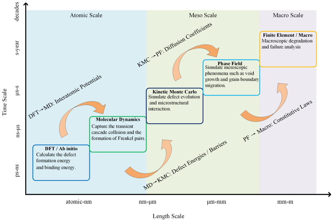
Irradiation damage is a typical scientific issue spanning multiple temporal and spatial scales. As shown in Figure 8, this process involves an extreme range from atomic nucleus collisions (10−15 s, 10−10 m) to the evolution of microscale defects, and further to the degradation of service performance of macroscale components (10−8 s, 1 m). To compensate for the limitations of a single experimental method in temporal and spatial resolution, modern nuclear materials research has developed a hierarchical multiscale coupling system.
In radiation damage research, multiscale simulation methods are essential for bridging microscopic defect evolution and macroscopic performance. First-principles calculations based on density functional theory (DFT) serve as fundamental tools for revealing the core characteristics of radiation-induced defects, particularly at symmetry-breaking boundaries. Their primary value lies in the precise quantification of defect energetics and structural stability near surfaces. DFT allows for the direct calculation of formation energies, binding energies, and migration barriers for vacancies and interstitials, providing reliable physical parameters for subsequent scales [66].
For example, in body-centered cubic iron (BCC-Fe), DFT calculations clarify the stability of self-interstitial atom (SIA) configurations, such as dumbbells and crowdions, and their migration behaviors, which are essential for calibrating Mendelev-type potentials near boundaries [67]. In the wide-bandgap semiconductor β-Ga2O3, this method can reveal significant differences in defect formation energy among different crystal planes (e.g., (100), (010)), and elaborate on the electronic structure characteristics and formation energy evolution laws of gallium vacancies, oxygen interstitials, and antisite defects [68]. For goethite (α-FeO(OH)), a material relevant to environmental applications, DFT can be used to quantify the regulatory effect of aluminum substitution and radiation damage on helium atom diffusion channels, clarifying the role of vacancies as He trapping sites and their impact on diffusion barriers [69]. Furthermore, in zinc oxide (ZnO) systems, by analyzing how oxygen vacancies and zinc interstitials modulate the electronic band structure, DFT provides a theoretical basis for enhancing the radiation tolerance of optoelectronic devices through surface-state engineering [70].
Molecular dynamics (MD) simulations excel at capturing the transient evolution of displacement cascades and cascade cooling processes [71]. By coupling high-precision force fields, such as machine learning-based and ZBL repulsive potentials, MD simulations track the atomic-scale trajectory of energy deposition and defect nucleation near boundaries. In silicon (Si) studies, MD successfully resolves surface crater evolution and localized cluster aggregation, directly linking single-ion impact dynamics to macroscopic surface pattern formation [72,73]. For antiprotons decelerating through polymer and metal-coated foils, MD utilizing quantum chemistry-derived potentials captures nuclear scattering-induced deflection and annihilation. This approach quantifies transmission probabilities across complex interfaces, providing a mechanistic understanding of how surface coatings and functional thin films modulate particle–matter interactions at the extreme vacuum–solid boundary [74,75].
Kinetic Monte Carlo (KMC), particularly Object-oriented KMC (OKMC), bridges the gap between atomic-scale dynamics and experimental timescales by employing statistical mechanics [76]. In the study of tungsten (W) as a plasma-facing material, OKMC parameterized by DFT accurately reproduces the implantation and thermal desorption spectra (TDS) of hydrogen isotopes [77]. It reveals how near-surface trapping sites, such as vacancies and impurities, govern the retention and recycling of isotopes at the vacuum–solid interface. Furthermore, in Fe-C systems, KMC successfully quantifies the dose-rate effects and the competition between surface-sink absorption and bulk recombination [78]. By simulating the flux-dependent evolution of interstitial loops and their interaction with microstructural features such as grain boundaries and dislocations, KMC clarifies how surface-mediated defect loss offsets radiation hardening at high doses. This provides a mechanistic link between surface-proximity kinetics and macroscopic performance degradation, enabling the prediction of material evolution trends under prolonged irradiation by accurately modeling the interactions between defects and diverse microstructural sinks.
For Gd2Ti2O7 pyrochlore, a candidate material for nuclear waste storage, adaptive KMC (aKMC) extends simulations to the second time scale, capturing rare events such as high-barrier ionic diffusion that MD cannot resolve. It clarifies the long-term healing process of radiation damage, showing that defect counts can be reduced to 30% of the initial value, providing key support for evaluating material long-term stability [79]. In Fe-Cu alloys, KMC simulates the nucleation and growth of copper-rich precipitates under neutron irradiation, accurately reproducing the saturation trend of precipitates with dose and offering a quantitative basis for understanding alloy embrittlement mechanisms [80].
As a macroscopic simulation tool, phase field models link microstructural evolution with macroscopic material responses through the introduction of order parameters. This method efficiently simulates the spatiotemporal distribution of defects, the growth of voids/bubbles, and grain boundary migration in large-scale systems without explicitly tracking atomic movements [81].
Currently, PF models are mainly used to study void and bubble evolution in radiation effects. They integrate thermodynamic and kinetic properties (such as chemical free energy and vacancy mobility) as functions of temperature and defect concentrations, enabling accurate reproduction of key phenomena like the quasi-bell-shaped temperature dependence of void swelling [82]. For void nucleation, advanced models incorporate stochastic fluctuations from collision cascades and diffusion jumps, treating it as precipitation in metastable point-defect solutions and achieving consistency with sharp-interface model results [83]. In radiation-induced segregation (RIS) studies, PF models successfully couple dislocation climb with point defect and chemical species transport, predicting Cr enrichment/depletion near dislocations and symmetric tilt grain boundaries (STGBs) in Fe-Cr alloys, which aligns with atom probe tomography data [84]. Aside from the aforementioned methods, Basaran developed the entropy-based Unified Mechanics Theory (UMT), which has been proven effective in evaluating the damage state of electronic packaging materials and structures [85,86,87]. Future studies can be performed using UMT to assess radiation damage evaluation of materials.

4.2. Characterization Techniques

In radiation damage research, characterization techniques are essential for revealing the changes in material microstructure, defect evolution, and performance degradation. Various characterization methods provide detailed information at multiple scales, from microscopic to macroscopic, thereby helping to elucidate the underlying mechanisms of radiation-induced damage.
Transmission electron microscopy (TEM) is a classic technique for investigating the microstructural changes induced by radiation damage, allowing for the direct observation of point defects, dislocations, defect clusters, and other microstructural features in materials. TEM, through high-resolution imaging and electron diffraction techniques, can accurately characterize the evolution of the microstructure of materials during irradiation, including changes in grain size, defect aggregation, and the formation of new phases [88]. It enables the quantification of defect number density, size, and spatial distribution. However, it is important to account for potential radiation damage induced by the electron beam itself, such as atomic displacement, sputtering, and radiation-induced degradation, which can be minimized by carefully controlling parameters like incident energy and dose.
X-ray photoelectron spectroscopy (XPS) serves as a critical tool for quantifying the chemical state and elemental distribution within the top 1–10 nm of irradiated surfaces, providing insights into radiation-induced segregation (RIS) and the evolution of surface oxide layers. By utilizing XPS depth profiling combined with ion beam etching techniques—such as monoatomic Ar+ or gas cluster ion beams (GCIB)—researchers can precisely map the spatial distribution of elements and chemical gradients from the surface into the bulk, which is essential for understanding the stability of functional coatings and nuclear cladding materials [89]. Furthermore, XPS can detect subtle shifts in binding energy and valence band structures to reveal irradiation-induced defects and phase transitions, though care must be taken to distinguish between radiation-induced damage and potential chemical reduction or structural alterations caused by the sputtering process itself during depth analysis.
Atomic force microscopy (AFM) is a powerful technique for evaluating the surface topographical and mechanical modifications induced by radiation damage at the nanoscale. By utilizing a high-resolution probe to scan the material surface, AFM can provide three-dimensional morphological data to identify radiation-induced features such as increased surface roughness, crater formation, and structural fragmentation. Beyond topographic imaging, AFM-based nanoindentation and peak force tapping modes enable the quantification of localized mechanical changes, including variations in Young’s modulus and adhesion, which reflect the underlying degradation of the material’s molecular or crystalline network [90]. Unlike electron-based techniques, AFM operates effectively under ambient conditions and provides a non-destructive means to assess surface integrity, though its observations are primarily limited to surface-level effects and can be influenced by the tip–sample interaction and environmental humidity.
Scanning electron microscopy (SEM) is a versatile technique for investigating the surface morphological evolution and macro-structural degradation induced by radiation damage, providing direct visualization of surface modifications such as roughening, cracking, and the formation of irradiation-induced craters or hillocks. Through secondary electron imaging and backscattered electron analysis, SEM can effectively characterize the structural integrity of materials under various irradiation conditions, including changes in surface porosity, grain boundary decoration, and the mechanical failure of low-dimensional structures like nanowires [91,92]. It enables the evaluation of large-scale damage patterns and surface-to-bulk transitions that are often inaccessible via higher-resolution but smaller-scale microscopy. However, high-magnification SEM observations must carefully manage the electron beam current and dwell time to prevent beam-induced surface charging or carbon contamination, which can obscure the authentic irradiation-induced features and lead to misinterpretation of the surface damage state.
Beyond traditional microscopy, Electron Backscatter Diffraction (EBSD) has emerged as a powerful tool for the quantitative mapping of surface-localized structural evolution under irradiation. As demonstrated by Trager-Cowan et al. [93], EBSD offers a high spatial resolution (~20 nm) capable of detecting subtle orientation changes as small as 0.02, which is critical for characterizing irradiation-induced surface tilt and lattice distortions. In the context of surface damage, EBSD allows for the statistical analysis of microstructural features such as dislocations and grain boundaries near the vacuum–solid interface, providing a direct link between micro-scale defect accumulation and macroscopic surface roughening. Furthermore, when combined with electron channeling contrast imaging (ECCI), EBSD facilitates the in situ observation of individual dislocations and their interaction with the surface strain field, offering indispensable insights into the surface-mediated mechanical degradation of irradiated materials.
Raman spectroscopy is a powerful non-destructive technique for quantifying structural disorder and phase transformations in materials subjected to radiation damage, particularly in carbon-based materials and ceramics. By analyzing characteristic vibrational modes, such as the evolution of the D-band to G-band intensity ratio (I_D/I_G) and G-band position shifts, Raman spectroscopy can accurately evaluate changes in crystallite size, defect density, and the degree of amorphization [37,94,95]. It enables the detection of lattice strain and the quantification of total irradiation damage even in specimens with steep damage gradients, making it ideal for characterizing depth-dependent structural evolution from the surface to the bulk. However, while Raman spectroscopy provides excellent sensitivity to crystalline quality and chemical bonding, the interpretation of spectra can be complex due to the overlapping of defect-induced peaks and potential fluorescence interference, requiring sophisticated spectral deconvolution to distinguish between different types of radiation-induced microstructural features.
Nanoindentation and electrical resistivity testing are effective techniques for characterizing macroscopic material properties. Nanoindentation testing, through continuous stiffness measurement and other methods, can evaluate the impact of radiation damage on mechanical properties such as hardness and elastic modulus, making it particularly useful for assessing the mechanical response of the shallow subsurface regions affected by irradiation [96]. During testing, it is crucial to consider the matching of indenter geometry, the size of the plastic zone, and the thickness of the irradiated layer to avoid substrate effects. Electrical resistivity testing, on the other hand, monitors changes in conductivity to reflect the influence of defects on electronic transport. Defects and transmutation elements scatter conduction electrons, resulting in an increase in resistivity [97]. This method can quantify the degradation of electrical properties induced by irradiation, and when combined with the rule of Matthiessen, it allows the separation of contributions from defects, transmutation elements, and microstructural changes to the resistivity.
Atom probe tomography (APT), with its atomic-scale spatial resolution and elemental identification capabilities, allows for precise three-dimensional analysis of defects, element distribution, and their relationship to radiation damage in materials. APT can capture atomic-level element segregation, defect–element interactions, and the formation of small precipitates, providing a clear representation of the coupling between radiation-induced compositional fluctuations and defect evolution [98]. It is particularly advantageous for studying radiation-induced element enrichment or depletion in multi-component alloys and compositional segregation around defects, offering direct atomic-scale evidence for understanding the microstructure-macroscopic performance relationship.
The integrated application of various characterization techniques provides a more comprehensive understanding of the microstructural mechanisms and macroscopic performance changes induced by radiation damage, offering critical data to support the optimization of material radiation tolerance.

5. Future Challenges and Radiation Resistance Strategies

5.1. Future Challenges

Future research on radiation damage faces the dual challenge of extreme environments and complex damage mechanisms, with the core focus on coupling effects, adaptation to extreme conditions, and cross-scale predictions. In fusion reactors and advanced fission reactors, the synergistic effects of high-energy particle irradiation and the transformation gases, such as hydrogen and helium intensify damage evolution. In fusion reactors, 14.1 MeV neutron irradiation generates numerous defects, while helium atoms promote bubble nucleation and growth, threatening the stability of the first wall and cladding materials [99]. In fourth-generation fission reactors, operating at temperatures ranging from 825 to 1275 K and under high doses exceeding 200 dpa, lattice distortion, phase separation, and radiation creep interactions lead to rapid degradation of mechanical properties [100].
In space applications, the low-dose rate mixed irradiation presents unique challenges. Long-term exposure to galactic cosmic rays and solar high-energy particles results in slow accumulation of damage, while the high linear energy transfer (LET) of heavy ions causes localized severe damage [101]. The low-dose rate damage mechanisms differ significantly from those observed in terrestrial experiments, with limited data supporting predictions of long-term service life. Furthermore, balancing radiation resistance with the need for lightweight and high mechanical performance remains a critical issue.
Additionally, the cross-scale correlations of multi-scale damage mechanisms are not yet fully understood. The coupling between atomic-scale point defect evolution and macroscopic performance degradation remains unclear, hindering the accurate development of predictive models linking micro-defects to macroscopic performance. This limits the efficient design of radiation-resistant materials [102].

5.2. Radiation Resistance Strategies

The development of radiation-resistant strategies has progressed toward an integrated, three-tiered system focused on material design, structural control, and mechanism optimization. At its core, this strategy relies on multi-scale regulation to achieve the dual objectives of defect suppression and annihilation.
In material composition design, high-entropy alloys (HEAs) and refractory high-entropy alloys (RHEAs) have demonstrated significant potential. The high mixing entropy and low diffusion coefficients of these multi-principal-element alloys form homogeneous solid solutions, which effectively hinder defect migration and aggregation. For instance, AlxCoCrFeNi HEAs maintain remarkable phase stability even at doses exceeding 80 dpa, where the disordered FCC/BCC phases inhibit the formation of large defect clusters that could otherwise lead to surface hardening and micro-cracking [102]. The W-Ta-Cr-V series RHEAs, through the synergistic effect of Cr and V, modulate defect migration energy barriers to suppress the growth of large dislocation loops, thereby maintaining structural and topographical integrity at 8 dpa [103]. Furthermore, atomistic simulations of Ti-based alloys indicate that higher displacement threshold energies (e.g., 55 eV in Ti-15V-3Cr-3Sn-3Al) effectively reduce the initial defect production rate, which is a key factor in minimizing the accumulation of radiation-induced damage near the material surface [104]. In addition, the introduction of carbon nanomaterials into diverse matrices has emerged as a high-performance strategy to enhance surface stability and structural integrity. For metal-matrix composites, the uniform dispersion of carbon nanotubes (CNTs) in aluminum significantly improves radiation resistance by providing a high density of one-dimensional sinks that promote rapid defect recombination, effectively suppressing the formation of large dislocation loops and voids that would otherwise trigger surface roughening and micro-cracking [105]. In polymer-based systems, hybrid carbon fillers demonstrate superior synergistic effects in maintaining surface chemistry. For instance, the incorporation of nanodiamonds (NDs) and graphene nanoplatelets (GNPs) into poly(ethylene terephthalate) (PET) enhances resistance to 3 MeV proton irradiation by stabilizing the polymer chains and reducing surface oxidative degradation [106]. Similarly, graphene-reinforced epoxy composites exhibit excellent stability under γ-radiation, where graphene sheets act as effective radical scavengers to prevent surface ablation and preserve the mechanical integrity of the outermost layers even at high cumulative doses [107].
In terms of microstructure regulation, nano-structural engineering and phase stability design have shown significant effects. Oxide dispersion strengthening (ODS) technology, which introduces nano-oxide particles into ferritic/martensitic steels, effectively captures defects and suppresses bubble formation [100]. In martensitic steels, B2-ordered superlattice nano-precipitates are used to facilitate in situ defect recombination through reversible disorder-order transitions, maintaining zero swelling even at high doses of 2350 dpa [108]. SiC/SiC composite materials, through optimized interface bonding and processing techniques (such as chemical vapor infiltration and nanopowder infiltration), enhance mechanical stability and thermal conductivity retention under high-temperature irradiation [99]. Carbon nanotube-based complementary metal–oxide–semiconductor (CMOS) electronics, fabricated on paper substrates, demonstrate remarkable robustness against total ionizing dose (TID) effects. These devices maintain stable voltage gains and high noise margins even in extreme radiation environments, benefiting from the inherent structural stability of the CNT channels and the optimized enhancement-mode (E-mode) configuration [109].
Interface and defect management play a crucial role in these strategies. Interface engineering is a key approach. Cu-Nb nanolayered composites, fabricated via accumulative roll bonding (ARB), use heterogeneous interfaces to trap defects and promote defect recombination, thus inhibiting helium bubble growth [110]. By combining atom probe tomography (APT), transmission electron microscopy (TEM) characterization, and molecular dynamics (MD) and kinetic Monte Carlo (KMC) simulations, multi-scale tracking of defect evolution can be achieved, providing precise guidance for strategy optimization [102,103].

6. Conclusions

This review systematically summarizes the surface effects of irradiation damage in metallic, ceramic, and polymeric systems, emphasizing the dominant role of the surface as a non-equilibrium sink in defect evolution kinetics. It is demonstrated that the surface is not only a center for point defect annihilation, but its energetic state and structural characteristics also directly determine the critical conditions for morphological instabilities, such as blistering, spallation, and ablation. By analyzing the radiation resistance mechanisms of high-entropy alloys, nano-structural engineering, and carbon-based composites, it is found that utilizing high-density interfaces to induce near-surface defect recombination, reducing atomic diffusion rates, and introducing chemical radical scavengers are effective strategies for maintaining surface integrity. Notably, the sub-2 nm helium bubble distribution observed in high-entropy ceramics at high temperatures provides crucial empirical evidence for developing extreme-environment materials that possess both structural stability and surface damage resistance.
Despite significant progress in this field, precise prediction and proactive design of radiation-resistant surfaces remain challenging. Future research should focus on the following directions: First, advanced in situ characterization techniques must be developed to achieve real-time monitoring of surface atomic reconstruction and crack initiation under the coupling of irradiation, mechanical loading, and corrosive environments. Second, a machine learning-based multi-scale computational framework should be established to quantitatively describe the correlation between near-surface microscopic defect evolution and macroscopic service life. Finally, a paradigm shift from “passive radiation resistance” to “active self-healing” should be explored, leveraging irradiation-induced energy to drive surface lattice reorganization for proactive damage recovery. In summary, in-depth clarification of surface-mediated irradiation damage mechanisms will provide core theoretical support for the reliability assessment and material development of next-generation nuclear energy systems, deep-space exploration equipment, and high-performance microelectronic devices.

Author Contributions

The contributions from each author are listed: Conceptualization, X.W. and J.Z.; investigation, J.Y., Y.H., Y.Z. and H.Z.; resources H.Z.; writing—original draft preparation, J.Y. and J.Z.; writing—review and editing, J.Y., Y.H., J.Z., T.R. and K.J.; supervision, Y.Z. and J.Z.; funding acquisition, J.Z. and H.Z. All authors have read and agreed to the published version of the manuscript.

Funding

This work is financially sponsored by the Open Fund of National Key Laboratory of Materials Behavior and Evaluation Technology in Space Environment (Program No. JZJJXN20240004), the National Natural Science Foundation of China (Program No. 52505211), the Natural Science Basic Research Program of Shaanxi Province (Program No. 2023-JC-QN-0005), Fundamental Research Funds for the Central Universities (ZYTS24032), the Youth Talent Support Program of the China Association for Science and Technology (YESS20230523), the Youth Talent Promotion Project of Gansu Province (GXH20210611-05), the Excellent Youth Foundation of Gansu Province (26JRRA031) the Science and Technology Major Project of Gansu Province (24 ZD13GA007), and the Young Talents of China Aerospace Science and Technology Corporation.

Institutional Review Board Statement

Not applicable.

Informed Consent Statement

Not applicable.

Data Availability Statement

No new data were created or analyzed in this study.

Conflicts of Interest

Author Yaqian Huang was employed by the company Amazon AWS Finance, Amazon re:Invent. The remaining authors declare that the research was conducted in the absence of any commercial or financial relationships that could be construed as a potential conflict of interest.

References

  1. Zinkle, S.J.; Busby, J.T. Structural materials for fission & fusion energy. Mater. Today 2009, 12, 12–19. [Google Scholar] [CrossRef]
  2. Duzellier, S. Radiation effects on electronic devices in space. Aerosp. Sci. Technol. 2005, 9, 93–99. [Google Scholar] [CrossRef]
  3. Kiritani, M.; Yoshiie, T.; Kojima, S.; Satoh, Y.; Hamada, K. Fission-fusion correlation by fission reactor irradiation with improved control. J. Nucl. Mater. 1990, 174, 327–351. [Google Scholar] [CrossRef]
  4. Short, M.P.; Yip, S. Materials aging at the mesoscale: Kinetics of thermal, stress, radiation activations. Curr. Opin. Solid State Mater. Sci. 2015, 19, 245–252. [Google Scholar] [CrossRef]
  5. Kiritani, M. Microstructure evolution during irradiation. J. Nucl. Mater. 1994, 216, 220–264. [Google Scholar] [CrossRef]
  6. Barabash, V.; Federici, G.; Linke, J.; Wu, C. Material/plasma surface interaction issues following neutron damage. J. Nucl. Mater. 2003, 313, 42–51. [Google Scholar] [CrossRef]
  7. Look, D.C.; Reynolds, D.; Hemsky, J.W.; Jones, R.; Sizelove, J. Production and annealing of electron irradiation damage in ZnO. Appl. Phys. Lett. 1999, 75, 811–813. [Google Scholar] [CrossRef]
  8. Was, G.; Jiao, Z.; Getto, E.; Sun, K.; Monterrosa, A.; Maloy, S.; Anderoglu, O.; Sencer, B.; Hackett, M. Emulation of reactor irradiation damage using ion beams. Scr. Mater. 2014, 88, 33–36. [Google Scholar] [CrossRef]
  9. Torres, E.; Maxwell, C. Effect of irradiation damage on the tensile deformation of α-zirconium systems: A molecular dynamics study. Comput. Mater. Sci. 2023, 222, 112088. [Google Scholar] [CrossRef]
  10. Haag, J.V., IV; Fu, Y.; Jiang, W.; Matthews, B.E.; Olszta, M.J.; Edwards, D.J.; Setyawan, W. Quantitative assessment of Ni+ and He+ ion irradiation damage in a tungsten heavy alloy under the simulated nuclear fusion environment. Sci. Rep. 2025, 15, 7066. [Google Scholar] [CrossRef]
  11. Nordlund, K.; Zinkle, S.J.; Sand, A.E.; Granberg, F.; Averback, R.S.; Stoller, R.E.; Suzudo, T.; Malerba, L.; Banhart, F.; Weber, W.J. Primary radiation damage: A review of current understanding and models. J. Nucl. Mater. 2018, 512, 450–479. [Google Scholar] [CrossRef]
  12. Averback, R. Atomic displacement processes in irradiated metals. J. Nucl. Mater. 1994, 216, 49–62. [Google Scholar] [CrossRef]
  13. Zinkle, S.; Singh, B. Analysis of displacement damage and defect production under cascade damage conditions. J. Nucl. Mater. 1993, 199, 173–191. [Google Scholar] [CrossRef]
  14. Baumer, R.; Demkowicz, M. Radiation response of amorphous metal alloys: Subcascades, thermal spikes and super-quenched zones. Acta Mater. 2015, 83, 419–430. [Google Scholar] [CrossRef]
  15. Wei, X.-P.; Li, H.; Zhang, Y.; Li, X.; Li, C. Molecular dynamics simulations of primary damage formation and tensile properties of irradiated Cu–Ni binary alloys. J. Mater. Res. Technol. 2025, 38, 2148–2167. [Google Scholar] [CrossRef]
  16. Zhang, T.; Zeng, Y.; Li, Y.-F.; Huang, H.-Z.; Gul Niazi, S. Multi-Directional Displacement Threshold Energy and Crystal Irradiation Damage Model. Appl. Sci. 2023, 13, 10701. [Google Scholar] [CrossRef]
  17. Azevedo, C.R.d.F. A review on neutron-irradiation-induced hardening of metallic components. Eng. Fail. Anal. 2011, 18, 1921–1942. [Google Scholar] [CrossRef]
  18. Ye, T.; Wu, Y.; Wang, Z.; Zhang, J.; Wang, M.; Chen, P.; Tian, W.; Su, G.; Qiu, S. Simulation of threshold displacement energy in Fe-Cr-Al alloys using molecular dynamics. J. Nucl. Mater. 2024, 588, 154821. [Google Scholar] [CrossRef]
  19. Pan, R.; Zhou, M.; Cui, J.; Fu, B.; Hou, Q.; Qin, J.; Kong, X.; Ma, C.; Wang, Q.; Wu, L. Unveiling the characteristics of the residual point defects of collision cascade in Zr-xNb binary system: A molecular dynamics study. J. Nucl. Mater. 2023, 584, 154546. [Google Scholar] [CrossRef]
  20. Yu, H.; Yi, X.; Chen, Q.; Fang, H.; Dong, S.; Li, S.; Han, W.; Liu, P.; Ohnuki, S.; Wan, F. Low-dose damage evolution in pure magnesium under electron irradiation: Effect of foil orientation and pre-existing dislocations. Nucl. Mater. Energy 2025, 42, 101845. [Google Scholar] [CrossRef]
  21. Huang, H.; Jiang, Y.; Li, Z.; Shen, S. High-dose Au3+ ion irradiation of Ni-17Mo-7Cr alloy at 650 °C: Understanding the evolution of damaged microstructure. J. Alloys Compd. 2025, 1047, 185005. [Google Scholar] [CrossRef]
  22. Kamlade, P.K.; Panta, J.; Mammone, M.; Yang, R.C.; Mildren, R.P.; Wang, J.; Ibrahim, M.; Thomson, R.; Zhang, Y. Degradation behaviour and damage mechanisms of carbon fibre reinforced polymer composite laminates subjected to laser irradiation. Compos. Part C Open Access 2025, 17, 100605. [Google Scholar] [CrossRef]
  23. Hao, J.; Zhang, P.; Li, X.; Wang, B.; Cai, Z.; Ma, L.; Mei, X. Evaluation of damage effects in 304 high-boron austenitic stainless steel under only and simultaneous Fe, He ions irradiation. J. Nucl. Mater. 2026, 621, 156378. [Google Scholar] [CrossRef]
  24. Saini, S.; Menon, R.; Sharma, S.K.; Nabhiraj, P.Y.; Neogy, S.; Srivastava, A.P. A comprehensive study on the impact of He ion irradiation on the microstructure and mechanical properties of a Ni-Cr-Mo alloy. Sci. Rep. 2025, 15, 28733. [Google Scholar] [CrossRef] [PubMed]
  25. Yang, X.; Li, Y.; Peng, J.; Liang, X.; Liu, W.; Liu, X.; Huang, H. Effect of proton irradiation on ferrite microstructure in aged 308L stainless steel. J. Mater. Sci. Technol. 2026, 244, 180–185. [Google Scholar] [CrossRef]
  26. Kalia, S.; Kumar, R.; Dhiman, R.; Singh, R.K. Ion irradiation/implantation induced defect engineering and modification in graphene derivatives-based nanocomposites: Energy storage/conversion and sensor. J. Energy Storage 2024, 83, 110650. [Google Scholar] [CrossRef]
  27. Xing, X.; Liu, Y.; Han, J.; Liu, W.; Wei, Z. Preparation of High Damage Threshold Device Based on Bi2Se3 Film and Its Application in Fiber Lasers. ACS Photonics 2023, 10, 2264–2271. [Google Scholar] [CrossRef]
  28. Wang, Y.; Wang, J.; Wang, J.; Kang, L.; Zhang, T.; Li, X.; Tang, Y. Micro and macro damage defects in heavy-ion irradiated MgO-Nd2Zr2O7 composite ceramics used for inert matrix fuel. J. Eur. Ceram. Soc. 2024, 44, 4804–4815. [Google Scholar] [CrossRef]
  29. Xin, X.-T.; Bao, W.; Wang, X.-G.; Guo, X.-J.; Lu, Y.; Zhu, C.; Liu, J.-X.; Li, Q.; Xu, F.; Zhang, G.-J. Reduced He ion irradiation damage in ZrC-based high-entropy ceramics. J. Adv. Ceram. 2023, 12, 916–929. [Google Scholar] [CrossRef]
  30. Corso, A.J.; Padovani, M.; Santi, G.; Hübner, R.; Kentsch, U.; Bazzan, M.; Pelizzo, M.G. Optical Thin Films in Space Environment: Investigation of Proton Irradiation Damage. ACS Appl. Mater. Interfaces 2024, 16, 38645–38657. [Google Scholar] [CrossRef]
  31. Fan, S.; Mi, B.; Wang, J.; Liu, P.; Ma, X.; Chen, T.; Li, W. Research progress on the preparation of irradiation-resistant coating based on PVD technology. J. Mater. Res. Technol. 2024, 32, 4069–4091. [Google Scholar] [CrossRef]
  32. Xia, L.; Cao, Y.; Liu, K.; Chen, D.; Jiang, C. Effect of Fe addition and ion irradiation on surface hardness in zirconium alloys: Experiments and modeling. Appl. Surf. Sci. 2024, 669, 160480. [Google Scholar] [CrossRef]
  33. Bilal, M.; Shahzad, K.; Lv, P.; Ejaz, A.; Wang, F.; Ahmad, Z.; Shah, A.; Rehman, M.; Ahmad, K.; Mian, A. Effect of helium ion irradiation on the microstructure, mechanical properties and surface morphology of Inconel 625 alloy. Mater. Chem. Phys. 2024, 319, 129286. [Google Scholar] [CrossRef]
  34. Shi, Z.; Zou, C.; Zhou, F.; Zhao, J. Analysis of the mechanical properties and damage mechanism of carbon fiber/epoxy composites under UV aging. Materials 2022, 15, 2919. [Google Scholar] [CrossRef] [PubMed]
  35. Xiao, Y.; Zhang, Y.; She, W.; Zhan, Z.; Yang, J.; Guo, L.; Deng, H. Highly efficient damage recovery in MgO: Insights from plasma-enabled atomic-scale reconstruction. J. Mater. Process. Technol. 2025, 341, 118886. [Google Scholar] [CrossRef]
  36. Chen, Y.; Xu, J.; Shen, L.; Zhao, J.; Chen, J.; Yang, Y.; Wang, L.; Zhao, J.; Wang, C. Tunable wettability of jet electrodeposited micro-nano structures modified by laser radiation. Surf. Coat. Technol. 2022, 446, 128763. [Google Scholar] [CrossRef]
  37. Jiang, M.; Ammigan, K.; Lolov, G.; Pellemoine, F.; Liu, D. A novel method for quantifying irradiation damage in nuclear graphite using Raman spectroscopy. Carbon 2023, 213, 118181. [Google Scholar] [CrossRef]
  38. Kim, Y.J.; Moon, Y.; Moon, J.H.; Kim, H.W.; Bahng, W.; Park, H.; Yoon, Y.J.; Seo, J.H. Displacement damage effect of proton irradiation on vertical β-Ga2O3 and SiC Schottky barrier diodes (SBDs). J. Sci. Adv. Mater. Devices 2024, 9, 100765. [Google Scholar] [CrossRef]
  39. Cao, Y.; Guo, H.; Ma, W.; Zhu, W.; Zhong, X.; Feng, Y.; Peng, C.; Lei, Z. Study on Proton Irradiation Damage of Diamond Schottky Barrier Diodes. IEEE Trans. Electron Devices 2026, 73, 1163–1168. [Google Scholar] [CrossRef]
  40. Tang, Y.; Zhu, H.; Wang, C.; Bu, J.; Li, M.; Zhou, H.; Zhang, J.; Ma, Y.; Wu, Z.; Liu, F. High-efficiency removal of surface adsorbates on carbon nanotubes via electron irradiation. J. Alloys Compd. 2025, 1041, 183713. [Google Scholar] [CrossRef]
  41. You, T.; Dai, Y.; Yu, J.; Ye, H.X.; Dai, W.J.; Cao, L.F. Damage regularity and multifractal analysis of sol-gel reflection coating of KDP crystal under low UV irradiation flux. Microsc. Res. Tech. 2024, 87, 2850–2861. [Google Scholar] [CrossRef] [PubMed]
  42. Wang, F.; Zhang, P.; Li, Y.; Yuan, Q.; Sun, L.; Lu, L.H.; Yuan, X. Degradation of optical components induced by production on aluminum alloy surface irradiated by stray light. Opt. Express 2024, 32, 47330–47346. [Google Scholar] [CrossRef]
  43. Zhang, W.; Deng, J.; Zhu, C.; Zhong, Y.; Yang, J.; Xin, H.; Liu, G.; Tang, R.; Feng, K.; Liu, N. Au-ion irradiation effects on microstructure and deuterium permeation resistance of Al/Al2O3 coating. Nucl. Fusion 2022, 62, 086039. [Google Scholar] [CrossRef]
  44. Liao, L.; Wan, Q.; Wang, H.; Yang, B.; Mei, Q. Irradiation enhanced corrosion resistance of CrN/TiSiN multilayers synthesized by cathodic arc ion plating. Mater. Today Commun. 2023, 35, 106155. [Google Scholar] [CrossRef]
  45. Zhang, S.; Dong, Y.; Sun, Y.; Liu, Y.; Sun, L.; Zhao, H.; Gao, N.; Wang, Z. Improved radiation resistance in metals via adaptive martensitic transformation. Nat. Commun. 2025, 16, 10995. [Google Scholar] [CrossRef]
  46. Dai, C.; Saidi, P.; Langelier, B.; Wang, Q.; Judge, C.; Daymond, M.; Mattucci, M. Radiation-induced segregation on dislocation loops in austenitic Fe-Cr-Ni alloys. Phys. Rev. Mater. 2022, 6, 053606. [Google Scholar] [CrossRef]
  47. Liski, A.; Lu, E.; Makkonen, I.; Chen, Z.; Mizohata, K.; Tuomisto, F. Migration and clustering of early-stage irradiation damage in vanadium. Phys. Rev. Mater. 2024, 8, 113602. [Google Scholar] [CrossRef]
  48. Daghbouj, N.; Sen, H.; Callisti, M.; Vronka, M.; Karlik, M.; Duchoň, J.; Čech, J.; Havránek, V.; Polcar, T. Revealing nanoscale strain mechanisms in ion-irradiated multilayers. Acta Mater. 2022, 229, 117807. [Google Scholar] [CrossRef]
  49. Li, N.; Fu, E.; Wang, H.; Carter, J.; Shao, L.; Maloy, S.; Misra, A.; Zhang, X. He ion irradiation damage in Fe/W nanolayer films. J. Nucl. Mater. 2009, 389, 233–238. [Google Scholar] [CrossRef]
  50. Cui, B.; Luo, C.; Chen, X.; Zou, C.; Li, M.; Xu, L.; Yang, J.; Meng, X.; Zhang, H.; Zhou, X. Superior radiation resistance of ZrO2-modified W composites. Materials 2022, 15, 1985. [Google Scholar] [CrossRef] [PubMed]
  51. Zhu, Y.; Chai, J.; Wang, Z.; Shen, T.; Niu, L.; Li, S.; Jin, P.; Zhang, H.; Li, J.; Cui, M. Microstructural damage evolution of (WTiVNbTa)C5 high-entropy carbide ceramics induced by self-ions irradiation. J. Eur. Ceram. Soc. 2022, 42, 2567–2576. [Google Scholar] [CrossRef]
  52. Giniyatova, S.G.; Kozlovskiy, A.L.; Rspayev, R.M.; Borgekov, D.B.; Zdorovets, M.V. Study of the kinetics of radiation damage in CeO2 ceramics upon irradiation with heavy ions. Materials 2023, 16, 4653. [Google Scholar] [CrossRef]
  53. Speckmann, C.; Lang, J.; Madsen, J.; Monazam, M.R.A.; Zagler, G.; Leuthner, G.T.; McEvoy, N.; Mangler, C.; Susi, T.; Kotakoski, J. Combined electronic excitation and knock-on damage in monolayer MoS2. Phys. Rev. B 2023, 107, 094112. [Google Scholar] [CrossRef]
  54. Liang, G.; Xu, B.; Wei, X. Collision Cascade in a Silicon-Based Device under Energetic Ar Ions Irradiation. Coatings 2023, 13, 1828. [Google Scholar] [CrossRef]
  55. Ding, J.; Zhou, Y.; Xu, W.; Yang, F.; Zhao, D.; Zhang, Y.; Jiang, Z.; Wang, Q. Ultraviolet-irradiated all-organic nanocomposites with polymer dots for high-temperature capacitive energy storage. Nano-Micro Lett. 2024, 16, 59. [Google Scholar] [CrossRef]
  56. Qu, C.; Hu, J.; Liu, X.; Li, Z.; Ding, Y. Morphology and mechanical properties of polyimide films: The effects of UV irradiation on microscale surface. Materials 2017, 10, 1329. [Google Scholar] [CrossRef]
  57. Xu, S.; Zhang, Y.; Zhao, Z.; Zhan, J.; Tian, G.; Wu, D. Effect of chemical structure on properties evolution and molecular mechanism of polyimide films under ultraviolet irradiation. Polym. Degrad. Stab. 2025, 240, 111503. [Google Scholar] [CrossRef]
  58. Naikwadi, A.T.; Sharma, B.K.; Bhatt, K.D.; Mahanwar, P.A. Gamma radiation processed polymeric materials for high performance applications: A review. Front. Chem. 2022, 10, 837111. [Google Scholar] [CrossRef] [PubMed]
  59. Wu, X.; Shu, C.; Zhong, M.; Fan, X.; Yu, Z.; Huang, W.; Yan, D. Irradiation tolerance of an optically transparent polyimide film under 1 MeV electron beam. Appl. Surf. Sci. 2022, 583, 152558. [Google Scholar] [CrossRef]
  60. Zaharescu, T.; Mariş, M. Irradiation Effects in Polymer Composites for Their Conversion into Hybrids. J. Compos. Sci. 2022, 6, 109. [Google Scholar] [CrossRef]
  61. Arregui-Mena, J.D.; Koyanagi, T.; Cullen, D.A.; Zachman, M.J.; Lin, Y.-R.; Edmondson, P.D.; Katoh, Y. Comprehensive characterization of the irradiation effects of glassy carbon. Acta Mater. 2024, 281, 120441. [Google Scholar] [CrossRef]
  62. Zong, M.; Chen, F.; Tang, X.; Ge, G.; Li, C.; Liu, Y. Molecular dynamics simulation of radiation defect evolution mechanism of NiFe-graphene nanocomposite. Appl. Surf. Sci. 2022, 584, 152503. [Google Scholar] [CrossRef]
  63. Xu, Z.; Liu, Y.; Zhang, J.; Tao, Z.; Jia, H.; Zhao, H.; Sun, L. Study on the irradiation damage effect of heavy ions on carbon foil. EPJ Web Conf. 2025, 327, 01013. [Google Scholar] [CrossRef]
  64. Tomić Luketić, K.; Hanžek, J.; Mihalcea, C.G.; Dubček, P.; Gajović, A.; Siketić, Z.; Jakšić, M.; Ghica, C.; Karlušić, M. Charge state effects in swift-heavy-ion-irradiated nanomaterials. Crystals 2022, 12, 865. [Google Scholar] [CrossRef]
  65. Liu, M.; Li, R.; Wang, J.; Wang, H.; Zhang, Y.; Zhang, R.; Li, X. Strength and fracture behaviors of ultralong carbon nanotubes with defects. Carbon 2022, 199, 300–317. [Google Scholar] [CrossRef]
  66. Ge, G.; Chen, F.; Tang, X.; Wang, Y.; Li, C. Irradiation damage versus lattice distortion in AlNbTiVCrx (x= 0, 0.5, 1) high-entropy alloys from first-principles calculations and irradiation experiments. J. Nucl. Mater. 2022, 563, 153630. [Google Scholar] [CrossRef]
  67. Malerba, L.; Marinica, M.C.; Anento, N.; Björkas, C.; Nguyen, H.; Domain, C.; Djurabekova, F.; Olsson, P.; Nordlund, K.; Serra, A.; et al. Comparison of empirical interatomic potentials for iron applied to radiation damage studies. J. Nucl. Mater. 2010, 406, 19–38. [Google Scholar] [CrossRef]
  68. Liu, T.; Li, Z.; Zhao, J.; Fei, X.; Feng, J.; Zuo, Y.; Hua, M.; Guo, Y.; Liu, S.; Zhang, Z. Orientation-dependent surface radiation damage in β-Ga2O3 explored by atomistic simulations. Acta Mater. 2025, 300, 121484. [Google Scholar] [CrossRef]
  69. Bassal, F.; Heller, B.; Roques, J.; Balout, H.; Tassan-Got, L.; Allard, T.; Gautheron, C. Revealing the radiation damage and Al-content impacts on He diffusion in goethite. Chem. Geol. 2022, 611, 121118. [Google Scholar] [CrossRef]
  70. Han, X.; Li, R.; Pan, S.; Liu, Y.; Niu, C.; Crespillo, M.L.; Zarkadoula, E.; Liu, P. Tailoring the electronic structures and spectral properties of ZnO with irradiation defects generated under intense electronic excitation: A combined experimental and DFT approach. Adv. Funct. Mater. 2024, 34, 2405885. [Google Scholar] [CrossRef]
  71. Yang, S.; Kim, S.; Oda, T. Sticking, reflection, and abstraction behavior of hydrogen irradiated on (110) tungsten surfaces at 0.1–100 eV by molecular dynamics simulations using a machine learning potential. Acta Mater. 2025, 297, 121306. [Google Scholar] [CrossRef]
  72. Norris, S.A.; Samela, J.; Bukonte, L.; Backman, M.; Djurabekova, F.; Nordlund, K.; Madi, C.S.; Brenner, M.P.; Aziz, M.J. Molecular dynamics of single-particle impacts predicts phase diagrams for large scale pattern formation. Nat. Commun. 2011, 2, 276. [Google Scholar] [CrossRef]
  73. Nordlund, K. Molecular dynamics simulation of ion ranges in the 1–100 keV energy range. Comput. Mater. Sci. 1995, 3, 448–456. [Google Scholar] [CrossRef]
  74. Nordlund, K.; Hori, M.; Sundholm, D. Large nuclear scattering effects in antiproton transmission through polymer and metal-coated foils. Phys. Rev. A 2022, 106, 012803. [Google Scholar] [CrossRef]
  75. Kesälä, E.; Kuronen, A.; Nordlund, K. Molecular dynamics simulation of pressure dependence of cluster growth in inert gas condensation. Phys. Rev. B 2007, 75, 174121. [Google Scholar] [CrossRef][Green Version]
  76. Hou, J.; Kong, X.; Hu, W.; Deng, H.; Nguyen-Manh, D.; Song, J. Deuterium trapping and desorption by vacancy clusters in irradiated Mo from object kinetic Monte Carlo simulations. Acta Mater. 2024, 274, 120014. [Google Scholar] [CrossRef]
  77. Wang, P.; Cao, Q.; Hou, J.; Kong, X.-S.; Chen, L.; Xie, Z. Implantation and desorption of H isotopes in W revisited by object kinetic Monte Carlo simulation. J. Nucl. Mater. 2022, 561, 153576. [Google Scholar] [CrossRef]
  78. Li, J.; Zhang, C.; Yang, Y.; Wang, T.; Martin-Bragado, I. Irradiation dose-rate effect in Fe-C system: An Object Kinetic Monte Carlo simulation. J. Nucl. Mater. 2022, 561, 153529. [Google Scholar] [CrossRef]
  79. Purton, J.A.; Hull, S.; Teobaldi, G. Adaptive kinetic Monte Carlo analysis of healing mechanisms in radiation damaged pyrochlore Gd2Ti2O7. Acta Mater. 2024, 267, 119726. [Google Scholar] [CrossRef]
  80. Domain, C.; Becquart, C.; Malerba, L. Simulation of radiation damage in Fe alloys: An object kinetic Monte Carlo approach. J. Nucl. Mater. 2004, 335, 121–145. [Google Scholar] [CrossRef]
  81. Millett, P.C.; Tonks, M. Application of phase-field modeling to irradiation effects in materials. Curr. Opin. Solid State Mater. Sci. 2011, 15, 125–133. [Google Scholar] [CrossRef]
  82. Li, Y.; Hu, S.; Sun, X.; Gao, F.; Henager, C.H., Jr.; Khaleel, M. Phase-field modeling of void evolution and swelling in materials under irradiation. Sci. China Phys. Mech. Astron. 2011, 54, 856–865. [Google Scholar] [CrossRef]
  83. Semenov, A.; Woo, C. Modeling void development in irradiated metals in the phase-field framework. J. Nucl. Mater. 2014, 454, 60–68. [Google Scholar] [CrossRef]
  84. Moladje, G.B.; Thuinet, L.; Becquart, C.; Legris, A. Radiation induced segregation near dislocations and symmetric tilt grain boundaries in Fe-Cr alloys: A phase-field study. Acta Mater. 2022, 225, 117523. [Google Scholar] [CrossRef]
  85. Basaran, C. Introduction to Unified Mechanics Theory with Applications; Springer Nature: Cham, Switzerland, 2023. [Google Scholar]
  86. Yao, W.; Basaran, C. Electromigration in lead-free solder joints under high-frequency pulse current: An experimental study. Int. J. Damage Mech. 2013, 22, 1127–1143. [Google Scholar] [CrossRef]
  87. Yao, W.; Basaran, C. Electromigration damage mechanics of lead-free solder joints under pulsed DC: A computational model. Comput. Mater. Sci. 2013, 71, 76–88. [Google Scholar] [CrossRef]
  88. Egerton, R.F.; Li, P.; Malac, M. Radiation damage in the TEM and SEM. Micron 2004, 35, 399–409. [Google Scholar] [CrossRef]
  89. Li, D.; Chen, Y.; Zhou, C.; Shi, C.; Xu, Z.; Miao, Z.; Xi, Z.; Han, J. XPS depth profiling of functional materials: Applications of ion beam etching techniques. Mater. Chem. Front. 2024, 8, 715–731. [Google Scholar] [CrossRef]
  90. Vadrucci, M.; Cicero, C.; Parisse, P.; Casalis, L.; De Bellis, G. Surface evaluation of the effect of X-rays irradiation on parchment artefacts through AFM and SEM. Appl. Surf. Sci. 2020, 513, 145881. [Google Scholar] [CrossRef]
  91. Cheng, Y.; Yao, H.; Duan, J.; Xu, L.; Zhai, P.; Lyu, S.; Chen, Y.; Maaz, K.; Mo, D.; Sun, Y. Surface modification and damage of MeV-energy heavy ion irradiation on gold nanowires. Nanomaterials 2017, 7, 108. [Google Scholar] [CrossRef]
  92. Nita, N.; Schaeublin, R.; Victoria, M.; Valiev, R. Effects of irradiation on the microstructure and mechanical properties of nanostructured materials. Philos. Mag. 2005, 85, 723–735. [Google Scholar] [CrossRef]
  93. Trager-Cowan, C.; Sweeney, F.; Trimby, P.; Day, A.; Gholinia, A.; Schmidt, N.-H.; Parbrook, P.; Wilkinson, A.J.; Watson, I. Electron backscatter diffraction and electron channeling contrast imaging of tilt and dislocations in nitride thin films. Phys. Rev. B 2007, 75, 085301. [Google Scholar] [CrossRef]
  94. Agarwal, S.; Chen, Q.; Koyanagi, T.; Zhao, Y.; Zinkle, S.J.; Weber, W.J. Revealing irradiation damage along with the entire damage range in ion-irradiated SiC/SiC composites using Raman spectroscopy. J. Nucl. Mater. 2019, 526, 151778. [Google Scholar] [CrossRef]
  95. Yoshida, M.; Tanabe, T.; Ohno, N.; Yoshimi, M.; Takamura, S. High temperature irradiation damage of carbon materials studies by laser Raman spectroscopy. J. Nucl. Mater. 2009, 386, 841–843. [Google Scholar] [CrossRef]
  96. Hardie, C.D.; Roberts, S.G.; Bushby, A.J. Understanding the effects of ion irradiation using nanoindentation techniques. J. Nucl. Mater. 2015, 462, 391–401. [Google Scholar] [CrossRef]
  97. Echols, J.R.; Garrison, L.M.; Reid, N.; Parish, C.M.; Hasegawa, A.; Bhattacharya, A.; Zhong, W.; Morrall, D.; Lance, M.; Katoh, Y. Degradation of electrical resistivity of tungsten following shielded neutron irradiation. Acta Mater. 2023, 257, 119025. [Google Scholar] [CrossRef]
  98. Jin, Y.; Xue, J.; Chang, X.; Chen, M.; Luan, B.; Hu, R. Microstructural evolution under irradiation in V-Ti-Ta-(Hf) refractory high-entropy alloys: Temperature effects revealed by atom probe tomography. J. Mater. Sci. Technol. 2025, 263, 243–253. [Google Scholar] [CrossRef]
  99. Muroga, T.; Gasparotto, M.; Zinkle, S.J. Overview of materials research for fusion reactors. Fusion Eng. Des. 2002, 61–62, 13–25. [Google Scholar] [CrossRef]
  100. Yvon, P.; Carré, F. Structural materials challenges for advanced reactor systems. J. Nucl. Mater. 2009, 385, 217–222. [Google Scholar] [CrossRef]
  101. Naito, M.; Kodaira, S. Considerations for practical dose equivalent assessment of space radiation and exposure risk reduction in deep space. Sci. Rep. 2022, 12, 13617. [Google Scholar] [CrossRef]
  102. Xia, S.; Gao, M.C.; Yang, T.; Liaw, P.K.; Zhang, Y. Phase stability and microstructures of high entropy alloys ion irradiated to high doses. J. Nucl. Mater. 2016, 480, 100–108. [Google Scholar] [CrossRef]
  103. El-Atwani, O.; Li, N.; Li, M.; Devaraj, A.; Baldwin, J.K.S.; Schneider, M.M.; Sobieraj, D.; Wróbel, J.S.; Nguyen-Manh, D.; Maloy, S.A. Outstanding radiation resistance of tungsten-based high-entropy alloys. Sci. Adv. 2019, 5, eaav2002. [Google Scholar] [CrossRef] [PubMed]
  104. Roy, A.; Senor, D.J.; Edwards, D.J.; Casella, A.M.; Devanathan, R. Insights into radiation resistance of titanium alloys from displacement cascade simulations. J. Nucl. Mater. 2023, 586, 154695. [Google Scholar] [CrossRef]
  105. So, K.P.; Chen, D.; Kushima, A.; Li, M.; Kim, S.; Yang, Y.; Wang, Z.; Park, J.G.; Lee, Y.H.; Gonzalez, R.I. Dispersion of carbon nanotubes in aluminum improves radiation resistance. Nano Energy 2016, 22, 319–327. [Google Scholar] [CrossRef]
  106. Borjanović, V.; Bistričić, L.; Pucić, I.; Mikac, L.; Slunjski, R.; Jakšić, M.; McGuire, G.; Stanković, A.T.; Shenderova, O. Proton-radiation resistance of poly (ethylene terephthalate)–nanodiamond–graphene nanoplatelet nanocomposites. J. Mater. Sci. 2016, 51, 1000–1016. [Google Scholar] [CrossRef][Green Version]
  107. Wang, H.; Pei, X.; Shao, R.; Liu, S.; Wang, W.; Zhao, C.; Xu, Z. Resistance of graphene/epoxy resin—Based composite materials to γ radiation damage and their mechanical properties. Coatings 2023, 13, 1536. [Google Scholar] [CrossRef]
  108. Du, J.; Jiang, S.; Cao, P.; Xu, C.; Wu, Y.; Chen, H.; Fu, E.; Lu, Z. Superior radiation tolerance via reversible disordering–ordering transition of coherent superlattices. Nat. Mater. 2023, 22, 442–449. [Google Scholar] [CrossRef]
  109. Wang, X.; Zhu, M.; Li, X.; Qin, Z.; Lu, G.; Zhao, J.; Zhang, Z. Ultralow-Power and Radiation-Tolerant Complementary Metal-Oxide-Semiconductor Electronics Utilizing Enhancement-Mode Carbon Nanotube Transistors on Paper Substrates. Adv. Mater. 2022, 34, 2204066. [Google Scholar] [CrossRef]
  110. Beyerlein, I.J.; Caro, A.; Demkowicz, M.J.; Mara, N.A.; Misra, A.; Uberuaga, B.P. Radiation damage tolerant nanomaterials. Mater. Today 2013, 16, 443–449. [Google Scholar] [CrossRef]
Figure 1. Synergistic mechanisms of surface radiation damage: From primary displacement to structural failure.
Surfaces 09 00040 g001
Figure 2. Multi-stage microstructural evolution of primary radiation damage in crystalline materials. The red arrow on the left indicates the displacement of a PKA from the lattice site when E > Ed.
Surfaces 09 00040 g002
Figure 3. BF TEM images of (a) CG and (b) GNS samples and (c) Dislocation Loop (DL) density and size were determined from TEM bright-field (BF) and weak-beam dark-field (WBDF) observations (N ≥ 3 independent measurements per dose). The orange circles mark α’ grains induced by irradiation in (a). The red circles show DLs in (b) [45]. (Note: Error bars indicate the standard deviation of the measurements. For several data points, error bars are not visually discernible because the variability among measurements is very small, reflecting the uniform formation of DLs at those doses. This does not affect the overall trends.)
Surfaces 09 00040 g003
Figure 4. Cross-sectional BF over-focused and under-focused images (a,b) ZrC and (c,d) (Zr0.2Ti0.2Nb0.2Ta0.2W0.2)C annealed under 1500 °C [29].
Surfaces 09 00040 g004
Figure 5. Depth dependence of vacancy probability density function (VPDF) (a) and interstitial atoms distribution (b) for 100 keV, 300 keV, 500 keV, 700 keV, and 900 keV Ar ion irradiation on the silicon target [54].
Surfaces 09 00040 g005
Figure 6. (a) Conduction current density as a function of electric field at 200 °C of pPAES(poly(aryl ether sulfone))-UV, pPAES/PD and pPAES/PD-UV. (b) Electric field dependent resistivity of pPAES, pPAES/PD and pPAES/PD-UV at 200 °C. Circles with arrows indicate the corresponding y-axis: left-pointing arrows denote discharged energy density, and right-pointing arrows denote charge-discharge efficiency [55].
Surfaces 09 00040 g006
Figure 7. Ratio of the peak intensities ID/IG as a function of the applied ion fluence and charge state Q for monolayer graphene (blue), bilayer graphene (green), trilayer graphene (orange), and HOPG (black) [64].
Surfaces 09 00040 g007
Figure 8. Correlation of multiscale simulation methods.
Surfaces 09 00040 g008
Table 1. Quantitative correlation between different types of defects and specific macroscopic performance degradation.
Defect TypePrimary Effect on
Macroscopic Properties
Core Mechanism of
Influence
Point Defects (Vacancies, Interstitials)Degraded near-surface electrical, thermal, and
optical properties
Carrier/phonon scattering at boundaries;
introduction of electronic states
Extended Defects (Voids, Clusters, Loops)Surface swelling;
irradiation hardening;
and loss of ductility
Dislocation pinning;
density reduction; lattice strain fields
Gas-filled Bubbles (He, H)Surface embrittlement;
reduced fracture
toughness
Grain boundary weakening; stress concentration; bubble nucleation
Solute and Phase Changes (Precipitates, Segregation)Surface hardening;
accelerated corrosion; phase instability
Altered local chemistry; phase boundary scattering
Boundary Damage (Grain/Phase interfaces)Intergranular failure;
environmental instability
Facilitated diffusion
pathways; boundary
decohesion
Structural Disorder (Amorphization, Chain scission)Softening; abnormal
fluctuations in physical
properties
Loss of long-range order; molecular chain alteration
Table 2. Cross-scale Comparison of Irradiation Response Behaviors in Major Material Systems.
Material SystemCore Damage CharacteristicsDominant Damage MechanismsTypical Macroscopic Performance Degradation
Metals and AlloysDefect Migration, Aggregation and Microstructural InstabilityLong-Range Migration and Aggregation of point defects (dislocation loops, voids, He bubbles)Hardening, Embrittlement, Volumetric Swelling
Ceramics and OxidesAltered Lattice Order and Structural Phase TransitionsLattice Distortion, Order-Disorder Transitions, Amorphization/RecrystallizationEmbrittlement, Decrease in Thermal/Electrical Conductivity, Phase Transition Cracking
Silicon-Based MaterialsCarrier Property Degradation and Interface DamagePoint defects as recombination/scattering centers, Sharp increase in interface state densityElectrical Parameter Drift, Device Performance Degradation
Polymers and Carbon-Based MaterialsMolecular/Atomic-Scale Structural ModificationMolecular Chain Scission/Cross-linking (polymers), Atomic Displacement/Phase Transformation (carbon materials)Drastic Changes in Mechanical/Electrical Properties, Evolution of Dielectric Properties
Disclaimer/Publisher’s Note: The statements, opinions and data contained in all publications are solely those of the individual author(s) and contributor(s) and not of MDPI and/or the editor(s). MDPI and/or the editor(s) disclaim responsibility for any injury to people or property resulting from any ideas, methods, instructions or products referred to in the content.