Review

On the Formation and Characterization of Nanoplastics During Surface Wear Processes

1
Marine Engineering Department, Bandirma Onyedi Eylul University, 10200 Balikesir, Türkiye
2
Marian Smoluchowski Institute of Physics, Jagiellonian University, 30-348 Krakow, Poland
*
Author to whom correspondence should be addressed.
Surfaces 2025, 8(2), 27; https://doi.org/10.3390/surfaces8020027
Submission received: 1 March 2025 / Revised: 11 April 2025 / Accepted: 15 April 2025 / Published: 18 April 2025
(This article belongs to the Special Issue Surface Science: Polymer Thin Films, Coatings and Adhesives)

Abstract

:
The invasive presence of nanoplastics in various ecosystems makes them a significant environmental problem nowadays. One of the main production mechanisms of nanoplastics is mechanical wear. The combination of friction, abrasion, and shear forces can indeed lead to the progressive fragmentation of polymeric materials. The high surface area–volume ratio of the resulting nanoparticles not only alters the physicochemical properties of the polymers but also leads to increased interaction with biological systems, which raises questions about the persistence of nanoplastics in the environment and their potential toxicity. Despite the growing body of research on microplastics, studies specifically addressing the formation, characterization, and impact of wear-induced nanoplastics remain limited. This article describes current research on the formation mechanisms of nanoplastics generated by mechanical wear, highlighting the tribological processes underlying their release. Advanced characterization techniques used to identify the morphology and composition of these particles are also mentioned. The techniques include atomic force microscopy (AFM), scanning electron microscopy (SEM), and, to some extent, Raman spectroscopy. In the case of AFM, an example of application to the extrusion of nanoplastics from polystyrene surfaces subjected to repeated nanoscratching is also provided.

1. Introduction

Microplastics, recognized as an important environmental pollutant of the Anthropocene, are widely studied due to their presence in marine and terrestrial ecosystems [1,2]. Defined as plastic particles less than 5 mm in size, microplastics can fragment due to mechanical wear and environmental degradation, leading to the formation of even smaller particles known as nanoplastics [3]. This definition is usually limited to plastic fragments usually below 100 nm in size [4], and definitely not above 1000 nm [5], as we assume in the following. Compared with microplastics, nanoplastics have different physicochemical properties, and they interact more strongly with biological systems [6]. Because of their size, nanoplastics can indeed penetrate deeply into organisms, raising concerns about their potential toxicological and environmental impact. Their detection and characterization require advanced techniques such as atomic force microscopy (AFM) [7], scanning electron microscopy (SEM) [8], or Raman spectroscopy [9]. In this work, we review the mechanisms of nanoplastic formation due to mechanical wear, briefly describe the aforementioned techniques, and exemplify the extrusion of nanoplastics in the case of a standard polymer surface scratched and imaged by AFM.
The wear of polymers is one of the most important mechanisms for the release of secondary micro- and nanoplastics into the environment [10,11]. Mechanical processes, such as friction, abrasion, and various shear forces, consume polymeric materials and release plastic particles from them into air, water, and soil [12,13]. These processes, which lead to progressive plastic fragmentation [14], are found in different environments: industrial machinery, vehicle tires, marine equipment, and consumer products. In this way, micro- and nanoplastics end up in oceans, fresh waters, and atmospheric samples [15]. Because of their tiny size, nanoplastics move easily in ecological systems and biological organisms [16]. For instance, they can enter the human body through inhalation or ingestion, potentially triggering inflammation, oxidative stress, and cellular damage [17]. New mitigation strategies for processes accompanied by polymer surface wear are therefore required to stem the flow of nanoplastics into the ecosystem and the resulting pollution [18]. To effectively reduce nanoplastic release associated with polymer surface wear, targeted mitigation strategies based on material science advancements are essential [19]. One approach involves surface modifications, such as low-friction coatings (e.g., diamond-like carbon coatings), which can significantly reduce mechanical degradation by minimizing direct polymer-to-surface contact [20,21]. Additionally, reinforced polymer composites, incorporating carbon fibers, glass fibers, or nanofillers, enhance structural integrity and wear resistance, thereby reducing fragmentation under stress [22,23]. Another promising strategy is the use of biodegradable or high-durability thermoplastics, such as polyetheretherketone (PEEK) or bio-based polylactic acid (PLA), which offer improved mechanical stability and reduced wear under repetitive loading [24,25]. Furthermore, processing techniques like plasma treatment or UV stabilization can enhance polymer resistance to environmental stressors, preventing premature degradation and subsequent nanoplastic generation [26,27]. By integrating these material science approaches, the release of nanoplastics due to wear can be significantly mitigated, contributing to a more sustainable polymer usage framework.
Studies on the environmental impacts of nanoplastics are rapidly increasing, with a strong focus on toxicological research. Extensive literature exists on the effects of nanoplastics on biological organisms, their accumulation in aquatic ecosystems, and their interactions with chemical pollutants [28,29,30]. However, there are relatively limited data on the formation mechanisms of nanoplastics via mechanical wear and the physical-mechanical dynamics of this process. Therefore, the fragmentation of plastics due to wear, the morphology of the resulting particles, and their environmental dispersion remain insufficiently understood.
The results outlined below emphasize that wear resistance and surface deformation characteristics significantly influence the amount and morphology of nanoplastic debris generated from polymer surfaces. Nevertheless, long-term dynamics under mechanical loads and the environmental transport of wear-generated nanoplastics remain largely unexplored. Future investigations should integrate tribological, spectroscopic, and computational modeling approaches to predict nanoplastic generation across various real-world applications. Moreover, expanding experimental studies on different polymer types and their degradation behavior under mechanical stress will be essential for a comprehensive understanding of nanoplastic pollution.

2. Recent Advances in Nanoplastics Research

2.1. Formation Mechanisms of Nanoplastics

The main mechanisms of nanoplastic generation by mechanical wear involve repeated mechanical stresses, including friction, shear forces, and abrasion, which lead to the gradual fragmentation of larger polymer structures [31]. In brittle polymers, such as polystyrene (PS), mechanical stresses exceeding the material strength threshold can result in the formation of microcracks, which propagate due to cyclic loading, fatigue, or external environmental factors such as temperature fluctuations and UV exposure [32]. These cracks may eventually lead to the detachment of nanometer-sized plastic particles [33,34]. In contrast, ductile polymers, such as polyethylene (PE), primarily undergo plastic deformation under mechanical stress, where material removal occurs through mechanisms such as tearing and plowing rather than crack formation [35]. This distinction is important in understanding how different polymer types contribute to nanoplastic generation under wear conditions.
Recent tribological studies have demonstrated that nanoplastics are released through different wear mechanisms, including abrasive wear, adhesive wear, and fatigue wear [11,36]. Pin-on-disk tribometry and reciprocating wear tests have been employed to simulate real-world plastic wear conditions, such as tire–road interactions and synthetic textile friction [37,38]. Findings from these studies indicate that thermoplastics with lower molecular weight and weaker intermolecular forces tend to release more nanoplastics under mechanical stress. Further, Alkhadra et al. [35] highlighted that tire wear particles (TWPs), a significant source of microplastic emissions, enter the environment through multiple pathways, including atmospheric deposition, wastewater effluents, and surface runoff. Their work demonstrated that smaller TWP fragments exhibit longer transport potential, contributing to the widespread dispersion of nanoplastics in marine ecosystems. The study also emphasized the role of pyrolysis-GC-MS in detecting benzothiazole as a molecular marker for TWPs, underscoring the need for more refined analytical methods to quantify nanoplastic release. Yu et al. [16] illustrated in Figure 1 the process of secondary micro- and nanoplastic (MNP) release through surface interactions, shear forces, compression, and repeated cycles of wear acting externally. Similar to natural beach settings, where wave action and sediment interactions accelerate plastic fragmentation, daily mechanical wear in industrial and consumer spaces intensifies nanoplastic production. While Figure 1 categorizes tire wear under adhesive wear, it is important to note that tire wear is not limited to adhesive wear but also involves abrasive wear. Adhesive wear occurs due to repeated friction between the tire and the road surface, leading to material detachment, whereas abrasive wear results from rough road textures progressively eroding the tire material. Furthermore, personal protective equipment (PPE) and personal care products, also depicted in Figure 1, contribute to micro- and possibly nano-plastic release through different wear mechanisms. PPE items, such as gloves and face masks, are subject to adhesive wear due to frequent frictional forces during use, leading to gradual degradation. On the other hand, personal care products, such as exfoliating scrubs, sponges, and brushes, experience abrasive wear, where continuous mechanical action causes material fragmentation [39].
The morphology of these fine particles differs depending on the direction of motion. Forces applied perpendicularly to the polymer surface (normal forces) create localized stress concentrations, initiating cracks that propagate and lead to tile-like secondary fragments. In contrast, shear forces acting parallel to the surface induce material fatigue and peeling, leading to flake-like detachments. This differentiation is consistent with reported fragmentation scenarios, where vertically applied forces tend to create cracks and sharp-edged particles, while shear forces promote surface peeling and flake-like structures, as noted in studies investigating particle formation during plastic wear and environmental stress [16]. Such processes are critical in effective mitigation strategies, especially in high-friction environments such as those associated with vehicle tires, synthetic textiles, and plastic packaging.
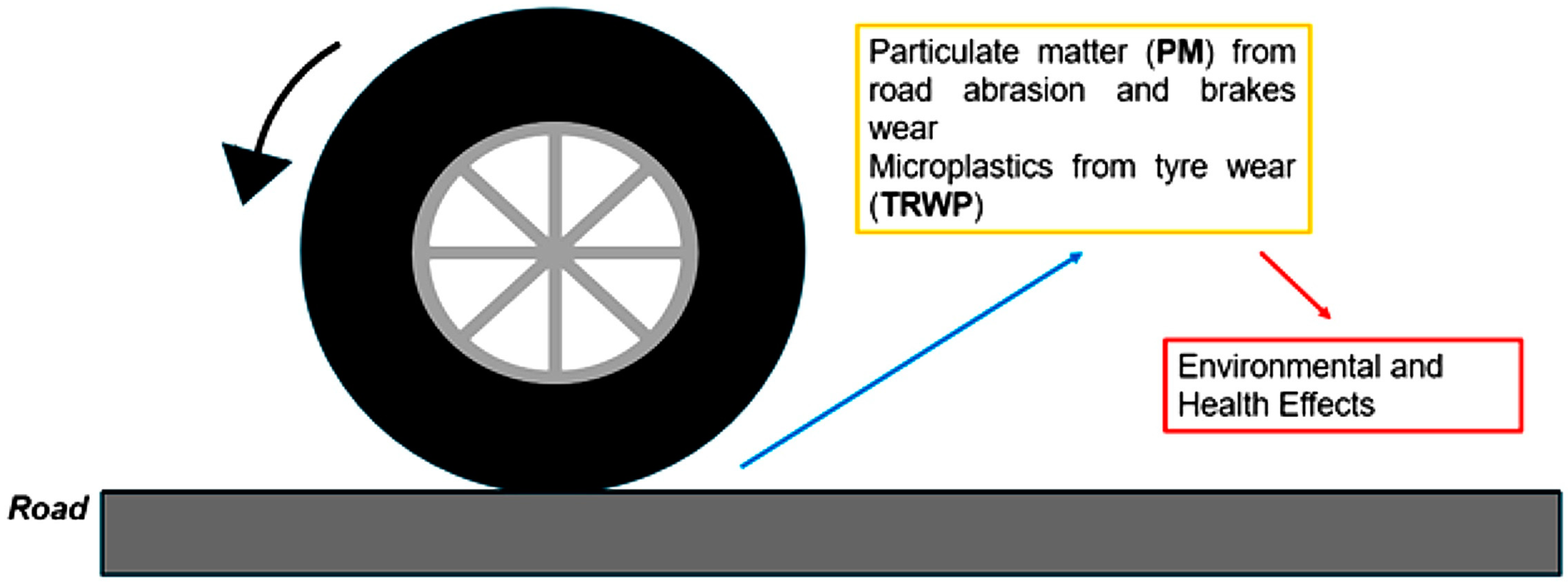
Particulate matter arising from tire road wear (TRWP) is considered one of the significant factors in micro- and nanoplastic pollution (in Figure 2). According to recent estimations, about 10% of the plastic pollution stemming from tire wear may be found in the world’s oceans. Although direct evidence is still lacking for the formation of nanoplastics from TRWPs, these particles are comprised of heavy metals and organic additives that may increase their risk to the environment and toxicologically. Nonetheless, importantly, there is still little investigation into the nanoscale dynamics of tire wear, hence the need for new hypotheses and modeling approaches for a much better understanding of the fragmentation processes taking place [40,41].
It is assumed that under actual road conditions, abraded tires actually produce nanoplastics through localized stress concentration and interfacial debonding at rubber–filler interfaces [41]. Repeated mechanical contact, high-pressure loading, and lateral shear between the tire tread and road surface may create microscale fatigue, leading to nanoscale fragments being detached from the viscoelastic matrix itself [42]. This process might be further affected by fillers such as carbon black or silica, which are points of stress concentration facilitating the development of cracks and release of nanoparticles. Finally, we also recognize the fact that nanoplastic particles can be caused by ultraviolet-induced destruction of pre-existing microplastic tire fragments. This photodegradation pathway is not abrasion-dependent but could significantly contribute to secondary nanoplastic pollution in sun-exposed environments. Therefore, both mechanical and photo-induced fragmentation pathways could be treated as complementary contributors to tire-derived nanoplastics.
The events associated with plastic weathering in the natural environment are dynamic and complex, since the degradation of polymers from UV irradiation and mechanical weathering leads progressively to the formation of microplastics and nanoplastics and the release of chemical additives [43]. Yang et al. showed that extended exposure to environmental stressors oxidizes the surface of the polymer, rendering it brittle and initiating crack or flake formation, which allows the detachment of nanoscale plastic particles [44]. The presence of reactive oxygen species (ROS) during photo-oxidation further exacerbates nanoplastic release, emphasizing the importance of environmental conditions in wear-related pollution. Nanoplastic release is closely linked to the aging of plastics, with smaller sized fragments becoming more predominant over time. While infrared and Raman analyses indicate that aging also promotes the leaching of polymer additives, this process is distinct from mechanical fragmentation. Additive leaching occurs due to polymer oxidation and degradation, whereas nanoplastic formation primarily results from surface cracking and flake detachment under mechanical stress. Both phenomena contribute to environmental pollution, but nanoplastic formation is directly tied to the physical breakdown of polymer structures. These findings are also relevant for tire road wear particles, as their ongoing mechanical wear against road surfaces similarly leads to progressive micro- and nanoplastic generation. Understanding these degradation mechanisms is essential for predicting nanoplastic pollution and designing effective mitigation strategies.
Figure 3 illustrates the surface morphological changes in aged polystyrene (PS) plastics, as observed through SEM imaging. The images show the progressive development of surface roughness, granular oxidation, and ultimately the formation of cracks and lamellar projections. As aging advances, cracks widen, increasing the likelihood of nanoplastic detachment. These observations support the idea that surface degradation mechanisms—such as crack formation and flake detachment—are primary contributors to the release of nanoplastics from weathered polymer surfaces. While this process is particularly evident in PS, similar degradation patterns have been reported for other thermoplastics under prolonged environmental exposure [44].
In this context, it is also worth mentioning the multi-scale abrasion mechanics model developed by Li et al. [45] to describe the formation of particulate matter during wear processes. Their findings indicate that nanoplastic generation is directly linked to fracture mechanics, where fatigue-induced microcrack propagation plays a critical role. Their model quantitatively relates abrasion-induced emissions to material properties, revealing that materials with lower fracture toughness tend to release higher concentrations of smaller plastic fragments. This approach provides a robust theoretical foundation for predicting and mitigating nanoplastic emissions from polymeric surfaces under repeated stress. In addition to mechanical wear, environmental factors also contribute to the degradation of TWPs over time.
As highlighted by Gnecco et al. [46], nanoripples can emerge due to periodic instabilities occurring under shear stress of polymeric surfaces. Their formation and evolution are governed by the interplay between lateral forces and the plastic response of the material. This process is particularly significant for PS, a commonly found plastic in the environment, and repeated mechanical stresses can trigger nanoparticle detachment. As shown by Hennig et al. [47], nanoplastics are nucleated from the crests of the ripples and easily displaced by the scratching tool (i.e., an AFM tip, see below). Similar results were recently obtained by Khaksar et al. on polymeric blends [12]. In both cases, the release of nanoplastics is possibly the result of crazing mechanisms in the polymer surfaces under tension. Although these studies were conducted using AFM under controlled nanoscratching conditions (constant loading forces in the sub µN range and constant scan velocities of few tens of µm/s at most), it is not excluded that similar mechanisms of nanoparticle detachment and surface instabilities occur in everyday life. For instance, tire abrasion on roads introduces continuous and fluctuating mechanical stresses, leading to progressive polymer degradation and nanoplastic detachment [48]. Processes similar to those observed under rather idealized laboratory conditions may also occur, although, at the time of writing, we are not aware of any systematic investigations extending the aforementioned studies in this direction. The same can be said for synthetic polymer coatings on ship hulls and industrial equipment, which experience mechanical erosion due to abrasive contact with sediment contributing to nanoplastic release, and for the wear of polymer fibers in textiles during washing cycles.
Altogether, these studies illustrate that nanoplastic formation during mechanical wear is influenced by intrinsic material properties, wear mechanics, and external environmental factors. A comprehensive understanding of these parameters is essential for developing sustainable materials and wear-resistant polymers to mitigate nanoplastic pollution. Despite these advancements, significant research gaps remain in fully elucidating the relationship between mechanical wear parameters and nanoplastic release rates. In addition to mechanical wear, environmental aging mechanisms—particularly UV radiation and elevated temperatures—play a crucial role in the progressive degradation of polymeric materials [27]. Studies have shown that prolonged exposure to sunlight and heat promotes surface oxidation and polymer chain scission, resulting in embrittlement and enhanced fragmentation potential [49,50]. This photo-oxidative degradation pathway facilitates the detachment of nanoscale fragments, even in the absence of direct mechanical abrasion. Notably, Yang et al. [44] demonstrated that the generation of reactive oxygen species (ROS) during UV-induced aging exacerbates crack formation and flake detachment, further contributing to nanoplastic release. These aging effects can also interact with wear mechanisms: for example, aged and oxidized surfaces are typically more susceptible to fragmentation under mechanical stress. Therefore, a comprehensive understanding of nanoplastic generation must account for the synergistic effects between environmental aging and tribological wear.

2.2. Mechanical Properties and Wear Behavior

Nanoplastics exhibit distinct mechanical properties that influence their formation, durability, and environmental persistence [51]. The elastic modulus, hardness, and wear resistance of plastic films determine how easily they are fragmented under mechanical stress. Different polymers respond to mechanical wear differently, affecting the amount and morphology of nanoplastics they generate [52].
Polymers with high brittleness and low tensile strength release more nanoplastics under wear tests [11,53]. Thermoplastics such as PE, PP, and PS exhibit varying degrees of mechanical properties and wear mechanisms. PE and PP are widely regarded as ductile polymers with moderate tensile strength, exhibiting plowing wear behavior, characteristic of ductile materials [13,54,55]. While they are mechanically softer than high-performance engineering polymers, they do not possess high brittleness. In contrast, PS and other amorphous thermoplastics demonstrate greater brittleness and are more prone to cutting wear behavior [56]. The distinction in wear mechanisms is crucial for selecting appropriate materials in tribological applications. Additionally, studies have demonstrated that mechanical degradation and fragmentation of thermoplastics like PE, PP, and PS can lead to the formation of microplastics and nanoplastics under environmental stressors [57,58,59]. A key aspect in this context is the mechanical breakdown of the materials, which is influenced by the molecular weight and structural stiffness of the polymers [60]. Environmental stressors also include ultraviolet (UV) light, which weakens polymer chains and increases fragmentation susceptibility, and consequently worsens the degradation of PE and other thermoplastics [61].
Frictional forces and roughness also play a crucial role in wear-induced nanoplastic generation [62]. Pin-on-disk tribological tests have demonstrated that increased sliding speed and applied load results in higher nanoplastic release rates. Surface modification techniques, such as adding fillers or reinforcing fibers, have been explored to reduce polymer wear and mitigate nanoplastic generation [63]. Despite these insights, challenges remain in accurately quantifying nanoplastic generation under real-world conditions. Many studies focus on controlled laboratory settings [64], whereas real-world factors such as temperature variations, humidity, and UV exposure can significantly alter polymer wear behavior [65,66].

2.3. Characterization Techniques

The accurate detection and characterization of nanoplastics are essential for understanding their formation, environmental fate, and toxicity [67], and a few analytical techniques can be used to identify and quantify nanoplastics.
Atomic force microscopy (AFM) is one of the most important tools for studying the morphology of polymer surfaces on the nanoscale [68]. It also plays a crucial role in understanding the nanoscale fragmentation process [69]. As an example, Figure 4 shows the formation of surface ripples as observed while scratching initially flat polystyrene surfaces with an AFM tip. Scratching was repeated one, three, or five times along a series of parallel lines, left to right only in the so-called “hover mode” [70]. The resulting particles had diameters in the order of 100 nm.
As seen in the previous example, AFM provides very accurate images of surface changes caused by wear and allows the process of generating individual nanoplastics to be observed. In addition, AFM plays a key role in quantifying nanoplastic removal mechanisms [71]. AFM-based nanoscratching is a powerful technique for investigating wear mechanisms at the nanoscale, offering valuable insights into how polymeric materials respond to mechanical interactions under controlled conditions [72]. The method employs diamond-coated tips under precisely controlled normal loads, effectively replicating abrasive wear commonly observed in real-world applications [73]. For instance, in marine environments, ship coatings endure continuous exposure to sand and debris [74]; in biomedical applications, polymeric implants experience frictional wear [75]; and in industrial settings, polymer seals and gaskets undergo tribological degradation due to repetitive mechanical contact [76]. One of the significant advantages of AFM-based nanoscratching is its ability to provide precise control over normal force, sliding speed, and environmental conditions such as humidity and temperature, which are crucial factors affecting polymer wear behavior [77,78]. These controlled experiments allow for a systematic evaluation of how material properties—such as cross-linking density, molecular structure, and the presence of reinforcing fillers—impact wear resistance in conditions that resemble those encountered in real-world applications.
Furthermore, studies have shown that wear behavior observed through AFM nanoscratching correlates well with macroscopic tribological tests, reinforcing its effectiveness as a predictive tool [79,80]. By enabling the analysis of plastic deformation, material transfer, and debris formation at the nanoscale, this method provides valuable data on the early-stage wear mechanisms that contribute to the long-term degradation of polymeric materials. Thus, AFM-based nanoscratching serves as a bridge between fundamental nanoscale wear analysis and the broader understanding of wear mechanisms in practical applications. AFM plays a crucial role in understanding nanoscale fragmentation processes and mechanical wear behavior [81]. However, obtaining high-resolution images and conducting well-controlled nanoscratching experiments require extensive fine-tuning [82]. Key factors include tip characterization, contact mechanics, and force modulation, all of which influence the accuracy of wear track analysis [83]. Tip radius, material properties, and cantilever stiffness must be carefully selected to ensure reproducibility in nanoplastic detection. Additionally, advanced AFM modes, such as PeakForce Tapping mode, provide further advantages in force control and nanoscale mechanical property mapping [84]. Unlike conventional contact-mode AFM, PeakForce mode dynamically modulates the tip–sample interaction forces, minimizing sample damage while simultaneously capturing quantitative mechanical properties such as adhesion, modulus, and deformation at a nanometer resolution [85]. In this context, PeakForce mode is particularly relevant for nanoplastic characterization, as it allows for real-time tracking of dynamic wear processes. Furthermore, AFM-IR (Atomic Force Microscopy-Infrared Spectroscopy) has emerged as a powerful tool for simultaneously obtaining morphological and chemical characterization of nanoplastics [86]. This hybrid technique overcomes AFM’s limitation in chemical identification by integrating IR absorption mapping, which can detect polymer-specific spectral fingerprints with nanometer-scale spatial resolution [87]. Given its ability to combine topographical imaging with molecular identification, AFM-IR provides a more comprehensive approach to nanoplastic analysis, particularly in distinguishing different polymer types in mixed samples. Altogether, these AFM-based characterizations provide fundamental insights into the mechanical processes governing the formation of nanoplastics and their potential dispersion in the environment. A limitation of AFM remains, however, in the inability to chemically identify the surfaces under investigation, a limitation that can be mitigated by complementary spectroscopic methods [88]. It is worth noting, for instance, that the integration of AFM with infrared (IR) and Raman spectroscopy allows for simultaneous morphological and chemical characterization of surfaces [86].
Nanoplastics’ size and shape can also be investigated using SEM [89] and TEM [90]. While SEM provides high-resolution surface imaging [91], TEM offers superior resolution for sub-micron polymer debris, making it particularly suitable for detecting nanoplastics smaller than 100 nm [92]. Additionally, Energy-Dispersive X-ray Spectroscopy (EDS) can be combined with both SEM and TEM to determine elemental composition, offering valuable insights into the chemical characteristics of nanoplastics [93]. High-resolution transmission-mode SEM (T-SEM) has also been employed (in Figure 5) to enhance the detection of nanoplastic particles at the nanoscale [94,95]. However, the quality of SEM images can be affected by various factors, such as accelerating voltage, beam current, and sample charge [96]. The physical state of some polymeric materials can be modified by the vacuum conditions used for SEM, which is especially critical for nanoplastics, as a high vacuum might distort polymeric structures or cause volatile components to evaporate [97]. To address these issues, low-voltage SEM methods and cryo-SEM are often used to preserve the natural structure of sensitive materials [98]. Furthermore, adjusting the working distance, detection angle, and beam energy can further improve SEM imaging quality while minimizing sample damage. Advances in SEM-EDS technology, particularly the use of high-sensitivity silicon drift detectors (SDDs), have significantly improved elemental mapping at the nanoscale. Similarly, TEM-EDS allows for precise elemental characterization of nanoplastics at much smaller dimensions, making it a complementary technique for detecting wear-induced plastic debris [99,100].
Raman spectroscopy and Fourier Transform Infrared (FTIR) spectroscopy are also useful for determining the chemical composition of polymeric fragments, although their application is, admittedly, limited to microplastics [101]. Raman spectroscopy allows for the analysis of those particles non-destructively and for distinguishing between various polymer components. However, it can occasionally be difficult to interpret Raman spectra due to fluorescence interference. Functional groups and polymer-specific spectral fingerprints can be easily identified using FTIR spectroscopy, especially for weakly Raman-active polymers [102]. Because of its sensitivity to surface characteristics, the attenuated total reflectance (ATR-FTIR) mode is widely utilized and a crucial tool for microplastic investigation. Furthermore, Raman spectroscopy can be improved by employing metal nanoparticles for surface-enhanced Raman scattering (SERS), which provides ultra-sensitive detection and broadens its uses in biomedical domains [103]. To fully characterize microplastics in environmental samples, both approaches are frequently applied in tandem, and for best results, they should be combined.
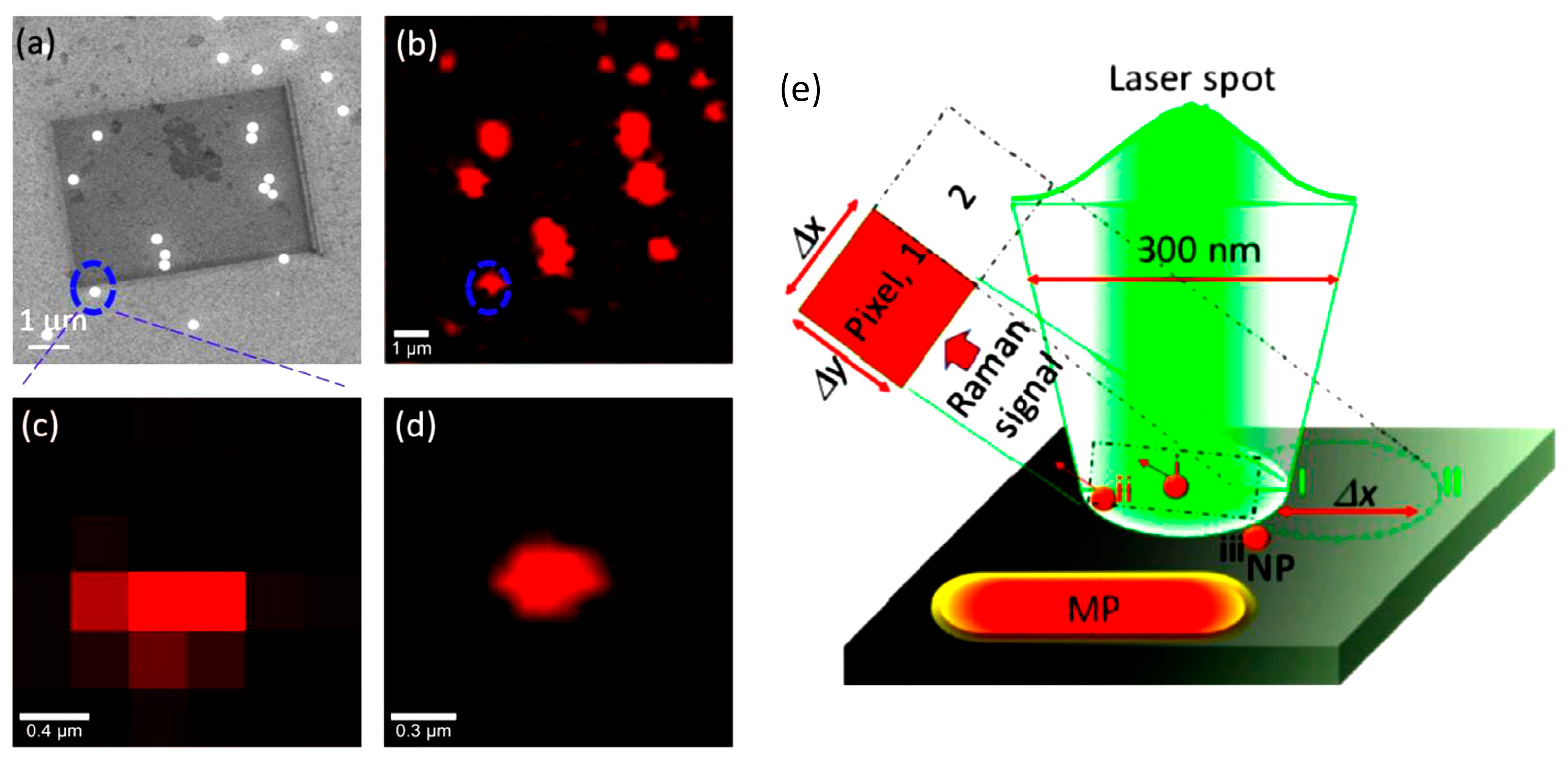
Besides mapping techniques based on AFM and SEM, Raman imaging has recently emerged as a powerful technique to identify and visualize nanoplastics down to 100 nm. As shown by Sobhani et al. [104], Raman mapping enables nanoplastics’ chemical characterization by detecting polymer-specific vibrational fingerprints at the nanoscale. This study involved the identification of polystyrenic nanoplastics with a size of down to 100 nm using a confocal Raman microscope with high-resolution pixel stepping (100 nm × 100 nm). Figure 6 clearly demonstrates that even a single nanoplastic particle smaller than the laser spot (~300 nm) can be visualized through pixel-specific spectral analysis. The immense advantages of this imaging method compared to conventional ones, especially in finding nanoplastics from complex environmental samples like polishing dust from vehicle paints, cannot be overstated. The ability to discriminate between microplastics and nanoplastics through their Raman spectra is opening new gateways in environmental risk assessment. Thus, Raman characterization supports AFM and SEM in carrying out simultaneous chemical identification and spatial resolution of plastic debris on the nanoscale.
Although nanoscale experiments, such as AFM-based nanoscratching, enhance our understanding of surface instabilities and the development of nanoplastic under very well-defined conditions, it must be noted that neither the type nor the extent of results would be in any way comparable to those formed with engineered or natural environments. Parameters such as contact pressure, asperity size, and force magnitudes vary greatly in real-world scenarios—ranging from nN at the tip–sample interface in AFM to N or kN ranges in applications such as tire–road interactions or marine equipment wear. Thus, direct quantitative extrapolation remains challenging. Nonetheless, the ripple formation and nanoparticle detachment observed at the nanoscale represent fundamental surface phenomena that may also manifest during the early stages of macroscale wear, especially under cyclic or repetitive loading. To effectively close the scale gap, such future endeavors will combine nanoscale understanding with multiscale computational modeling approaches and upscaled experimental work to predict wear behavior for various conditions. Also, we acknowledge that definitions of abrasive wear in natural environments have not yet gained a quantitative foundation; thus, much more research on reliable metrics—such as energy dissipation, particle emission rates, and force distributions under uncontrolled environmental stress—is needed to serve as an input for further developments.

3. Conclusions

Because of their distinct physicochemical characteristics and tiny size, nanoplastics cause serious environmental and health risks. These particles are commonly found in different ecosystems because of the fragmentation of larger plastic materials caused by friction, abrasion, and shear pressures. Their high surface area–volume ratio makes them more prone to adsorb contaminants and interact with biological systems. Determining the long-term environmental fate and potential hazards of nanoplastics requires an understanding of the mechanisms behind their generation, but investigations focusing on wear-induced nanoplastic formation remain limited so far. Thus, comprehensive studies are needed to establish direct correlations between material properties and fragmentation behavior. Integrating tribological analyses with spectroscopic and microscopic techniques can enhance the accuracy of nanoplastic detection and provide a deeper understanding of their release dynamics under real-world conditions. In this context, AFM is particularly useful for investigating the wear-related mechanisms leading to nanoplastic release, while SEM, Raman spectroscopy, and IR spectroscopy provide crucial insights into the size, morphology, and chemical composition of the generated nanoplastics.
To sum up, nanoplastics produced by mechanical wear processes are a new environmental problem that needs to be addressed urgently. Developing successful mitigation techniques will require filling in the current information gaps regarding the toxicity, characterization, and generation of nanoplastics. To fully evaluate nanoplastic contamination, future studies should concentrate on interdisciplinary techniques that integrate environmental chemistry, material science, and toxicology. To minimize the release of nanoplastics into the environment, it will also be essential to promote sustainable material alternatives and enhance waste management techniques.
A final remark is necessary. Although our focus remained on nanoplastics, it is important to note that the number of direct studies on their wear-induced formation is still limited compared to the literature on microplastics. Consequently, findings from microplastic research cannot be downscaled a priori. However, recent evidence by Jurkschat et al. [105] demonstrated that vehicle tires—historically known to release microplastics—can also contribute to nanoplastic pollution. In their study, TWPs accounted for the largest proportion of nanoplastic mass in remote Alpine snow samples. These results justify the inclusion of tire wear in discussions of nanoplastic generation, even if mechanistic studies are still developing in this area.

Author Contributions

Conceptualization, O.D., H.K. and E.G.; writing—original draft preparation, O.D.; writing—review and editing, E.G.; visualization, H.K.; funding acquisition, E.G. All authors have read and agreed to the published version of the manuscript.

Funding

This research was partially funded by the Strategic Program Excellence Initiative at the Jagiellonian University ‘SciMat’, grant number U1U/P05/NO/01.05.

Data Availability Statement

No new data were created or analyzed in this study. Data sharing is not applicable to this article.

Conflicts of Interest

The authors declare no conflicts of interest. The funders had no role in the design of the study; in the collection, analyses, or interpretation of data; in the writing of the manuscript; or in the decision to publish the results.

References

  1. Hurley, R.; Woodward, J.; Rothwell, J.J. Microplastic Contamination of River Beds Significantly Reduced by Catchment-Wide Flooding. Nat. Geosci. 2018, 11, 251–257. [Google Scholar] [CrossRef]
  2. Yildiz, R.O.; Koc, E.; Der, O.; Aymelek, M. Unveiling the Contemporary Research Direction and Current Business Management Strategies for Port Decarbonization Through a Systematic Review. Sustainability 2024, 16, 10959. [Google Scholar] [CrossRef]
  3. Thompson, R.C.; Olsen, Y.; Mitchell, R.P.; Davis, A.; Rowland, S.J.; John, A.W.G.; McGonigle, D.; Russell, A.E. Lost at Sea: Where Is All the Plastic? Science 2004, 304, 838. [Google Scholar] [CrossRef] [PubMed]
  4. Koelmans, A.A.; Besseling, E.; Shim, W.J. Nanoplastics in the Aquatic Environment. Critical Review. In Marine Anthropogenic Litter; Springer International Publishing: Cham, Switzerland, 2015; pp. 325–340. [Google Scholar]
  5. Frias, J.P.G.L.; Nash, R. Microplastics: Finding a Consensus on the Definition. Mar. Pollut. Bull. 2019, 138, 145–147. [Google Scholar] [CrossRef]
  6. Mitrano, D.M.; Wick, P.; Nowack, B. Placing Nanoplastics in the Context of Global Plastic Pollution. Nat. Nanotechnol. 2021, 16, 491–500. [Google Scholar] [CrossRef]
  7. Li, W.; Luo, Y.; Pan, X. Identification and Characterization Methods for Microplastics Basing on Spatial Imaging in Micro-/Nanoscales. In Microplastics in Terrestrial Environments; Springer: Berlin/Heidelberg, Germany, 2020; pp. 25–37. [Google Scholar]
  8. Wagner, J.; Wang, Z.-M.; Ghosal, S.; Rochman, C.; Gassel, M.; Wall, S. Novel Method for the Extraction and Identification of Microplastics in Ocean Trawl and Fish Gut Matrices. Anal. Methods 2017, 9, 1479–1490. [Google Scholar] [CrossRef]
  9. Schiavi, S.; Parmigiani, M.; Galinetto, P.; Albini, B.; Taglietti, A.; Dacarro, G. Plasmonic Nanomaterials for Micro- and Nanoplastics Detection. Appl. Sci. 2023, 13, 9291. [Google Scholar] [CrossRef]
  10. Adetuyi, B.O.; Mathew, J.T.; Inobeme, A.; Falana, Y.O.; Adetunji, C.O.; Shahnawaz, M.; Oyewole, O.A.; Eniola, K.I.T.; Yerima, M.B.; Popoola, O.A. Tyres, Bitumen Wear and Plastic Bottles, Other Single Used Plastic as Major Sources of Microplastic. In Microplastic Pollution; Springer Nature: Singapore, 2024; pp. 141–160. [Google Scholar]
  11. Rahman, E.; BinAhmed, S.; Keyes, P.; Alberg, C.; Godfreey-Igwe, S.; Haugstad, G.; Xiong, B. Nanoscale Abrasive Wear of Polyethylene: A Novel Approach To Probe Nanoplastic Release at the Single Asperity Level. Environ. Sci. Technol. 2024, 58, 13845–13855. [Google Scholar] [CrossRef]
  12. Khaksar, H.; Ma, C.; Janiszewska, N.; Awsiuk, K.; Budkowski, A.; Gnecco, E. Nanoscale Wear Evolution on a Polystyrene/Poly (n-Butyl Methacrylate) Blend. Wear 2024, 536–537, 205160. [Google Scholar] [CrossRef]
  13. Ordu, M.; Der, O. Polymeric Materials Selection for Flexible Pulsating Heat Pipe Manufacturing Using a Comparative Hybrid MCDM Approach. Polymers 2023, 15, 2933. [Google Scholar] [CrossRef]
  14. Sipe, J.M.; Bossa, N.; Berger, W.; von Windheim, N.; Gall, K.; Wiesner, M.R. From Bottle to Microplastics: Can We Estimate How Our Plastic Products Are Breaking Down? Sci. Total Environ. 2022, 814, 152460. [Google Scholar] [CrossRef] [PubMed]
  15. Musa, I.O.; Auta, H.S.; Ilyasu, U.S.; Aransiola, S.A.; Makun, H.A.; Adabara, N.U.; Abioye, O.P.; Aziz, A.; Jayanthi, B.; Maddela, N.R.; et al. Micro- and Nanoplastics in Environment: Degradation, Detection, and Ecological Impact. Int. J. Environ. Res. 2024, 18, 1. [Google Scholar] [CrossRef]
  16. Yu, Y.; Craig, N.; Su, L. A Hidden Pathway for Human Exposure to Micro- and Nanoplastics—The Mechanical Fragmentation of Plastic Products during Daily Use. Toxics 2023, 11, 774. [Google Scholar] [CrossRef] [PubMed]
  17. da Silva Antunes, J.C.; Sobral, P.; Branco, V.; Martins, M. Uncovering Layer by Layer the Risk of Nanoplastics to the Environment and Human Health. J. Toxicol. Environ. Health Part B 2025, 28, 63–121. [Google Scholar] [CrossRef]
  18. Suzuki, G.; Uchida, N.; Tuyen, L.H.; Tanaka, K.; Matsukami, H.; Kunisue, T.; Takahashi, S.; Viet, P.H.; Kuramochi, H.; Osako, M. Mechanical Recycling of Plastic Waste as a Point Source of Microplastic Pollution. Environ. Pollut. 2022, 303, 119114. [Google Scholar] [CrossRef]
  19. Bulut, M.S.; Ordu, M.; Der, O.; Basar, G. Sustainable Thermoplastic Material Selection for Hybrid Vehicle Battery Packs in the Automotive Industry: A Comparative Multi-Criteria Decision-Making Approach. Polymers 2024, 16, 2768. [Google Scholar] [CrossRef]
  20. Nilsson, D.; Stavlid, N.; Lindquist, M.; Hogmark, S.; Wiklund, U. The Role of Aluminum Additions in the Oxidation and Wear of a TaC:C Low-Friction Coating. Surf. Coat. Technol. 2009, 203, 2989–2994. [Google Scholar] [CrossRef]
  21. Yoshida, K.; Horiuchi, T.; Kano, M.; Kumagai, M. Or0404—Effect of a Tribochemical Reacted Film on Friction and Wear Properties of DLC Coatings. Plasma Process. Polym. 2009, 6, S96–S101. [Google Scholar] [CrossRef]
  22. Fan, X.; Li, G.; Guo, Y.; Zhang, L.; Xu, Y.; Zhao, F.; Zhang, G. Role of Reinforcement Types and Silica Nanoparticles on Tribofilm Growth at PTFE-Steel Interface. Tribol. Int. 2020, 143, 106035. [Google Scholar] [CrossRef]
  23. Venkatesan, M.; Palanikumar, K.; Boopathy, S.R. Experimental Investigation and Analysis on the Wear Properties of Glass Fiber and CNT Reinforced Hybrid Polymer Composites. Sci. Eng. Compos. Mater. 2018, 25, 963–974. [Google Scholar] [CrossRef]
  24. Reyes Acosta, Y.K.; Cruz Martinez, W.E.; Reyes Acosta, A.V.; Cepeda Tovar, V.A.; Contreras Esquivel, J.C.; Aguilar Gonzales, C.N.; Narro Cespedes, R.I.; Martinez, R.R. Thermal Properties by Adding Natural Oils, Foods, Organic Materials, Fibers, and Nanocomposites in the PLA, and Applications in 3D Printing. In Engineering Principles for Food Processing Technology and Product Realization; Apple Academic Press: New York, NY, USA, 2024; pp. 87–110. [Google Scholar]
  25. Reyes Acosta, Y.K.; Martinez, E.C.; Reyes Acosta, A.V.; Torre, L.S.; Aguilar Gonzales, C.N.; Narro Cespedes, R.I. Release Studies to Improve the Mechanical Properties of the Biopolymer, Polylactic Acid (PLA) for Food Packaging Applications. In Engineering Principles for Food Processing Technology and Product Realization; Apple Academic Press: New York, NY, USA, 2024; pp. 67–85. [Google Scholar]
  26. Levchenko, I.; Xu, S.; Baranov, O.; Bazaka, O.; Ivanova, E.; Bazaka, K. Plasma and Polymers: Recent Progress and Trends. Molecules 2021, 26, 4091. [Google Scholar] [CrossRef]
  27. Maraveas, C.; Kyrtopoulos, I.V.; Arvanitis, K.G.; Bartzanas, T. The Aging of Polymers under Electromagnetic Radiation. Polymers 2024, 16, 689. [Google Scholar] [CrossRef]
  28. Yong, C.; Valiyaveettil, S.; Tang, B. Toxicity of Microplastics and Nanoplastics in Mammalian Systems. Int. J. Environ. Res. Public Health 2020, 17, 1509. [Google Scholar] [CrossRef] [PubMed]
  29. Shi, C.; Liu, Z.; Yu, B.; Zhang, Y.; Yang, H.; Han, Y.; Wang, B.; Liu, Z.; Zhang, H. Emergence of Nanoplastics in the Aquatic Environment and Possible Impacts on Aquatic Organisms. Sci. Total Environ. 2024, 906, 167404. [Google Scholar] [CrossRef]
  30. Zhang, L.; García-Pérez, P.; Muñoz-Palazon, B.; Gonzalez-Martinez, A.; Lucini, L.; Rodriguez-Sanchez, A. A Metabolomics Perspective on the Effect of Environmental Micro and Nanoplastics on Living Organisms: A Review. Sci. Total Environ. 2024, 932, 172915. [Google Scholar] [CrossRef] [PubMed]
  31. Gulati, S.; Amar, A.; Olihan, S. Environmental Fate, Behavior, and Risk Management Approaches of Nanoplastics in the Environment. In Solid Waste Treatment Technologies; CRC Press: Boca Raton, FL, USA, 2024; pp. 148–172. [Google Scholar]
  32. Vettegren’, V.I.; Lyashkov, A.I.; Savitskii, A.V.; Shcherbakov, I.P.; Vasil’ev, K.D. Microcrack Dynamics in a Polymer Composite Material during Friction. Tech. Phys. 2012, 57, 1445–1448. [Google Scholar] [CrossRef]
  33. Ito, M.M.; Gibbons, A.H.; Qin, D.; Yamamoto, D.; Jiang, H.; Yamaguchi, D.; Tanaka, K.; Sivaniah, E. Structural Colour Using Organized Microfibrillation in Glassy Polymer Films. Nature 2019, 570, 363–367. [Google Scholar] [CrossRef]
  34. Zhou, H.; Zhang, Z. Evolution of Silicon Particle Damage on Fatigue Crack Initiation and Early Propagation in an Aluminum Alloy. Rare Met. 2023, 42, 2470–2476. [Google Scholar] [CrossRef]
  35. Alkhadra, M.A.; Root, S.E.; Hilby, K.M.; Rodriquez, D.; Sugiyama, F.; Lipomi, D.J. Quantifying the Fracture Behavior of Brittle and Ductile Thin Films of Semiconducting Polymers. Chem. Mater. 2017, 29, 10139–10149. [Google Scholar] [CrossRef]
  36. Su, Y.; Yang, C.; Wang, S.; Li, H.; Wu, Y.; Xing, B.; Ji, R. Mechanochemical Formation of Poly(Melamine-Formaldehyde) Microplastic Fibers During Abrasion of Cleaning Sponges. Environ. Sci. Technol. 2024, 58, 10764–10775. [Google Scholar] [CrossRef]
  37. Solasa, K.C.; Venkataraman, N.V.; Choudhury, P.R.; Schueller, J.K.; Bhattacharyya, A. On the Use of Alternative Measurement Methods in the Estimation of Wear Rates in Rotary-Pin-on-Disk Tribometry. Tribol. Lett. 2024, 72, 45. [Google Scholar] [CrossRef]
  38. Orgeldinger, C.; Rosnitschek, T.; Tremmel, S. Unveiling an Additively Manufactured Open Hardware Pin-on-Disc Tribometer Considering Its High Reproducibility. Wear 2024, 552–553, 205437. [Google Scholar] [CrossRef]
  39. Budinski, K.G. Adhesive Transfer to Abrasive Particles in Abrasion Testing. Wear 2011, 271, 1258–1263. [Google Scholar] [CrossRef]
  40. Morreale, M.; La Mantia, F.P. Current Concerns about Microplastics and Nanoplastics: A Brief Overview. Polymers 2024, 16, 1525. [Google Scholar] [CrossRef] [PubMed]
  41. Giechaskiel, B.; Grigoratos, T.; Mathissen, M.; Quik, J.; Tromp, P.; Gustafsson, M.; Franco, V.; Dilara, P. Contribution of Road Vehicle Tyre Wear to Microplastics and Ambient Air Pollution. Sustainability 2024, 16, 522. [Google Scholar] [CrossRef]
  42. Yu, L.; Hu, J.; Li, R.; Yang, Q.; Guo, F.; Pei, J. Tire-Pavement Contact Pressure Distribution Analysis Based on ABAQUS Simulation. Arab. J. Sci. Eng. 2022, 47, 4119–4132. [Google Scholar] [CrossRef]
  43. Cao, Y.; Lin, H.; Zhang, K.; Xu, S.; Yan, M.; Leung, K.M.Y.; Lam, P.K.S. Microplastics: A Major Source of Phthalate Esters in Aquatic Environments. J. Hazard. Mater. 2022, 432, 128731. [Google Scholar] [CrossRef]
  44. Yang, N.; Men, C.; Zhang, Y.; Xie, Z.; Zuo, J. Exploring Polystyrene Weathering Behavior: From Surface Traits to Micro(Nano)Plastics and Additives Release. J. Environ. Manag. 2024, 367, 121880. [Google Scholar] [CrossRef]
  45. Li, K.; Zhang, Y.; Yu, K.; Du, H.; Sioutas, C.; Wang, Q. Mechanics of Abrasion-Induced Particulate Matter Emission. J. Mech. Phys. Solids 2024, 188, 105661. [Google Scholar] [CrossRef]
  46. Gnecco, E.; Pedraz, P.; Nita, P.; Dinelli, F.; Napolitano, S.; Pingue, P. Surface Rippling Induced by Periodic Instabilities on a Polymer Surface. New J. Phys. 2015, 17, 032001. [Google Scholar] [CrossRef]
  47. Hennig, J.; Litschko, A.; Mazo, J.J.; Gnecco, E. Nucleation and Detachment of Polystyrene Nanoparticles from Plowing-Induced Surface Wrinkling. Appl. Surf. Sci. Adv. 2021, 6, 100148. [Google Scholar] [CrossRef]
  48. Heinrich, G.; Klüppel, M. Basic Mechanisms and Predictive Testing of Tire-Road Abrasion. In Degradation of Elastomers in Practice, Experiments and Modeling; Springer: Berlin/Heidelberg, Germany, 2022; pp. 1–14. [Google Scholar]
  49. Lisci, C.; Sitzia, F.; Pires, V.; Mirão, J. Building Stones Durability by UVA Radiation, Moisture and Spray Accelerated Weathering. J. Build. Pathol. Rehabil. 2022, 7, 60. [Google Scholar] [CrossRef]
  50. Gu, X.; Michaels, C.A.; Drzal, P.L.; Jasmin, J.; Martin, D.; Nguyen, T.; Martin, J.W. Probing Photodegradation beneath the Surface: A Depth Profiling Study of UV-Degraded Polymeric Coatings with Microchemical Imaging and Nanoindentation. J. Coat. Technol. Res. 2007, 4, 389–399. [Google Scholar] [CrossRef]
  51. El Hadri, H.; Gigault, J.; Maxit, B.; Grassl, B.; Reynaud, S. Nanoplastic from Mechanically Degraded Primary and Secondary Microplastics for Environmental Assessments. NanoImpact 2020, 17, 100206. [Google Scholar] [CrossRef]
  52. Arinstein, A.; Zussman, E. Electrospun Polymer Nanofibers: Mechanical and Thermodynamic Perspectives. J. Polym. Sci. B Polym. Phys. 2011, 49, 691–707. [Google Scholar] [CrossRef]
  53. Wohlleben, W.; Bossa, N.; Mitrano, D.M.; Scott, K. Everything Falls Apart: How Solids Degrade and Release Nanomaterials, Composite Fragments, and Microplastics. NanoImpact 2024, 34, 100510. [Google Scholar] [CrossRef]
  54. Mourad, A.-H.I. Impact of Blending Ratio and Thermal Treatment on the Mechanical Behaviour of Polyethylene/Polypropylene Blends. In Volume 6: Materials and Fabrication, Parts A and B, Proceedings of the ASME 2008 Pressure Vessels and Piping Conference, Chicago, IL, USA, 27–31 July 2008; ASMEDC: New York, NY, USA, 2008; pp. 145–155. [Google Scholar]
  55. Mourad, A.-H.I. Thermo-Mechanical Characteristics of Thermally Aged Polyethylene/Polypropylene Blends. Mater. Des. 2010, 31, 918–929. [Google Scholar] [CrossRef]
  56. Bermúdez, M.D.; Brostow, W.; Carrión-Vilches, F.J.; Cervantes, J.J.; Pietkiewicz, D. Wear of Thermoplastics Determined by Multiple Scratching. e-Polymers 2005, 5, 1–9. [Google Scholar] [CrossRef]
  57. Kadac-Czapska, K.; Knez, E.; Gierszewska, M.; Olewnik-Kruszkowska, E.; Grembecka, M. Microplastics Derived from Food Packaging Waste—Their Origin and Health Risks. Materials 2023, 16, 674. [Google Scholar] [CrossRef]
  58. Mattsson, K.; Björkroth, F.; Karlsson, T.; Hassellöv, M. Nanofragmentation of Expanded Polystyrene Under Simulated Environmental Weathering (Thermooxidative Degradation and Hydrodynamic Turbulence). Front. Mar. Sci. 2021, 7, 578178. [Google Scholar] [CrossRef]
  59. Song, Y.K.; Hong, S.H.; Jang, M.; Han, G.M.; Jung, S.W.; Shim, W.J. Corrections to “Combined Effects of UV Exposure Duration and Mechanical Abrasion on Microplastic Fragmentation by Polymer Type”. Environ. Sci. Technol. 2018, 52, 3831–3832. [Google Scholar] [CrossRef] [PubMed]
  60. Kierkels, J.T.A.; Dona, C.-L.; Tervoort, T.A.; Govaert, L.E. Kinetics of Re-embrittlement of (Anti)Plasticized Glassy Polymers after Mechanical Rejuvenation. J. Polym. Sci. B Polym. Phys. 2008, 46, 134–147. [Google Scholar] [CrossRef]
  61. Sun, J.; Zheng, H.; Xiang, H.; Fan, J.; Jiang, H. The Surface Degradation and Release of Microplastics from Plastic Films Studied by UV Radiation and Mechanical Abrasion. Sci. Total Environ. 2022, 838, 156369. [Google Scholar] [CrossRef] [PubMed]
  62. Wang, P.; Liang, H.; Jiang, L.; Qian, L. Effect of Nanoscale Surface Roughness on Sliding Friction and Wear in Mixed Lubrication. Wear 2023, 530–531, 204995. [Google Scholar] [CrossRef]
  63. Chelliah, N.; Kailas, S.V. Synergy between Tribo-Oxidation and Strain Rate Response on Governing the Dry Sliding Wear Behavior of Titanium. Wear 2009, 266, 704–712. [Google Scholar] [CrossRef]
  64. Randhawa, K.S.; Patel, A. The Effect of Environmental Humidity/Water Absorption on Tribo-Mechanical Performance of Polymers and Polymer Composites—A Review. Ind. Lubr. Tribol. 2021, 73, 1146–1158. [Google Scholar] [CrossRef]
  65. Kalin, M.; Kupec, A. The Dominant Effect of Temperature on the Fatigue Behaviour of Polymer Gears. Wear 2017, 376–377, 1339–1346. [Google Scholar] [CrossRef]
  66. Korku, M.; İlhan, R.; Feyzullahoğlu, E. Investigation of Effects of Environmental Conditions on Wear Behaviors of Glass Fiber Reinforced Polyester Composite Materials. Polym. Compos. 2025, 46, 355–371. [Google Scholar] [CrossRef]
  67. Lai, Y.; Zhou, X.; Liu, J. Separation and Enrichment of Nanoplastics from Samples. In Analysis of Microplastics and Nanoplastics; Elsevier: Amsterdam, The Netherlands, 2025; pp. 281–293. [Google Scholar]
  68. Shere, L.; Zhang, Z.J.; Preece, J.A. Application of Atomic Force Microscopy in Formulation Engineering. Johns. Matthey Technol. Rev. 2018, 62, 438–452. [Google Scholar] [CrossRef]
  69. Ishida, N. Atomic Force Microscopy. In Non-Destructive Material Characterization Methods; Elsevier: Amsterdam, The Netherlands, 2024; pp. 89–125. [Google Scholar]
  70. Kammoun, M.; Dupres, V.; Landoulsi, J.; Subramaniam, M.; Hawse, J.; Bensamoun, S.F. Transversal Elasticity of TIEG1 KO Muscle and Tendon Fibers Probed by Atomic Force Microscopy. Comput. Methods Biomech. Biomed. Eng. 2019, 22, S308–S310. [Google Scholar] [CrossRef]
  71. Lv, S.; Wang, Q.; Li, Y.; Gu, L.; Hu, R.; Chen, Z.; Shao, Z. Biodegradation of Polystyrene (PS) and Polypropylene (PP) by Deep-Sea Psychrophilic Bacteria of Pseudoalteromonas in Accompany with Simultaneous Release of Microplastics and Nanoplastics. Sci. Total Environ. 2024, 948, 174857. [Google Scholar] [CrossRef] [PubMed]
  72. Shen, N.; Feigenbaum, E.; Suratwala, T.; Steele, W.; Wong, L.; Feit, M.D.; Miller, P.E. Nanoplastic Removal Function and the Mechanical Nature of Colloidal Silica Slurry Polishing. J. Am. Ceram. Soc. 2019, 102, 3141–3151. [Google Scholar] [CrossRef]
  73. Pöhl, F.; Hardes, C.; Theisen, W. Deformation Behavior and Dominant Abrasion Micro Mechanisms of Tempering Steel with Varying Carbon Content under Controlled Scratch Testing. Wear 2019, 422–423, 212–222. [Google Scholar] [CrossRef]
  74. Soon, Z.Y.; Tamburri, M.N.; Kim, T.; Kim, M. Estimating Total Microplastic Loads to the Marine Environment as a Result of Ship Biofouling In-Water Cleaning. Front. Mar. Sci. 2024, 11, 1502000. [Google Scholar] [CrossRef]
  75. Modica, F.; Basile, V.; Surace, R.; Fassi, I. Replication Study of Molded Micro-Textured Samples Made of Ultra-High Molecular Weight Polyethylene for Medical Applications. Micromachines 2023, 14, 523. [Google Scholar] [CrossRef]
  76. Zhang, Y.; Yang, X.; Wang, S.; Lv, G.; Gao, Y.; Chen, K.; Yang, H. Research Status and Prospect of Wear and Aging on Hydraulic Rubber Sealing Materials. Plast. Rubber Compos. 2023, 52, 249–266. [Google Scholar] [CrossRef]
  77. He, Y.; Yan, Y.; Geng, Y.; Hu, Z. Fabrication of None-Ridge Nanogrooves with Large-Radius Probe on PMMA Thin-Film Using AFM Tip-Based Dynamic Plowing Lithography Approach. J. Manuf. Process. 2017, 29, 204–210. [Google Scholar] [CrossRef]
  78. Colaço, R. An AFM Study of Single-Contact Abrasive Wear: The Rabinowicz Wear Equation Revisited. Wear 2009, 267, 1772–1776. [Google Scholar] [CrossRef]
  79. Reichelt, M.; Cappella, B. Wear Volume of Self-Mated Steel at the Submicron-Scale: An Atomic Force Microscopy Study. J. Tribol. 2022, 144, 061702. [Google Scholar] [CrossRef]
  80. Singh, V.P.; Kumar, A.; Kumar, D.; Kuriachen, B. Effect of Welding Speed on Microstructure, Nano-Mechanical and Nano-Tribological Characteristics of Dissimilar Friction Stir Welded AA6061-T6 and AZ31 Alloy. J. Adhes. Sci. Technol. 2024, 38, 2295–2317. [Google Scholar] [CrossRef]
  81. Stan, G.; King, S.W. Atomic Force Microscopy for Nanoscale Mechanical Property Characterization. J. Vac. Sci. Technol. B 2020, 38, 060801. [Google Scholar] [CrossRef]
  82. Lin, C.-T.; Liao, W.-C.; Chen, J.-Y.; Su, H.-C.; Chiang, K.-N. Design and Analysis of a Nano-Probe of the AFM Based on the Small/Large Deflection Theory. In Electronic and Photonic Packaging, Electrical Systems Design and Photonics, and Nanotechnology, Proceedings of the ASME 2004 International Mechanical Engineering Congress and Exposition, Anaheim, CA, USA, 13–19 November 2004; ASMEDC: New York, NY, USA, 2004; pp. 337–344. [Google Scholar]
  83. Krasnoborodko, S.Y.; Vysokikh, Y.E.; Bulatov, M.F.; Churikov, D.V.; Smagulova, S.A.; Shevyakov, V.I. Defocused Ion Beam Etching of the Silicon Probes for High Resolution Atomic-Force Microscopy. In Proceedings of the 2019 PhotonIcs & Electromagnetics Research Symposium—Spring (PIERS-Spring), Rome, Italy, 17–20 June 2019; IEEE: New York, NY, USA, 2019; pp. 1063–1066. [Google Scholar]
  84. Xu, K.; Sun, W.; Shao, Y.; Wei, F.; Zhang, X.; Wang, W.; Li, P. Recent Development of PeakForce Tapping Mode Atomic Force Microscopy and Its Applications on Nanoscience. Nanotechnol. Rev. 2018, 7, 605–621. [Google Scholar] [CrossRef]
  85. Pittenger, B.; Erina, N.; Su, C. Mechanical Property Mapping at the Nanoscale Using PeakForce QNM Scanning Probe Technique. In Nanomechanical Analysis of High Performance Materials; Springer: Berlin/Heidelberg, Germany, 2014; pp. 31–51. [Google Scholar]
  86. Kochan, K.; Peleg, A.Y.; Heraud, P.; Wood, B.R. Atomic Force Microscopy Combined with Infrared Spectroscopy as a Tool to Probe Single Bacterium Chemistry. J. Vis. Exp. 2020, 163, e61728. [Google Scholar] [CrossRef]
  87. Cho, H.; Felts, J.R.; Yu, M.-F.; Bergman, L.A.; Vakakis, A.F.; King, W.P. Improved Atomic Force Microscope Infrared Spectroscopy for Rapid Nanometer-Scale Chemical Identification. Nanotechnology 2013, 24, 444007. [Google Scholar] [CrossRef] [PubMed]
  88. Zhou, L.; Cai, M.; Tong, T.; Wang, H. Progress in the Correlative Atomic Force Microscopy and Optical Microscopy. Sensors 2017, 17, 938. [Google Scholar] [CrossRef]
  89. Vladár, A.E.; Hodoroaba, V.-D. Characterization of Nanoparticles by Scanning Electron Microscopy. In Characterization of Nanoparticles; Elsevier: Amsterdam, The Netherlands, 2020; pp. 7–27. [Google Scholar]
  90. Konomi, B.A.; Dhavala, S.S.; Huang, J.Z.; Kundu, S.; Huitink, D.; Liang, H.; Ding, Y.; Mallick, B.K. Bayesian Object Classification of Gold Nanoparticles. Ann. Appl. Stat. 2013, 7, 640–668. [Google Scholar] [CrossRef]
  91. Tsiper, S.; Dicker, O.; Kaizerman, I.; Zohar, Z.; Segev, M.; Eldar, Y.C. Sparsity-Based Super Resolution for SEM Images. Nano Lett. 2017, 17, 5437–5445. [Google Scholar] [CrossRef] [PubMed]
  92. John Mardinly, A. Electron Tomography and Three-Dimensional Aspects of Transmission Electron Microscopy. EDFA Tech. Artic. 2005, 7, 6–12. [Google Scholar] [CrossRef]
  93. Girão, A.V.; Caputo, G.; Ferro, M.C. Application of Scanning Electron Microscopy–Energy Dispersive X-Ray Spectroscopy (SEM-EDS). Compr. Anal. Chem. 2017, 75, 153–168. [Google Scholar]
  94. Hodoroaba, V.-D.; Rades, S.; Salge, T.; Mielke, J.; Ortel, E.; Schmidt, R. Characterisation of Nanoparticles by Means of High-Resolution SEM/EDS in Transmission Mode. IOP Conf. Ser. Mater. Sci. Eng. 2016, 109, 012006. [Google Scholar] [CrossRef]
  95. Buhr, E.; Senftleben, N.; Klein, T.; Bergmann, D.; Gnieser, D.; Frase, C.G.; Bosse, H. Characterization of Nanoparticles by Scanning Electron Microscopy in Transmission Mode. Meas. Sci. Technol. 2009, 20, 084025. [Google Scholar] [CrossRef]
  96. Pretorius, E. Influence of Acceleration Voltage on Scanning Electron Microscopy of Human Blood Platelets. Microsc. Res. Tech. 2010, 73, 225–228. [Google Scholar] [CrossRef] [PubMed]
  97. Albalak, R.J. (Ed.) Polymer Devolatilization; Routledge: Oxfordshire, UK, 2017; ISBN 9780203742914. [Google Scholar]
  98. Bell, D.C.; Mankin, M.; Day, R.W.; Erdman, N. Successful Application of Low Voltage Electron Microscopy to Practical Materials Problems. Ultramicroscopy 2014, 145, 56–65. [Google Scholar] [CrossRef]
  99. Zheng, K.; Gao, Y.; Bai, X.; Che, R.; Zhang, Z.; Han, X.; Bando, Y.; Yang, S.; Wang, E.; Cao, Q. In Situ TEM: Theory and Applications. In Progress in Nanoscale Characterization and Manipulation; Springer: Berlin/Heidelberg, Germany, 2018; pp. 381–477. [Google Scholar]
  100. Slater, T.J.A.; Lewis, E.A.; Haigh, S.J. Energy Dispersive X-Ray Tomography for 3D Elemental Mapping of Individual Nanoparticles. J. Vis. Exp. 2016, 113, e52815. [Google Scholar] [CrossRef]
  101. Kotula, A.P.; Orski, S.V.; Brignac, K.C.; Lynch, J.M.; Heilala, B.M.J. Time-Gated Raman Spectroscopy of Recovered Plastics. Mar. Pollut. Bull. 2022, 181, 113894. [Google Scholar] [CrossRef] [PubMed]
  102. Käppler, A.; Fischer, D.; Oberbeckmann, S.; Schernewski, G.; Labrenz, M.; Eichhorn, K.-J.; Voit, B. Analysis of Environmental Microplastics by Vibrational Microspectroscopy: FTIR, Raman or Both? Anal. Bioanal. Chem. 2016, 408, 8377–8391. [Google Scholar] [CrossRef] [PubMed]
  103. Gaur, R.; Manikandan, P.; Manikandan, D.; Umapathy, S.; Padhy, H.M.; Maaza, M.; Elayaperumal, M. Noble Metal Ion Embedded Nanocomposite Glass Materials for Optical Functionality of UV–Visible Surface Plasmon Resonance (SPR) Surface-Enhanced Raman Scattering (SERS) X-Ray and Electron Microscopic Studies: An Overview. Plasmonics 2021, 16, 1461–1493. [Google Scholar] [CrossRef]
  104. Sobhani, Z.; Zhang, X.; Gibson, C.; Naidu, R.; Megharaj, M.; Fang, C. Identification and Visualisation of Microplastics/Nanoplastics by Raman Imaging (i): Down to 100 Nm. Water Res. 2020, 174, 115658. [Google Scholar] [CrossRef]
  105. Jurkschat, L.; Gill, A.J.; Milner, R.; Holzinger, R.; Evangeliou, N.; Eckhardt, S.; Materić, D. Using a Citizen Science Approach to Assess Nanoplastics Pollution in Remote High-Altitude Glaciers. Sci. Rep. 2025, 15, 1864. [Google Scholar] [CrossRef] [PubMed]
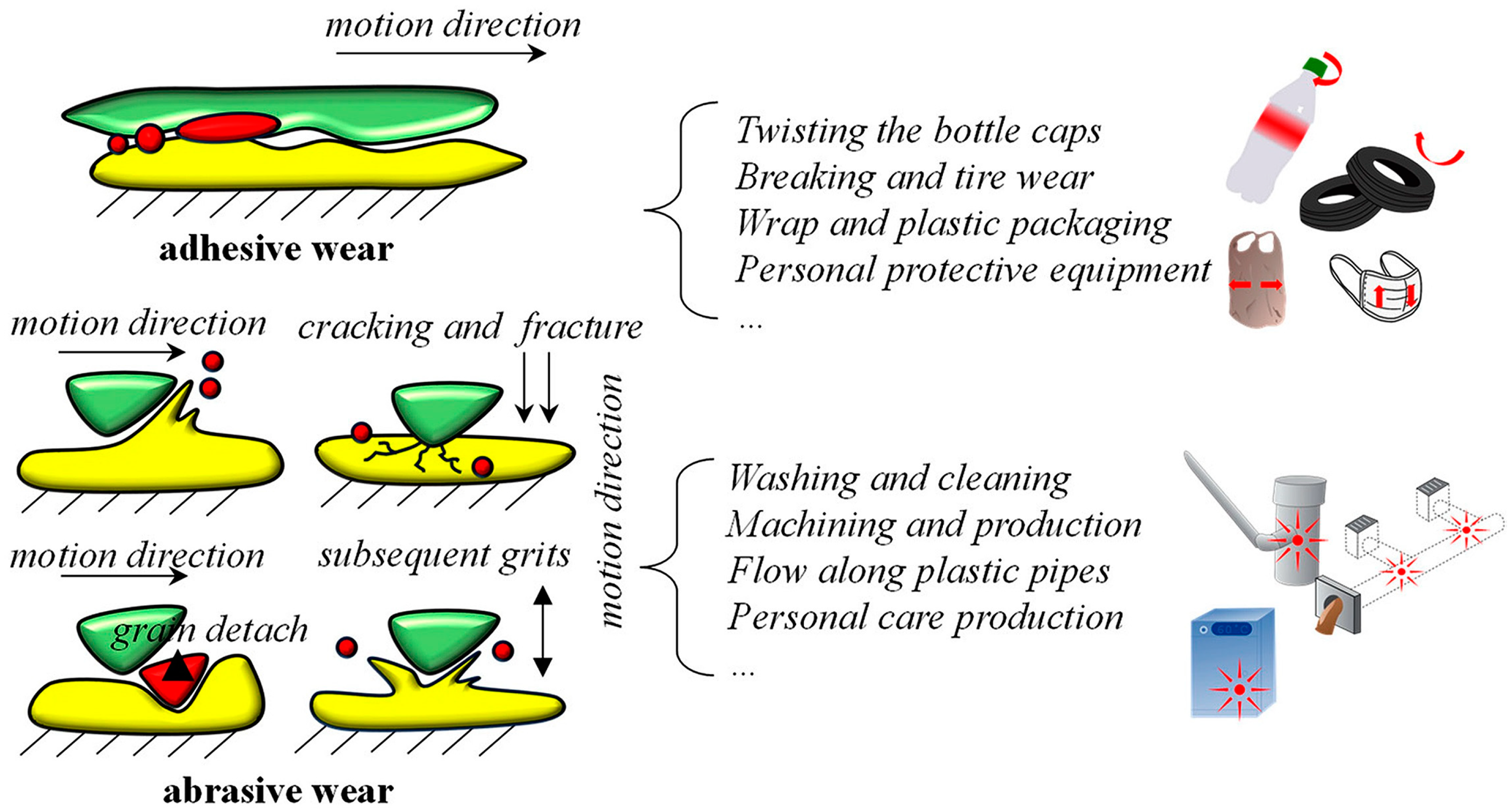
Figure 1. The formation of secondary MNPs through adhesive and abrasive wear processes and their corresponding activities. Reprinted with permission from Ref. [16]. Copyright 2024, MDPI.
Surfaces 08 00027 g001
Figure 2. Diagram illustrating the formation and effects of TRWPs. Reprinted with permission from Ref. [40]. Copyright 2024, MDPI.
Surfaces 08 00027 g002
Figure 3. SEM images of polystyrene (PS) at various stages of weathering (ae) along with an SEM image depicting the resulting nanoparticles (f). Note the formation of holes after 1 day and of granular protrusions after 5 days (highlighted by red circles and ovals). Adapted with permission from Ref. [44]. Copyright 2024, Elsevier.
Surfaces 08 00027 g003
Figure 4. (ac) Topography images of three polystyrene surfaces previously scratched in ambient condition (one, three, and five times) by a silicon probe (NSG-01 NT-MDT, spring constant k = 5.1 N/m) with a normal force FN = 100 nN and a scan velocity v = 15 µm/s. Frame sizes: 5 × 4 µm2. Reprinted with permission from Ref. [47]. Copyright 2021, Elsevier.
Surfaces 08 00027 g004
Figure 5. Schemes of SEM/EDS technique: (a) Bulk sample analysis in an SEM. (b) Thin sample analysis. (c) Thin sample analysis in transmission mode (T-SEM). (d) Photograph of the T-SEM setup in working position (close to the pole shoe). (e) Geometry of the annular SDD EDS. Reprinted with permission from Ref. [94]. Copyright 2016, IOP.
Surfaces 08 00027 g005
Figure 6. Raman technique for characterization of nanoplastics (NPs) or microplastics (MPs) on a gold surface: (a) SEM image, (b) Raman mapping image, (c,d) single nanoplastic without/with pixel color interpolation, (e) laser spot scans and excitation of the NP. Reprinted with permission from Ref. [104]. Copyright 2020, Elsevier.
Surfaces 08 00027 g006
Disclaimer/Publisher’s Note: The statements, opinions and data contained in all publications are solely those of the individual author(s) and contributor(s) and not of MDPI and/or the editor(s). MDPI and/or the editor(s) disclaim responsibility for any injury to people or property resulting from any ideas, methods, instructions or products referred to in the content.