Review

Electrospinning for Biomedical Applications: An Overview of Material Fabrication Techniques

by
Anastasiia D. Tsareva
1,
Valeriia S. Shtol
1,
Dmitriy V. Klinov
1,2 and
Dimitri A. Ivanov
1,3,*
1
Department of Genetics and Life Sciences, Sirius University of Science and Technology, Olympic Ave., 1., Sirius Federal Territory, 354340 Krasnodar, Russia
2
Lopukhin Federal Research and Clinical Center of Physical–Chemical Medicine of Federal Medical Biological Agency, Malaya Pirogovskaya 1a, 119435 Moscow, Russia
3
Institut de Sciences des Matériaux de Mulhouse–IS2M, CNRS UMR 7361, F-68057 Mulhouse, France
*
Author to whom correspondence should be addressed.
Surfaces 2025, 8(1), 7; https://doi.org/10.3390/surfaces8010007
Submission received: 17 November 2024 / Revised: 25 December 2024 / Accepted: 30 December 2024 / Published: 8 January 2025
(This article belongs to the Special Issue Bio-Inspired Surfaces)

Abstract

:
This review examines recent methodologies for fabricating nonwoven polymer materials through electrospinning, focusing on the underlying physical principles, including the effects of external parameters, experimental conditions, material selection, and primary operational mechanisms. Potential applications of electrospun polymer matrices in tissue engineering are analyzed, with particular emphasis on their utility in biomedical contexts. Key challenges in incorporating new materials into biomedical devices are discussed, along with recent advances in electrospinning techniques driving innovation in this field.

1. Introduction

Electrospinning, a method for producing polymer nonwoven materials, was first developed in the early 20th century [1], but has only recently gained widespread attention in biomedical applications [2,3,4,5]. This technique is used to fabricate polymeric matrices, or scaffolds, consisting of micro- or nanofibers. An electrospinning setup primarily consists of three core components: a syringe with a pump system for dispensing a polymer solution or melt, a high-voltage power supply, and a collector for gathering the formed fibers. Under the influence of the applied high voltage, the liquid forms a Taylor cone at the needle tip, from which fine polymer jets are ejected. These jets travel toward the collector, solidifying into fibers during their trajectory [6].
One of the most prominent applications of electrospinning lies in the fabrication of nonwoven materials for biomedical uses. The resulting structures consist of entangled micro- or nanofibers with varied surface morphologies. For tissue engineering scaffolds, biodegradable polymers are typically used, such as polylactic acid (PLA) [7], poly(lactic–co–glycolic acid) (PLGA) [7], poly(ε–caprolactone) (PCL) [8], and polyurethanes (PUs) [9,10]. These materials provide a supportive environment for cell growth and tissue regeneration, making electrospinning a valuable tool in the development of next-generation biomedical solutions [11].
Tissue engineering scaffolds, produced using electrospun nanofibrous materials, closely mimic the native extracellular matrix (ECM) in terms of mechanical and functional properties, providing a biomimetic structure that matches the architecture of natural protein fibers [12]. Key factors such as fiber diameter, porosity, and pore diameter influence cell attachment, distribution, proliferation, and differentiation [13,14]. However, challenges remain, such as uneven cell distribution and limited migration within the polymer scaffold, often due to the structural geometry, which may not be conducive to certain cell types [15,16].
To improve the bioactive properties of polymer nonwoven materials, various polymer combinations and biologically active components are employed to promote cellular proliferation and differentiation while reducing tissue scarring. Polymer scaffolds embedded with drugs or bioactive compounds are widely used in tissue engineering for treating burn wounds and preventing scarring [4]. Nanofiber-based biosensors, known for their enhanced sensitivity, broad detection range, and cost efficiency, are another promising application [5]. Electrospun materials are also utilized in drug delivery [17,18], biosensing [19,20], and tissue engineering [15,21,22,23]. The versatility of electrospinning in selecting polymer components, forming fiber structures, and functionalizing them with bioactive molecules has led to the development of nanofibrous scaffolds with optimized mechanical properties and biological characteristics [15,21,22,23].
This review will explore the design of electrospinning setups, the conditions for forming polymeric matrices, methods for producing nanofibers, polymers used in tissue engineering, and the latest trends in the development of polymeric matrices for biomedical applications.

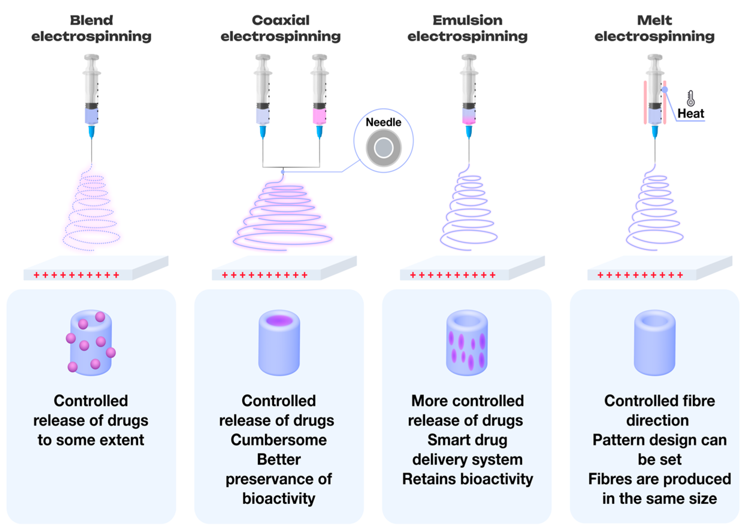
2. Methods for the Production of Polymer Nonwoven Materials

There are several methods for producing nanofibers, including both solution-based [24,25,26], and melt electrospinning techniques. The selection of a specific method depends on the research objectives, the apparatus design, and the required characteristics of the final product (Figure 1). Each spraying technique is carefully chosen based on the desired fiber structure, experimental conditions, and application goals.

2.1. Melt Electrospinning

Melt electrospinning is a widely utilized technique for generating micro- and nanofibers, where fiber formation is achieved under high temperatures and applied voltage. One of its primary advantages is its cost-effectiveness and environmental sustainability, as it eliminates the need for toxic solvents [27].
In this method, the polymer melt is drawn into fibers via a charged jet. The high viscosity of the melt allows for precise control of the jet, facilitating the creation of defined patterns and even three-dimensional structures—an advantage over solution electrospinning [28,29,30]. To further enhance fiber properties, additives such as nanoparticles can be incorporated into the polymer melt [31,32]. However, challenges arise when incorporating biologically active components, as high temperatures may lead to their degradation [33]. Despite this, the inclusion of proteins remains feasible for low-melting-point polymers [34]. The diameter of fibers produced via melt electrospinning typically ranges from 0.1 to 5 mm [35].

2.2. Electrospinning of Multicomponent Systems

Multicomponent system electrospinning combines polymers with biologically active components, including drugs [25,36], plant-based substances [37,38], amino acids [39], inorganic materials [40], and various other additives. In this process, biologically active agents or nanoparticles are either dissolved or dispersed within the polymer solution, allowing for the encapsulation of active components within the polymer fibers to enable controlled release. This method is particularly effective for small molecule delivery and has been successfully used for encapsulating antimicrobial peptides, among other applications [41].

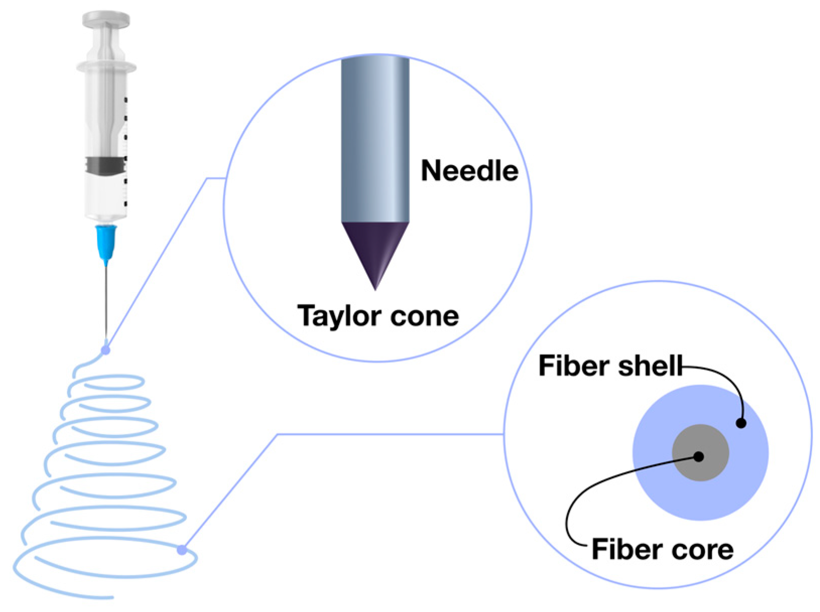
2.3. Coaxial Electrospinning: Advanced Fiber Formation Technique

In coaxial electrospinning, two distinct solutions are employed: one forms the core, while the other forms the shell of the fiber. Achieving optimal complementarity between these solutions is crucial, particularly regarding their viscosity, miscibility, conductivity, and flow rates [42]. A coaxial nozzle, usually comprising two or more concentric needles, facilitates this process (Figure 2). The core solution is dispensed through the inner needle, while the shell solution is delivered through the outer needle [43].
A key aspect of successful coaxial fiber formation is ensuring that the shell solution has higher viscosity [44], and that the flow rates and conductivity of both solutions are appropriately matched. The use of non-volatile solvents for the shell solution is essential to ensure the formation of a stable and uniform shell layer [45]. When volatile solvents or low polymer concentrations are utilized, hollow fibers may form due to rapid evaporation of the core solvent during fiber formation [46].
Both miscible and immiscible solutions can be used in coaxial electrospinning. Immiscible solutions prevent interactions between the core and shell components, thus avoiding conflicts between solvents and solutes. In the case of miscible solutions, distinct core–shell fibers can still form, but the diffusion and evaporation rates must be precisely controlled to maintain effective separation [43].

2.4. Emulsion-Based Electrospinning Techniques

Unlike blends, an emulsion consists of two or more distinct phases that remain separated throughout the electrospinning process. In this process, the continuous phase can form the fiber shell, while the dispersed droplet phase, usually aqueous, becomes the fiber core [24]. Unlike coaxial electrospinning, emulsion electrospinning does not require a specialized coaxial needle, making it a more environmentally sustainable option. This is achieved by minimizing the use of organic solvents, often substituting them with water [47]. Surfactants are commonly utilized to reduce surface tension between the immiscible phases, promoting the emulsification of the aqueous phase into the solvent phase, which enhances the fiber formation process and similarity to core–shell structure [48].

3. Polymers Utilized in Electrospinning for Tissue Engineering Applications

Polymers used in electrospinning for tissue engineering can be broadly categorized into two groups: natural polymers derived from animal and plant sources, and synthetic or semi-synthetic polymers. Natural polymers include proteins and polysaccharides, while synthetic and semi-synthetic polymers, though artificially produced, often demonstrate properties such as biocompatibility and biodegradability that make them suitable for biomedical applications, akin to their natural counterparts [49].

3.1. Applications of Natural and Plant-Based Polymers in Electrospinning for Tissue Engineering Applications

Natural and plant-based polymers have attracted considerable interest in tissue engineering because of their intrinsic biocompatibility, biodegradability, and their ability to closely replicate the structural features of native tissues. These materials support enhanced cellular interactions and promote better tissue integration, offering sustainable alternatives to synthetic polymers. Their use represents a promising path toward innovative solutions in regenerative medicine, combining environmental sustainability with advanced biomedical functionality [50].

3.1.1. Silk Fibroin

Silk fibroin is a natural protein polymer with exceptional properties, traditionally sourced from insect cocoons, predominantly from the silkworm (Bombyx mori) [51]. Its biocompatibility, biodegradability, high mechanical strength, and low immunogenicity make it a highly versatile material for various biomedical applications, such as vascular grafts [52,53,54], wound dressings [55,56], sutures [57], and other medical uses. Ongoing research continues to explore the modification of silk fibroin, particularly in the development of composite materials that combine fibroin with other biocompatible polymers or bioactive additives to enhance functionality [58,59,60]. For example, incorporating hydroxyapatite into fibroin scaffolds has been shown to improve osteointegration in bone implants [61], while the addition of silver nanoparticles offers antibacterial properties to prevent infections on biomaterial surfaces [62].
In a study by Phamornnak et al. [63], two-layer electrospun fibroin scaffolds were developed, with an aligned upper layer and a randomly oriented lower layer. Investigations using the NG108–15 hybrid cell line demonstrated that these scaffolds were biocompatible, enhanced metabolic activity, and promoted axonal growth, highlighting their potential in nerve tissue engineering.

3.1.2. Collagen

Collagen, a fundamental component of the ECM, is the most abundant protein in human and animal tissues, playing a key role in maintaining structural integrity [64]. Its low antigenicity, excellent biocompatibility, and biodegradability promote cell proliferation and tissue regeneration [65,66,67,68]. Sources of collagen are diverse, including bovine skin, tendons [69], and porcine bladder tissue [70], making it easily accessible for both research and practical applications. Collagen-based biomaterials have been extensively utilized in tissue engineering, particularly in areas such as burn treatment, wound healing [71,72,73], and bone tissue engineering [74,75].
In a study by García–Hernández et al. [76], electrospun polymer matrices were fabricated from a blend of PVA (polyvinyl alcohol), HC (collagen), and EEHP (ethanol extract of St. John’s wort). These matrices exhibited optimal porosity (67–90%), which is essential for supporting cell growth, interaction, and proliferation. Furthermore, the fibrous scaffolds demonstrated significant antimicrobial activity against S. aureus, indicating their potential for applications in wound healing and tissue regeneration.

3.1.3. Gelatin

Gelatin, a biopolymer derived through the partial acid or alkaline hydrolysis of collagen from sources such as skin, white connective tissue, and bone [77], is classified as a derivative protein since it does not exist in nature in its pure form and is produced through hydrolysis [78]. Gelatin is recognized for its key properties: biocompatibility, biodegradability, and low toxicity, which support cell proliferation. Additionally, it does not elicit immune responses, making it suitable for biomedical applications [79]. Despite these advantages, gelatin is thermally unstable, which can limit its applications.
Gelatin can be employed to fabricate hydrogels and nanofibers that promote cell attachment, proliferation, and differentiation, showing promise in regenerating skin, bone, cartilage, and even nerve tissue [80,81,82]. One of its emerging applications is in nerve tissue engineering, a field of growing interest.
In a study by Ahmadi et al. [82], poly(ε–caprolactone) (PCL)/gelatin (Gel) scaffolds incorporating layered double hydroxides (LDH) were evaluated for their effects on SH–SY5Y human neuroblastoma cells. The study demonstrated that the scaffold composition significantly improved cell attachment and proliferation. After three days of seeding, the scaffolds supported notable neuronal growth with a microstructure similar to the ECM, promoting cell migration and proliferation (Figure 3) [82].

3.1.4. Chitosan

Chitosan, a linear, semi-crystalline polysaccharide, is derived from the deacetylation of chitin [83], the second most abundant biopolymer after cellulose. Chitin is commonly sourced from the exoskeletons of crustaceans, insects, and certain fungi species [84,85]. Chitosan exhibits limited solubility in neutral and alkaline conditions and is insoluble in most organic solvents [86]. Chitosan offers numerous advantages, including wide availability, hydrophilicity, biocompatibility, biodegradability, and non-toxicity [86,87,88,89]. These properties make it an ideal candidate for bioactive materials used in tissue engineering applications, such as bone, vascular, and skin tissue development [90,91,92].
The performance of chitosan in tissue engineering can be significantly enhanced by combining it with other materials, improving both its bioactive and mechanical properties. One successful example is the combination of silk fibroin with chitosan, which enhances cell proliferation and adhesion to the scaffold [91]. Additionally, chitosan-based systems incorporating nanohydroxyapatite and polyethylene glycol have shown marked improvements in mechanical strength, promoting bone tissue regeneration [93].
In a study by Pezeshki–Modaress et al. [94], gelatin/chitosan (Chi) scaffolds in ratios of 100/0, 70/30, 60/40, and 50/50 were evaluated using human dermal fibroblasts (HDFs). The in vitro assessments demonstrated biocompatibility, adhesion, differentiation, and proliferation across all scaffold formulations. The DAPI staining results confirmed effective cell attachment and proliferative behavior on all studied scaffolds (Figure 4) [94].
As depicted in Figure 5, the number of cells increased consistently over the 14-day cultivation period for all polymer matrices. On days 7 and 14, the Chi 30 and Chi 40 matrices demonstrated the highest cell proliferation, while the sample without chitosan (Chi 0) exhibited the lowest proliferation rate.
The authors reported that the scaffolds had fiber diameters ranging from 180 to 196 nm, 92% porosity, and a tensile strength of 1.1 MPa. The MTS assay results demonstrated that the presence of chitosan positively affected HDF cell cultures. Based on these findings and the scaffold’s mechanical properties, the authors suggested that the gelatin/chitosan (70/30) scaffold holds significant promise for tissue engineering applications [94].

3.1.5. Alginate and Its Derivatives

Alginate is a natural polysaccharide valued for its biocompatibility, gel-forming capability, stimulation of cell proliferation, non-toxicity, biodegradability, high absorption capacity, and ease of processing [95,96]. It is primarily derived from brown seaweeds such as Laminaria, Sargassum, and Lessonia, as well as certain bacteria like Azotobacter and Pseudomonas [97,98]. In algae, alginate is typically found in the form of calcium, magnesium, and sodium salts.
To enhance its efficiency, researchers have developed various alginate-based composites. For instance, Wang et al. [99] synthesized sodium alginate with different oxidation levels to improve spinnability for electrospinning, incorporating zinc oxide nanoparticles to create fibrous membranes with optimized morphology, mechanical properties, and biocompatibility. Membranes containing up to 3% zinc oxide nanoparticles exhibited antibacterial activity against E. coli and S. aureus in vitro, reduced inflammatory responses, and promoted granulation tissue formation [99].
Recent studies have increasingly focused on developing polymer composites to improve the functional characteristics of biomaterials. Hajiabbas et al. [100] created a scaffold composed of oxidized alginate (OAL), gelatin (G), and silk fibroin (SF), which provided the appropriate porosity and structure for mesenchymal stem cell (AMSC) adhesion and distribution within the scaffold. Both the OAL–G and OAL–G–SF scaffolds were non-toxic and effectively promoted AMSC proliferation.

3.2. Applications of Synthetic Polymers in Electrospinning for Tissue Engineering

Synthetic polymers have become indispensable in tissue engineering, offering customizable properties and exceptional versatility that facilitate the design of scaffolds for a wide range of applications. Their ability to precisely regulate mechanical strength, degradation rates, and bioactivity makes them crucial in promoting cell proliferation and tissue regeneration across various clinical contexts. These attributes enable the development of advanced biomaterials that meet the specific demands of diverse therapeutic applications.

3.2.1. Polylactic Acid (PLA)

Polylactic acid (PLA) is a thermoplastic, biodegradable aliphatic polyester derived from lactic acid (2–hydroxypropionic acid) [101]. It undergoes biodegradation easily and decomposes in physiological environments, with its breakdown products excreted through the kidneys [102]. PLA exists in two main stereoisomeric forms: poly(L–lactic acid) (PLLA) and poly(D–lactic acid) (PDLA). The combination of these two forms in various proportions influences the polymer’s structural, thermal, barrier, and mechanical properties [103].
PLA’s solubility is influenced by its molecular weight and isomer content. Common solvents for PLA include dichloromethane (DCM) [104], hexafluoroisopropanol (HFIP) [105], acetone (AC) [106], chloroform (CF) [107], and tetrahydrofuran (THF) [108], among others. A common method is to use solvent mixtures, as combining volatile and less volatile solvents allows for the production of porous fibers [109]. The size and number of pores can be regulated by adjusting the solvent concentration [110]. The choice of solvent or solvent system also impacts a solution’s properties and conductivity [111]. Yin et al. successfully produced porous PLA fibers using a CF/DMF mixture in 90/10 and 80/20 ratios [109].
PLA is extensively utilized in various medical applications, including drug delivery systems [112], maxillofacial surgery [113,114], and tissue engineering [115]. Recent studies have focused on the development of composite scaffolds. For instance, Samokhin et al. [116] investigated the incorporation of polyethylene glycol (PEG) into nanofiber membranes composed of PLA/chitosan, fabricated using electrospinning. The addition of PEG led to the production of thinner fibers with reduced surface porosity, as well as increased metabolic activity in seeded cells, suggesting the enhanced hydrophilicity of the scaffold [116]. Similarly, Abdullah et al. [117] utilized coaxial electrospun fibers for partial bone tissue replacement, with cellulose acetate (CA) serving as the core and PLA as the shell. The coaxially spun fibers demonstrated greater strength and stiffness compared to pure CA fibers. By controlling flow rates, defect-free fibers were successfully fabricated. In vitro tests revealed the superior distribution and attachment of human osteoblasts on the matrix, along with higher cell proliferation, compared to monolithic PLA fibers and mixed PLA/CA fibers [117].
Polymer matrices composed of copolymers of polylactic acid and glycolic acid (PLGA) were utilized by Stachewicz et al. [118] to assess biocompatibility with osteoblast-like cells, including MC3T3-E1 and UMR106. The results demonstrated that the gene expression of UMR106 cells cultured on PLGA fibers was comparable to that of cells grown on conventional plastic substrates, indicating the preservation of their phenotypic characteristics. Additionally, the study found that randomly oriented polymer fibers enhanced cell migration due to increased porosity, attributed to the presence of free spaces between fibers.

3.2.2. Poly(ε–Caprolactone) (PCL)

Poly(ε–caprolactone) (PCL) is a semi-crystalline aliphatic polyester, with crystallinity reaching up to 50%, produced through the ring-opening polymerization of ε–caprolactone. Its key attributes include biocompatibility, high mechanical strength, and biodegradability [119,120]. PCL finds extensive use in tissue engineering [121,122,123], drug delivery systems [124,125,126,127], and bone regeneration applications [128], among other fields.
To produce nanofibers and minimize bead formation, Li et al. [129] introduced water into a PCL solution in glacial acetic acid. Water enhanced the conductivity of the solution and ionized the acetic acid, leading to the formation of ultrathin fibers. The proportion of fibers with a diameter of 500 nm diminished as the water content increased, and at a water concentration of 9%, the fibers became more uniform [129].
Research on pore formation in fibers has been a focus for many groups. For example, Katsogiannis et al. [130] investigated the effects of different solvents, including chloroform, dichloromethane, tetrahydrofuran (THF), and formic acid (FA). These solvents were identified as optimal for producing electrospinnable PCL solutions. Chloroform and dichloromethane were preferred due to their low boiling points, while THF’s miscibility with water and FA’s high dielectric constant contributed to their suitability. Additionally, dimethyl sulfoxide (DMSO), which has a low evaporation rate, was used to promote good phase separation. The study found that pores of various sizes and depths formed, with considerable variability in fiber diameter. Increasing DMSO concentration led to a reduction in fiber diameter from 2270 nm to 1470 nm, corresponding to increased solution conductivity from 0.42 to 0.67 μS/cm. However, the benefits of DMSO were limited to concentrations between 10% and 20%. At concentrations exceeding 30%, ribbon-like fibers were produced, and the pores disappeared [130].
Gao and Callanan [131] explored PCL-based scaffolds for HepG2 liver cells, creating fibers with varied surface morphologies using different solvent combinations. The resulting fibers featured surface depressions or pores, which significantly influenced cell viability and DNA content (Figure 6).
The HepG2 liver cells demonstrated optimal viability and DNA levels on scaffolds with small surface depressions or pores (Figure 7). This research underscores the potential of polymer scaffolds for developing platforms that mimic liver tissue.

3.2.3. Polyamide–6 (PA6)

Polyamide–6 (PA6) is known for its biocompatibility, biodegradability, excellent thermomechanical properties, and high wear resistance [132,133]. Due to these properties, PA6 is widely used in biomedicine [134,135], as well as in the automotive, electrical, and packaging industries. PA6 is obtained through the hydrolytic polymerization of caprolactone, with a structure similar to the collagen backbone [134].
Zhang and his research team [136] developed composite matrices using PA6 for biomedical applications. Cerium oxide (CeO2) was added to enhance biological activity. At CeO2 concentrations ranging from 0 to 5%, the fiber diameter decreased, but increased at 7 to 9%. The optimal concentration of CeO2 was found to be 5%, which resulted in minimal fiber diameter variability. Tests on mouse macrophages and osteoblasts demonstrated that the matrices exhibited the required biocompatibility and were non-toxic, with osteoblast proliferation outpacing that of macrophages [136].

3.2.4. Polyhydroxyalkanoates (PHA)

Polyhydroxyalkanoates (PHA) are a family of linear, thermoplastic, aliphatic polyesters, classified as biopolymers [137]. These polymers are synthesized by microorganisms as nutrient reserves [138]. The chemical structure of PHA depends on the metabolic capabilities of the microorganism [139,140]. PHAs can be classified based on chain length: short, medium, or long—each with distinct mechanical properties. For example, short-chain PHAs have poor mechanical properties, while medium-chain PHAs exhibit improved tensile strength and mechanical performance [141].
PHAs possess desirable traits such as renewability, biocompatibility, mechanical strength, and biodegradability under physiological conditions without producing toxic byproducts. Additionally, they have piezoelectric properties that stimulate bone tissue growth and wound healing [141,142,143,144].
PHA-based matrices are commonly used in wound healing. For instance, Li et al. [144] employed PHA in coaxial electrospinning to create wound dressings. The inner layer consisted of dodecyltrimethylammonium chloride and polyvinylpyrrolidone (PVP), while the outer layer was a PHA/polyethersulfone (PES) blend. Single fibers were also produced for comparison. The authors noted that the PHA/PES outer layer was hydrophobic, effectively preventing unwanted biocide release in physiological conditions. The single and coaxial nanofibers reduced the viability of P. aeruginosa bacteria by 97.4% and 86.9%, respectively, after 2 h of contact, and further reduced viability to 98.9% and 98.0% after 4 h. These findings suggest that the matrices could be effective for treating localized wound surfaces [144].

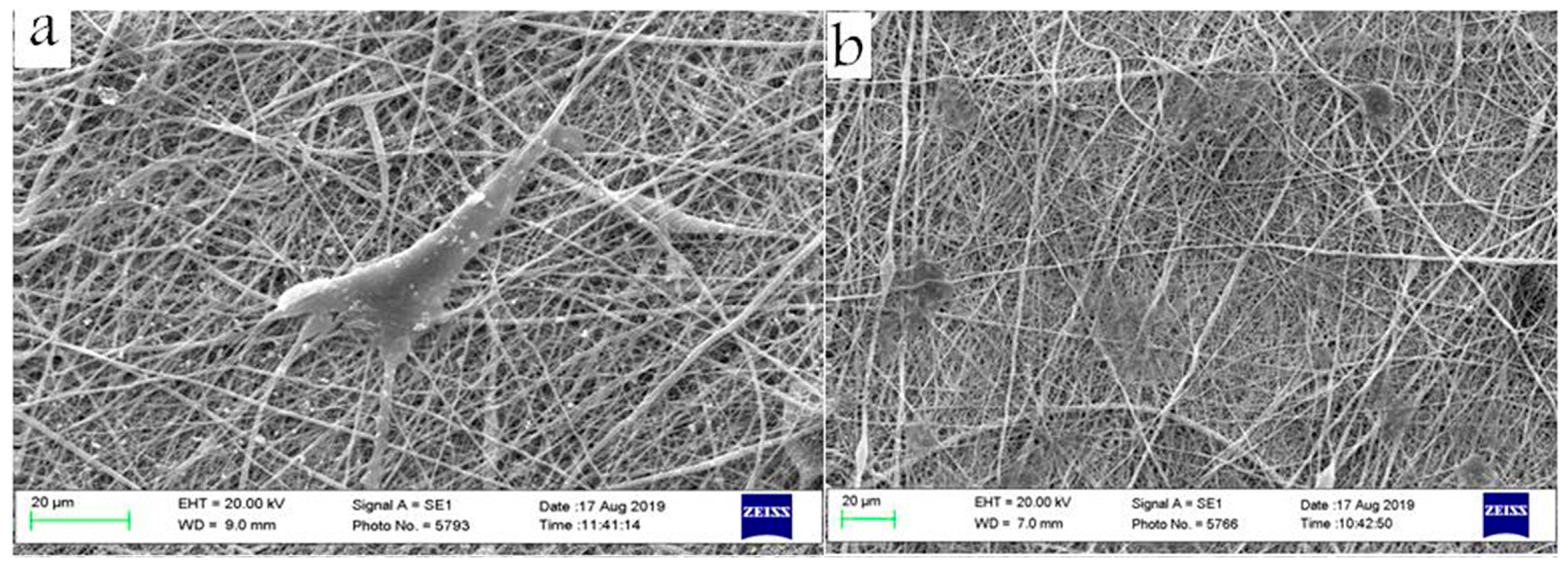
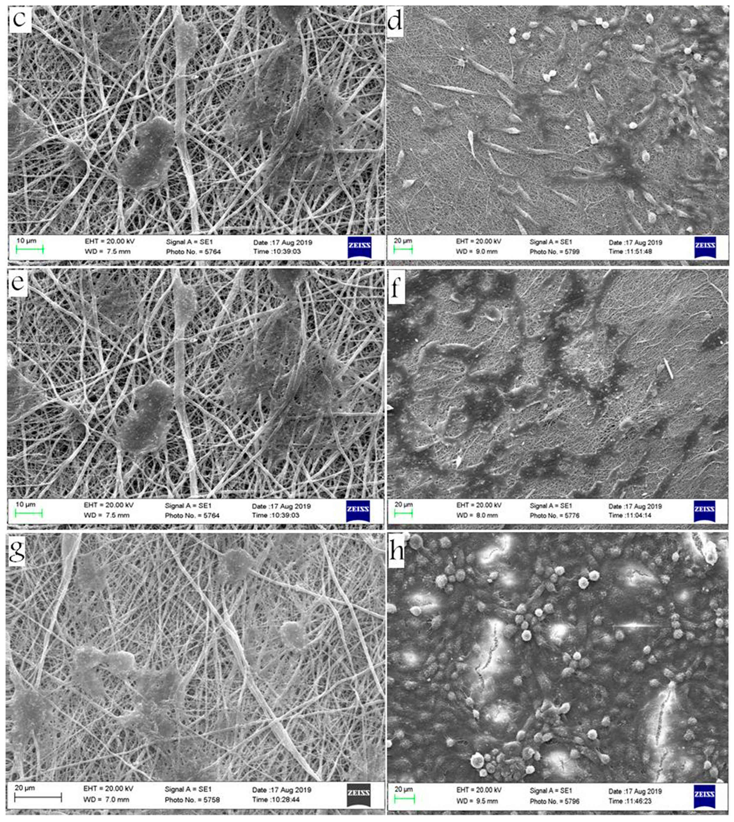
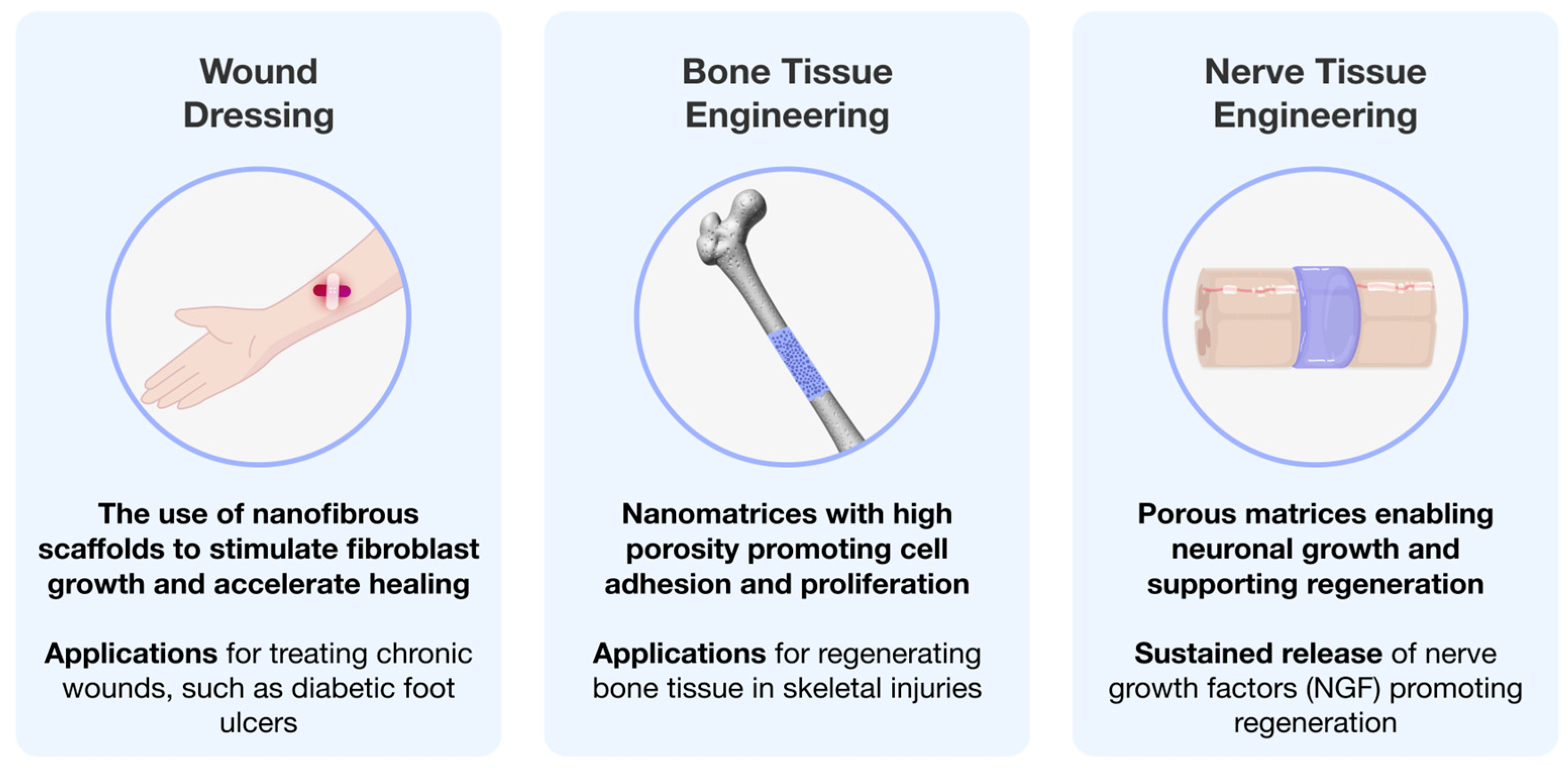
4. Tissue Engineering Using Electrospun Polymeric Matrices

The application of polymeric matrices in tissue engineering has emerged as a critical area of research, offering innovative solutions for repairing and regenerating various types of tissues [145,146]. These matrices play a significant role in wound healing, facilitating fast recovery while enhancing the integration of engineered tissues with host structures. Additionally, their utility extends into nerve tissue engineering, where they provide support for neuronal growth and repair, while also serving as scaffolds in bone tissue engineering to promote osteoconduction and facilitate the regeneration of bone tissue, thereby addressing the challenges associated with skeletal injuries. The following subsections examine key studies in tissue engineering, with a focus on applications in wound healing, nerve tissue engineering, and bone tissue engineering (Figure 8). These investigations highlight the use of multi-component systems based on biodegradable polymers for specific biomedical applications.

4.1. Wound Healing

Wound healing electrospinning is a cutting-edge technique that produces nanofibrous scaffolds, providing a supportive environment for tissue regeneration.
Ahn et al. [147] described the development of a polymer nonwoven material composed of a mixture of cellulose acetate (CA) and soy protein hydrolysate (SPH), using pure PCL and CA as comparative matrices. The addition of SPH to CA increased surface roughness and hydrophilicity. In vitro studies demonstrated that the CA/SPH matrices enhanced the proliferation, growth, migration, and infiltration of fibroblasts, while exhibiting low cytotoxicity in comparison to PCL and CA nanofibers. The authors suggest that such tissue-engineered constructs will represent the next generation of regenerative dressings, broadening the potential of nanofiber technology and the wound care market.
Chronic skin ulcers are a common occurrence in individuals with diabetes, highlighting the need for bioactive dressings. In this context, Chouhan et al. [148] investigated polymer matrices derived from various types of silk fibroin (SF) combined with polyvinyl alcohol (PVA). The polymer matrices included compositions of PVA, PVAAA = (PVA + Antheraea assama silk fibroin), PVABM = (PVA + Bombyx mori silk fibroin), and PVAPR = (PVA + Philosamia ricini silk fibroin). The hybrid matrices based on PVA/SF demonstrated superior performance when compared to pure PVA. Based on their findings, the authors concluded that the produced polymer matrices represent a reliable wound dressing material with significant potential for the treatment of chronic wounds, such as diabetic foot ulcers.

4.2. Nerve Tissue Engineering

Nerve tissue engineering (NTE) represents one of the most promising approaches to the restoration of the central nervous system. A key aspect of NTE is the three-dimensional distribution and growth of cells within a porous scaffold, which has significant clinical implications. Currently, there is no ideal strategy for nerve tissue restoration; however, this field demonstrates high potential for further development.
Hu et al. [149] investigated multi-component systems of PCL, nerve growth factor (NGF), and Bovine Serum Albumin (BSA) on rat pheochromocytoma (PC12) cells. The authors produced aligned and randomly oriented fibers in configurations of PCL, PCL/NGF, PCL/BSA, and PCL/NGF/BSA using an emulsified electrospinning technique. The release rate study indicated that there was almost no discernible difference between the randomly oriented and aligned fibers. The release profile of BSA after 56 days for the randomly oriented R–PCL–BSA and the aligned A–PCL–BSA was 92.57 ± 0.41% and 94.72 ± 1.94%, respectively, while a stable release of NGF from (R/A)–PCL–NGF/BSA was observed for 28 days. All polymeric matrices demonstrated high biocompatibility and showed no adverse effects on cell viability. The sustained release of NGF facilitated enhanced neuronal differentiation of the cells. The authors assert that the obtained results could be directed toward the development of improved constructs for nerve repair.
Biocomposite polymer matrices replicate the morphology of the ECM and can thus serve as nerve grafts in tissue engineering. Kijeńska et al. [150] developed matrices composed of poly(L–lactic acid)–co–poly(ε–caprolactone) or P(LLA–CL), collagen I, and collagen III, with P(LLA–CL) serving as a reference. Cellular assays were conducted using C17.2 nerve stem cells. The authors successfully produced aligned fibers of P(LLA–CL)/collagen I/collagen III, exhibiting an average diameter of 253 ± 102 nm. Notably, cell proliferation was found to be 22% higher for the matrix comprising P(LLA–CL)/collagen I/collagen III compared to the pure P(LLA–CL). Based on these findings, the authors conclude that composite matrices possess significant potential for enhancing nerve regeneration.

4.3. Bone Tissue Engineering

Bone tissue engineering has established itself as one of the most promising therapeutic approaches for the treatment of bone defects. The materials employed in the construction of scaffolds intended for bone tissue regeneration must possess a high specific surface area, significant porosity, and an optimal surface structure that promotes cell adhesion, proliferation, and differentiation [151].
Rethinam et al. [152] developed nanobiomembranes using the electrospinning method, employing PVA and nano-Demineralized Bone Matrix (nano-DBM), and incorporating carbon nanoparticles (CNP) to impart additional strength. To evaluate biocompatibility, the authors utilized the MG 63 osteoblast cell line. According to the obtained data, the polymer matrix composed of PVA, nano-DBM, and CNP (0.6%) exhibited the best mechanical properties, with a tensile strength of 14.58 ± 0.13 MPa and an elongation at break of 13.87 ± 0.05%. In vitro testing demonstrated higher cellular proliferation compared to the control. The antibacterial properties were assessed against both Gram-negative bacteria (E. coli) and Gram-positive bacteria (S. aureus), confirming the antibacterial activity of the resulting polymer matrices. Based on their findings, the authors concluded that scaffolds comprising PVA, nano-DBM, and CNP show promising potential for bone tissue regeneration.

5. Challenges and Perspectives

Electrospinning is a highly versatile method for producing polymer matrices with unique properties and diverse applications. However, the process is accompanied by several challenges, such as the selection of suitable polymers and additives, precise control over fiber morphology, and the limited productivity of laboratory-scale devices. Addressing these issues necessitates further research and the development of advanced technologies to enhance process efficiency and stability.
Recent advances in electrospinning research have focused on developing tissue-engineered constructs incorporating phytocomponents. For instance, Zhou et al. integrated Chinese herbs into polymer matrices to produce wound dressings, demonstrating excellent antibacterial properties and a prolonged release profile of the active ingredient, Yunnan Baiyao [153]. The inclusion of herbal extracts not only enhances antibacterial efficacy but also imparts inherent anti-inflammatory and antioxidant properties [154,155,156,157]
Additionally, efforts are being made to improve the performance of laboratory electrospinning setups while reducing their cost, making the technology more accessible [158,159].
Future advancements in electrospinning techniques hold significant potential for the development of functional materials, with promising applications in medicine and other industries.

6. Conclusions

Electrospinning is a straightforward and adaptable technique extensively used to fabricate nonwoven materials, particularly in tissue engineering. The morphology of electrospun nanofibers is influenced by multiple factors, including polymer concentration, viscosity, molecular weight, applied voltage, needle-to-collector distance, and solvent choice. Adjustments to humidity and solvent type can yield polymer fibers with a porous surface, and specialized collectors, like rotating drums or disks, can produce aligned fibers, which promote greater cell proliferation and differentiation than randomly oriented matrices.
Various electrospinning methods are available for creating polymer matrices, including melt extrusion and solution electrospinning, which encompasses blended, coaxial, and emulsion techniques. Blended electrospinning, being straightforward and widely used, is advantageous for its simplicity, requiring fewer parameter adjustments and less specialized equipment.
Polymers selected for tissue engineering applications must exhibit biocompatibility, biodegradability, and non-toxicity upon degradation. Polycaprolactone (PCL) and polylactic acid (PLA) are among the most commonly utilized polymers due to their biocompatibility and biodegradability, with PLA, in particular, valued for its mechanical strength and suitability in medical applications.
Despite these advantages, electrospinning faces limitations in efficiency that pose challenges for scaling up in industrial applications. Further research and technological advancements are required to improve production yields and facilitate the integration of electrospinning into larger-scale processes.
Electrospun nanofibrous scaffolds effectively replicate the extracellular matrix’s microstructure, enhancing cell adhesion, differentiation, and proliferation. These characteristics position electrospun polymer scaffolds as promising materials for current and future biomedical applications in tissue engineering.

Author Contributions

Conceptualization, A.D.T., D.V.K. and D.A.I.; Writing—Original Draft Preparation, A.D.T.; Writing—Review and Editing, D.A.I.; Visualization, V.S.S. All authors have read and agreed to the published version of the manuscript.

Funding

This work was supported by the grant of the state program of the «Sirius» Federal Territory «Scientific and technological development of the «Sirius» Federal Territory» (Agreement № 18-03 from 10 September 2024).

Conflicts of Interest

The authors declare no conflicts of interest.

References

  1. Cooley, J.F. Apparatus for Electrically Dispersing Fluids. U.S. Patent No. 692,631, 4 February 1902. [Google Scholar]
  2. Ding, H.; Cheng, Y.; Niu, X.; Hu, Y. Application of Electrospun Nanofibers in Bone, Cartilage and Osteochondral Tissue Engineering. J. Biomater. Sci. Polym. Ed. 2020, 32, 536–561. [Google Scholar] [CrossRef]
  3. Jiffrin, R.; Razak, S.I.A.; Jamaludin, M.I.; Hamzah, A.S.A.; Mazian, M.A.; Jaya, M.A.T.; Nasrullah, M.Z.; Majrashi, M.; Theyab, A.; Aldarmahi, A.A.; et al. Electrospun Nanofiber Composites for Drug Delivery: A Review on Current Progresses. Polymers 2022, 14, 3725. [Google Scholar] [CrossRef] [PubMed]
  4. Mulholland, E.J. Electrospun Biomaterials in the Treatment and Prevention of Scars in Skin Wound Healing. Front. Bioeng. Biotechnol. 2020, 8, 481. [Google Scholar] [CrossRef]
  5. Liu, Y.; Hao, M.; Chen, Z.; Liu, L.; Liu, Y.; Yang, W.; Ramakrishna, S. A Review on Recent Advances in Application of Electrospun Nanofiber Materials as Biosensors. Curr. Opin. Biomed. Eng. 2020, 13, 174–189. [Google Scholar] [CrossRef]
  6. Kretov, E.I.; Zapolotsky, E.N.; Tarkova, A.R.; Prokhorikhin, A.A.; Boykov, A.A.; Malaev, D.U. Electrospinning for the Design of Medical Supplies. Bull. Sib. Med. 2020, 19, 153–162. [Google Scholar] [CrossRef]
  7. You, Y.; Min, B.M.; Lee, S.J.; Lee, T.S.; Park, W.H. In Vitro Degradation Behavior of Electrospun Polyglycolide, Polylactide, and Poly(Lactide-Co-Glycolide). J. Appl. Polym. Sci. 2005, 95, 193–200. [Google Scholar] [CrossRef]
  8. Yoshimoto, H.; Shin, Y.M.; Terai, H.; Vacanti, J.P. A Biodegradable Nanofiber Scaffold by Electrospinning and Its Potential for Bone Tissue Engineering. Biomaterials 2003, 24, 2077–2082. [Google Scholar] [CrossRef]
  9. Jaganathan, S.K.; Prasath Mani, M.; Ayyar, M.; Rathanasamy, R. Biomimetic Electrospun Polyurethane Matrix Composites with Tailor Made Properties for Bone Tissue Engineering Scaffolds. Polym. Test. 2019, 78, 105955. [Google Scholar] [CrossRef]
  10. Gabriel, L.P.; Rodrigues, A.A.; Macedo, M.; Jardini, A.L.; Maciel Filho, R. Electrospun Polyurethane Membranes for Tissue Engineering Applications. Mater. Sci. Eng. C 2017, 72, 113–117. [Google Scholar] [CrossRef] [PubMed]
  11. Rahmati, M.; Mills, D.K.; Urbanska, A.M.; Saeb, M.R.; Venugopal, J.R.; Ramakrishna, S.; Mozafari, M. Electrospinning for Tissue Engineering Applications. Prog. Mater. Sci. 2021, 117, 100721. [Google Scholar] [CrossRef]
  12. Sell, S.A.; Wolfe, P.S.; Garg, K.; McCool, J.M.; Rodriguez, I.A.; Bowlin, G.L. The Use of Natural Polymers in Tissue Engineering: A Focus on Electrospun Extracellular Matrix Analogues. Polymers 2010, 2, 522–553. [Google Scholar] [CrossRef]
  13. Nam, J.; Huang, Y.; Agarwal, S.; Lannutti, J. Improved Cellular Infiltration in Electrospun Fiber via Engineered Porosity. Tissue Eng. 2007, 13, 2249–2257. [Google Scholar] [CrossRef] [PubMed]
  14. Şimşek, M. Tuning Surface Texture of Electrospun Polycaprolactone Fibers: Effects of Solvent Systems and Relative Humidity. J. Mater. Res. 2020, 35, 332–342. [Google Scholar] [CrossRef]
  15. Lannutti, J.; Reneker, D.; Ma, T.; Tomasko, D.; Farson, D. Electrospinning for Tissue Engineering Scaffolds. Mater. Sci. Eng. C 2007, 27, 504–509. [Google Scholar] [CrossRef]
  16. Huang, W.Y.; Suye, S.I.; Fujita, S. Cell Trapping via Migratory Inhibition within Density-Tuned Electrospun Nanofibers. ACS Appl. Bio. Mater. 2021, 4, 7456–7466. [Google Scholar] [CrossRef]
  17. Sill, T.J.; von Recum, H.A. Electrospinning: Applications in Drug Delivery and Tissue Engineering. Biomaterials 2008, 29, 1989–2006. [Google Scholar] [CrossRef] [PubMed]
  18. Pant, B.; Park, M.; Park, S.J. Drug Delivery Applications of Core-Sheath Nanofibers Prepared by Coaxial Electrospinning: A Review. Pharmaceutics 2019, 11, 305. [Google Scholar] [CrossRef]
  19. Sawicka, K.; Gouma, P.; Simon, S. Electrospun Biocomposite Nanofibers for Urea Biosensing. Sens. Actuators B Chem. 2005, 108, 585–588. [Google Scholar] [CrossRef]
  20. Halicka, K.; Cabaj, J. Electrospun Nanofibers for Sensing and Biosensing Applications—A Review. Int. J. Mol. Sci. 2021, 22, 6357. [Google Scholar] [CrossRef]
  21. Zhao, G.; Zhang, X.; Lu, T.J.; Xu, F. Recent Advances in Electrospun Nanofibrous Scaffolds for Cardiac Tissue Engineering. Adv. Funct. Mater. 2015, 25, 5726–5738. [Google Scholar] [CrossRef]
  22. Nguyen, T.H.; Bao, T.Q.; Park, I.; Lee, B.T. A Novel Fibrous Scaffold Composed of Electrospun Porous Poly(ε-Caprolactone) Fibers for Bone Tissue Engineering. J. Biomater. Appl. 2013, 28, 514–528. [Google Scholar] [CrossRef] [PubMed]
  23. Abrigo, M.; McArthur, S.L.; Kingshott, P. Electrospun Nanofibers as Dressings for Chronic Wound Care: Advances, Challenges, and Future Prospects. Macromol. Biosci. 2014, 14, 772–792. [Google Scholar] [CrossRef] [PubMed]
  24. Buzgo, M.; Mickova, A.; Doupnik, M.; Rampichova, M. Blend Electrospinning, Coaxial Electrospinning, and Emulsion Electrospinning Techniques. In Core-Shell Nanostructures for Drug Delivery and Theranostics: Challenges, Strategies and Prospects for Novel Carrier Systems; Elsevier: Amsterdam, The Netherlands, 2018; pp. 325–347. ISBN 9780081021989. [Google Scholar]
  25. Cornejo Bravo, J.M.; Villarreal Gómez, L.J.; Serrano Medina, A. Electrospinning for Drug Delivery Systems: Drug Incorporation Techniques. In Electrospinning—Material, Techniques, and Biomedical Applications; InTech: Bronx, NY, USA, 2016; Volume 14. [Google Scholar] [CrossRef]
  26. Voniatis, C.; Manikion, K.; Gyulai, G.; Jedlovszky-Hajdu, A. Synergistic properties of polysuccinimide/poly (lactic acid) co-electrospun and blended-electrospun nanofibers. J. Mol. Liq. 2023, 390, 123150. [Google Scholar] [CrossRef]
  27. Alghoraibi, I.; Alomari, S. Different Methods for Nanofiber Design and Fabrication. In Handbook of Nanofibers; Springer International Publishing: Berlin/Heidelberg, Germany, 2018; pp. 1–46. [Google Scholar] [CrossRef]
  28. Dayan, C.B.; Afghah, F.; Okan, B.S.; Yıldız, M.; Menceloglu, Y.; Culha, M.; Koc, B. Modeling 3D Melt Electrospinning Writing by Response Surface Methodology. Mater. Des. 2018, 148, 87–95. [Google Scholar] [CrossRef]
  29. Zhang, L.H.; Duan, X.P.; Yan, X.; Yu, M.; Ning, X.; Zhao, Y.; Long, Y.Z. Recent Advances in Melt Electrospinning. RSC Adv. 2016, 6, 53400–53414. [Google Scholar] [CrossRef]
  30. Brown, T.D.; Dalton, P.D.; Hutmacher, D.W. Melt Electrospinning Today: An Opportune Time for an Emerging Polymer Process. Prog. Polym. Sci. 2016, 56, 116–166. [Google Scholar] [CrossRef]
  31. Bachs-Herrera, A.; Yousefzade, O.; Del Valle, L.J.; Puiggali, J. Melt Electrospinning of Polymers: Blends, Nanocomposites, Additives and Applications. Appl. Sci. 2021, 11, 1808. [Google Scholar] [CrossRef]
  32. Li, X.; Liu, H.; Wang, J.; Li, C. Preparation and Properties of PET/SiO2 Composite Micro/Nanofibers by a Laser Melt-Electrospinning System. J. Appl. Polym. Sci. 2012, 125, 2050–2055. [Google Scholar] [CrossRef]
  33. Hengsawas Surasarang, S.; Keen, J.M.; Huang, S.; Zhang, F.; McGinity, J.W.; Williams, R.O. Hot Melt Extrusion versus Spray Drying: Hot Melt Extrusion Degrades Albendazole. Drug Dev. Ind. Pharm. 2017, 43, 797–811. [Google Scholar] [CrossRef]
  34. Hewitt, E.; Mros, S.; McConnell, M.; Cabral, J.D.; Ali, A. Melt-Electrowriting with Novel Milk Protein/PCL Biomaterials for Skin Regeneration. Biomed. Mater. 2019, 14, 055013. [Google Scholar] [CrossRef]
  35. Hutmacher, D.W.; Dalton, P.D. Melt Electrospinning. Chem. Asian J. 2011, 6, 44–56. [Google Scholar] [CrossRef] [PubMed]
  36. Vlachou, M.; Siamidi, A.; Kyriakou, S. Electrospinning and Drug Delivery. In Electrospinning and Electrospraying-Techniques and Applications; InTech: Bronx, NY, USA, 2019; pp. 1–22. [Google Scholar] [CrossRef]
  37. Suganya, S.; Senthil Ram, T.; Lakshmi, B.S.; Giridev, V.R. Herbal Drug Incorporated Antibacterial Nanofibrous Mat Fabricated by Electrospinning: An Excellent Matrix for Wound Dressings. J. Appl. Polym. Sci. 2011, 121, 2893–2899. [Google Scholar] [CrossRef]
  38. Liu, H.; Bai, Y.; Huang, C.; Wang, Y.; Ji, Y.; Du, Y.; Xu, L.; Yu, D.G.; Bligh, S.W.A. Recent Progress of Electrospun Herbal Medicine Nanofibers. Biomolecules 2023, 13, 184. [Google Scholar] [CrossRef] [PubMed]
  39. Ji, Y.; Song, W.; Xu, L.; Yu, D.G.; Bligh, S.W.A. A Review on Electrospun Poly(Amino Acid) Nanofibers and Their Applications of Hemostasis and Wound Healing. Biomolecules 2022, 12, 794. [Google Scholar] [CrossRef] [PubMed]
  40. El-Aassar, M.R.; Ibrahim, O.M.; Fouda, M.M.G.; El-Beheri, N.G.; Agwa, M.M. Wound Healing of Nanofiber Comprising Polygalacturonic/Hyaluronic Acid Embedded Silver Nanoparticles: In-Vitro and In-Vivo Studies. Carbohydr. Polym. 2020, 238, 116175. [Google Scholar] [CrossRef]
  41. Zehetmeyer, G.; Meira, S.; Scheibel, J.; Silva, C.; Rodembusch, F.; Brandelli, A.; Soares, R. Biodegradable and Antimicrobial Films Based on Poly(Butylene Adipate-Co-Terephthalate) Electrospun Fibers. Polym. Bull. 2017, 74, 3243–3268. [Google Scholar] [CrossRef]
  42. Dias, F.T.G.; Rempel, S.P.; Agnol, L.D.; Bianchi, O. The Main Blow Spun Polymer Systems: Processing Conditions and Applications. J. Polym. Res. 2020, 27, 205. [Google Scholar] [CrossRef]
  43. Heseltine, P.L.; Hosken, J.; Agboh, C.; Farrar, D.; Homer-Vanniasinkam, S.; Edirisinghe, M. Fiber Formation from Silk Fibroin Using Pressurized Gyration. Macromol. Mater. Eng. 2019, 304, 1800577. [Google Scholar] [CrossRef]
  44. Sun, Z.; Zussman, E.; Yarin, A.L.; Wendorff, J.H.; Greiner, A. Compound Core-Shell Polymer Nanofibers by Co-Electrospinning. Adv. Mater. 2003, 15, 1929–1932. [Google Scholar] [CrossRef]
  45. Chen, R.; Huang, C.; Ke, Q.; He, C.; Wang, H.; Mo, X. Preparation and Characterization of Coaxial Electrospun Thermoplastic Polyurethane/Collagen Compound Nanofibers for Tissue Engineering Applications. Colloids Surf. B Biointerfaces 2010, 79, 315–325. [Google Scholar] [CrossRef]
  46. Khalf, A.; Madihally, S.V. Recent Advances in Multiaxial Electrospinning for Drug Delivery. Eur. J. Pharm. Biopharm. 2017, 112, 1–17. [Google Scholar] [CrossRef] [PubMed]
  47. Avossa, J.; Herwig, G.; Toncelli, C.; Itel, F.; Rossi, R.M. Electrospinning Based on Benign Solvents: Current Definitions, Implications and Strategies. Green. Chem. 2022, 24, 2347–2375. [Google Scholar] [CrossRef]
  48. Zhang, J.X.; Chen, D.; Wang, S.J.; Zhu, K.J. Optimizing Double Emulsion Process to Decrease the Burst Release of Protein from Biodegradable Polymer Microspheres. J. Microencapsul. 2005, 22, 413–422. [Google Scholar] [CrossRef] [PubMed]
  49. Huang, Z.M.; Zhang, Y.Z.; Kotaki, M.; Ramakrishna, S. A Review on Polymer Nanofibers by Electrospinning and Their Applications in Nanocomposites. Compos. Sci. Technol. 2003, 63, 2223–2253. [Google Scholar] [CrossRef]
  50. Chinatangkul, N.; Limmatvapirat, C.; Limmatvapirat, S. Electrospun Nanofibers from Natural Polymers and Their Application. Sci. Eng. Health Stud. 2021, 15, 21010005. [Google Scholar]
  51. Wray, L.S.; Hu, X.; Gallego, J.; Georgakoudi, I.; Omenetto, F.G.; Schmidt, D.; Kaplan, D.L. Effect of Processing on Silk-Based Biomaterials: Reproducibility and Biocompatibility. J. Biomed. Mater. Res. B Appl. Biomater. 2011, 99B, 89–101. [Google Scholar] [CrossRef] [PubMed]
  52. Zhang, X.; Baughman, C.B.; Kaplan, D.L. In Vitro Evaluation of Electrospun Silk Fibroin Scaffolds for Vascular Cell Growth. Biomaterials 2008, 29, 2217–2227. [Google Scholar] [CrossRef]
  53. Zhou, J.; Cao, C.; Ma, X.; Lin, J. Electrospinning of Silk Fibroin and Collagen for Vascular Tissue Engineering. Int. J. Biol. Macromol. 2010, 47, 514–519. [Google Scholar] [CrossRef]
  54. Wang, D.; Liu, H.; Fan, Y. Silk Fibroin for Vascular Regeneration. Microsc. Res. Tech. 2017, 80, 280–290. [Google Scholar] [CrossRef] [PubMed]
  55. Çalamak, S.; Erdoǧdu, C.; Özalp, M.; Ulubayram, K. Silk Fibroin Based Antibacterial Bionanotextiles as Wound Dressing Materials. Mater. Sci. Eng. C 2014, 43, 11–20. [Google Scholar] [CrossRef]
  56. Patil, P.P.; Reagan, M.R.; Bohara, R.A. Silk Fibroin and Silk-Based Biomaterial Derivatives for Ideal Wound Dressings. Int. J. Biol. Macromol. 2020, 164, 4613–4627. [Google Scholar] [CrossRef] [PubMed]
  57. Choudhury, A.J.; Gogoi, D.; Chutia, J.; Kandimalla, R.; Kalita, S.; Kotoky, J.; Chaudhari, Y.B.; Khan, M.R.; Kalita, K. Controlled Antibiotic-Releasing Antheraea Assama Silk Fibroin Suture for Infection Prevention and Fast Wound Healing. Surgery 2016, 159, 539–547. [Google Scholar] [CrossRef]
  58. Tuwalska, A.; Grabska-Zielińska, S.; Sionkowska, A. Chitosan/Silk Fibroin Materials for Biomedical Applications—A Review. Polymers 2022, 14, 1343. [Google Scholar] [CrossRef] [PubMed]
  59. Yonesi, M.; Garcia-Nieto, M.; Guinea, G.V.; Panetsos, F.; Pérez-Rigueiro, J.; González-Nieto, D. Silk Fibroin: An Ancient Material for Repairing the Injured Nervous System. Pharmaceutics 2021, 13, 429. [Google Scholar] [CrossRef] [PubMed]
  60. Asadpour, S.; Kargozar, S.; Moradi, L.; Ai, A.; Nosrati, H.; Ai, J. Natural Biomacromolecule Based Composite Scaffolds from Silk Fibroin, Gelatin and Chitosan toward Tissue Engineering Applications. Int. J. Biol. Macromol. 2020, 154, 1285–1294. [Google Scholar] [CrossRef]
  61. Farokhi, M.; Mottaghitalab, F.; Samani, S.; Shokrgozar, M.A.; Kundu, S.C.; Reis, R.L.; Fatahi, Y.; Kaplan, D.L. Silk Fibroin/Hydroxyapatite Composites for Bone Tissue Engineering. Biotechnol. Adv. 2018, 36, 68–91. [Google Scholar] [CrossRef]
  62. Khan, R.S.; Rather, A.H.; Wani, T.U.; Rather, S.; Abdal-hay, A.; Sheikh, F.A. A Comparative Review on Silk Fibroin Nanofibers Encasing the Silver Nanoparticles as Antimicrobial Agents for Wound Healing Applications. Mater. Today Commun. 2022, 32, 103914. [Google Scholar] [CrossRef]
  63. Phamornnak, C.; Han, B.; Spencer, B.F.; Ashton, M.D.; Blanford, C.F.; Hardy, J.G.; Blaker, J.J.; Cartmell, S.H. Instructive Electroactive Electrospun Silk Fibroin-Based Biomaterials for Peripheral Nerve Tissue Engineering. Biomater. Adv. 2022, 141, 213094. [Google Scholar] [CrossRef] [PubMed]
  64. Shoulders, M.; Raines, R. Collagen Structure and Stability. Annu. Rev. Biochem. 2009, 78, 929–958. [Google Scholar] [CrossRef]
  65. Schmitt, F.O.; Levine, L.; Drake, M.P.; Rubin, A.L.; Pfahl, D.; Davison, P.F. The antigenicity of tropocollagen. Bull. Soc. Chim. Biol. 1964, 51, 493–497. [Google Scholar] [CrossRef]
  66. Parenteau-Bareil, R.; Gauvin, R.; Berthod, F. Collagen-Based Biomaterials for Tissue Engineering Applications. Materials 2010, 3, 1863–1887. [Google Scholar] [CrossRef]
  67. Lynn, A.K.; Yannas, I.V.; Bonfield, W. Antigenicity and Immunogenicity of Collagen. J. Biomed. Mater. Res. B Appl. Biomater. 2004, 71, 343–354. [Google Scholar] [CrossRef] [PubMed]
  68. Lee, C.H.; Singla, A.; Lee, Y. Biomedical Applications of Collagen. Int. J. Pharm. 2001, 221, 1–22. [Google Scholar] [CrossRef] [PubMed]
  69. Rodrigues, C.V.M.; Serricella, P.; Linhares, A.B.R.; Guerdes, R.M.; Borojevic, R.; Rossi, M.A.; Duarte, M.E.L.; Farina, M. Characterization of a Bovine Collagen-Hydroxyapatite Composite Scaffold for Bone Tissue Engineering. Biomaterials 2003, 24, 4987–4997. [Google Scholar] [CrossRef]
  70. Chen, F.; Yoo, J.J.; Atala, A. Acellular collagen matrix as a possible “off the shelf” biomaterial for urethral repair. Urology 1999, 54, 407–410. [Google Scholar] [CrossRef] [PubMed]
  71. Chattopadhyay, S.; Raines, R.T. Review Collagen-Based Biomaterials for Wound Healing. Biopolymers 2014, 101, 821–833. [Google Scholar] [CrossRef] [PubMed]
  72. Doillon, C.J.; Whyne, C.F.; Brandwein, S.; Silver, F.H. Collagen-based Wound Dressings: Control of the Pore Structure and Morphology. J. Biomed. Mater. Res. 1986, 20, 1219–1228. [Google Scholar] [CrossRef] [PubMed]
  73. Brett, D.W. A Review of Collagen and Collagen-Based Wound Dressings. Wounds 2008, 20, 347–356. [Google Scholar] [PubMed]
  74. Kon, E.; Delcogliano, M.; Filardo, G.; Busacca, M.; Di Martino, A.; Marcacci, M. Novel Nano-Composite Multilayered Biomaterial for Osteochondral Regeneration: A Pilot Clinical Trial. Am. J. Sports Med. 2011, 39, 1180–1190. [Google Scholar] [CrossRef] [PubMed]
  75. Pankajakshan, D.; Voytik-Harbin, S.L.; Nör, J.E.; Bottino, M.C. Injectable Highly Tunable Oligomeric Collagen Matrices for Dental Tissue Regeneration. ACS Appl. Bio. Mater. 2020, 3, 859–868. [Google Scholar] [CrossRef]
  76. García-Hernández, A.B.; Morales-Sánchez, E.; Berdeja-Martínez, B.M.; Escamilla-García, M.; Salgado-Cruz, M.P.; Rentería-Ortega, M.; Farrera-Rebollo, R.R.; Vega-Cuellar, M.A.; Calderón-Domínguez, G. PVA-Based Electrospun Biomembranes with Hydrolyzed Collagen and Ethanolic Extract of Hypericum Perforatum for Potential Use as Wound Dressing: Fabrication and Characterization. Polymers 2022, 14, 1981. [Google Scholar] [CrossRef]
  77. Djagny, K.B.; Wang, Z.; Xu, S. Gelatin: A Valuable Protein for Food and Pharmaceutical Industries: Review. Crit. Rev. Food Sci. Nutr. 2001, 41, 481–492. [Google Scholar] [CrossRef] [PubMed]
  78. Elzoghby, A.O. Gelatin-Based Nanoparticles as Drug and Gene Delivery Systems: Reviewing Three Decades of Research. J. Control. Release 2013, 172, 1075–1091. [Google Scholar] [CrossRef] [PubMed]
  79. Echave, M.C.; Sánchez, P.; Pedraz, J.L.; Orive, G. Progress of Gelatin-Based 3D Approaches for Bone Regeneration. J. Drug Deliv. Sci. Technol. 2017, 42, 63–74. [Google Scholar] [CrossRef]
  80. Yazdanpanah, A.; Madjd, Z.; Pezeshki-Modaress, M.; Khosrowpour, Z.; Farshi, P.; Eini, L.; Kiani, J.; Seifi, M.; Kundu, S.C.; Ghods, R.; et al. Bioengineering of Fibroblast-Conditioned Polycaprolactone/Gelatin Electrospun Scaffold for Skin Tissue Engineering. Artif. Organs 2022, 46, 1040–1054. [Google Scholar] [CrossRef] [PubMed]
  81. Gautam, S.; Sharma, C.; Purohit, S.D.; Singh, H.; Dinda, A.K.; Potdar, P.D.; Chou, C.F.; Mishra, N.C. Gelatin-Polycaprolactone-Nanohydroxyapatite Electrospun Nanocomposite Scaffold for Bone Tissue Engineering. Mater. Sci. Eng. C 2021, 119, 111588. [Google Scholar] [CrossRef] [PubMed]
  82. Ahmadi, S.; Shafiei, S.S.; Sabouni, F. Electrospun Nanofibrous Scaffolds of Polycaprolactone/Gelatin Reinforced with Layered Double Hydroxide Nanoclay for Nerve Tissue Engineering Applications. ACS Omega 2022, 7, 28351–28360. [Google Scholar] [CrossRef] [PubMed]
  83. Wang, J.; Zhuang, S. Chitosan-Based Materials: Preparation, Modification and Application. J. Clean. Prod. 2022, 355, 131825. [Google Scholar] [CrossRef]
  84. Kim, S.K. Chitin, Chitosan, Oligosaccharides and Their Derivatives: Biological Activities and Applications; CRC Press: Boca Raton, FL, USA, 2010. [Google Scholar] [CrossRef]
  85. Arbia, W.; Arbia, L.; Adour, L.; Amrane, A. Chitin Extraction from Crustacean Shells Using Biological Methods—A Review. Food Technol. Biotechnol. 2013, 51, 12–25. [Google Scholar]
  86. Wang, W.; Meng, Q.; Li, Q.; Liu, J.; Zhou, M.; Jin, Z.; Zhao, K. Chitosan Derivatives and Their Application in Biomedicine. Int. J. Mol. Sci. 2020, 21, 487. [Google Scholar] [CrossRef] [PubMed]
  87. Pavoni, J.M.F.; Luchese, C.L.; Tessaro, I.C. Impact of Acid Type for Chitosan Dissolution on the Characteristics and Biodegradability of Cornstarch/Chitosan Based Films. Int. J. Biol. Macromol. 2019, 138, 693–703. [Google Scholar] [CrossRef] [PubMed]
  88. Kaczmarek, B.; Owczarek, A.; Nadolna, K.; Sionkowska, A. The Film-Forming Properties of Chitosan with Tannic Acid Addition. Mater. Lett. 2019, 245, 22–24. [Google Scholar] [CrossRef]
  89. Kritchenkov, A.S.; Egorov, A.R.; Kurasova, M.N.; Volkova, O.V.; Meledina, T.V.; Lipkan, N.A.; Tskhovrebov, A.G.; Kurliuk, A.V.; Shakola, T.V.; Dysin, A.P.; et al. Novel Non-Toxic High Efficient Antibacterial Azido Chitosan Derivatives with Potential Application in Food Coatings. Food Chem. 2019, 301, 125247. [Google Scholar] [CrossRef] [PubMed]
  90. Balagangadharan, K.; Dhivya, S.; Selvamurugan, N. Chitosan Based Nanofibers in Bone Tissue Engineering. Int. J. Biol. Macromol. 2017, 104, 1372–1382. [Google Scholar] [CrossRef]
  91. Islam, M.M.; Shahruzzaman, M.; Biswas, S.; Nurus Sakib, M.; Rashid, T.U. Chitosan Based Bioactive Materials in Tissue Engineering Applications—A Review. Bioact. Mater. 2020, 5, 164–183. [Google Scholar] [CrossRef] [PubMed]
  92. Zhang, X.; Jia, C.; Qiao, X.; Liu, T.; Sun, K. Silk Fibroin Microfibers and Chitosan Modified Poly (Glycerol Sebacate) Composite Scaffolds for Skin Tissue Engineering. Polym. Test. 2017, 62, 88–95. [Google Scholar] [CrossRef]
  93. Vishwanath, V.; Pramanik, K.; Biswas, A. Optimization and Evaluation of Silk Fibroin-Chitosan Freeze-Dried Porous Scaffolds for Cartilage Tissue Engineering Application. J. Biomater. Sci. Polym. Ed. 2016, 27, 657–674. [Google Scholar] [CrossRef] [PubMed]
  94. Pezeshki-Modaress, M.; Zandi, M.; Rajabi, S. Tailoring the Gelatin/Chitosan Electrospun Scaffold for Application in Skin Tissue Engineering: An in Vitro Study. Prog. Biomater. 2018, 7, 207–218. [Google Scholar] [CrossRef] [PubMed]
  95. Sahoo, D.R.; Biswal, T. Alginate and Its Application to Tissue Engineering. SN Appl. Sci. 2021, 3, 30. [Google Scholar] [CrossRef]
  96. Sudarsan, S.; Franklin, D.S.; Guhanathan, S. Imbibed Salts and PH-Responsive Behaviours of Sodium Alginate Based Eco-Friendly Biopolymeric Hydrogels-A Solventless Approach. MMAIJ 2015, 11, 24–29. [Google Scholar]
  97. Clementi, F. Alginate Production by Azotobacter Vinelandii. Crit. Rev. Biotechnol. 1997, 17, 327–361. [Google Scholar] [CrossRef] [PubMed]
  98. Govan, J.R.W.; Fyfe, J.A.M.; Jarman, T.R. Isolation of Alginate-Producing Mutants of Pseudomonas Fluorescens, Pseudomonas Putida and Pseudomonas Mendocina. J. Gen. Microbiol. 1981, 125, 217–220. [Google Scholar] [CrossRef] [PubMed]
  99. Wang, W.; Liu, M.Y.; Shafiq, M.; Li, H.Y.; Hashim, R.; EL-Newehy, M.; EL-Hamshary, H.; Morsi, Y.; Mo, X. Synthesis of Oxidized Sodium Alginate and Its Electrospun Bio-Hybrids with Zinc Oxide Nanoparticles to Promote Wound Healing. Int. J. Biol. Macromol. 2023, 232, 123480. [Google Scholar] [CrossRef] [PubMed]
  100. Hajiabbas, M.; Alemzadeh, I.; Vossoughi, M. A Porous Hydrogel-Electrospun Composite Scaffold Made of Oxidized Alginate/Gelatin/Silk Fibroin for Tissue Engineering Application. Carbohydr. Polym. 2020, 245, 116465. [Google Scholar] [CrossRef] [PubMed]
  101. Lim, L.T.; Auras, R.; Rubino, M. Processing Technologies for Poly(Lactic Acid). Prog. Polym. Sci. 2008, 33, 820–825. [Google Scholar] [CrossRef]
  102. Pawar, R.P.; Tekale, S.U.; Shisodia, S.U.; Totre, J.T.; Domb, A.J. Biomedical Applications of Poly(Lactic Acid). Recent. Pat. Regen. Med. 2014, 4, 40–51. [Google Scholar] [CrossRef]
  103. Ahmed, J.; Zhang, J.X.; Song, Z.; Varshney, S.K. Thermal Properties of Polylactides: Effect of Molecular Mass and Nature of Lactide Isomer. J. Therm. Anal. Calorim. 2009, 95, 957–964. [Google Scholar] [CrossRef]
  104. Cao, X.; Wang, W.; Hu, J.; Wan, J.; Cui, L. Effect of Mixed Solvents on the Structure and Properties of PLLA/PDLA Electrospun Fibers. Fibers Polym. 2020, 21, 970–977. [Google Scholar] [CrossRef]
  105. Chen, S.C.; Huang, X.B.; Cai, X.M.; Lu, J.; Yuan, J.; Shen, J. The Influence of Fiber Diameter of Electrospun Poly(Lactic Acid) on Drug Delivery. Fibers Polym. 2012, 13, 1120–1125. [Google Scholar] [CrossRef]
  106. Wang, P.; Mele, E. Effect of Antibacterial Plant Extracts on the Morphology of Electrospun Poly(Lactic Acid) Fibres. Materials 2018, 11, 923. [Google Scholar] [CrossRef]
  107. Arrieta, M.P.; López, J.; López, D.; Kenny, J.M.; Peponi, L. Development of Flexible Materials Based on Plasticized Electrospun PLA-PHB Blends: Structural, Thermal, Mechanical and Disintegration Properties. Eur. Polym. J. 2015, 73, 433–446. [Google Scholar] [CrossRef]
  108. Natarajan, L.; New, J.; Dasari, A.; Yu, S.; Manan, M.A. Surface Morphology of Electrospun PLA Fibers: Mechanisms of Pore Formation. RSC Adv. 2014, 4, 44082–44088. [Google Scholar] [CrossRef]
  109. Yin, J.; Xu, L.; Ahmed, A. Batch Preparation and Characterization of Electrospun Porous Polylactic Acid-Based Nanofiber Membranes for Antibacterial Wound Dressing. Adv. Fiber Mater. 2022, 4, 832–844. [Google Scholar] [CrossRef]
  110. Nguyen, T.T.T.; Ghosh, C.; Hwang, S.G.; Chanunpanich, N.; Park, J.S. Porous Core/Sheath Composite Nanofibers Fabricated by Coaxial Electrospinning as a Potential Mat for Drug Release System. Int. J. Pharm. 2012, 439, 296–306. [Google Scholar] [CrossRef]
  111. Casasola, R.; Thomas, N.L.; Trybala, A.; Georgiadou, S. Electrospun Poly Lactic Acid (PLA) Fibres: Effect of Different Solvent Systems on Fibre Morphology and Diameter. Polymer 2014, 55, 4728–4737. [Google Scholar] [CrossRef]
  112. Fattahi, F.S.; Khoddami, A.; Avinc, O. Poly (Lactic Acid) Nano-Fibers as Drug-Delivery Systems: Opportunities and Challenges. Nanomed. Res. J. 2019, 4, 130–140. [Google Scholar] [CrossRef]
  113. Matsumine, H.; Sasaki, R.; Yamato, M.; Okano, T.; Sakurai, H. A Polylactic Acid Non-Woven Nerve Conduit for Facial Nerve Regeneration in Rats. J. Tissue Eng. Regen. Med. 2014, 8, 454–462. [Google Scholar] [CrossRef] [PubMed]
  114. Murgia, D.; Mauceri, R.; Campisi, G.; De Caro, V. Advance on Resveratrol Application in Bone Regeneration: Progress and Perspectives for Use in Oral and Maxillofacial Surgery. Biomolecules 2019, 9, 94. [Google Scholar] [CrossRef] [PubMed]
  115. Maleki, H.; Azimi, B.; Ismaeilimoghadam, S.; Danti, S. Poly(Lactic Acid)-Based Electrospun Fibrous Structures for Biomedical Applications. Appl. Sci. 2022, 12, 3192. [Google Scholar] [CrossRef]
  116. Samokhin, Y.; Varava, Y.; Diedkova, K.; Yanko, I.; Husak, Y.; Radwan-Pragłowska, J.; Pogorielova, O.; Janus, Ł.; Pogorielov, M.; Korniienko, V. Fabrication and Characterization of Electrospun Chitosan/Polylactic Acid (CH/PLA) Nanofiber Scaffolds for Biomedical Application. J. Funct. Biomater. 2023, 14, 414. [Google Scholar] [CrossRef] [PubMed]
  117. Abdullah, M.F.; Andriyana, A.; Muhamad, F.; Ang, B.C. Fabrication of Poly(Lactic Acid)-Cellulose Acetate Core-Shell Electrospun Fibers with Improved Tensile Strength and Biocompatibility for Bone Tissue Engineering. J. Polym. Res. 2023, 30, 257. [Google Scholar] [CrossRef]
  118. Stachewicz, U.; Qiao, T.; Rawlinson, S.C.F.; Almeida, F.V.; Li, W.Q.; Cattell, M.; Barber, A.H. 3D Imaging of Cell Interactions with Electrospun PLGA Nanofiber Membranes for Bone Regeneration. Acta Biomater. 2015, 27, 88–100. [Google Scholar] [CrossRef]
  119. Sisson, A.L.; Ekinci, D.; Lendlein, A. The Contemporary Role of ε-Caprolactone Chemistry to Create Advanced Polymer Architectures. Polymer 2013, 54, 4333–4350. [Google Scholar] [CrossRef]
  120. Neppalli, R.; Marega, C.; Marigo, A.; Bajgai, M.P.; Kim, H.Y.; Causin, V. Poly(ε-Caprolactone) Filled with Electrospun Nylon Fibres: A Model for a Facile Composite Fabrication. Eur. Polym. J. 2010, 46, 968–976. [Google Scholar] [CrossRef]
  121. Ghomi, E.R.; Lakshminarayanan, R.; Chellappan, V.; Verma, N.K.; Chinnappan, A.; Neisiany, R.E.; Amuthavalli, K.; Poh, Z.S.; Wong, B.H.S.; Dubey, N.; et al. Electrospun Aligned PCL/Gelatin Scaffolds Mimicking the Skin ECM for Effective Antimicrobial Wound Dressings. Adv. Fiber Mater. 2023, 5, 968–976. [Google Scholar] [CrossRef]
  122. Shie Karizmeh, M.; Poursamar, S.A.; Kefayat, A.; Farahbakhsh, Z.; Rafienia, M. An in Vitro and in Vivo Study of PCL/Chitosan Electrospun Mat on Polyurethane/Propolis Foam as a Bilayer Wound Dressing. Biomater. Adv. 2022, 135, 112667. [Google Scholar] [CrossRef] [PubMed]
  123. Lin, M.; Liu, Y.; Gao, J.; Wang, D.; Xia, D.; Liang, C.; Li, N.; Xu, R. Synergistic Effect of Co-Delivering Ciprofloxacin and Tetracycline Hydrochloride for Promoted Wound Healing by Utilizing Coaxial PCL/Gelatin Nanofiber Membrane. Int. J. Mol. Sci. 2022, 23, 1895. [Google Scholar] [CrossRef]
  124. Mitxelena-Iribarren, O.; Riera-Pons, M.; Pereira, S.; Calero-Castro, F.J.; Castillo Tuñón, J.M.; Padillo-Ruiz, J.; Mujika, M.; Arana, S. Drug-Loaded PCL Electrospun Nanofibers as Anti-Pancreatic Cancer Drug Delivery Systems. Polym. Bull. 2023, 80, 7763–7778. [Google Scholar] [CrossRef]
  125. Haroosh, H.J.; Dong, Y.; Jasim, S.; Ramakrishna, S. Morphological Structures and Drug Release Effect of Multiple Electrospun Nanofibre Membrane Systems Based on PLA, PCL, and PCL/Magnetic Nanoparticle Composites. J. Nanomater. 2022, 1, 5190163. [Google Scholar] [CrossRef]
  126. Eskitoros-Togay, M.; Bulbul, Y.E.; Tort, S.; Demirtaş Korkmaz, F.; Acartürk, F.; Dilsiz, N. Fabrication of Doxycycline-Loaded Electrospun PCL/PEO Membranes for a Potential Drug Delivery System. Int. J. Pharm. 2019, 565, 83–94. [Google Scholar] [CrossRef]
  127. Darbasizadeh, B.; Mortazavi, S.A.; Kobarfard, F.; Jaafari, M.R.; Hashemi, A.; Farhadnejad, H.; Feyzi-barnaji, B. Electrospun Doxorubicin-Loaded PEO/PCL Core/Sheath Nanofibers for Chemopreventive Action against Breast Cancer Cells. J. Drug Deliv. Sci. Technol. 2021, 64, 102576. [Google Scholar] [CrossRef]
  128. Heydari, Z.; Mohebbi-Kalhori, D.; Afarani, M.S. Engineered Electrospun Polycaprolactone (PCL)/Octacalcium Phosphate (OCP) Scaffold for Bone Tissue Engineering. Mater. Sci. Eng. C 2017, 81, 127–132. [Google Scholar] [CrossRef]
  129. Li, W.; Shi, L.; Zhang, X.; Liu, K.; Ullah, I.; Cheng, P. Electrospinning of Polycaprolactone Nanofibers Using H2O as Benign Additive in Polycaprolactone/Glacial Acetic Acid Solution. J. Appl. Polym. Sci. 2018, 135, 45578. [Google Scholar] [CrossRef]
  130. Katsogiannis, K.A.G.; Vladisavljević, G.T.; Georgiadou, S. Porous Electrospun Polycaprolactone (PCL) Fibres by Phase Separation. Eur. Polym. J. 2015, 69, 284–295. [Google Scholar] [CrossRef]
  131. Gao, Y.; Callanan, A. Influence of Surface Topography on PCL Electrospun Scaffolds for Liver Tissue Engineering. J. Mater. Chem. B 2021, 9, 8081–8093. [Google Scholar] [CrossRef]
  132. Heikkilä, P.; Harlin, A. Parameter Study of Electrospinning of Polyamide-6. Eur. Polym. J. 2008, 44, 3067–3079. [Google Scholar] [CrossRef]
  133. Tsou, S.Y.; Lin, H.S.; Cheng, P.J.; Huang, C.L.; Wu, J.Y.; Wang, C. Rheological Aspect on Electrospinning of Polyamide 6 Solutions. Eur. Polym. J. 2013, 49, 3619–3629. [Google Scholar] [CrossRef]
  134. Niu, X.; Wang, L.; Xu, M.; Qin, M.; Zhao, L.; Wei, Y.; Hu, Y.; Lian, X.; Liang, Z.; Chen, S.; et al. Electrospun Polyamide-6/Chitosan Nanofibers Reinforced Nano-Hydroxyapatite/Polyamide-6 Composite Bilayered Membranes for Guided Bone Regeneration. Carbohydr. Polym. 2021, 260, 117769. [Google Scholar] [CrossRef] [PubMed]
  135. Fatma Nur, P.; Pınar, T.; Uğur, P.; Ayşenur, Y.; Murat, E.; Kenan, Y. Fabrication of Polyamide 6/Honey/Boric Acid Mats by Electrohydrodynamic Processes for Wound Healing Applications. Mater. Today Commun. 2021, 29, 102921. [Google Scholar] [CrossRef]
  136. Zhang, X.; Chen, X. Preparation of Polyamide 6/CeO2 Composite Nanofibers through Electrospinning for Biomedical Applications. Int. J. Polym. Sci. 2019, 2019, 2494586. [Google Scholar] [CrossRef]
  137. Patel, S.K.S.; Sandeep, K.; Singh, M.; Singh, G.P.; Lee, J.K.; Bhatia, S.K.; Kalia, V.C. Biotechnological Application of Polyhydroxyalkanoates and Their Composites as Anti-Microbials Agents. In Biotechnological Applications of Polyhydroxyalkanoates; Springer: Singapore, 2019; pp. 207–225. [Google Scholar] [CrossRef]
  138. Urtuvia, V.; Villegas, P.; Fuentes, S.; González, M.; Seeger, M. Burkholderia Xenovorans LB400 Possesses a Functional Polyhydroxyalkanoate Anabolic Pathway Encoded by the Pha Genes and Synthesizes Poly(3-Hydroxybutyrate) under Nitrogen-Limiting Conditions. Int. Microbiol. 2018, 21, 47–57. [Google Scholar] [CrossRef] [PubMed]
  139. Jia, K.; Cao, R.; Hua, D.H.; Li, P. Study of Class i and Class III Polyhydroxyalkanoate (PHA) Synthases with Substrates Containing a Modified Side Chain. Biomacromolecules 2016, 17, 1477–1485. [Google Scholar] [CrossRef]
  140. Mudhoo, A.; Sharma, S.K. A Handbook of Applied Biopolymer Technology: Synthesis, Degradation and Applications; RSC Green Chemistry: College Park, MD, USA, 2011; Volume 12. [Google Scholar]
  141. Chen, G.Q.; Wu, Q. The Application of Polyhydroxyalkanoates as Tissue Engineering Materials. Biomaterials 2005, 26, 6565–6578. [Google Scholar] [CrossRef] [PubMed]
  142. Rai, R.; Keshavarz, T.; Roether, J.A.; Boccaccini, A.R.; Roy, I. Medium Chain Length Polyhydroxyalkanoates, Promising New Biomedical Materials for the Future. Mater. Sci. Eng. R Rep. 2011, 72, 29–47. [Google Scholar] [CrossRef]
  143. Sudesh, K.; Lee, Y.-F.; Sridewi, N.; Ramanathan, S. The Influence of Electrospinning Parameters and Drug Loading on Polyhydroxyalkanoate (PHA) Nanofibers for Drug Delivery. Int. J. Biotechnol. Wellness Ind. 2016, 4, 103–113. [Google Scholar] [CrossRef]
  144. Li, W.; Cicek, N.; Levin, D.B.; Logsetty, S.; Liu, S. Bacteria-Triggered Release of a Potent Biocide from Core-Shell Polyhydroxyalkanoate (PHA)-Based Nanofibers for Wound Dressing Applications. J. Biomater. Sci. Polym. Ed. 2020, 31, 394–406. [Google Scholar] [CrossRef] [PubMed]
  145. Dhandayuthapani, B.; Yoshida, Y.; Maekawa, T.; Kumar, D.S. Polymeric Scaffolds in Tissue Engineering Application: A Review. Int. J. Polym. Sci. 2011, 2011, 290602. [Google Scholar] [CrossRef]
  146. Ozdil, D.; Aydin, H.M. Polymers for Medical and Tissue Engineering Applications. J. Chem. Technol. Biotechnol. 2014, 89, 1793–1810. [Google Scholar] [CrossRef]
  147. Ahn, S.; Chantre, C.O.; Gannon, A.R.; Lind, J.U.; Campbell, P.H.; Grevesse, T.; O’Connor, B.B.; Parker, K.K. Soy Protein/Cellulose Nanofiber Scaffolds Mimicking Skin Extracellular Matrix for Enhanced Wound Healing. Adv. Healthc. Mater. 2018, 7, 1701175. [Google Scholar] [CrossRef]
  148. Chouhan, D.; Janani, G.; Chakraborty, B.; Nandi, S.K.; Mandal, B.B. Functionalized PVA-Silk Blended Nanofibrous Mats Promote Diabetic Wound Healing via Regulation of Extracellular Matrix and Tissue Remodelling. J. Tissue Eng. Regen. Med. 2018, 12, e1559–e1570. [Google Scholar] [CrossRef]
  149. Hu, J.; Tian, L.; Prabhakaran, M.P.; Ding, X.; Ramakrishna, S. Fabrication of Nerve Growth Factor Encapsulated Aligned Poly(ε-Caprolactone) Nanofibers and Their Assessment as a Potential Neural Tissue Engineering Scaffold. Polymers 2016, 8, 54. [Google Scholar] [CrossRef] [PubMed]
  150. Kijeńska, E.; Prabhakaran, M.P.; Swieszkowski, W.; Kurzydlowski, K.J.; Ramakrishna, S. Electrospun Bio-Composite P(LLA-CL)/Collagen I/Collagen III Scaffolds for Nerve Tissue Engineering. J. Biomed. Mater. Res. B Appl. Biomater. 2012, 100B, 1093–1102. [Google Scholar] [CrossRef]
  151. Meng, C.; Tang, D.; Liu, X.; Meng, J.; Wei, W.; Gong, R.H.; Li, J. Heterogeneous Porous PLLA/PCL Fibrous Scaffold for Bone Tissue Regeneration. Int. J. Biol. Macromol. 2023, 235, 123781. [Google Scholar] [CrossRef] [PubMed]
  152. Rethinam, S.; Basaran, B.; Vijayan, S.; Mert, A.; Bayraktar, O.; Aruni, A.W. Electrospun Nano-Bio Membrane for Bone Tissue Engineering Application—A New Approach. Mater. Chem. Phys. 2020, 249, 123010. [Google Scholar] [CrossRef]
  153. Zhou, J.; Wang, L.; Gong, W.; Wang, B.; Yu, D.G.; Zhu, Y. Integrating Chinese herbs and western medicine for new wound dressings through handheld electrospin-ning. Biomedicines 2023, 11, 2146. [Google Scholar] [CrossRef] [PubMed]
  154. Ersanli, C.; Voidarou, C.; Tzora, A.; Fotou, K.; Zeugolis, D.I.; Skoufos, I. Electrospun Scaffolds as Antimicrobial Herbal Extract Delivery Vehicles for Wound Healing. J. Funct. Biomater. 2023, 14, 481. [Google Scholar] [CrossRef]
  155. Wang, Y.; Meng, Q.; Li, Y.; Chen, S.; Wu, S. Electrospun herbal extract-loaded poly (3-hydroxy butyric acid-co-3-hydroxy valeric acid) nanofiber mats as potential wound dressing materials. Mater. Today Commun. 2024, 41, 110300. [Google Scholar] [CrossRef]
  156. Maleki, H.; Doostan, M.; Khoshnevisan, K.; Baharifar, H.; Maleki, S.A.; Fatahi, M.A. Zingiber officinale and thymus vulgaris extracts co-loaded polyvinyl alcohol and chitosan electrospun nanofibers for tackling infection and wound healing promotion. Heliyon 2024, 10, e23719. [Google Scholar] [CrossRef]
  157. Ramamoorthy, R.; Andra, S.; Balu, S.K.; Damiri, F.; Krishnan, G.; Andiappan, M.; Muthalagu, M.; Berrada, M. Flavonoids, phenolics, and tannins loaded polycaprolactone nanofibers (NF) for wound dressing applications. Results Mater. 2023, 18, 100407. [Google Scholar] [CrossRef]
  158. Owida, H.; Moh’d, B.A.; Al-Naimat, F. Fabricating orientated nanofibrous meshes with a bespoke ultra-cost-effective electrospinning machine. HardwareX 2023, 16, e00483. [Google Scholar] [CrossRef]
  159. Switz, A.; Mishra, A.; Jabech, K.; Prasad, A. Affordable lab-scale electrospinning setup with interchangeable collectors for targeted fiber formation. HardwareX 2024, 17, e00501. [Google Scholar] [CrossRef]
Figure 1. Schematic illustration of conventional electrospinning techniques for polymer fiber fabrication.
Surfaces 08 00007 g001
Figure 2. Schematic representation of fiber formation through coaxial electrospinning.
Surfaces 08 00007 g002
Figure 3. SEM micrographs of SH–SY5Y neuroblastoma cells cultured on electrospun scaffolds for 1 and 3 days. (a,b) PCL/Gel, (c,d) PCL/Gel 0.1% LDH, (e,f) PCL/Gel 1% LDH, and (g,h) PCL/Gel 10% LDH scaffolds [82]. Reproduced from reference [82].
Surfaces 08 00007 g003aSurfaces 08 00007 g003b
Figure 4. DAPI staining of HDF cells on electrospun gelatin/chitosan scaffold after 1 and 7 days of culturing. (A) Chi 50; (B) Chi 40; (C) Chi 30 [94]. Reproduced from reference [94].
Surfaces 08 00007 g004
Figure 5. MTS assay after HDF cells were cultured on nanofibrous scaffolds. Formazan absorbance expressed as a function of culture time [94]. Reproduced from reference [94].
Surfaces 08 00007 g005
Figure 6. SEM images of electrospun fibers reveal PCL scaffolds displaying diverse surface topographies (A); cross-sectional SEM images illustrate fibers with prominent, minor, and no surface depressions (B) [131]. Reproduced from reference [131].
Surfaces 08 00007 g006
Figure 7. (A) Cell viability results for HepG2 on separate scaffold groups, measured via Cell Titer Blue assay. (B) HepG2 dsDNA quantity, measured via Picogreen assay. N = 5, error bars ± SD. (C) Cell viability normalized to DNA content. Statistics performed: one-way ANOVA Tukey post hoc test, * = p value < 0.05, ** = p value < 0.01 [131]. Reproduced from reference [131].
Surfaces 08 00007 g007
Figure 8. Tissue engineering using electrospun polymeric matrices: wound dressing, nerve tissue engineering, and bone tissue engineering.
Surfaces 08 00007 g008
Disclaimer/Publisher’s Note: The statements, opinions and data contained in all publications are solely those of the individual author(s) and contributor(s) and not of MDPI and/or the editor(s). MDPI and/or the editor(s) disclaim responsibility for any injury to people or property resulting from any ideas, methods, instructions or products referred to in the content.

