Article

Identification of Materials and Kirazuri Decorative Technique in Japanese Ukiyo-e Prints Using Non-Invasive Spectroscopic Tools

by
Laura Rampazzi
1,2,*,
Valentina Brunello
3,
Francesco Paolo Campione
1,2,4,
Cristina Corti
1,2,
Ludovico Geminiani
2,3,
Sandro Recchia
3 and
Moira Luraschi
2,4
1
Dipartimento di Scienze Umane e dell’Innovazione per il Territorio, Università degli Studi dell’Insubria, Via Sant’Abbondio 12, 22100 Como, Italy
2
Centro Speciale di Scienze e Simbolica dei Beni Culturali, Università degli Studi dell’Insubria, Via Sant’Abbondio 12, 22100 Como, Italy
3
Dipartimento di Scienza e Alta Tecnologia, Università degli Studi dell’Insubria, Via Valleggio 11, 22100 Como, Italy
4
Museo delle Culture (MUSEC), Villa Malpensata, Riva Antonio Caccia 5, 6900 Lugano, Switzerland
*
Author to whom correspondence should be addressed.
Heritage 2025, 8(9), 349; https://doi.org/10.3390/heritage8090349
Submission received: 25 June 2025 / Revised: 4 August 2025 / Accepted: 22 August 2025 / Published: 27 August 2025
(This article belongs to the Section Artistic Heritage)

Abstract

Ten ukiyo-e woodblock prints from the collection of the Museo delle Culture in Lugano (Switzerland) were analyzed to identify the materials used in their production. These Japanese artworks were traditionally created with colors derived from minerals and plants, mixed with diluted animal glue and applied to paper using wooden matrices. Due to their fragility, non-invasive external reflection infrared spectroscopy and imaging analysis were employed. Spectral data were compared with reference samples of Japanese pigments and existing literature, reflecting the growing interest in the characterization of ukiyo-e prints. Within the limits of the non-invasive approach, several colorants were identified, including akane (madder), suo (sappanwood), yamahaji (Japanese sumac), kariyasu (Eulalia), and kio (orpiment), along with a proteinaceous binding medium. The extensive use of bero-ai (Prussian blue), applied both as a pure pigment and in mixtures, was confirmed. Notably, mica was detected in the background of one print, providing the first analytical evidence of the kirazuri decorative technique, which produces a sparkling, silver-like effect. Ultraviolet-induced fluorescence imaging further contributed to the assessment of conservation status, revealing faded decorative motifs and signs of previous water damage.

1. Introduction

Ukiyo-e (“images of the floating world”, early 17th to late 19th century) woodblock prints are among the most iconic forms of Japanese art [1,2,3]. Xylographic printing had been practiced in Japan since the 8th century, primarily for religious texts and images in black and white. At the beginning of the ukiyo-e period in the 17th century, prints (sumi-e) were still produced using only black ink (sumi, lamp black, consisting primarily of amorphous carbon C). As printing technology evolved, color was gradually introduced—initially red and green around 1740 (benizuri-e), followed by a wider palette.
Color printing employed purpose-carved wooden matrices, typically made from cherry wood (yamazakura). Printing began with the key block (omohan or sumita) for black outlines, followed by successive color blocks (iroita) applied one at a time. Common subjects included beautiful women (bijinga), kabuki actors (yakusha-e), samurai heroes (musha-e), shunga (erotic scenes), landscapes, and nature motifs such as birds and flowers (kacho-e), all representing the Floating World (ukiyo) [1,2,3].
During the Edo period (1603–1868), prints served as an affordable medium to disseminate images. They promoted kabuki performances, restaurants, and geisha districts, and were also sold as souvenirs and collectibles. Some were produced as illustrations in popular or erotic novels (ehon, “illustrated books”). Richly colored prints produced from the late 18th century onwards were known as nishiki-e (“brocade prints”), due to their refined visual texture [3,4]. Higher color counts increased production costs, and luxurious editions employed specialized, labor-intensive techniques such as black lacquer accents (urushi-e), embossing for three-dimensional effects (karazuri), and mica applications for glittering backgrounds (kirazuri).
The selection of prints in this study spans the full nishiki-e period, from the late 18th to early 20th centuries (Figure 1).
Although artists and publishers often signed or sealed the works, engravers and printers typically remained anonymous [5,6,7]. The colors were of natural origin, prepared daily from plants or minerals and bound with nikawa, a glue derived from animal collagen. Mineral materials such as gofun (calcium carbonate, CaCO3), kira (mica), and gold powder were also used for their reflective or textural properties.
Scientific analysis of prints is essential for conservation planning, helping museum professionals optimize exhibition and storage conditions. Moreover, material identification contributes to our understanding of traditional Japanese printmaking and enriches interpretive content for museum audiences. In recent years, interest in the material characterization of Japanese prints has grown, and various analytical techniques have been applied, including X-ray fluorescence, Raman spectroscopy, and infrared spectroscopy [8,9,10,11,12,13,14,15,16,17].
Reflectance spectroscopy has been particularly useful for identifying natural dyes, assessing fading and pigment stability, and, when possible, distinguishing individual pigments [18,19,20,21,22,23,24]. Combined approaches—such as reflectance and fluorescence spectroscopy—offer more comprehensive insights. Leona first applied fiber-optic reflectance spectroscopy to ukiyo-e prints by Hokusai, identifying bero-ai (Prussian blue, KFe[Fe(CN)6), ai (indigo, C16H10N2O2), and enji (kermes, mainly composed of kermesic acid C18H12O9) [25]. More recently, non-invasive techniques such as Raman [26,27,28], FTIR [29], and multimodal combinations [10,15,16,30,31,32,33,34] have overcome the limitations of single methods. Imaging techniques—infrared reflectography, UV-induced fluorescence, false-color infrared, and hyperspectral imaging—further assist in visualizing pigment behavior and distribution across the surface. For example, Fiske et al. [35] preliminarily identified red and blue pigments via UV and IR imaging, while Villafana et al. [36] developed a database on Japanese colors based on their spectral responses. Hyperspectral imaging, which collects a spectrum for each pixel, has also proven highly effective [37,38].
Previous research on ukiyo-e woodblock prints has mainly focused on identifying the dyestuffs used by the artists. While microsampling recently allowed obtaining very complete and reliable identification of dyes used by the ancient artists by using HPLC-MS/MS [15] and SERS [27], the majority of the works focused on the development of non-invasive analytical techniques due to the fragile nature of ukiyo-e. It was possible to confirm the use of dyestuff coming from the Japanese tradition [15,19,33], to trace the introduction of synthetic dyestuffs in Japan during the late Edo and Meiji periods [15,27], as well as to identify Prussian blue, which tells us about the trade relations existing between East and West [33]. Another common finding of the most recent works is that colorants were often combined, either through mixture in a bowl or on the printing block, or by two-step overprinting [29,33].
This study aimed to identify the pigments and binding media, as well as to assess the conservation state, of ten single-sheet ukiyo-e prints from the Museo delle Culture in Lugano, Switzerland (Table 1, Table S1 (Supplementary Materials)), dating from the late 18th to early 20th century. The selected works were not part of bound ehon volumes and were chosen based on conservation needs.
Due to the fragility of the works, only non-invasive techniques were employed. Imaging, visible reflectance, and external reflection infrared spectroscopy (ER-FTIR) were selected based on their successful application in prior studies of Japanese photographs and painted surfaces [39,40]. Among the critical aspects of ER-FTIR, which is increasingly used for the analysis of cultural heritage, are the derivative signals that complicate interpretation [41]. In reflection ER-FTIR measurements, derivative bands can in fact arise due to the combined effects of surface and volume reflection, influenced by the material’s optical and morphological properties; smooth surfaces cause more specular reflection, while rougher ones lead to diffuse reflection. In cases where derivative-like bands were observed, the spectra were processed using the Kramers–Kronig (KK) transformation to obtain absorbance-like bands and to enable comparison with conventional infrared spectra reported in the literature [42]. We also employed ultraviolet and infrared imaging, a widely available and low-tech resource especially useful for detecting faded or altered organic colorants [35], as well as for revealing damage not visible to the naked eye. Reflectance spectroscopy combined with imaging techniques had been used to reveal those compounds that cannot be revealed by infrared reflection mode too, which sometimes presented overlapping and distortion of signals. Although visible reflectance spectra can be influenced by factors such as the binder or pigment dilution—sometimes resulting in inconclusive or misleading identifications—we chose this technique due to its speed, affordability, and portability. The instrument for visible reflectance measurements is in fact small and easy to handle and is very useful in preliminary analyses, especially when performed in situ. To address its limitations, we created a custom reference database of the most common Japanese pigments and dyes applied on paper with glue-based binders and in various proportions. This enabled more accurate comparisons and identifications.
To support interpretation, a custom-built database of Japanese and Western traditional pigments was used. Reference spectra, especially for Japanese materials, were drawn from previous research [29,36,40], filling gaps in the existing literature.

The Japanese Art of Woodblock Prints

Most woodblocks used for ukiyo-e printing were carved from yamazakura (Prunus donarium), a wild mountain cherry tree prized for its hardness and ability to hold fine details [7,43]. The printing paper was typically made from the inner bark of the mulberry tree (kōzo, Broussonetia kajinoki). To enhance the smoothness and luster of the surface, the paper was often sized with finely milled rice starch, and occasionally with white clay or calcium carbonate [43]. A preparatory coating known as dōsa—a mixture of animal glue (nikawa) and alum KAl(SO4)2·12H2O (myōban)—was applied to the paper to increase its strength and reduce ink absorption. For printing, the matrix was brushed with ink, and the moistened paper was laid on top and pressed using a circular motion with a baren (a flat, hand-held pad). This process produced different saturation levels and textures depending on the ink, paper, and pressure applied. The operation was repeated for each color with a corresponding block, and the prints were then air-dried. Pigments of mineral origin were ground and mixed with water and binders such as nikawa or himenori (rice starch) in ceramic bowls to prepare the inks. Various printing techniques and materials were employed to create special visual effects and textures, enhancing the flat woodblock with the illusion of three-dimensionality [7,43]. For example, karazuri (“empty printing”) created an embossed effect without pigment by moistening the paper and pressing a dry block to raise certain areas, especially white highlights. In the gofun-chirashi (“shell white spattering”) technique, powdered calcium carbonate (gofun) was sprinkled across the surface to simulate falling snow. Metallic and iridescent effects were achieved through kinginzuri (printing with gold or silver), which used metallic powders mixed with nikawa, and kirazuri (mica printing), where mica powder was applied to produce a shimmering, reflective finish. Depending on the pigment mixed with mica (kira or unmo), different tones could be achieved, such as white (shiro kira), black (kuro kira), or red (beni kira) [43].

2. Materials and Methods

2.1. Reflectance Spectroscopy

Diffuse reflectance measurements of selected colored areas were performed using a portable Lovibond® SP60 spectrophotometer, equipped with an integrating sphere. The instrument operated with standard daylight illumination (D65) and a CIE 1964 10° standard observer. Calibration was conducted using a white ceramic reference disk (white standard) and a dispersion cone (black trap). Spectral acquisition was carried out in the 400–700 nm range, with a 10 nm step interval. The geometry was d/8° in SPIN (specular-included) mode, analyzing a circular area 8 mm in diameter (8 mm viewing/12 mm illumination). Spectra were compared with those reported in the literature [10,22,31,33,36,37,44,45] and with a custom-built reference database composed of pigments and dyes applied to drawing paper, including selected mixtures previously identified through literature review [40]. In some cases, spectra were normalized to enhance the visibility of diagnostic features and facilitate comparison, as recommended in the literature [46,47]. When the Standard Normal Variate (SNV) transformation was applied, the resulting spectra were plotted in arbitrary units (a.u.) on the y-axis, while “reflectance” was replaced by “modified reflectance” in the figure labels. This terminology emphasizes that the data originate from reflectance measurements but have been altered through post-processing. While raw reflectance values cannot be negative, modified reflectance values may include negative values due to the applied mathematical treatment.

2.2. Imaging Analysis

Imaging analyses were performed in situ using two Nikon digital single-lens reflex (DSLR) cameras:
-
A modified Nikon D90 full-spectrum camera (sensitivity extended to ~1000 nm), equipped with a CMOS sensor (12.3 MP, 23.6 × 15.8 mm) and a high-pass interference filter (850 nm) for near-infrared (NIR) reflectography.
-
A Nikon D7200 DSLR with a UV-blocking filter and a yellow long-pass filter for ultraviolet-induced visible fluorescence imaging. This camera featured a CMOS sensor (24.2 MP, 23.6 × 15.8 mm).
Images were acquired in RAW format to ensure maximum data quality. Illumination was provided by a 1000 W halogen lamp (QL-1000W model) for visible and infrared imaging, and two Wood lamps (peak emission at 365 nm) for UV-induced fluorescence. Infrared false color (IRFC) images were obtained by combining the red and green channels of the RGB visible image with the corresponding NIR image.

2.3. External Reflection Infrared Spectroscopy

Fourier-transform infrared (FTIR) spectra were acquired in external reflection mode using a Bruker Alpha portable spectrometer equipped with a DTGS detector. Measurements were carried out contactless over the 7500–375 cm−1 range, with a spectral resolution of 4 cm−1 and 200 co-added scans. Background spectra were periodically collected using a flat gold mirror. The instrument was positioned approximately 1–1.5 cm from the print surface. The optimal working distance was first established using the onboard camera, then refined via software by maximizing the interferogram signal. The sampled area was approximately 6 mm in diameter. Spectra were processed in pseudo-absorbance units [log(1/R), where R = reflectance]. Only diagnostically relevant spectral regions are presented in the figures. When necessary, baseline correction was applied to enhance spectral readability. The Kramers–Kronig Transformation (KKT) was applied in a single instance to extract derivative band positions from the original reflectance spectrum. Spectra were compared with a custom reference database of Japanese and Western pigments, binding media, and mixtures developed by the authors [40], as well as relevant literature data. Due to the natural aging and transformation of organic binders, spectra of aged materials were also considered where applicable [48].

3. Results and Discussion

3.1. Investigation of External Reflection FTIR and Visible Reflectance Spectroscopy on Prints

Japanese woodblock prints were traditionally produced on paper derived from kōzo (mulberry tree), which was sometimes treated with organic coatings to enhance surface properties. As a result, the spectra acquired by external reflection FTIR included characteristic signals from both the paper substrate and any organic treatments applied.
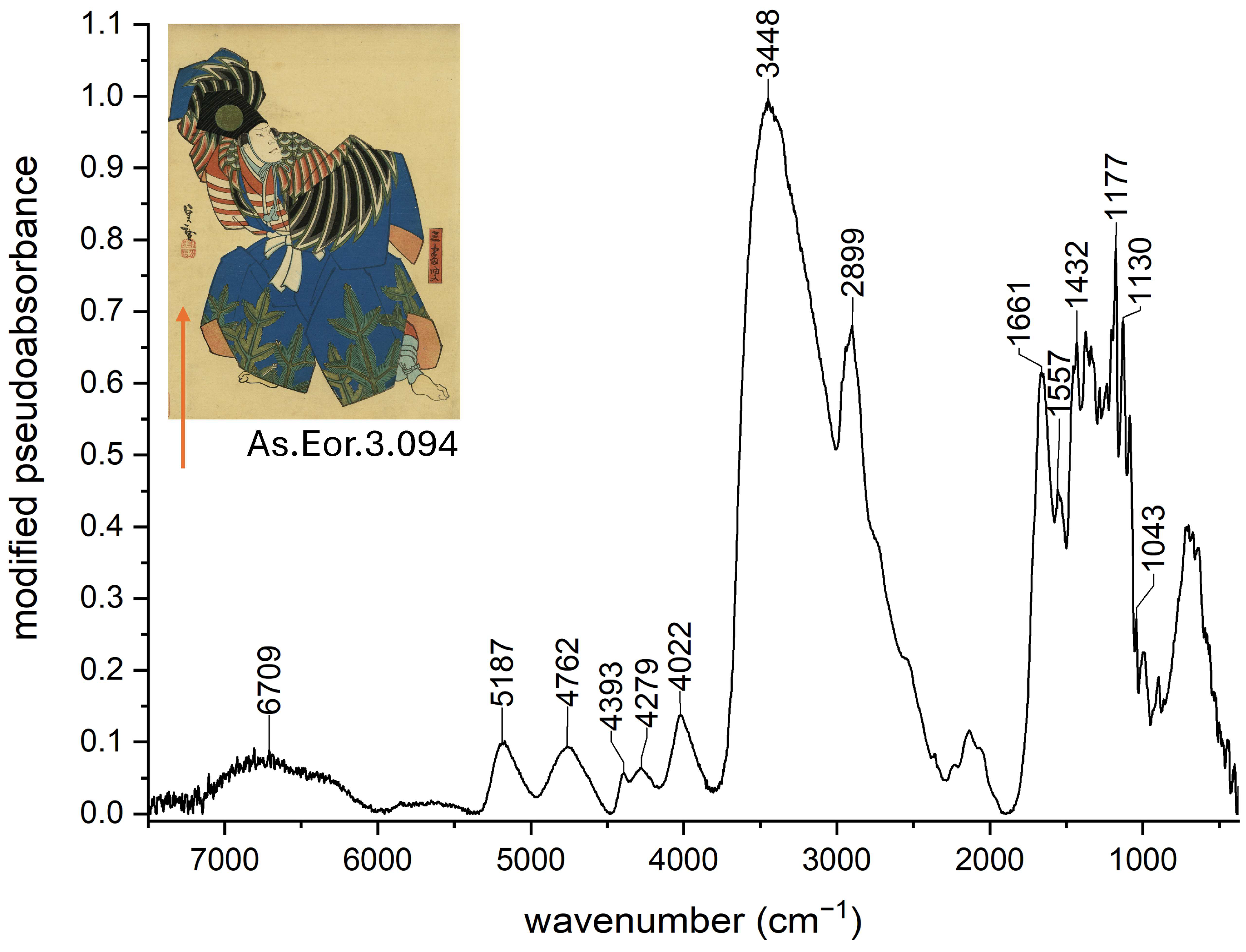
To differentiate these background signals from those of pigments or binding media, non-colored areas of each print were analyzed as a baseline reference (Figure 2). These spectra were compared with the literature and with those previously obtained from similar paper substrates in earlier studies by the authors [29,39,40,48,49,50]. Signals attributed to the paper support were systematically excluded during pigment analysis.
The support’s spectrum predominantly exhibits bands characteristic of cellulose [(C6H10O5)n], the primary constituent of paper, whose structure has been discussed elsewhere [51]. The signals at 1177, 1130, and 1043 cm−1 were due to C-O-C bending absorption. The combination of CH bending and stretching at 4279 cm−1 and of CH and C-C stretching at 4022 cm−1, the combination of OH stretching and C-O bending at 4762 and 4393 cm−1 and the O(2)H…O(6) intramolecular band at 3448 cm−1 were also due to the paper. The band at 6709 cm−1 was due to OH stretching and the one at 5187 cm−1 to the combination of OH bending and stretching. As for the remaining bands, the signals at 1661 cm−1 (Amide I C = O stretching) and 1557 cm−1 (Amide II mode) suggested the occurrence of a proteinaceous compound, as confirmed by (CH) bending at 1432 cm−1 and the CH stretching mode at 2899 cm−1. These protein-related signals were consistently detected across all analyzed areas, including the colored regions, often with high intensity. Their presence supports the use of animal glue [–NH–CHR–CO–]n as a possible sizing or reinforcing treatment applied to the paper. Because the paper and proteinaceous components generated dominant spectral features, the identification of pigments was based on the detection of additional, diagnostic signals specific to functional groups associated with individual colorants.
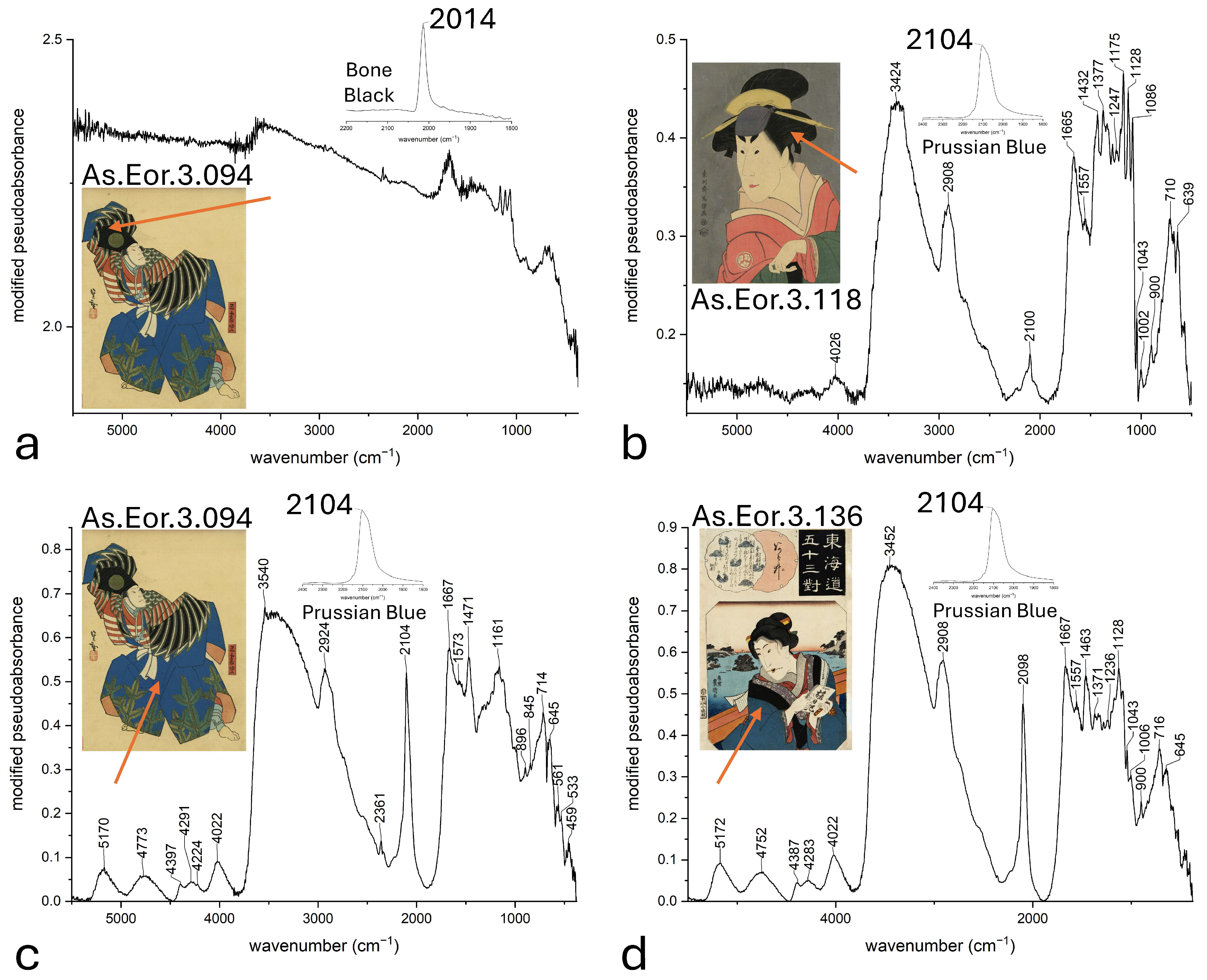
Infrared spectra collected from black areas of the prints showed primarily the characteristic spectral pattern of the treated paper substrate (Figure 3a). No bands corresponding to typical black pigments of animal origin, such as Western bone black or ivory black-composed of amorphous carbon (C) mixed with calcium phosphate compounds such as Ca3(PO4)2 or hydroxyapatite (Ca5(PO4)3(OH))-, were detected. This absence suggests that the black pigments used were most likely mineral- or plant-based, consistent with the traditional Japanese sumi (lamp black, consisting primarily of amorphous carbon C). Unfortunately, sumi pigments lack distinctive features within the spectral range accessible by external reflection FTIR, making their direct identification challenging. An exception was observed in a black area of print As.Eor.3.118, where a band near 2100 cm−1 (triple bond CN stretching) was detected (Figure 3b). This feature is indicative of the presence of bero-ai (Prussian blue, KFe[Fe(CN)6]), suggesting that this pigment was possibly mixed with the black to produce a nuanced tone with greater depth.
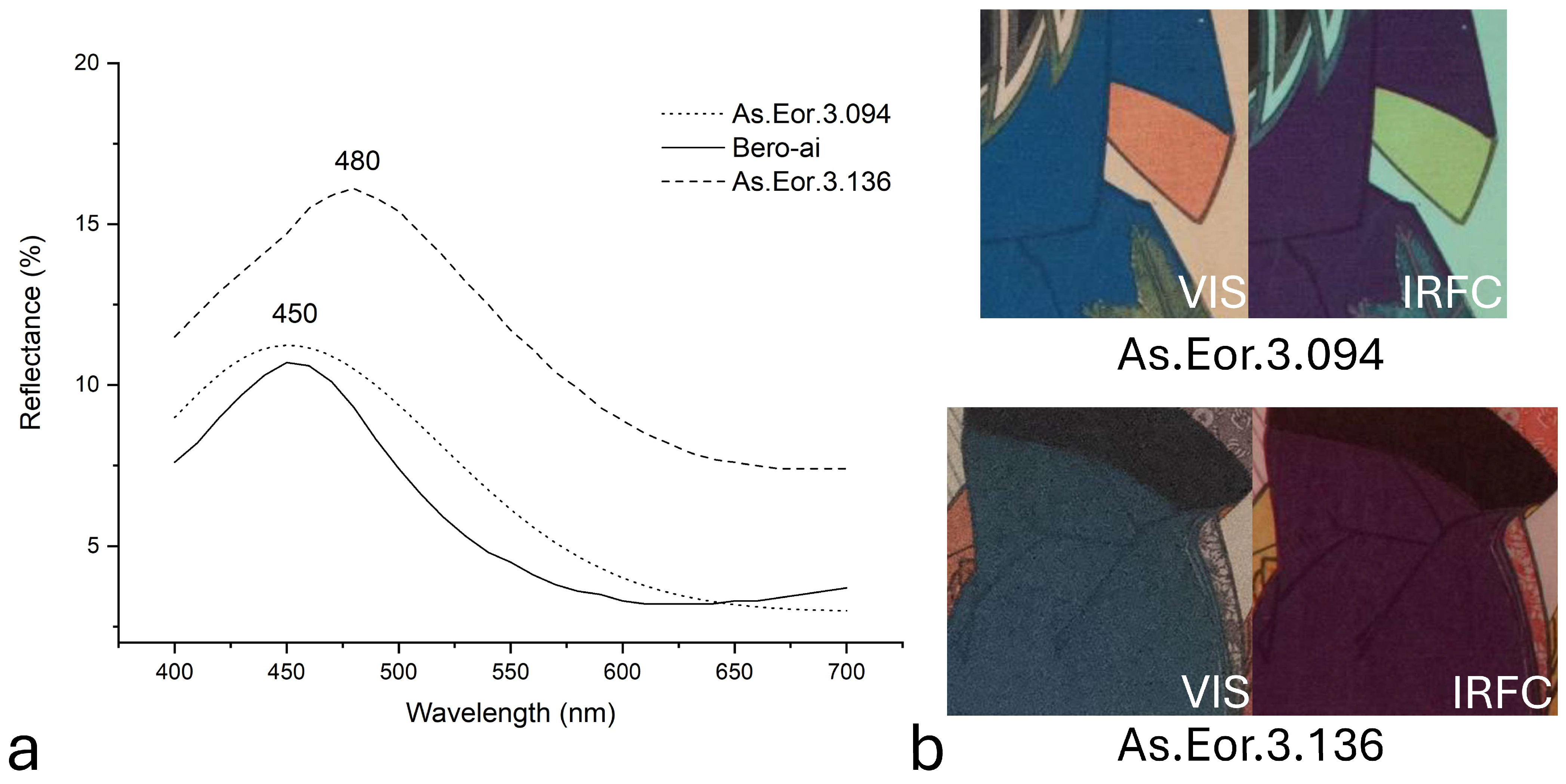
Infrared spectra collected from the blue areas consistently exhibited again a strong, sharp band near 2090 cm−1. This feature allowed an unambiguous identification of bero-ai (Prussian blue) (Figure 3c,d) [39]. The presence of bero-ai was further supported by visible reflectance spectroscopy results on the same areas. Figure 4a presents a comparison between reflectance spectra recorded from the blue regions of prints As.Eor.3.094 and As.Eor.3.136 and that of a bero-ai reference pigment. A clear overlap of the characteristic broad reflectance band centered around 450 nm is evident and consistent with the literature reports [22,44]. Interestingly, the blue area of print As.Eor.3.136 displayed a shifted reflectance maximum near 480 nm. According to Biron et al. [44], the reflectance band of bero-ai shifts towards longer wavelengths (up to ~475 nm) as pigment concentration decreases within the paint layer. This shift correlates with the increased reflectance observed in the spectrum of As.Eor.3.136, suggesting a lower pigment concentration in this sample. The presence of bero-ai (Prussian blue) was further confirmed by infrared false color (IRFC) imaging (Figure 4b). The blue areas in the prints displayed a characteristic violet hue in the IRFC images, consistent with the known signature of Prussian blue [36,39,44].
While the blue birds adorning the kimono of the figure in print As.Eor.3.105 were identified as bero-ai (Prussian blue) based on FTIR analysis, the brown areas likely consist of pigment mixtures, as suggested by reflectance spectroscopy (Figure 5a). The absence of characteristic Prussian blue spectral features in the FTIR spectra of the brown regions excludes its presence. The reflectance spectrum of the brown areas, showing absorbance bands near 590 and 640 nm and a reflectance maximum around 620 nm, suggests the possible use of aigami (dayflower, containing commelinin, a metal–flavonoid complex). However, the overall spectral profile is also consistent with a mixture of ai (indigo, C16H10N2O2) combined with an unidentified red pigment (Figure 5a). Similar interpretations have been reported by Mounier et al. and Shimoyama et al. [23,33]. Infrared false color (IRFC) imaging supports these conclusions: the blue birds appear violet, characteristic of Prussian blue, while the brown areas show a bright red hue, comparable to the reference spectra of aigami (dayflower) and indigo.
Infrared spectra from the green areas of prints As.Eor.3.098, As.Eor.3.094, and As.Eor.3.118 showed a characteristic band near 2093 cm−1, confirming the presence of bero-ai (Prussian blue) (Figure 6a). Reflectance spectra recorded on the same regions exhibited maxima between 520 and 540 nm. This shift toward longer wavelengths is consistent with the known spectral behavior of bero-ai mixed with yellow pigments, as reported in the literature [22,31,33,38]. Following Vermeulen et al. and Mounier et al. [16,33], the presence of an inflection point between 450 and 490 nm in green mixtures is indicative of a blue pigment combined with a yellow colorant or pigment. While the yellow component in As.Eor.3.098 remains uncertain, the green areas in As.Eor.3.094 and As.Eor.3.118 seem to match with a reference mixture of kio (orpiment, As2S3) and bero-ai (Prussian blue) (Figure 5b), suggesting the use of orpiment. This interpretation is supported by the maximum at approximately 530 nm, along with inflection points at 480 and 580 nm, which align closely with those observed in the reference mixture. Minor deviations between the sample and reference spectra can be attributed to experimental limitations, including dye fading and paper yellowing. These findings are consistent with those reported in other studies [16,33]. It should be noted that in our reconstructions of green mixtures—obtained from the most commonly used at the time starting materials—variations in yellow dye nature significantly affect spectral features, including the positions of maxima, minima, and inflection points. The only mixture that matches the experimental data is the proposed combination of Prussian blue and orpiment. In contrast, diluting the mixture tends to result in only minor spectral variations. This observation supports the hypothesis that, within the Japanese artistic tradition, hues were likely fine-tuned by modifying pigment ratios and dilution levels. We followed a similar approach in our experimental reconstructions, aiming to reproduce a green hue closely resembling that observed in the print under investigation.
Regarding purple areas, the reflectance spectrum of As.Eor.3.098 displayed a broad minimum between 530 and 600 nm, with a pronounced minimum near 570 nm, characteristic of dyes such as crystal violet (C25H30ClN3) or methyl violet (C24H28ClN3) [13,22] (Figure 5c). These pigments have been documented in Japan since the late 19th century (1883 and 1866, respectively) [27]. The corresponding infrared reflection spectrum exhibited a band at 2096 cm−1, confirming the presence of bero-ai (Prussian blue) (Figure 6b). This indicates that Prussian blue was mixed with a red or purple pigment to achieve the desired hue, as similarly reported in the literature [27]. The use of methyl violet is consistent with the print’s dating (1882), although alternative mixtures of Prussian blue with red dyes producing comparable reflectance spectra cannot be excluded.
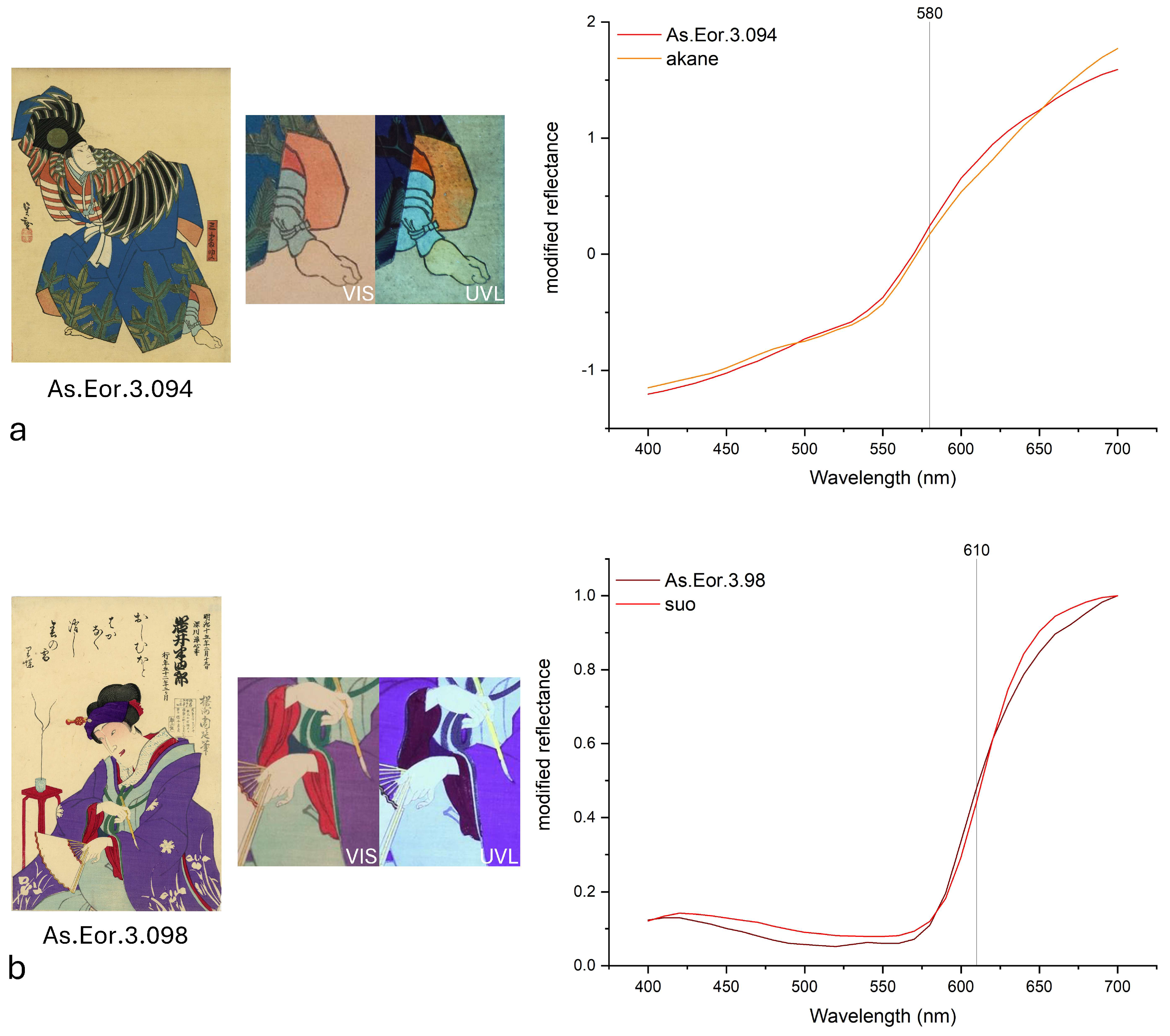
Reflectance spectra of red areas in prints As.Eor.3.094 and As.Eor.3.051 exhibited an inflection point around 580 nm, consistent with the presence of akane (madder, primary coloring compound alizarin C14H8O4) (Figure 7a). This attribution is further supported by the characteristic orange fluorescence response under UV light, in agreement with Tamburini’s observations [52]. Conversely, the red area in print As.Eor.3.098 showed an absorption pattern with an inflection point near 610 nm, indicative of suo (sappanwood, actual colorant brazilein C16H12O5) (Figure 7b). Additional evidence for this assignment comes from the black appearance of the same area under UV illumination, as reported in the literature [52].
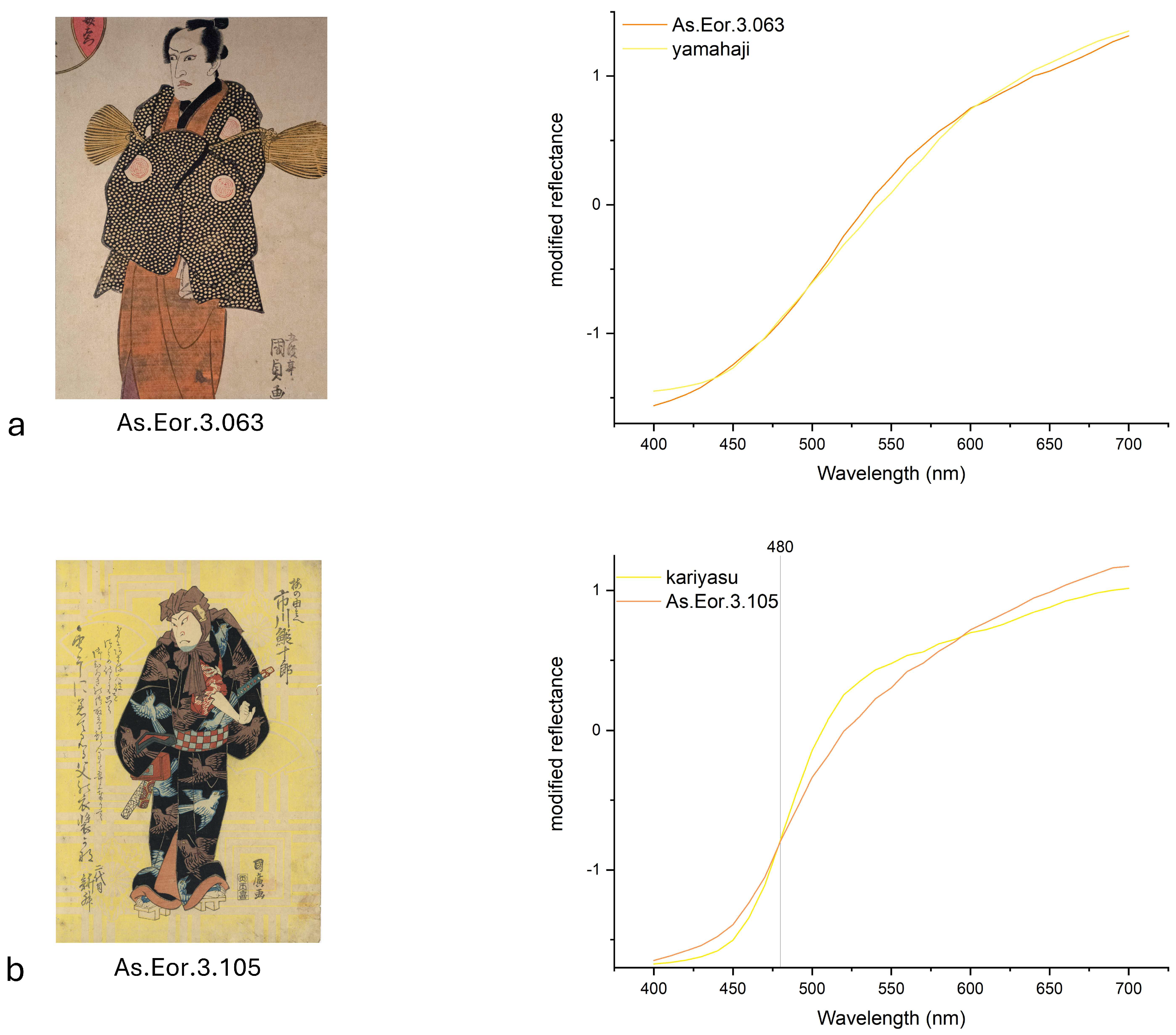
Interpreting reflectance spectra of yellow colorants is challenging due to the lack of distinct spectral features. Nevertheless, tentative identifications can be proposed based on spectral trends. The reflectance spectra of yellow areas in prints As.Eor.3.118, As.Eor.3.169, and As.Eor.3.063 suggest the presence of yamahaji (Japanese sumac, composed by complex polyphenols, e.g., gallotannic acid C76H52O46) (Figure 8a). In contrast, the yellow background of print As.Eor.3.105 is likely attributable to kariyasu (Eulalia, made of flavonoids, primarily luteolin C15H10O6), as indicated by an inflection point near 480 nm (Figure 8b). This interpretation is supported by comparisons of UV-induced fluorescence responses with those of reference pigments.
The grey background of print As.Eor.3.118 exhibited a prominent band at 3642 cm−1 and derivative features at 1097, 1046, 685, and 552 cm−1 (Figure 9).
The observed spectral pattern is characteristic of muscovite [KAl2(AlSi3O10)(OH)2], the main mineral component of kira (mica), traditionally used in the kirazuri decorative technique to create a shimmering effect on the print surface [53].
The sharp band at 3642 cm−1 corresponds to the typical OH stretching vibration. After KK transformation, the derivative-like signals yielded bands roughly at 1097 and 1046 cm−1, corresponding to the characteristic Si–O stretching modes typically observed in conventional IR spectra [53,54,55]. There are SiOAl vibrations at around 765, 685, and 552 (derivative, KK-processed as above) cm−1 [53,54]. Additional SiOAl vibrations appear around 765, 704, and 628 cm−1 (derivative, KK-processed as above), and confirmatory bending modes ascribed to OH and Si–O are present at 932 and 492 cm−1 (derivative, KK-processed as above), respectively. The strong spectral features of mica tend to dominate and often obscure signals from the paper substrate and binder.
The analyzed yakusha-e print is a later original reprint made from Sharaku’s matrices, depicting the half-length bust of kabuki actor Osagawa Tsuneyo II (1753–1808) in an onnagata (female) role performed in 1794. Sharaku produced 28 okubi-e (“large-head images”) featuring mica backgrounds in ōban format, all associated with plays staged in 1794. The presence of a mica shimmering background indicates that this print was a luxury edition, similar to Sharaku’s original works. Kabuki actors were immensely popular in Japan, and deluxe editions of yakusha-e prints were purchased by affluent collectors. Reprinting popular prints was a common practice among publishers to maximize profits. Although Sharaku’s sole publisher, Tsutaya Jūzaburō, was highly influential and promoted luxurious editions, Sharaku’s distinctive style was not widely appreciated during his brief career, which lasted less than a year between 1794 and 1795 [3]. Nonetheless, Sharaku’s works gained posthumous admiration, particularly in the West, where they were reissued in new deluxe editions, often featuring kirazuri.
The identification of mica in this study is particularly significant. Despite its frequent mention in literature on Japanese art techniques [7,43], this represents the first analytical confirmation of the kirazuri technique on ukiyo-e prints. Kira (mica) was traditionally mixed with rice starch and either combined with the main colorants or applied to the background to produce a semi-silvery, translucent, lustrous, and iridescent effect. Mica, a group of sheet silicate (phyllosilicate) minerals, is primarily represented by muscovite [KAl2(AlSi3O10)(OH)2] in painting applications [56,57]. Its light-reflective properties produce a glimmering effect that varies with viewing angle and illumination, enhancing the soft, opaque tones of the figures against the sparkling background.
The use of mica as a pigment to achieve glittering, silver-like effects is also well documented in Western painting treatises [58,59]. Analytical techniques such as SEM-EDX and Raman spectroscopy have identified mica as bright specks in iron gall inks on 17th-century paper documents [56], as well as in 17th-century Persian wall paintings [57]. Other reports note its hypothesized presence in Persian plasters and manuscripts, 13th-century Chinese wall paintings, and 15th-century Indian miniatures [57]. Modern mica-based powders are commonly employed in conservation and restoration for retouching damaged metallic surfaces or gildings [60].
To the best of our knowledge, this study presents the first documented evidence of the kirazuri technique’s use on an ukiyo-e print.

3.2. Investigation of Visible Fluorescence Induced by UV Radiation on Prints

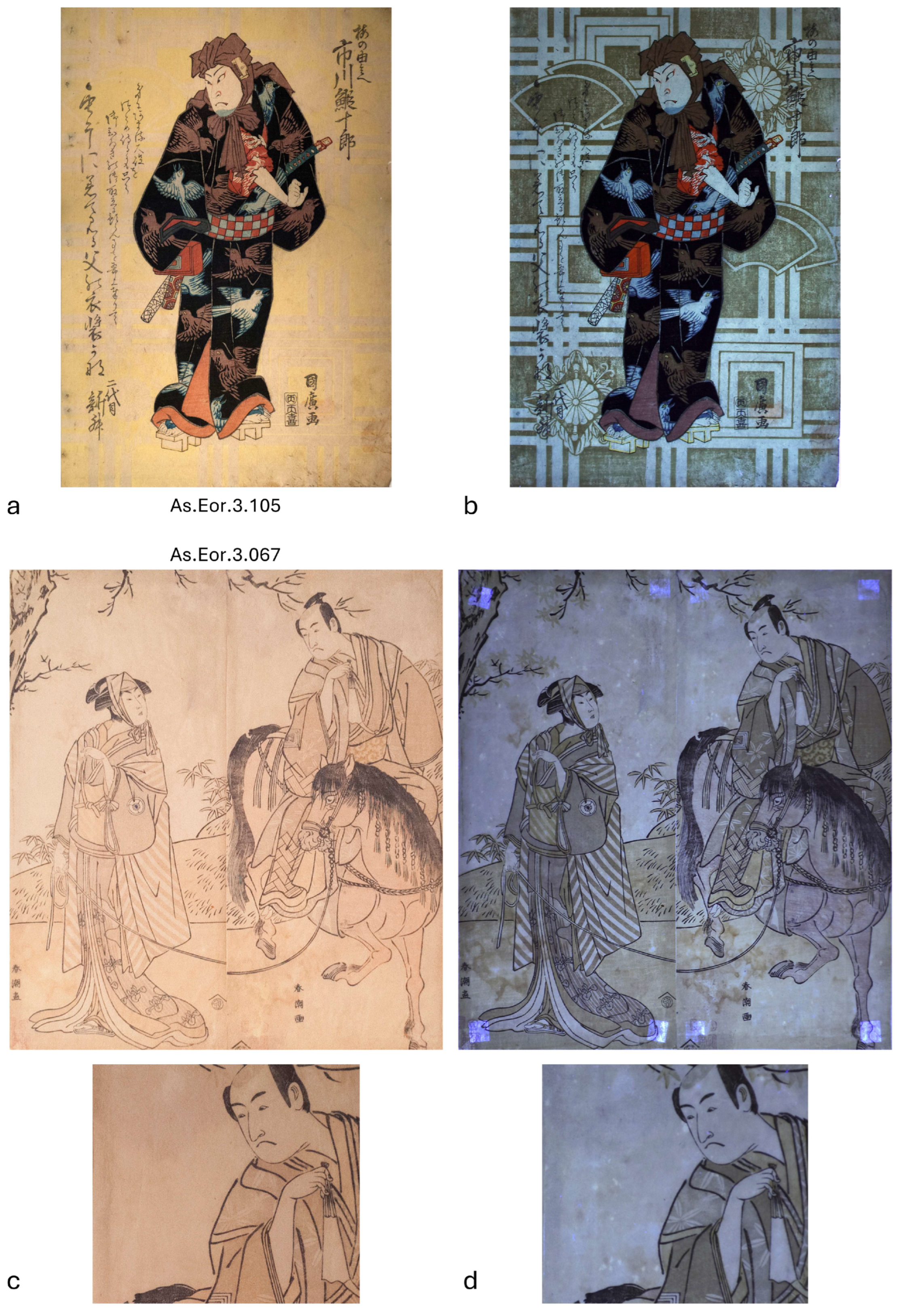
The yellow geometric pattern in the background of print As.Eor.3.105 is barely perceptible under visible light (Figure 10a). However, ultraviolet-induced visible fluorescence imaging reveals the richness and intricate detail of these decorative motifs (Figure 10b). While such yellow geometric patterns are not uniquely characteristic of a specific artist or publisher, they were occasionally employed in yakusha-e prints of Osaka actors, the most renowned kabuki performers in Japan.
The UV fluorescence analysis of the oldest print studied, As.Eor.3.067, proved particularly informative regarding its conservation status. The print exhibits significant fading under visible light (Figure 10c), and attempts to identify pigments using IR spectroscopy and reflectance were unsuccessful. However, UV fluorescence imaging restored visibility to the details of clothing and background vegetation (Figure 10d). Notably, water stains are evident, likely caused by inadequate conservation or display conditions. These stains manifest as lighter spots where pigment degradation has exposed the paper substrate.
The deterioration primarily affects the sky area, indicating the original use of a blue pigment. Among the blue colorants traditionally employed in ukiyo-e, aigami (dayflower blue, also known as tsuyukusa) is recognized as particularly sensitive to moisture [61]. Prolonged humidity exposure results in pale brown discoloration in areas originally painted with this pigment. Sasaki et al. demonstrated that aigami’s distinctive fading pattern can be visually identified post-water damage [62]. This observation aligns with the current case, suggesting the use of aigami (dayflower).
Further support arises from the slight grey fluorescence of the sky under UV light, consistent with previous studies by Edwards et al. and Fiske et al., who associated this feature with aigami (dayflower) [10,35]. Historically, aigami (dayflower) was favored in the 18th century for printing blue shades and, when mixed with yellow or red, for green and purple hues, respectively. The colorant was absorbed on paper and used after deliberate dilution, giving a wide range of colors, gradations and blurred effects from brilliant blue, dark and mild, to semitransparent and greyish blue.
However, according to both historical treatises and recent studies, dayflower blue is highly sensitive to humidity, gradually fading to a pale beige or brown hue when exposed to moisture [20]. Historically, aigami (dayflower) was widely used by dyers for the precise “underdrawing” of textile designs because it could easily be removed by spraying with water [63]. Its fugitive nature was even referenced in the famous 8th-century Japanese poetry anthology Man’yōshū as a metaphor for ephemeral love [63]. This inherent instability likely contributed to its reduced use in later woodblock printing.
The decay of dayflower blue has sometimes been mistaken for fading due to light exposure; however, aging experiments do not support this [62]. The coloring principle of aigami (dayflower) is commelinin, a metalloanthocyanin composed of six molecules of anthocyanin (malonylawobanin), six molecules of flavocommelin, and two magnesium ions. When in contact with moisture, the glucose components of malonylawobanin and flavocommelin undergo changes, turning grey [63]. The pigment tends to migrate rather than smear in the direction of moisture, leaving the paper almost unpainted. When mixed with beni (safflower, main coloring compound carthamin C43H42O22) to obtain purple hues, the yellowing of aigami (dayflower) produces a brownish-purple tone or leaves only the red hue of beni (safflower).
UV analysis of the same print also revealed eight highly fluorescent areas along the sheet edges (Figure 10d), corresponding to organic residues likely left by adhesive tape from previous framing.

4. Conclusions

The non-invasive techniques discussed in this paper proved to be promising for investigating the painting materials used in fragile Japanese ukiyo-e prints. Most of the pigments were identified based on reflectance spectroscopy results, while reflection FTIR confirmed the identification and, in some cases, provided insights into the composition of the painting materials, the paper support, and the proteinaceous sizing treatment. The results confirmed the use of traditional Japanese pigments, such as akane (madder), suo (sappanwood), yamahaji (Japanese sumac), kariyasu (Eulalia), and kio (orpiment).
A large quantity of bero-ai (Prussian blue) was found, used alone or mixed to obtain violets and greens. Prussian blue arrived in Japan at the end of the 18th century, probably imported from the Netherlands through the Nagasaki harbor [27,33,37,40,64,65]. However, the results presented here show that several artists used Prussian blue extensively even before the official opening of Japanese harbors in 1853–1854, as also reported by other authors [27,33]. This may indicate that Prussian blue was relatively cheap and/or easier to obtain than previously believed. Further research into Japan’s trade dynamics during its period of seclusion would help clarify how this pigment circulated so widely.
A key result of this study is the analytical identification of the kirazuri technique, based on the detection of kira (mica). While the use of mica to create iridescent effects has been visually recognized in prints and described in historical sources, this is the first time its presence has been analytically demonstrated through non-invasive techniques. The muscovite minerals identified by reflection FTIR confirm the use of mica to create pearlescent backgrounds, contributing to the understanding of luxury editions of ukiyo-e prints.
The combined use of reflectance spectroscopy, infrared imaging, and reflection FTIR proved effective for identifying many traditional Japanese pigments and materials but also revealed important limitations. Yellow colorants such as yamahaji (Japanese sumac) and kariyasu (Eulalia) often produce broad, featureless spectra in the visible range, making their identification tentative without supporting data. Pigment mixtures, degradation products, and overlapping layers further complicate spectral interpretation. In reflection FTIR, derivative bands caused by surface reflection can mask weaker signals from the paper substrate or organic colorants.
These challenges highlight the need to complement instrumental analyses with historical and stylistic knowledge of materials and printing techniques. Spectral data nonetheless provide valuable information for conservation: they can reveal hidden features and pigment degradation, such as the fading of dayflower blue (aigami), informing risk assessment and preventive measures (e.g., humidity control and light exposure limits). Ultraviolet-induced fluorescence imaging has also proved particularly useful for visualizing faded motifs, residues from past restoration, and hidden decorative patterns, supporting detailed documentation and treatment planning.
From a methodological perspective, ER-FTIR is well suited to detecting inorganic pigments such as mica and Prussian blue but is strongly influenced by paper and surface roughness. Reflectance spectroscopy, although better at identifying a wider range of pigments, requires careful interpretation and reliable reference spectra. Building and sharing spectral libraries is therefore essential for accurate pigment attribution.

Supplementary Materials

The following supporting information can be downloaded at: https://www.mdpi.com/article/10.3390/heritage8090349/s1.

Author Contributions

Conceptualization, V.B., L.G., M.L., and L.R.; Investigation, V.B., C.C., L.G., and L.R.; Writing—original draft preparation, C.C., L.G., M.L., and L.R.; Writing—review and editing, V.B., F.P.C., and S.R. All authors have read and agreed to the published version of the manuscript.

Funding

This research received no specific grant from any funding agency in the public, commercial, or not-for-profit sectors.

Data Availability Statement

The data presented in this study are available on request from the corresponding author.

Acknowledgments

The authors gratefully acknowledge Museo delle Culture (Lugano, Switzerland), Marco Fagioli, Fondazione culture e musei, Lugano, Fondazione “Ada Ceschin and Rosanna Pilone”, Zurich, Kremer Pigmente (Germany), Tokyo Pigment (Japan), Akiko Izawa, and Giovanna Colombo. Cristina Corti gratefully acknowledges “Università degli Studi dell’Insubria—Dipartimento di Scienze Umane e dell’Innovazione per il Territorio” for funding her research grant “Caratterizzazione chimico-mineralogica di pigmenti e coloranti pittorici tradizionali giapponesi”.

Conflicts of Interest

The authors declare no conflicts of interest.

References

  1. Hillier, J.R. The Art of the Japanese Book; Sotheby’s Publications: London, UK, 1987. [Google Scholar]
  2. Hillier, J.R. Japanese Colour Prints; Phaidon: London, UK, 1991. [Google Scholar]
  3. Lane, R. Images from the Floating World: The Japanese Print; Dorset Press: New York, NY, USA, 1978. [Google Scholar]
  4. Takahashi, S. Traditional Woodblock Prints of Japan; Heibonsha: Tokyo, Japan, 1976; Volume 22. [Google Scholar]
  5. Marks, A. Publishers of Japanese Woodblock Prints: A Compendium; Hotei Publishing: Amsterdam, The Netherlands, 2011. [Google Scholar]
  6. Marks, A. Japanese Woodblock Prints: Artists, Publishers and Masterworks: 1680-1900; Tuttle Publishing: Tokyo, Japan, 2010; ISBN 1462905994. [Google Scholar]
  7. Salter, R. Japanese Woodblock Printing; University of Hawaii Press: Honolulu, Hawaii, 2002; ISBN 0824825535. [Google Scholar]
  8. Benedetti, D.P.; Zhang, J.; Tague, T.J.; Lombardi, J.R.; Leona, M. In Situ Microanalysis of Organic Colorants by Inkjet Colloid Deposition Surface-enhanced Raman Scattering. J. Raman Spectrosc. 2014, 45, 123–127. [Google Scholar] [CrossRef]
  9. Cesaratto, A.; Leona, M.; Pozzi, F. Recent Advances on the Analysis of Polychrome Works of Art: SERS of Synthetic Colorants and Their Mixtures With Natural Dyes. Front. Chem. 2019, 7, 105. [Google Scholar] [CrossRef] [PubMed]
  10. Edwards, G.; Villafana, T. Multi-Analytic Characterization of Colorants in Two Impressions of an Utagawa Toyoharu Perspective Print. J. Cult. Herit. 2020, 45, 48–58. [Google Scholar] [CrossRef]
  11. Huang, J. A Technical Study of A Painting by Suzuki Harunobu at the Museum of Fine Arts, Boston: Artist’s Palette and Techniques. Book. Pap. Group. Annu. 2007, 26, 151–153. [Google Scholar]
  12. Kissell, L.N.; Quady, T.K.; Durastanti, D.; Springer, S.; Kenmotsu, J.; Clare, T.L. A Multi-Analytical Approach to Identify Red Colorants on Woodblock Prints Attributed to Suzuki Harunobu. Herit. Sci. 2022, 10, 94. [Google Scholar] [CrossRef]
  13. Leona, M.; Decuzzi, P.; Kubic, T.A.; Gates, G.; Lombardi, J.R. Nondestructive Identification of Natural and Synthetic Organic Colorants in Works of Art by Surface Enhanced Raman Scattering. Anal. Chem. 2011, 83, 3990–3993. [Google Scholar] [CrossRef]
  14. Sakamoto, A.; Ochiai, S.; Higashiyama, H.; Masutani, K.; Kimura, J.; Koseto-Horyu, E.; Tasumi, M. Raman Studies of Japanese Art Objects by a Portable Raman Spectrometer Using Liquid Crystal Tunable Filters. J. Raman Spectrosc. 2012, 43, 787–791. [Google Scholar] [CrossRef]
  15. Vermeulen, M.; Burgio, L.; Vandeperre, N.; Driscoll, E.; Viljoen, M.; Woo, J.; Leona, M. Beyond the Connoisseurship Approach: Creating a Chronology in Hokusai Prints Using Non-Invasive Techniques and Multivariate Data Analysis. Herit. Sci. 2020, 8, 62. [Google Scholar] [CrossRef]
  16. Vermeulen, M.; Leona, M. Evidence of Early Amorphous Arsenic Sulfide Production and Use in Edo Period Japanese Woodblock Prints by Hokusai and Kunisada. Herit. Sci. 2019, 7, 73. [Google Scholar] [CrossRef]
  17. Zaleski, S.; Takahashi, Y.; Leona, M. Natural and Synthetic Arsenic Sulfide Pigments in Japanese Woodblock Prints of the Late Edo Period. Herit. Sci. 2018, 6, 32. [Google Scholar] [CrossRef]
  18. Connors, S.A.; Whitmore, P.M.; Keyes, R.S.; Coombs, E.I. The Identification and Light Sensitivity of Japanese Woodblock Print Colorants: The Impact on Art History and Preservation. In Scientific Research on the Pictorial Art of Asia. Second Fobers Symposium at the Freer Gallery of Art London; Archetype Publications Ltd.: London, UK, 2005; pp. 35–47. [Google Scholar]
  19. Derrick, M.; Newman, R.; Wright, J. Characterization of Yellow and Red Natural Organic Colorants on Japanese Woodblock Prints by EEM Fluorescence Spectroscopy. J. Am. Inst. Conserv. 2017, 56, 171–193. [Google Scholar] [CrossRef]
  20. Feller, R.L.; Curran, M.; Bailie, C. Identification of Traditional Organic Colorants Employed in Japanese Prints and Determination of Their Rates of Fading. In Japanese Woodblock Prints: A Catalogue of the Mary A. Ainsworth Collection; Keyes, R.S., Allen Memorial Art Museum, Eds.; Allen Memorial Art Museum, Oberlin College; Indiana University Press: Oberlin, OH, USA; Bloomington, IN, USA, 1984; ISBN 978-0942946017. [Google Scholar]
  21. Nakamura, R.; Tanaka, Y.; Ogata, A.; Naruse, M. Dye Analysis of Shosoin Textiles Using Excitation−Emission Matrix Fluorescence and Ultraviolet−Visible Reflectance Spectroscopic Techniques. Anal. Chem. 2009, 81, 5691–5698. [Google Scholar] [CrossRef]
  22. Pérez-Arantegui, J.; Rupérez, D.; Almazán, D.; Díez-de-Pinos, N. Colours and Pigments in Late Ukiyo-e Art Works: A Preliminary Non-Invasive Study of Japanese Woodblock Prints to Interpret Hyperspectral Images Using in-Situ Point-by-Point Diffuse Reflectance Spectroscopy. Microchem. J. 2018, 139, 94–109. [Google Scholar] [CrossRef]
  23. Shimoyama, S.; Matsui, H.; Shimoyama, Y. Non-Destructive Identification of Blue Colorants in Ukiyo-e Prints by Visible-Near Infrared Reflection Spectrum Obtained with a Portable Spectrophotometer Using Fiber Optics. Bunseki Kagaku 2006, 55, 121–126. [Google Scholar] [CrossRef]
  24. Whitmore, P.M.; Cass, G.R. The Ozone Fading of Traditional Japanese Colorants. Stud. Conserv. 1988, 33, 29. [Google Scholar] [CrossRef]
  25. Leona, M.; Winter, J. Fiber Optics Reflectance Spectroscopy: A Unique Tool for the Investigation of Japanese Paintings. Stud. Conserv. 2001, 46, 153. [Google Scholar] [CrossRef]
  26. Cañamares, M.V.; Mieites-Alonso, M.G.; Leona, M. Fourier Transform-Raman and Surface-enhanced Raman Spectroscopy Analysis of Safflower Red-dyed Washi Paper: PH Study and Bands Assignment. J. Raman Spectrosc. 2020, 51, 903–909. [Google Scholar] [CrossRef]
  27. Cesaratto, A.; Luo, Y.-B.; Smith, H.D.; Leona, M. A Timeline for the Introduction of Synthetic Dyestuffs in Japan during the Late Edo and Meiji Periods. Herit. Sci. 2018, 6, 22. [Google Scholar] [CrossRef]
  28. Minamikawa, T.; Nagai, D.; Kaneko, T.; Taniguchi, I.; Ando, M.; Akama, R.; Takenaka, K. Analytical Imaging of Colour Pigments Used in Japanese Woodblock Prints Using Raman Microspectroscopy. J. Raman Spectrosc. 2017, 48, 1887–1895. [Google Scholar] [CrossRef]
  29. Biron, C.; Le Bourdon, G.; Pérez-Arantegui, J.; Servant, L.; Chapoulie, R.; Daniel, F. Probing Some Organic Ukiyo-e Japanese Pigments and Mixtures Using Non-Invasive and Mobile Infrared Spectroscopies. Anal. Bioanal. Chem. 2018, 410, 7043–7054. [Google Scholar] [CrossRef] [PubMed]
  30. Gargano, M.; Longoni, M.; Pesce, V.; Palandri, M.C.; Canepari, A.; Ludwig, N.; Bruni, S. From Materials to Technique: A Complete Non-Invasive Investigation of a Group of Six Ukiyo-E Japanese Woodblock Prints of the Oriental Art Museum E. Chiossone (Genoa, Italy). Sensors 2022, 22, 8772. [Google Scholar] [CrossRef]
  31. Korenberg, C.F.; Pereira-Pardo, L.; McElhinney, P.J.; Dyer, J. Developing a Systematic Approach to Determine the Sequence of Impressions of Japanese Woodblock Prints: The Case of Hokusai’s ‘Red Fuji’. Herit. Sci. 2019, 7, 9. [Google Scholar] [CrossRef]
  32. Luo, Y.; Basso, E.; Smith, H.D.; Leona, M. Synthetic Arsenic Sulfides in Japanese Prints of the Meiji Period. Herit. Sci. 2016, 4, 17. [Google Scholar] [CrossRef]
  33. Mounier, A.; Le Bourdon, G.; Aupetit, C.; Lazare, S.; Biron, C.; Pérez-Arantegui, J.; Almazán, D.; Aramendia, J.; Prieto-Taboada, N.; Fdez-Ortiz de Vallejuelo, S.; et al. Red and Blue Colours on 18th–19th Century Japanese Woodblock Prints: In Situ Analyses by Spectrofluorimetry and Complementary Non-Invasive Spectroscopic Methods. Microchem. J. 2018, 140, 129–141. [Google Scholar] [CrossRef]
  34. Newman, R.; Derrick, M.; Mysak, E. EEM Fluorescence Spectroscopy of Natural Red and Yellow Organic Colorants in Japanese Woodblock Prints. In Springer Series on Fluorescence; Springer: Cham, Switzerland, 2022. [Google Scholar] [CrossRef]
  35. Fiske, B.; Morenus, L.S. Ultraviolet and Infrared Examination of Japanese Woodblock Prints: Identifying Reds and Blues. Book. Pap. Group. Annu. 2004, 23, 21–32. [Google Scholar]
  36. Villafana, T.; Edwards, G. Creation and Reference Characterization of Edo Period Japanese Woodblock Printing Ink Colorant Samples Using Multimodal Imaging and Reflectance Spectroscopy. Herit. Sci. 2019, 7, 94. [Google Scholar] [CrossRef]
  37. Biron, C.; Mounier, A.; Arantegui, J.P.; Le Bourdon, G.; Servant, L.; Chapoulie, R.; Roldán, C.; Almazán, D.; Díez-de-Pinos, N.; Daniel, F. Colours of the «images of the Floating World». Non-Invasive Analyses of Japanese Ukiyo-e Woodblock Prints (18th and 19th Centuries) and New Contributions to the Insight of Oriental Materials. Microchem. J. 2020, 152, 104374. [Google Scholar] [CrossRef]
  38. Biron, C.; Mounier, A.; Le Bourdon, G.; Servant, L.; Chapoulie, R.; Daniel, F. Revealing the Colours of Ukiyo-e Prints by Short Wave Infrared Range Hyperspectral Imaging (SWIR). Microchem. J. 2020, 155, 104782. [Google Scholar] [CrossRef]
  39. Rampazzi, L.; Brunello, V.; Corti, C.; Lissoni, E. Non-Invasive Techniques for Revealing the Palette of the Romantic Painter Francesco Hayez. Spectrochim. Acta A Mol. Biomol. Spectrosc. 2017, 176, 142–154. [Google Scholar] [CrossRef]
  40. Rampazzi, L.; Brunello, V.; Campione, F.P.; Corti, C.; Geminiani, L.; Recchia, S.; Luraschi, M. Non-Invasive Identification of Pigments in Japanese Coloured Photographs. Microchem. J. 2020, 157, 105017. [Google Scholar] [CrossRef]
  41. Miliani, C.; Rosi, F.; Daveri, A.; Brunetti, B.G. Reflection Infrared Spectroscopy for the Non-Invasive in Situ Study of Artists’ Pigments. Appl. Phys. A 2012, 106, 295–307. [Google Scholar] [CrossRef]
  42. Estupiñán Méndez, D.; Allscher, T. Advantages of External Reflection and Transflection over ATR in the Rapid Material Characterization of Negatives and Films via FTIR Spectroscopy. Polymers 2022, 14, 808. [Google Scholar] [CrossRef]
  43. Newland, A.R. (Ed.) The Hotei Encyclopedia of Japanese Woodblock Prints; Hotei Publishing: Amsterdam, The Netherlands, 2005. [Google Scholar]
  44. Biron, C.; Mounier, A.; Le Bourdon, G.; Servant, L.; Chapoulie, R.; Daniel, F. A Blue Can Conceal Another! Noninvasive Multispectroscopic Analyses of Mixtures of Indigo and Prussian Blue. Color. Res. Appl. 2020, 45, 262–274. [Google Scholar] [CrossRef]
  45. Aceto, M.; Agostino, A.; Fenoglio, G.; Idone, A.; Gulmini, M.; Picollo, M.; Ricciardi, P.; Delaney, J.K. Characterisation of Colourants on Illuminated Manuscripts by Portable Fibre Optic UV-Visible-NIR Reflectance Spectrophotometry. Anal. Methods 2014, 6, 1488. [Google Scholar] [CrossRef]
  46. Geminiani, L.; Campione, F.P.; Corti, C.; Luraschi, M.; Recchia, S.; Rampazzi, L. New Evidence of Traditional Japanese Dyeing Techniques: A Spectroscopic Investigation. Heritage 2024, 7, 3610–3629. [Google Scholar] [CrossRef]
  47. De Ferri, L.; Campanella, B.; Martignon, A.; Vallotto, D.; Tomaini, B.; Legnaioli, S.; Pojana, G. Coupling of Electron and Vibrational Spectroscopy with False Colour Imaging for the Investigation of Natural Dyes in Historical Fabrics (15th–18th Centuries). Eur. Phys. J. Plus 2023, 138, 634. [Google Scholar] [CrossRef]
  48. Meilunas, R.J.; Bentsen, J.G.; Steinberg, A. Analysis of Aged Paint Binders by FTIR Spectroscopy. Stud. Conserv. 1990, 35, 33–51. [Google Scholar] [CrossRef]
  49. Geminiani, L.; Paolo Campione, F.; Corti, C.; Giussani, B.; Gorla, G.; Luraschi, M.; Recchia, S.; Rampazzi, L. Non-Invasive Identification of Historical Textiles and Leather by Means of External Reflection FTIR Spectroscopy. Spectrochim. Acta A Mol. Biomol. Spectrosc. 2025, 326, 125184. [Google Scholar] [CrossRef]
  50. Schwanninger, M.; Rodrigues, J.C.; Fackler, K. A Review of Band Assignments in near Infrared Spectra of Wood and Wood Components. J. Near Infrared Spectrosc. 2011, 19, 287–308. [Google Scholar] [CrossRef]
  51. Geminiani, L.; Campione, F.P.; Corti, C.; Luraschi, M.; Motella, S.; Recchia, S.; Rampazzi, L. Differentiating between Natural and Modified Cellulosic Fibres Using ATR-FTIR Spectroscopy. Heritage 2022, 5, 4114–4139. [Google Scholar] [CrossRef]
  52. Tamburini, D.; Dyer, J. Fibre Optic Reflectance Spectroscopy and Multispectral Imaging for the Non-Invasive Investigation of Asian Colourants in Chinese Textiles from Dunhuang (7th-10th Century AD). Dye. Pigment. 2019, 162, 494–511. [Google Scholar] [CrossRef]
  53. Sayin, M.; Graf von Reichenbach, H. Infrared Spectra of Muscovites as Affected by Chemical Composition, Heating and Particle Size. Clay Min. 1978, 13, 241–254. [Google Scholar] [CrossRef]
  54. Farmer, V.C. The Layer Silicates. In The Infrared Spectra of Minerals; Farmer, V.C., Ed.; The Mineralogical Society of Great Britain: London, UK, 1974; Volume 4, ISBN 0903056054. [Google Scholar]
  55. Šontevska, V.; Jovanovski, G.; Makreski, P.; Raškovska, A.; Šoptrajanova, B. Minerals From Macedonia. XXI. Vibrational Spectroscopy as Identificational Tool for Some Phyllosilicate Minerals. Acta Chim. Slov. 2008, 55, 757–766. [Google Scholar]
  56. Odlyha, M.; Walker, R.M.; Liddell, W.R. A Study of the Effects of Conservation Treatment on the Fanshawe Archive. Stud. Conserv. 1992, 37, 104–111. [Google Scholar] [CrossRef]
  57. Karimy, A.-H.; Holakooei, P. Looking Like Silver: Mica as a Pigment in Mid-Seventeenth Century Persian Wall Decorations. Iran 2021, 59, 109–118. [Google Scholar] [CrossRef]
  58. Gettens, R.J.; Stout, G.L. Painting Materials: A Short Encyclopaedia; Dover Art Instruction Series; Dover Publications: New York, NY, USA, 1966; ISBN 9780486215976. [Google Scholar]
  59. Eastaugh, N. Pigment Compendium: A Dictionary and Optical Microscopy of Historical Pigments; Routledge Conservation and Museology; Routledge: London, UK, 2008; ISBN 9780750689809. [Google Scholar]
  60. Bailão, A.; Šustić, S. Retouching with Mica Pigments. E-Conserv. J. 2013, 1, 45–60. [Google Scholar] [CrossRef]
  61. Smith, H.D. Hokusai and the Blue Revolution in Edo Prints. In Hokusai and His Age: Ukiyo-e Painting, Print-Making, and Book Illustration in Late Edo Japan; Carpenter, J.H., Ed.; Hotei Publishing: Amsterdam, The Netherlands, 2005; pp. 234–269. [Google Scholar]
  62. Sasaki, S.; Coombs, E.I. Dayflower Blue: Its Appearance and Lightfastness in Traditional Japanese Prints. In Scientific Research on the Pictorial Arts of Asia: Proceedings of the Second Forbes Symposium at the Freer Gallery of Art; Archetype: London, UK, 2005; pp. 48–57. [Google Scholar]
  63. Sasaki, S.; Webber, P. A Study of Dayflower Blue Used in Ukiyo-e Prints. Stud. Conserv. 2002, 47, 185–188. [Google Scholar] [CrossRef]
  64. Bickford, L.R. Prussian Blue and the Dating of Hokusai’s Fuji Series. Andon 1987, 7, 20–21. [Google Scholar]
  65. FitzHugh, E.W. A Database of Pigments on Japanese Ukiyo-e Paintings in the Freer Gallery of Art. In Pigments in Later Japanese Paintings, Washington, Freer Gallery of Art; FitzHugh, E.W., Winter, J., Leona, M., Eds.; Smithsonian Institution: Washington, DC, USA, 2003; pp. 1–56. [Google Scholar]
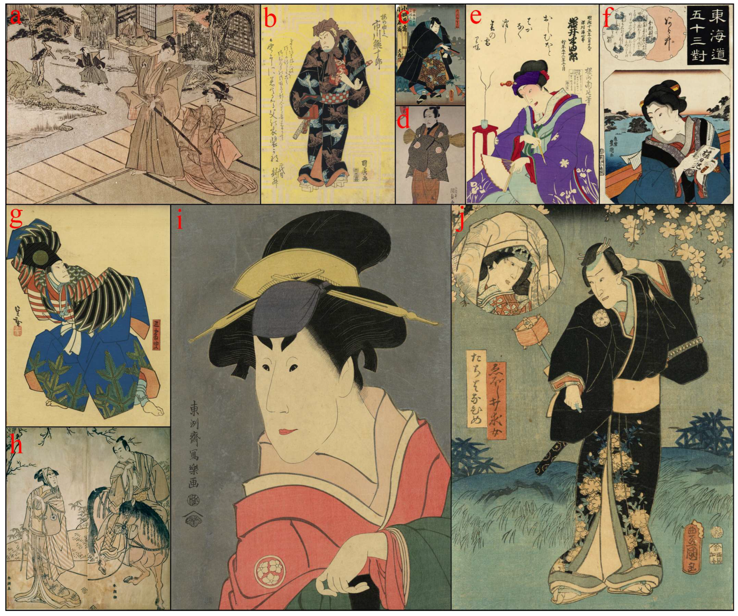
Figure 1. The ukiyo-e woodblock prints analyzed and discussed in the paper.
Heritage 08 00349 g001
Figure 2. FTIR spectra in modified pseudoabsorbance of the support showing the features of paper and glue.
Heritage 08 00349 g002
Figure 3. FTIR spectra in modified pseudoabsorbance of (a) the black area of As.Eor.3.094; (b) the black area in As.Eor.3.118, showing the presence of Prussian blue; (c) the blue area in As.Eor.3.094, showing the presence of Prussian blue; (d) the blue area in As.Eor.3.136, showing the presence of Prussian blue.
Heritage 08 00349 g003
Figure 4. (a) Visible spectra of the blue areas in As.Eor.3.094 and As.Eor.3.136, showing the shift of the maximum reflection. (b) close-up of the blue areas under analysis in As.Eor.3.094 and As.Eor.3.136, comparing the visible image and the infrared false color images.
Heritage 08 00349 g004
Figure 5. (a) Visible spectrum of the brown area in print As.Eor.3.105; (b) visible spectrum of the green areas in As.Eor.3.094 and As.Eor.3.118, showing the presence of kio (orpiment) and bero-ai (Prussian blue); (c) visible spectrum of the purple area in As.Eor.3098.
Heritage 08 00349 g005
Figure 6. FTIR spectra in modified pseudoabsorbance of the print As.Eor.3.098, showing the presence of bero-ai (Prussian blue) in (a) the green area and (b) the purple area.
Heritage 08 00349 g006
Figure 7. Visible spectra of (a) the red areas in print As.Eor.3.94, recognized as akane (madder); (b) the red area in print As.Eor.3.98, recognized as suo (sappanwood).
Heritage 08 00349 g007
Figure 8. Visible spectra of (a) the yellow areas of print As.Eor.3.063, recognized as yamahaji (Japanese sumac); (b) the yellow background of As.Eor.3.105, recognized as kariyasu (Eulalia).
Heritage 08 00349 g008
Figure 9. (a) FTIR spectrum in modified pseudoabsorbance of the grey area in print As.Eor.3.118 showing the features of kira (mica). The derivative bands, identified through Kramers–Kronig transformation (KKT) applied to the original reflectance spectrum, are indicated in grey at 1097, 1046, 685, 552, and 492 cm−1; (b) detail of the area.
Heritage 08 00349 g009
Figure 10. Pictures in visible light and visible ultraviolet fluorescence of print As.Eor.3.105 [(a) and (b), respectively] and of print As.Eor.3.067 [(c) and (d), respectively].
Heritage 08 00349 g010
Table 1. Description of the prints and areas analyzed by reflectance spectroscopy and external reflection FTIR.
Print
Inventory Number
Painter, YearAreas Analyzed
As.Eor.3.067
(Figure 1h)
Yushido Shuncho,1780–1796Green, grey, red
As.Eor.3.051
(Figure 1a)
Utagawa Toyokuni, 1812Green, grey, red, yellow
As.Eor.3.063
(Figure 1d)
Utagawa Kunisada, 1816Orange, red, yellow
As.Eor.3.105
(Figure 1b)
Utagawa Kunihiro, 1828Black, blue, brown, red, yellow
As.Eor.3.094
(Figure 1g)
Konishi (?) Hirosada, 1840Black, blue, green, red, white, yellow
As.Eor.3.136
(Figure 1f)
Utagawa Kunisada, 1845–1846Black, blue, red, white
As.Eor.3.169
(Figure 1c)
Utagawa Kunisada, 1854Black, brown, green, grey, red, yellow
As.Eor.3.083
(Figure 1j)
Utagawa Toyokuni III,1859Black, blue, yellow
As.Eor.3.098
(Figure 1e)
Toyohara Chikanobu, 1882Brown, green, purple, red
As.Eor.3.118
(Figure 1i)
Toshusai Sharaku, after 1906Black, green, grey, purple, red, white, yellow
Disclaimer/Publisher’s Note: The statements, opinions and data contained in all publications are solely those of the individual author(s) and contributor(s) and not of MDPI and/or the editor(s). MDPI and/or the editor(s) disclaim responsibility for any injury to people or property resulting from any ideas, methods, instructions or products referred to in the content.