
Hidden Treasures: Precious Textiles from the St Eustace Head Reliquary


Abstract
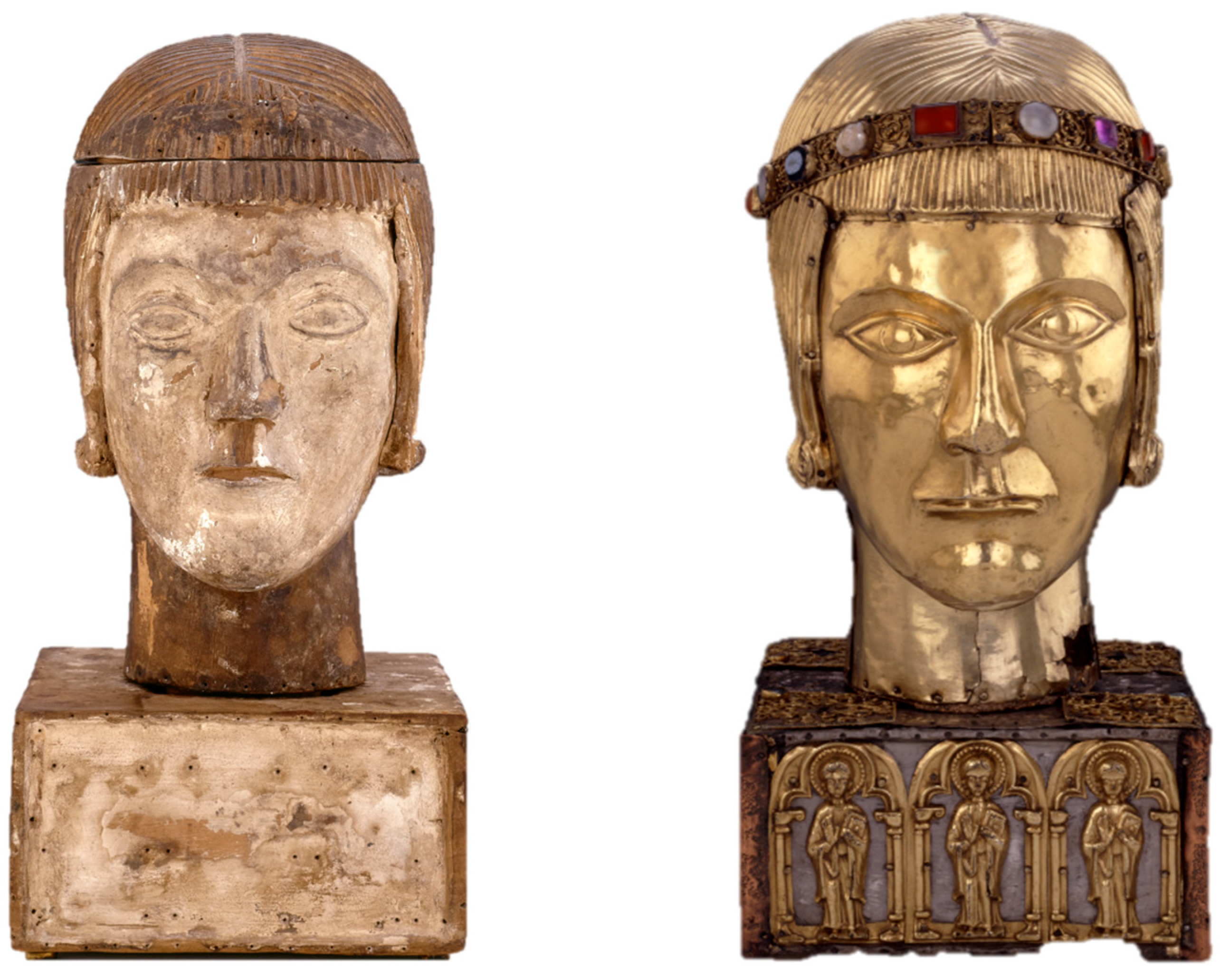
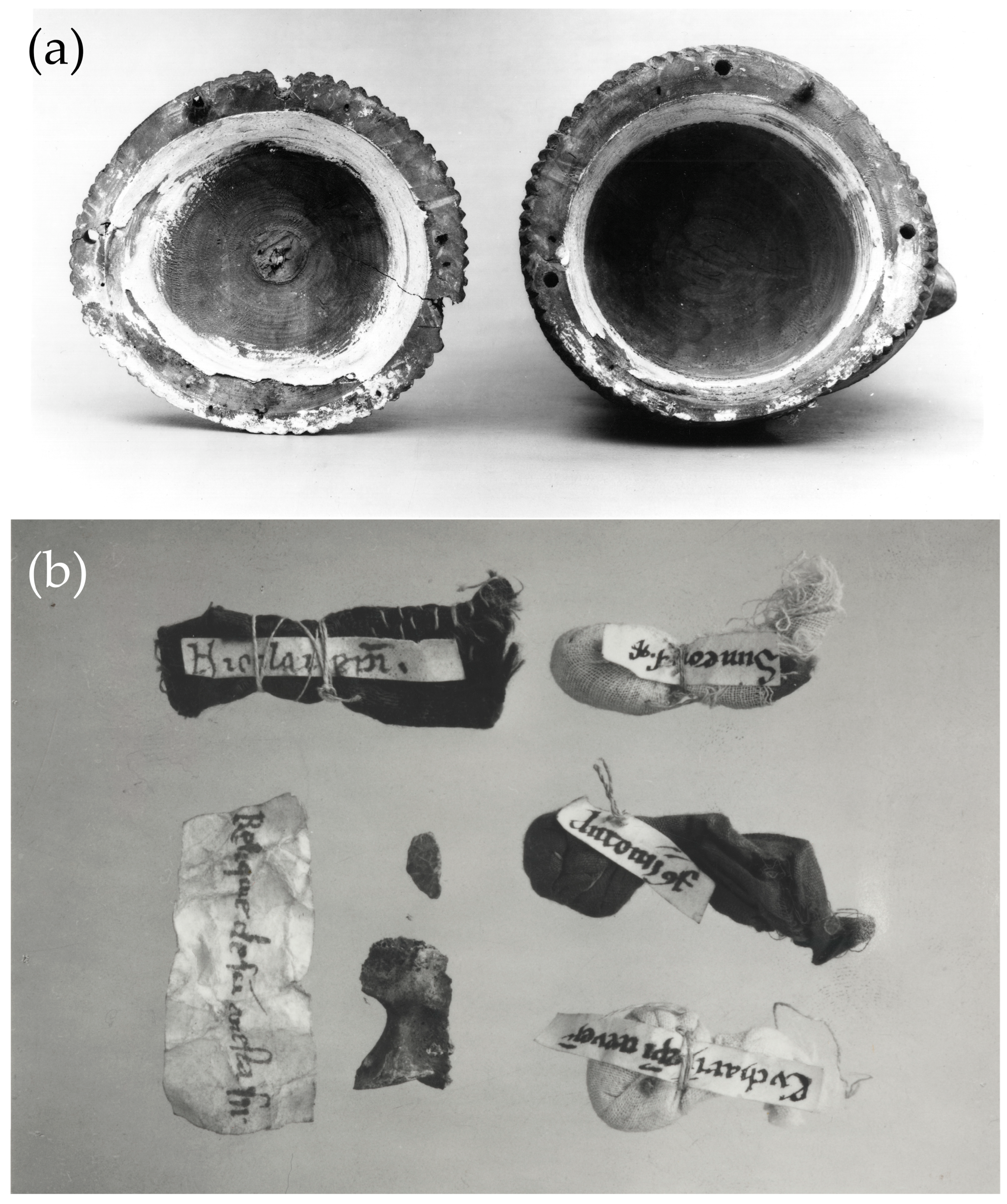
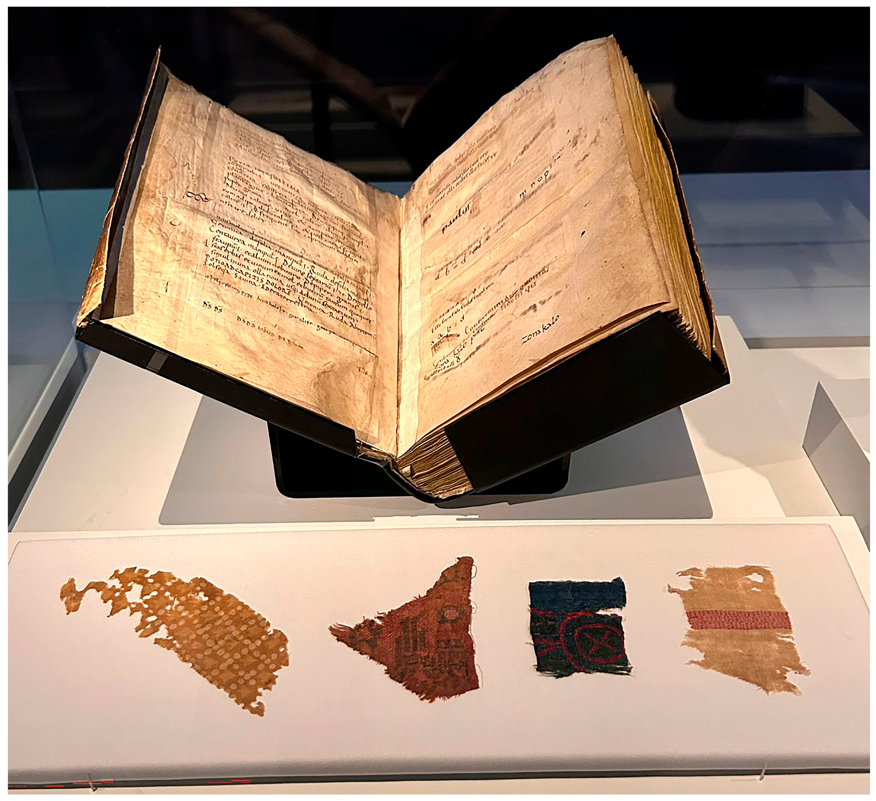
1. Introduction
2. Materials and Methods
2.1. The Textile Fragments
2.1.1. Fragment of Resist-Dyed Silk from the Relic Wrapping of St Anastasius (Accession Number 1850,1127.1.a)
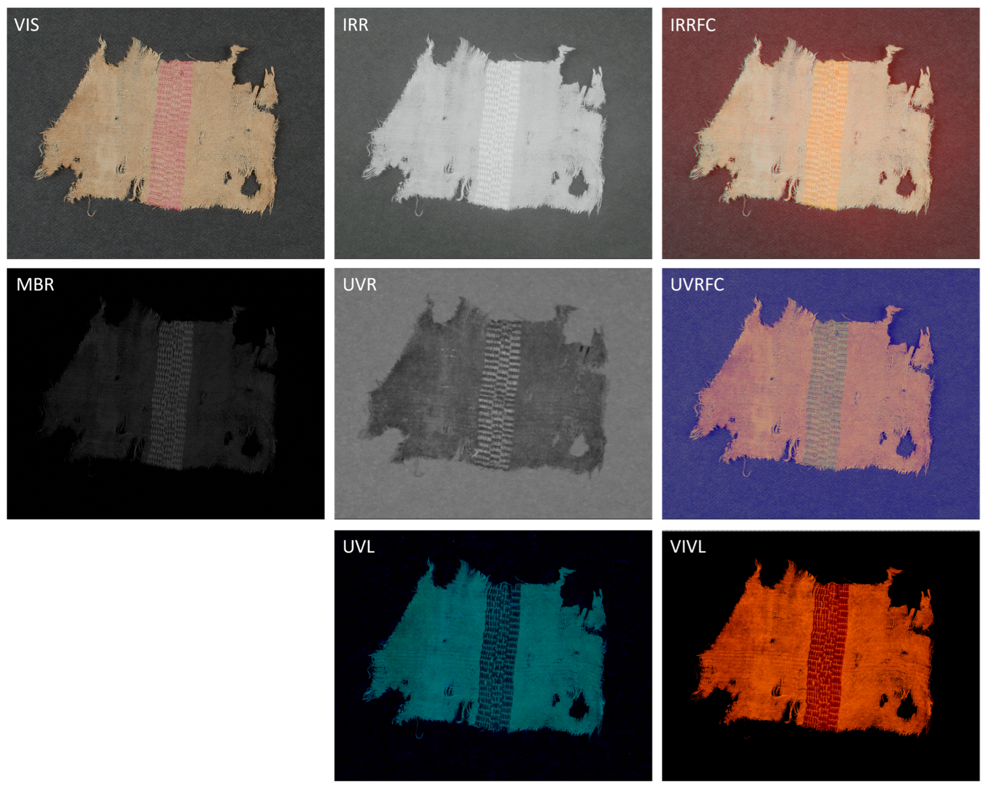
2.1.2. Fragment of White Silk Textile with a Mauve Band from the Relic Wrapping of St Benedict (Accession Number 1850,1127.1.c)
2.1.3. A Fragment of a Twill Silk Textile from the Relic Wrapping of St Nicholas (Accession Number 1850,1127.1.f)
2.1.4. Fragment of Twill Silk Textile from the Relic Wrapping of St Nicholas of Myra (Accession Number 1850,1127.1.g)
2.2. Methodology
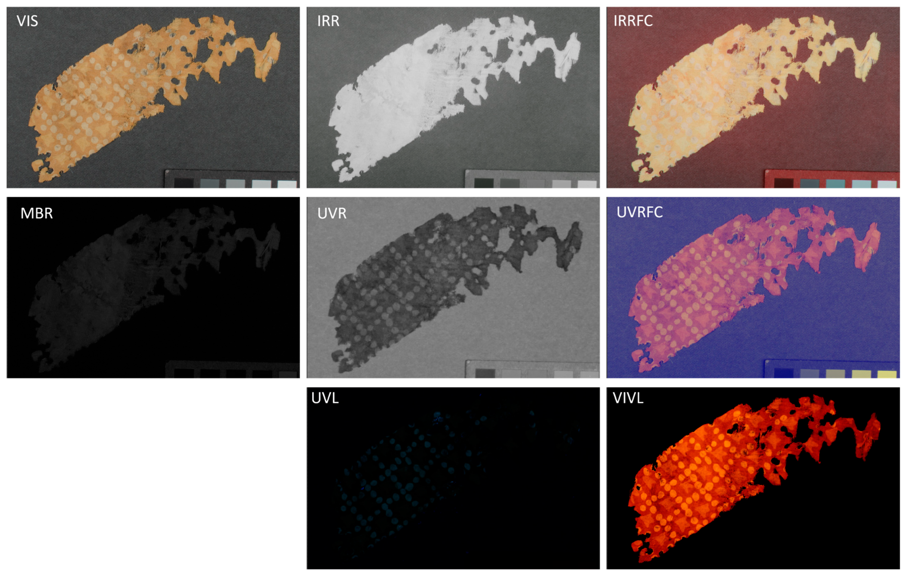
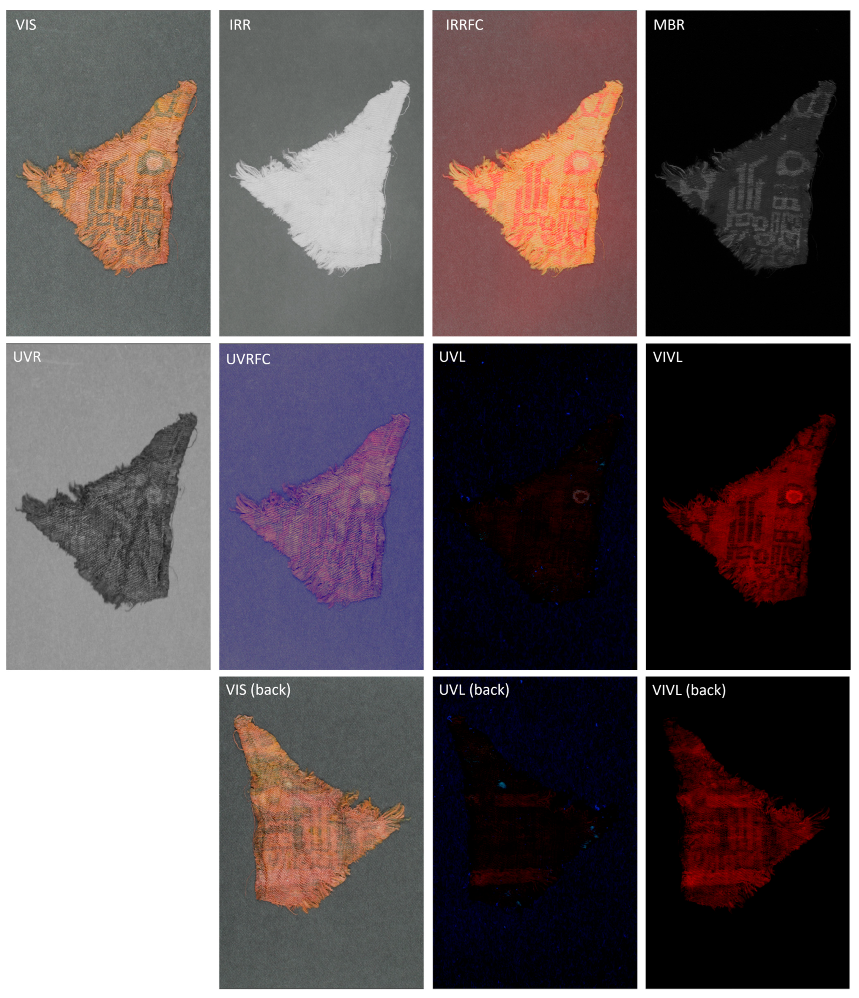
2.2.1. Multiband Imaging (MBI)
2.2.2. Fibre Optic Reflectance Spectroscopy (FORS)
2.2.3. Scanning Electron Microscopy—Energy Dispersive X-Ray Spectroscopy (SEM-EDX)
2.2.4. High Pressure Liquid Chromatography—Diode Array Detector—Electrospray Ionisation—Quadrupole—Time of Flight (HPLC-DAD-ESI-Q-ToF)
3. Results
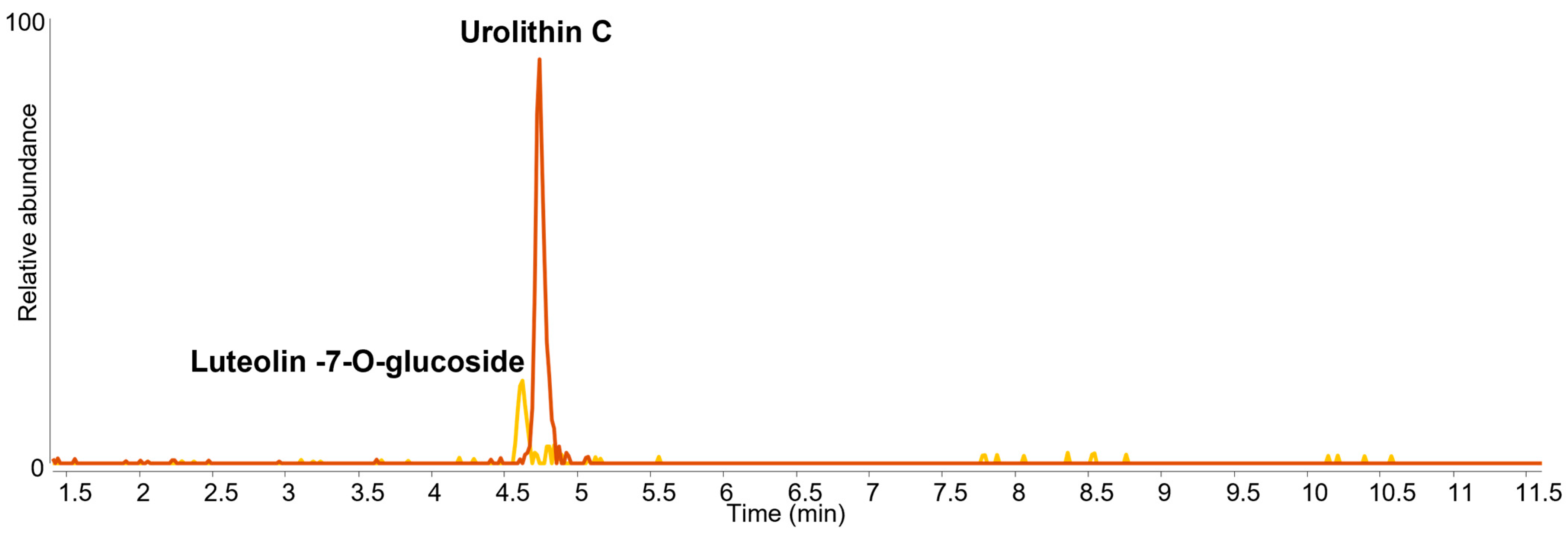
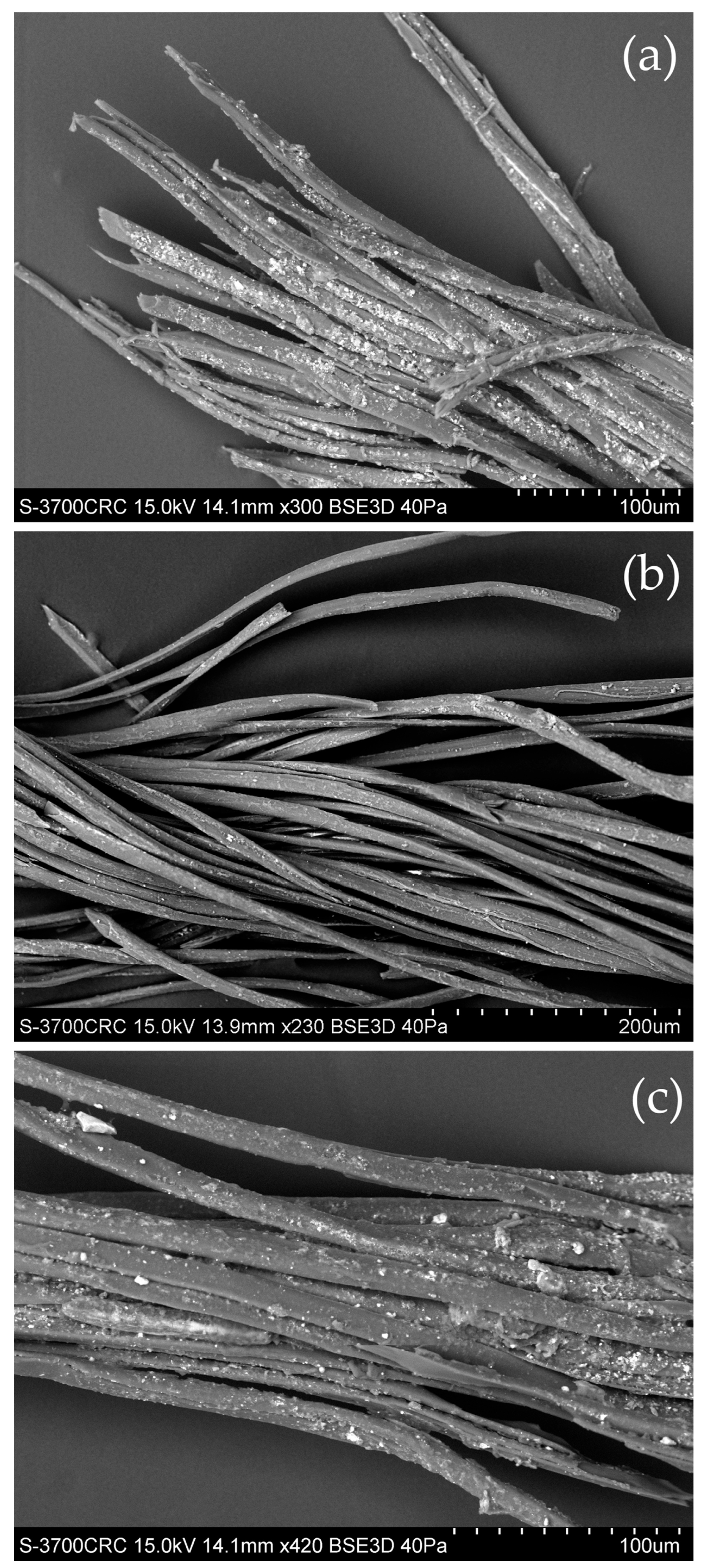
3.1. Textile Fragment 1850,1127.1.a
- Fibres with a smooth texture and flattened cylinder shape, often ribbon-like but with occasional swellings or areas of irregular thickness;
- A triangular or wedge-shaped cross-section to each fibre;
- Fibre ends that are often sharply pointed, bifurcated, angled or have a rounded point;
- Sometimes silk fibres may be in paired form (naturally gummed together).
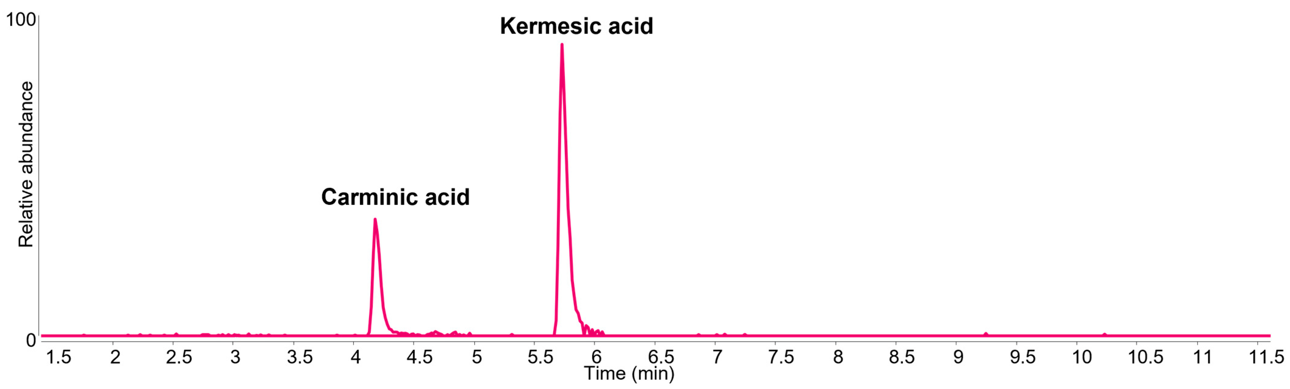
3.2. Textile Fragment 1850,1127.1.c
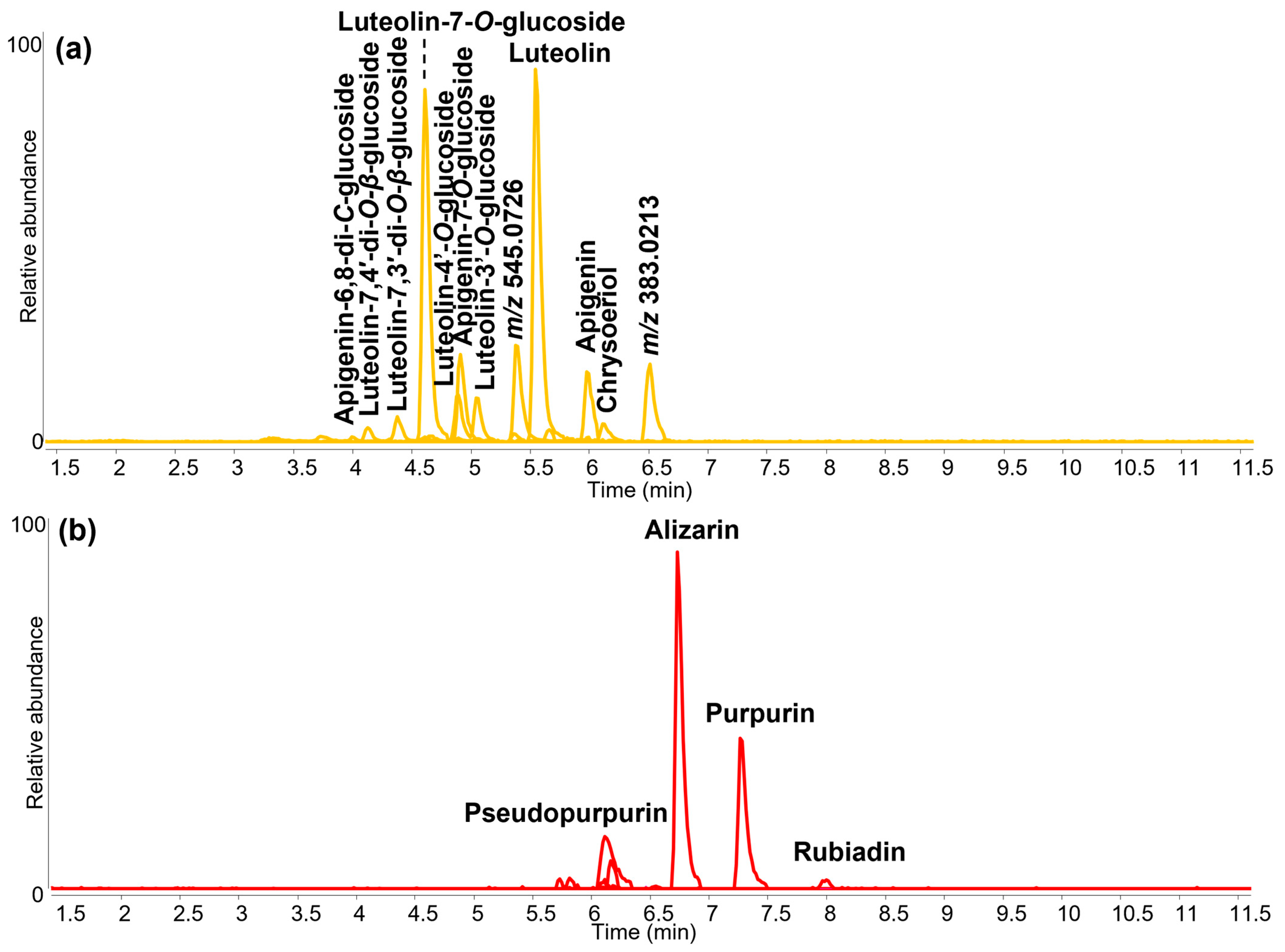
3.3. Textile Fragment 1850,1127.1.f
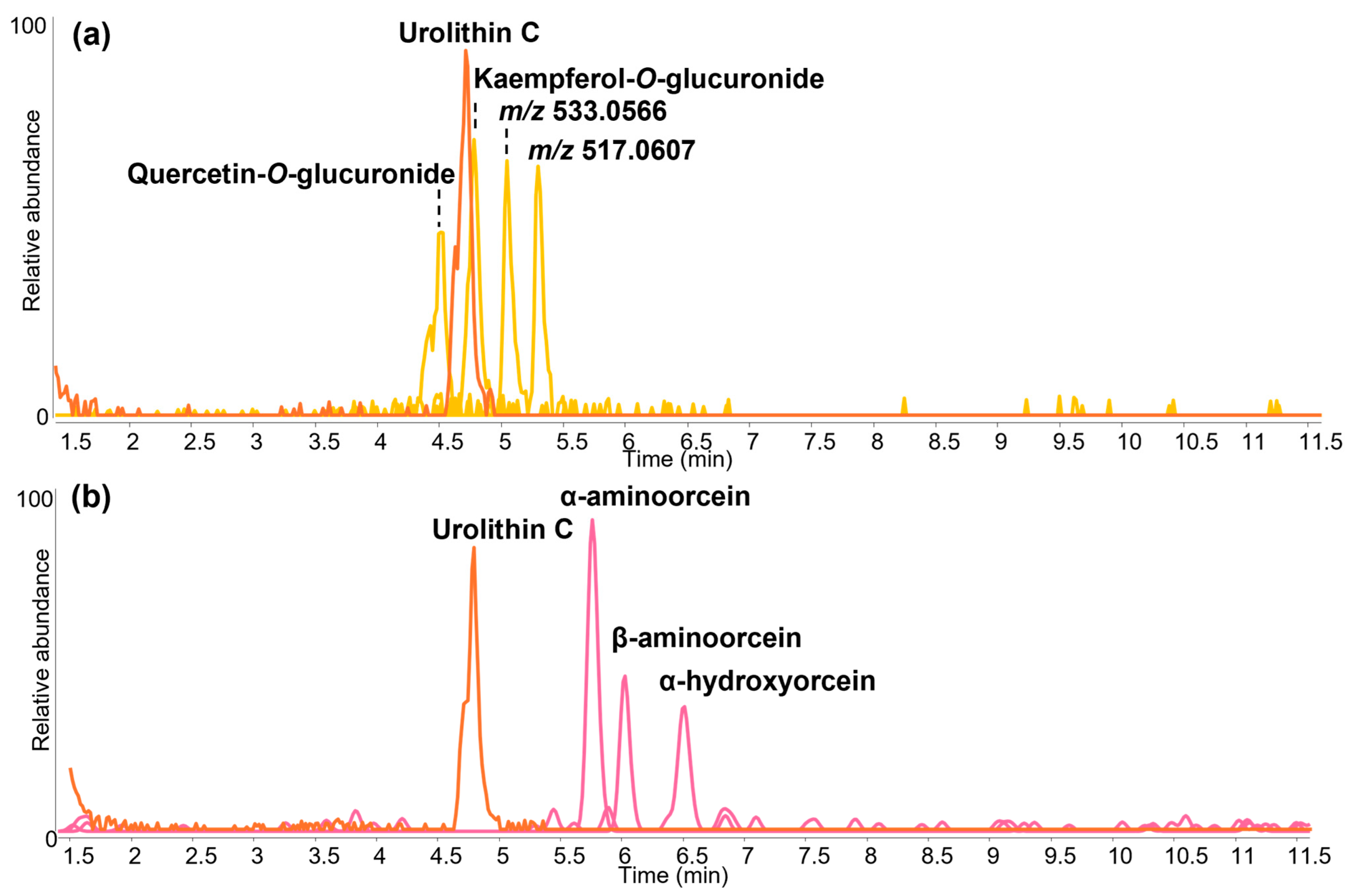
3.4. Textile Fragment 1850,1127.1.g
3.5. Textile Fragments 1850,1127.1.b, d, h, j and k
4. Discussion
5. Conclusions
Supplementary Materials
Author Contributions
Funding
Data Availability Statement
Acknowledgments
Conflicts of Interest
References
- Falk, B. Bildnisreliquiare: Zur Entstehung und Entwicklung der metallenen Kopf-, Büsten-und Halbfigurenreliquiare im Mittelalter. Aachen. Kunstbl. 1991–1993, 59, 99–238, at no. 52, pp. 206–209. [Google Scholar] [CrossRef]
- Burckhardt, R.F. Die Kunstdenkmäler des Kantons Basel-Stadt. Bd 2. In Der Basler Münsterschatz; Verlag E. Birkhäuser & Cie: Basel, Switzerland, 1933; no. 5; pp. 62–68. [Google Scholar]
- Husband, T. 9. Reliquary Head of Saint Eustace. In The Treasury of Basel Cathedral; Husband, T., Chapuis, J., Eds.; Metropolitan Museum of Art: New York, NY, USA, 2001; pp. 54–55. [Google Scholar]
- Robinson, J. Masterpieces of Medieval Art; British Museum Press: London, UK, 2008; pp. 74–75. [Google Scholar]
- Speakman, N. 104. Reliquary Head of St. Eustace. In Treasures of Heaven: Saints, Relics and Devotion in Medieval Europe; Bagnoli, M., Klein, H.A., Mann, C.G., Robinson, J., Eds.; British Museum Press: London, UK, 2011; p. 191. [Google Scholar]
- Cherry, J. 13. Kopfreliquiar des hl. Eustachius. In Der Basler Münsterschatz; Meles, B., Ed.; Christoph Merian Verlag: Basel, Switzerland, 2001; pp. 60–64. [Google Scholar]
- Joyner, L.; Freestone, I.; Robinson, J. Crowning glory: The identification of gems on the head reliquary of St Eustace from the Basle Cathedral Treasury. J. Gemmol. 2006, 30, 169–182. [Google Scholar] [CrossRef]
- Cartwright, C.R.; Joyner, L.; Ambers, J.C. Report on the 13th Century Silver Gilt Head Reliquary from Basle Cathedral; Unpublished British Museum DSR Project 0857 Report; The British Museum: London, UK, 1999. [Google Scholar]
- Folda, J. Crusader Art in the Holy Land, from the Third Crusade to the Fall of Acre; Cambridge University Press: Cambridge, UK, 2005; pp. 69–71. [Google Scholar]
- Gunther, P. The Capture of Constantinople: The Hystoria Constantinopolitana of Gunther of Pairis; Adams, A., Ed. and Translator; University of Pennsylvania Press: Philadelphia, PA, USA, 1997; pp. 123–124. [Google Scholar]
- Novelties in the British Museum. The Daily News, 22 April 1851; p. 3.
- Muthesius, A.M. Eastern Silks in Western Shrines and Treasuries Before 1200; Courtauld Institute of Art (University of London): London, UK, 1980; pp. 309–310. [Google Scholar]
- Granger-Taylor, H. Textile Fragments From the Basel Reliquary Head, British Museum (MLA 50 11-27 1); Unpublished British Museum Report; The British Museum: London, UK, 1989. [Google Scholar]
- Green, L.R. Analysis of Dyes From Silk Textile Fragments Found with the Basel Reliquary Head (MLA 50 11-27 1); Unpublished British Museum Conservation Research Report, LR1990-21; The British Museum: London, UK, 1990. [Google Scholar]
- Daniels, V.D. Analysis of Dyes on Eight Textile Samples; Unpublished British Museum Conservation Research Report, LR1991-19; The British Museum: London, UK, 1991. [Google Scholar]
- Brunning, S.; Luk, Y.P.; O’Connell, E.R.; Williams, T. Silk Roads; The British Museum: London, UK, 2024; pp. 256–257. [Google Scholar]
- Dyer, J.; Tamburini, D.; O’Connell, E.R.; Harrison, A. A multispectral imaging approach integrated into the study of Late Antique textiles from Egypt. PLoS ONE 2018, 13, e0204699. [Google Scholar] [CrossRef] [PubMed]
- Tamburini, D.; Dyer, J. Fibre optic reflectance spectroscopy and multispectral imaging for the non-invasive investigation of Asian colourants in Chinese textiles from Dunhuang (7th–10th century AD). Dye. Pigment. 2019, 162, 494–511. [Google Scholar] [CrossRef]
- Tamburini, D.; Dyer, J.; Cartwright, C.; Green, A. Changes in the production materials of Burmese textiles in the nineteenth century—Dyes, mordants and fibres of Karen garments from the British Museum’s collection. Herit. Sci. 2023, 11, 150–181. [Google Scholar] [CrossRef]
- Dyer, J.; Verri, G.; Cupitt, J. Multispectral Imaging in Reflectance and Photo-Induced Luminescence Modes: A User Manual; 2013. Available online: https://www.researchgate.net/publication/267266175_Multispectral_Imaging_in_Reflectance_and_Photo-induced_Luminescence_modes_A_User_Manual (accessed on 28 May 2025).
- Tamburini, D.; Cartwright, C.R.; Pullan, M.; Vickers, H. An investigation of the dye palette in Chinese silk embroidery from Dunhuang (Tang dynasty). Archaeol. Anthropol. Sci. 2019, 11, 1221–1239. [Google Scholar] [CrossRef]
- Ilvessalo-Pfäffli, M.-S. Fiber Atlas: Identification of Papermaking Fibers; Springer Science & Business Media: Berlin/Heidelberg, Germany, 1995. [Google Scholar]
- Cartwright, C.R.; Duffy, C.; Wang, H. Microscopical examination of fibres used in Ming dynasty paper money. Br. Mus. Tech. Res. Bull. 2014, 8, 105–116. [Google Scholar]
- Rast-Eicher, A. Fibres: Microscopy of Archaeological Textiles and Furs; Archaeolingua Alapítvány: Budapest, Hungary, 2016. [Google Scholar]
- Tamburini, D. Investigating Asian colourants in Chinese textiles from Dunhuang (7th–10th century AD) by high performance liquid chromatography tandem mass spectrometry—Towards the creation of a mass spectra database. Dye. Pigment. 2019, 163, 454–474. [Google Scholar] [CrossRef]
- van der Klift, E.; Villela, A.; Derksen, G.C.H.; Lankhorst, P.P.; van Beek, T.A. Microextraction of Reseda luteola-Dyed Wool and Qualitative Analysis of Its Flavones by UHPLC-UV, NMR and MS. Molecules 2021, 26, 3787. [Google Scholar] [CrossRef]
- Marques, R.; Sousa, M.M.; Oliveira, M.C.; Melo, M.J. Characterization of weld (Reseda luteola L.) and spurge flax (Daphne gnidium L.) by high-performance liquid chromatography–diode array detection–mass spectrometry in Arraiolos historical textiles. J. Chromatogr. A 2009, 1216, 1395–1402. [Google Scholar] [CrossRef]
- Zhang, X.; Good, I.; Laursen, R. Characterization of dyestuffs in ancient textiles from Xinjiang. J. Archaeol. Sci. 2008, 35, 1095–1103. [Google Scholar] [CrossRef]
- Liu, J.; Li, W.; Kang, X.; Zhao, F.; He, M.; She, Y.; Zhou, Y. Profiling by HPLC-DAD-MSD reveals a 2500-year history of the use of natural dyes in Northwest China. Dye. Pigment. 2021, 187, 109143. [Google Scholar] [CrossRef]
- Doherty, B.; Degano, I.; Romani, A.; Higgitt, C.; Peggie, D.; Colombini, M.P.; Miliani, C. Identifying Brazilwood’s Marker Component, Urolithin C, in Historical Textiles by Surface-Enhanced Raman Spectroscopy. Heritage 2021, 4, 1415–1428. [Google Scholar] [CrossRef]
- Wouters, J.; Verhecken, A. The coccid insect dyes: Hplc and computerized diode-array analysis of dyed yarns. Stud. Conserv. 1989, 34, 189–200. [Google Scholar] [CrossRef]
- Mantzouris, D.; Karapanagiotis, I. Armenian cochineal (Porphyrophora hamelii) and purpurin-rich madder in ancient polychromy. Color. Technol. 2015, 131, 370–373. [Google Scholar] [CrossRef]
- Serrano, A.; van den Doel, A.; van Bommel, M.; Hallett, J.; Joosten, I.; van den Berg, K.J. Investigation of crimson-dyed fibres for a new approach on the characterization of cochineal and kermes dyes in historical textiles. Anal. Chim. Acta 2015, 897, 116–127. [Google Scholar] [CrossRef]
- Mouri, C.; Laursen, R. Identification of anthraquinone markers for distinguishing Rubia species in madder-dyed textiles by HPLC. Microchim. Acta 2012, 179, 105–113. [Google Scholar] [CrossRef]
- Aceto, M.; Calà, E.; Agostino, A.; Fenoglio, G.; Gulmini, M.; Idone, A.; Porter, C.; Hofmann, C.; Rabitsch, S.; Denoël, C.; et al. Mythic dyes or mythic colour? New insight into the use of purple dyes on codices. Spectrochim. Acta Part A Mol. Biomol. Spectrosc. 2019, 215, 133–141. [Google Scholar] [CrossRef]
- Mouri, C.; Mozaffarian, V.; Zhang, X.; Laursen, R. Characterization of flavonols in plants used for textile dyeing and the significance of flavonol conjugates. Dye. Pigment. 2014, 100, 135–141. [Google Scholar] [CrossRef]
- Calà, E.; Benzi, M.; Gosetti, F.; Zanin, A.; Gulmini, M.; Idone, A.; Serafini, I.; Ciccola, A.; Curini, R.; Whitworth, I.; et al. Towards the identification of the lichen species in historical orchil dyes by HPLC-MS/MS. Microchem. J. 2019, 150, 104140. [Google Scholar] [CrossRef]
- Clementi, C.; Doherty, B.; Gentili, P.L.; Miliani, C.; Romani, A.; Brunetti, B.G.; Sgamellotti, A. Vibrational and electronic properties of painting lakes. Appl. Phys. A 2008, 92, 25–33. [Google Scholar] [CrossRef]
- Gulmini, M.; Idone, A.; Diana, E.; Gastaldi, D.; Vaudan, D.; Aceto, M. Identification of dyestuffs in historical textiles: Strong and weak points of a non-invasive approach. Dye. Pigment. 2013, 98, 136–145. [Google Scholar] [CrossRef]
- Tamburini, D.; Dyer, J.; Vandenbeusch, M.; Borla, M.; Angelici, D.; Aceto, M.; Oliva, C.; Facchetti, F.; Aicardi, S.; Davit, P.; et al. A multi-scalar investigation of the colouring materials used in textile wrappings of Egyptian votive animal mummies. Herit. Sci. 2021, 9, 106. [Google Scholar] [CrossRef]
- Bagnoli, M. Dressing the Relics: Some Thoughts on the Custom of Relic Wrapping in Medieval Christianity. In Matter of Faith: An Interdisciplinary Study of Relics and Relic Veneration in the Medieval Period; Robinson, J., De Beer, L., Harnden, A., Eds.; The British Museum: London, UK, 2014; pp. 100–109. [Google Scholar]
- Fleming, R. Acquiring, Flaunting and Destroying Silk in late Anglo-Saxon England. Early Mediev. Eur. 2007, 15, 127–158. [Google Scholar] [CrossRef]
- Williams, E.D. The Mobility of Fabric: Textiles in and around Medieval Eurasia. In Bringing the Holy Land Home: The Crusades, Chertsey Abbey, and the Reconstruction of a Medieval Masterpiece; Luyster, A., Ed.; Harvey Miller Publishers: London, UK, 2023; pp. 177–200, at p. 191. [Google Scholar]
- Cardon, D. Natural Dyes: Sources, Tradition, Technology and Science; Archetype: London, UK, 2007. [Google Scholar]
- de Graaff, J.H.H.; Roelofs, W.G.T.; van Bommel, M.R. The Colourful Past: Origins, Chemistry and Identification of Natural Dyestuffs; Abegg-Stiftung: Riggisberg, Switzerland, 2004. [Google Scholar]
- Keijzer, R.H.; Bommel, M.R.; Joosten, I.; Bogensperger, I. Late Antique Textiles from the Papyrus Collection of the Austrian National Library: Scientific Investigation of Fibres, Dyes and Dyeing Techniques; De Gruyter: Berlin, Germany, 2024. [Google Scholar]
- Schmedding, B. Mittelalterliche Textilien in Kirchen und Klöstern der Schweiz: Katalog; Stämpfli: Bern, Switzerland, 1978; no. 134, pp. 164–166; nos. 118–119, pp. 147–150. [Google Scholar]
- Petroviciu, I.; Nabais, P.; Melo, M. Red Dyes from West to East in Medieval Europe: From Portuguese Manuscript Illuminations to Romanian Textiles. In Textile Crossroads: Exploring European Clothing, Identity, and Culture Across Millennia. Anthology of COST Action “CA 19131—EuroWeb”; Droß-Krüpe, K., Quillien, L., Sarri, K., Eds.; Zea Books: Lincoln, NE, USA, 2024. [Google Scholar]
- Cabrera Lafuente, A.; Tamburini, D.; Dyer, J. Developing a protocol using FORS for the selection and study of historical textiles in the Victoria and Albert Museum. In Dyes in History and Archaeology 36 (DHA 36); Hampton Court Palace: England, UK, 2017. [Google Scholar]
- Miller, L.E.; Lafuente, A.C. Silk: Fiber, Fabric, and Fashion; WW Norton: New York, NY, USA, 2021. [Google Scholar]
- Witkowski, B.; Ganeczko, M.; Hryszko, H.; Stachurska, M.; Gierczak, T.; Biesaga, M. Identification of orcein and selected natural dyes in 14th and 15th century liturgical paraments with high-performance liquid chromatography coupled to the electrospray ionization tandem mass spectrometry (HPLC-ESI/MS/MS). Microchem. J. 2017, 133, 370–379. [Google Scholar] [CrossRef]
- Vanden Berghe, I.; Gleba, M.; Mannering, U. Towards the identification of dyestuffs in Early Iron Age Scandinavian peat bog textiles. J. Archaeol. Sci. 2009, 36, 1910–1921. [Google Scholar] [CrossRef]
- Joosten, I.; van Bommel, M.R.; Keijzer, R.H.-d.; Reschreiter, H. Micro Analysis on Hallstatt Textiles: Colour and Condition. Microchim. Acta 2006, 155, 169–174. [Google Scholar] [CrossRef]
- Claisse, P.; Marembert, C.; Galluzzi, F.; Chapoulie, R.; Dallel, M.; Mounier, A. Ephemeral Orchil in the Lady and the Unicorn Tapestry: Recipe, Experimentation, and Characterisation. Heritage 2024, 7, 3455–3469. [Google Scholar] [CrossRef]
- Bivar, A.D.H. Sasanian Iconography on Textiles and Seals. In Central Asian Textiles and Their Contexts in the Early Middle Ages; Schorta, R., Ed.; Abegg-Stiftung: Riggisberg, Switzerland, 2006; pp. 9–21, at pp. 16–17. [Google Scholar]
- Kuhn, D. Medieval Chinese Silk Fabrics: Technical Versatility and Puzzling Terminology. In Oriental Silks in Medieval Europe; von Fircks, J., Schorta, R., Eds.; Abegg-Stiftung: Riggisberg, Switzerland, 2016; pp. 12–33, at p. 23. [Google Scholar]
- Meier, H.-R.; Schwarz, P.-A.; Ochsner, C. Die Grabfunde des 12. bis 19. Jahrhunderts aus dem Basler Münster: Repräsentation im Tod und kultureller Wandel im Spiegel der materiellen Kultur; Materialhefte zur Archäologie in Basel; Archäologische Bodenforschung des Kantons Basel-Stadt Place: Basel, Switzerland, 2013; Volume 23, pp. 1–392. [Google Scholar] [CrossRef]
- Husband, T. Relics Sacred and Profane. In The Treasury of Basel Cathedral; Husband, T., Chapuis, J., Eds.; Metropolitan Museum of Art: New York, NY, USA, 2001; nos. 45–46; pp. 120–122. [Google Scholar]
- Schorta, R. 3. Goldgemustertes Seidengewebe. 4. Zwei Seidengewebe. In Der Basler Münsterschatz; Meles, B., Ed.; Christoph Merian Verlag: Basel, Switzerland, 2001; pp. 26–29. [Google Scholar]


| Textile Fragment | FORS | SEM-EDX | HPLC-DAD-ESI-Q-ToF |
|---|---|---|---|
| 1850,1127.1.a | Buff and light yellow areas—apparent absorption maximum at c. 400 nm for both (unidentified yellow/light brown colourants) | Silk | Luteolin-7-O-glucoside Likely weld (Reseda luteola) or other luteolin-containing (flavonoid) yellow colourants Urolithin C Sappanwood (Biancaea sappan) |
| 1850,1127.1.c | Mauve band—apparent absorption maxima at c. 520 and c. 560 nm (insect-based anthraquinone dye) | Silk | Carminic and kermesic acids Cochineal (likely Porphyrophora sp.) and kermes (Kermes vermilio) |
| 1850,1127.1.f | Red areas—featureless apparent absorption at c. 500 nm (plant-based anthraquinone dye, highly concentrated) | Silk | Alizarin, purpurin, pseudopurpurin and rubiadin Madder (likely Rubia tinctorum) |
| Blue areas—apparent absorption maximum at c. 660 nm (indigoid dye) | Indigotin and indirubin Indigoid dye | ||
| Green areas—apparent absorption maximum at c. 660 nm (indigoid dye) with additional maximum at c. 410 nm (unidentified yellow colourant) | Indigotin and indirubin Indigoid dye Luteolin, apigenin and chrysoeriol, and associated glucosides Weld (Reseda luteola) | ||
| Yellow areas—apparent absorption maximum at c. 410 nm (unidentified yellow colourant) | Luteolin, apigenin and chrysoeriol, and associated glucosides Weld (Reseda luteola) | ||
| 1850,1127.1.g | Pink areas—features at c. 550 and c. 590 nm (orchil) | Silk | α-aminoorcein, β-aminoorcein and α-hydroxyorcein Orchil Urolithin C Sappanwood (Biancaea sappan) |
| Green/blue areas—apparent absorption maximum at c. 660 nm (indigoid dye) | Indigotin and indirubin Indigoid dye Kaempferol-O-glucuronide, quercetin-O-glucuronide and 2 other flavonoid glucuronides Unidentified Yellow |
Disclaimer/Publisher’s Note: The statements, opinions and data contained in all publications are solely those of the individual author(s) and contributor(s) and not of MDPI and/or the editor(s). MDPI and/or the editor(s) disclaim responsibility for any injury to people or property resulting from any ideas, methods, instructions or products referred to in the content. |
© 2025 by the authors. Licensee MDPI, Basel, Switzerland. This article is an open access article distributed under the terms and conditions of the Creative Commons Attribution (CC BY) license (https://creativecommons.org/licenses/by/4.0/).
Share and Cite
Dyer, J.; Tamburini, D.; Speakman, N.; Cartwright, C.R. Hidden Treasures: Precious Textiles from the St Eustace Head Reliquary. Heritage 2025, 8, 206. https://doi.org/10.3390/heritage8060206
Dyer J, Tamburini D, Speakman N, Cartwright CR. Hidden Treasures: Precious Textiles from the St Eustace Head Reliquary. Heritage. 2025; 8(6):206. https://doi.org/10.3390/heritage8060206
Chicago/Turabian StyleDyer, Joanne, Diego Tamburini, Naomi Speakman, and Caroline R. Cartwright. 2025. "Hidden Treasures: Precious Textiles from the St Eustace Head Reliquary" Heritage 8, no. 6: 206. https://doi.org/10.3390/heritage8060206
APA StyleDyer, J., Tamburini, D., Speakman, N., & Cartwright, C. R. (2025). Hidden Treasures: Precious Textiles from the St Eustace Head Reliquary. Heritage, 8(6), 206. https://doi.org/10.3390/heritage8060206