Article

Experimental Ancient Egyptian Human Mummification Tested in a Porcine Model: Excellent Preservation at a 13-Year Follow-Up

by
Andreas G. Nerlich
1,*,
Stephanie Panzer
1,2,3,
Florian Fischer
1 and
Oliver K. Peschel
1
1
Institute of Forensic Medicine, Ludwig-Maximilians-University Munich, D-80336 Munich, Germany
2
Department of Radiology, University Hospital Salzburg, Paracelsus Medical University Salzburg, 5020 Salzburg, Austria
3
Institute of Biomechanics, Paracelsus Medical University Salzburg, 5020 Salzburg, Austria
*
Author to whom correspondence should be addressed.
Heritage 2025, 8(6), 194; https://doi.org/10.3390/heritage8060194
Submission received: 23 February 2025 / Revised: 24 April 2025 / Accepted: 27 May 2025 / Published: 28 May 2025

Abstract

:
Aim of the study: Several previous experimental studies simulated ancient Egyptian mummification of human bodies using the embalming protocol described by Herodotus. Besides two human experiments, several animal studies have been performed with very different species, mostly over short observation periods. We used a human-sized piglet model that matches closely to humans and undertook a long-term experiment with two detailed examination time points over 13 years. This was conducted to test the efficacy of the Herodotus embalming method in the long term. Material and Methods: An 88 kg piglet, 1.30 m body length, obtained from a veterinary practice was chosen as the skin is similar to humans. Using the described formula, the carcass was cleaned, eviscerated, filled with spices and natron sachets, and then covered with 240 kg of natron for 40 days. It was then reopened, and most of the sachets were removed. The surface was cleaned with wine, the body cavity partly refilled with sachets and spices, the surface treated with oils, wax, honey and bitumen, and finally sealed with linen bandages. The body weight was regularly monitored over the 13-year period. At 7 and 13 years, re-examination, with a protocol including CT scanning, histology and microbiology, was performed. Results: The monitoring of the body weight showed a rapid loss of weight within the first year, gradually slowing, reaching more than 66% of its weight. In the final 6 years, the body weight was reduced by only 1.7 kg. The CT scans at 7 years and 13 years showed that the structures of the heart, muscle, skin, and soft tissue were well preserved, and the body significantly shrunken; only the musculature showed air inclusions. Histology and microbiology (examined at baseline, 7 years and 13 years) revealed excellently conserved tissue with anaerobic microbe spores, very limited tissue destruction, and no significant fungal or parasitic invasion. However, the preserved kidneys and internal genitalia had disappeared. Conclusions: This ongoing long-term project confirmed excellent mummification with near-perfect body conservation at 13 years, supporting the efficacy of the described Herodotus technique. This model is suitable for the assessment of special preservation techniques recently suggested for individual soft tissue organs.

1. Introduction

One of the hallmarks of Ancient Egyptian culture is the preservation of human bodies for the afterlife. By repeated refining of the techniques and the substances applied, ancient Egyptians achieved efficient conservation of the corpse. The constant hot and dry climate promoted the long-term conservation of cadavers, with more and more elaborate methods developed from the Old to the New Kingdom. At that time, excellent preservation of human bodies was achieved. However, in later periods, economic limitations and/or loss of knowledge led to deterioration in the mummification process until this custom was completely abolished by the advent of the Islamic religion [1,2].
The art and practice of ancient Egyptian embalming have been enigmatic. This was only possible by elaborate techniques, which, unfortunately, have not been documented in detail. The current knowledge of embalming mainly comes from the investigation of human remains in ancient Egyptian graves [3,4]. Further information exists in some fragments of written tradition, the most important ones being the reports that have been provided by descriptions of mummification by the Greek writer Herodotus (c.484 to c.425 BCE) and the Roman historian Diodorus Siclus (fl. 1st century BCE) who reported on exenteration, cleaning, dehydration, surface-treatment, and bandaging of the dead body [1,2]. However, both never went to Egypt, and their reports are only brief outlines of the embalming techniques. In this respect, many details of ancient Egyptian embalming remain unknown. Only very recently, new data have been obtained from chemical analyses of a Late Period embalming workshop in Saqqara [5], which were not available when the long-term experiment was performed.
In addition, there exist few studies which used an experimental approach that tried to simulate the embalming procedure. As early as 1909, Lucas performed a study on three chickens that he kept for about 13 years in soluble natron, different from Herodotus’ description of dry natron [6]. Lucas did not report the result of this experiment. He replicated his experiments using pigeons more than 20 years later [6], however, again, without any reporting the outcome. In 1963, Sandison [7] used human (amputated) toes but observed them for only 40 days. In 1979, Garner [8] performed embalming experiments on laboratory mice for 50 days. In 1985, Notman and Aufderheide [9] applied the Herodotus method to an adult dog that was re-examined after 34 weeks with excellent preservation noted. In the same year, Aturaliya [10] performed experimental embalming on rats and reported success without providing details of the methodology. Garcia-Jimenez [11] performed embalming studies on three rabbits, and Ikram used the Herodotus technique for experimental embalming also on rabbits with excellent outcomes [12]. Again, the follow-up periods of those experiments were very limited, and most were undertaken on small species of a different size to humans or with significantly different integuments, e.g., fur or plumage.
Very few experiments have been performed on human cadavers. In 1994, Brier and Wade were the first to use a complete human cadaver for the simulation of ancient Egyptian embalming [13,14,15]. This specimen, called MUMAB, presumably still exists today after 31 years. Although this is the only reported long-term human embalming experiment, only a few data have been published on it, indicating that the embalming technique was well performed and efficient, though costly. The timing and techniques described by Herodotus seemed to have been successful. Unfortunately, the limited reports do not record any monitoring of bodyweight over a long time period, which would have been easy to undertake and given useful data. Some information is available on CT findings, but there exists no data on histological or microbiological investigations.
In 2017, an English group performed a replication of the MUMAB experiment [16], however, with a reported follow-up period of only 3 months. Unfortunately, no information about its further outcome or any scientifically relevant data has been reported despite some indication of a successful outcome. Some more information comes from experiments on two human amputated legs, which were followed up by CT scanning, MRI, and microbiological, molecular and histological analyses. These studies provide interesting information in terms of gradual loss of water content, increasing destruction of tissue structures, and shrinkage of those structures [17,18,19]. These experiments seem to have been terminated after about 2 years.
All these studies provide very valuable insights into different aspects of human embalming. Accordingly, the Herodotus technique seems applicable and plausible. However, there are several limitations. In the non-human experimental setting, the sizes of the animals seem to be very different from adult human cadavers, and the body integument is very different between humans and animals, with fur and feathers in the latter. Ultimately, the determined parameters and the observation times seem either incomplete or of limited scientific value.
Given this, a long-term mummification experiment on an animal model similar to humans would be useful as a surrogate for human cadavers. The aim of this study was to test the described Herodotus technique on an animal model over a long time period with the purpose of demonstrating the quality of the preservation at different time points.

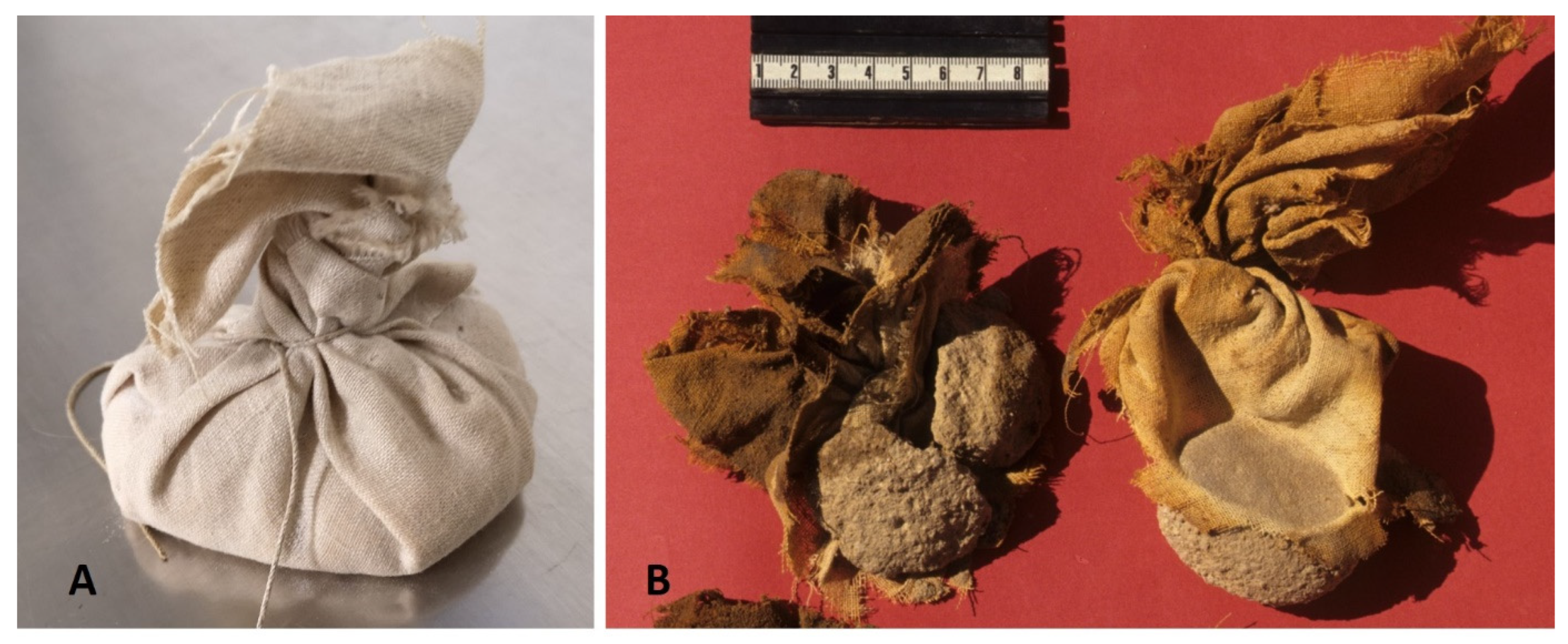
2. Material and Methods

The experiment was performed at the Institute of Pathology, Academic Clinic Schwabing, Ludwig-Maximilians-University Munich on 21 June 2011. A porcine model was chosen as it resembles the human closely with respect to size and skin [20]. Despite obvious differences in anatomy and some differences in the body composition, especially a slightly higher fat content, this model comes close to humans and is notable as it is the chosen animal for xenograft research human transplant surgery [21,22,23]. The animal was provided by the Department of Molecular Animal Breeding and Biotechnology, Faculty of Veterinary Medicine of the University, as it required euthanizing because of an eye infection. Under EU law, no further regulatory or ethical approval was required for the study.
The initial preparation was described in a conference report [24], where some details of the experimental set-up and embalming technique were recorded. In summary, a female piglet (Sus domesticus), aged 5 months and weighing 88 kg with a body length of 1.30 m, was used. Clinically, there was no evidence of systemic infection. The carcass was obtained within 24 h of death. It was first cleaned with tap water and then eviscerated according to Herodotus’ description via a left lateral abdominal incision, as described in the translated version by Feix [25] (for the various steps of preparation, see Figure 1). Through this approach, several internal organs (lungs, liver, intestines, stomach and spleen) were removed; all other organs, especially the heart, kidneys and internal genitalia, were left in situ. All removed organs were discarded. The skull cavity was opened by perforating the ethmoideal roof, and the brain was also removed and discarded. The cleansed body cavities were then filled with natron sachets, which corresponded to findings from ancient Egyptian mummy studies [26] (Figure 2) and a variety of herbs and spices (a mixture of dried herbs including peppermint, quendula, thyme, lavender, cloves and cubeb; since the Herodotus description did not provide any quantities for those components, they were mixed in 1:1 w/w concentrations). The body incision was closed with a suture. The body was then transferred into a wooden coffin (made of spruce), and the complete body was then covered with a total of 240 kg of synthetically prepared natron and left in place for 40 days (Figure 1B). This preparation of “natron” was necessary since naturally occurring natron in the Egyptian Wadi El-Natrun [Natron-Valley] was not available. Therefore, a mixture of sodium carbonate 81%, sodium bicarbonate 10%, sodium chloride 6%, and sodium sulfate 3%, as described by Notman and Aufderheide, was prepared [10]. After that period, the external natron was removed (Figure 1C), and the body was washed, first by running tap water and subsequently with white wine (commercial grape product, alcohol content 12% w/v) (Figure 1D). The incision of the cleansed body was reopened, and most of the natron sachets and the herb mixtures were removed from the abdomen and thorax. Furthermore, the internal surfaces were also cleaned with wine, as indicated above. Finally, several fresh natron sachets, along with further herbs and spices, were introduced into the chest and abdomen in an identical manner as before. At this point, a small (ca. 2 × 1 cm) skin biopsy was taken for microbiology and histology. Finally, the body surface was treated with a mixture of oils (cedarwood oil, cypress oil, pistacia oil, olive oil, 1:1 v/v mixture), wax (bees wax), honey (natural wild bee’s honey, commercial product) and bitumen (Pix tumens, commercial product) and completely sealed with linen bandages (modern fabrication of sterile bandages) as described by Herodotus [25]. [Unfortunately, we did not record the specific site of fabrication of the materials used and/or any specific production number when we started the experiment 13 years ago. However, we can state that the used materials were commercially available at the time of the experiment in Germany].
This body was then kept in the open wooden sarcophagus in a room with constantly controlled temperature at 24 °C and 45% humidity. The temperature was thereby comparable to the fairly constant in-door temperatures of ancient Egyptian tombs (“Valley of the Kings/Thebes-West” between 22 and 28 °C [27,28]).
The baseline for the experiment was day 41 after the initial preservation, on completion of the full preservation protocol. The body was kept intact and only moved from the sarcophagus for regular monitoring of the body weight, initially every month, and finally, after 3 years, 6 monthly. Two more detailed examinations were undertaken opportunistically when resources and funding were available when the body had to be moved from its storage place. The first termed 7-year follow-up was at 2498 days from the baseline and the second at 13 years (4749 days).

2.1. Follow-Up Imaging

In the two follow-up examinations, CT scanning (Siemens Healthcare, Erlangen, Germany) was undertaken in the supine position with a slice thickness of 0.625 mm, 120 kV and 200 mA in the standard algorithm [29]. Histology and microbiology were performed with an additional MRI scan [30,31] in the 7-year examination only [24]. A comparison of the results of the CT scans did not reveal any major differences, so the second MRI analysis was omitted since it would not have added any further information. The CT data of the two examinations were finally compared on a workstation with full access to around 2.200 body slices and 3D-rendering techniques [29].

2.2. Histological Investigation

For histological analysis, a baseline sample was taken on day 41 when the piglet had been removed from the natron as the formal embalming process was now completed. Two further samples for histology were procured at days 2498 (7 years) and 4749 (13 years) when the two reexaminations took place. At these time points, tissue samples were obtained with a 0.8 cm drill biopsy under sterile conditions from the dorsal lower thorax of the body at 7 years from the left side, then at 13 years from the right side. Therefore, an area of 2 cm2 of the superficial linen bindings was disinfected and removed using sterile instruments. Following a change to a further set of sterile instruments, the drill biopsies were taken.
From all three samples, histological analyses were performed following rehydration and fixation in 4–6% buffered formaldehyde, pH 7.4, as previously reported for mummified tissue [32]. The rehydrated samples were routinely embedded into paraffin wax for the preparation of histological sections (2–4 µm think). These were used for various histochemical stainings, in particular haematoxilin and eosin stain (HE), Elastica-van-Gieson stain for connective tissues, periodic-acid-Schiff’s-reagent staining (PAS) for the detection of mucopolysaccharides, but also fungal microbes, Prussian blue (Fe) for the potential identification of haemosiderin and Gram and May-Grünwald-Giemsa (MGG) staining for the detection of bacteria. All staining protocols had previously been tested successfully in human mummified tissue samples [33].

2.3. Microbiological Investigations

Sterile parallel samples of the three samples (day 41, 7 years and 13 years) were also used for microbiological analyses. These followed the stepwise routine protocols of microbiological investigations: first, smear preparations were stained with Gram and MGG staining for visual control of bacterial and/or fungal growth. Then, the material was exposed to differential growth media with particular reference to aerobic and anaerobic growth conditions, but also fungal growth according to routine protocols [34]. In parallel, extracted microbial DNA was identified by sequencing of 16S ribosomal DNA and also by routine protocols [35]. Ultimately, the material was subjected to MALDI-TOF mass spectrometry, where positive results in the aforementioned analyses were found [36] for further confirmation.

2.4. Evaluation of Preservation Quality
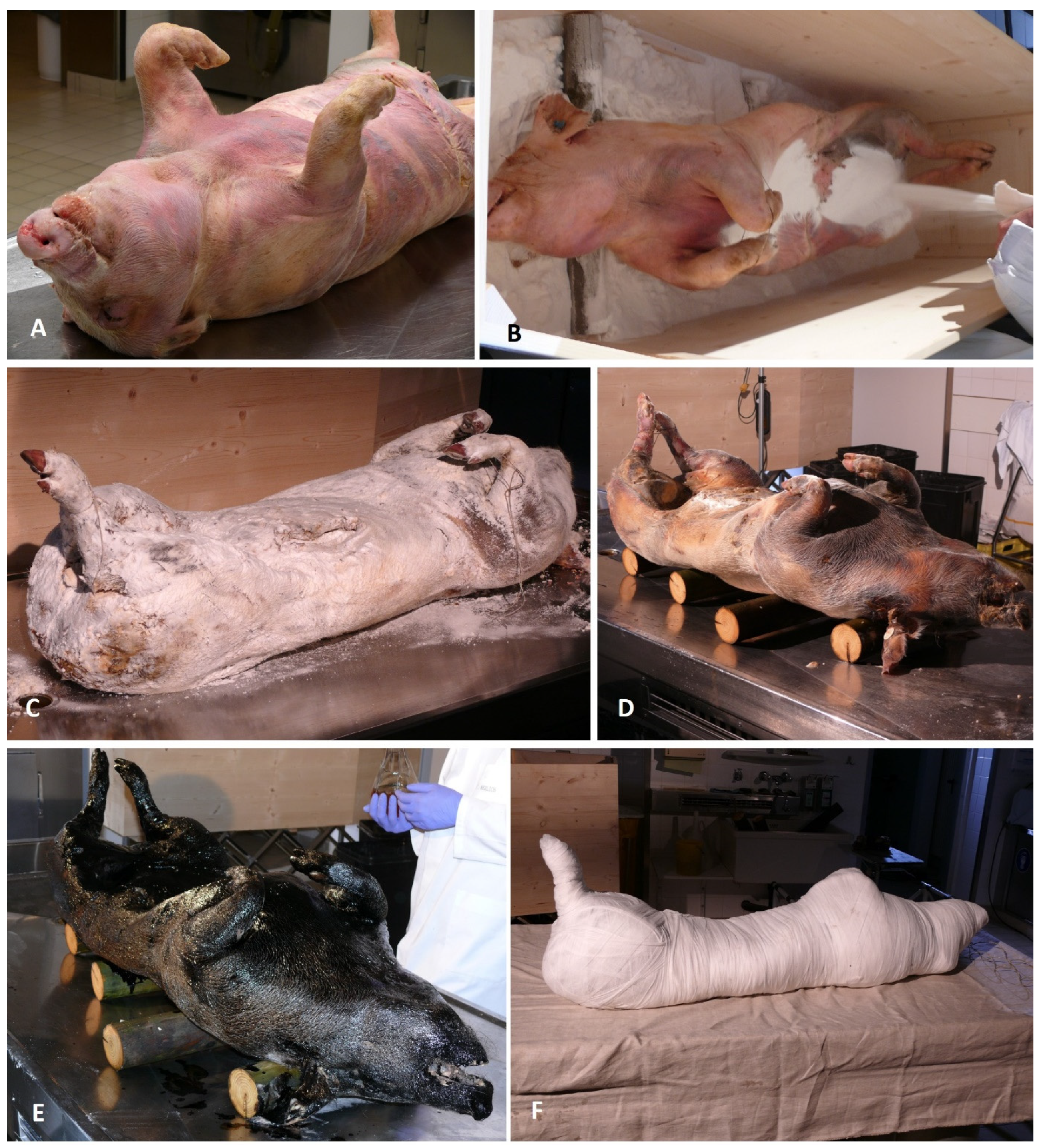
The quality of the preservation was assessed by the following criteria, which had been established after discussion with the study team (Table 1):

3. Results

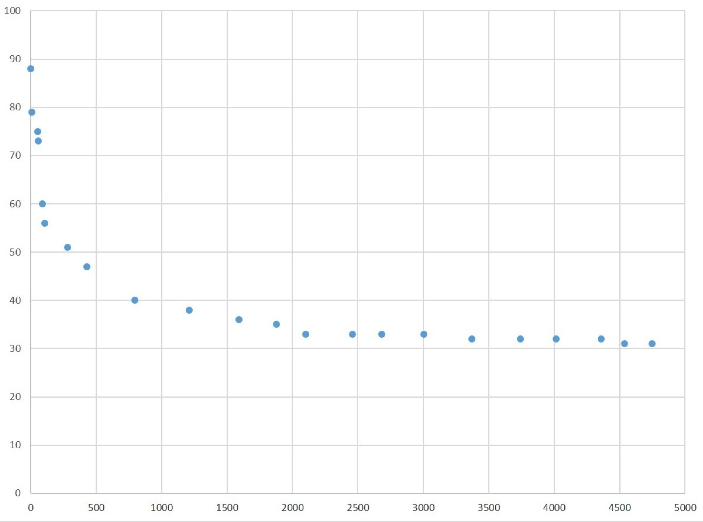
3.1. Changes in the Body Weight

The recording of the body weight revealed a significant initial loss of weight of about 30% within the first 200 days. After that, the weight change reduced, reaching 50% after around 650 days and about 38% of the initial weight after approximately 1600 days. After this period, the rate of weight loss was much lower at about 200 g every 6 months for the next 4 years. At the time of the 7-year review, the body weighed 32.3 kg (36.7% of the initial weight). The weight loss then slowed down further so that after 13 years, the body weighed 30.6 kg (34.7%)—an average final loss of weight of around 140 g every 6 months (Figure 3).

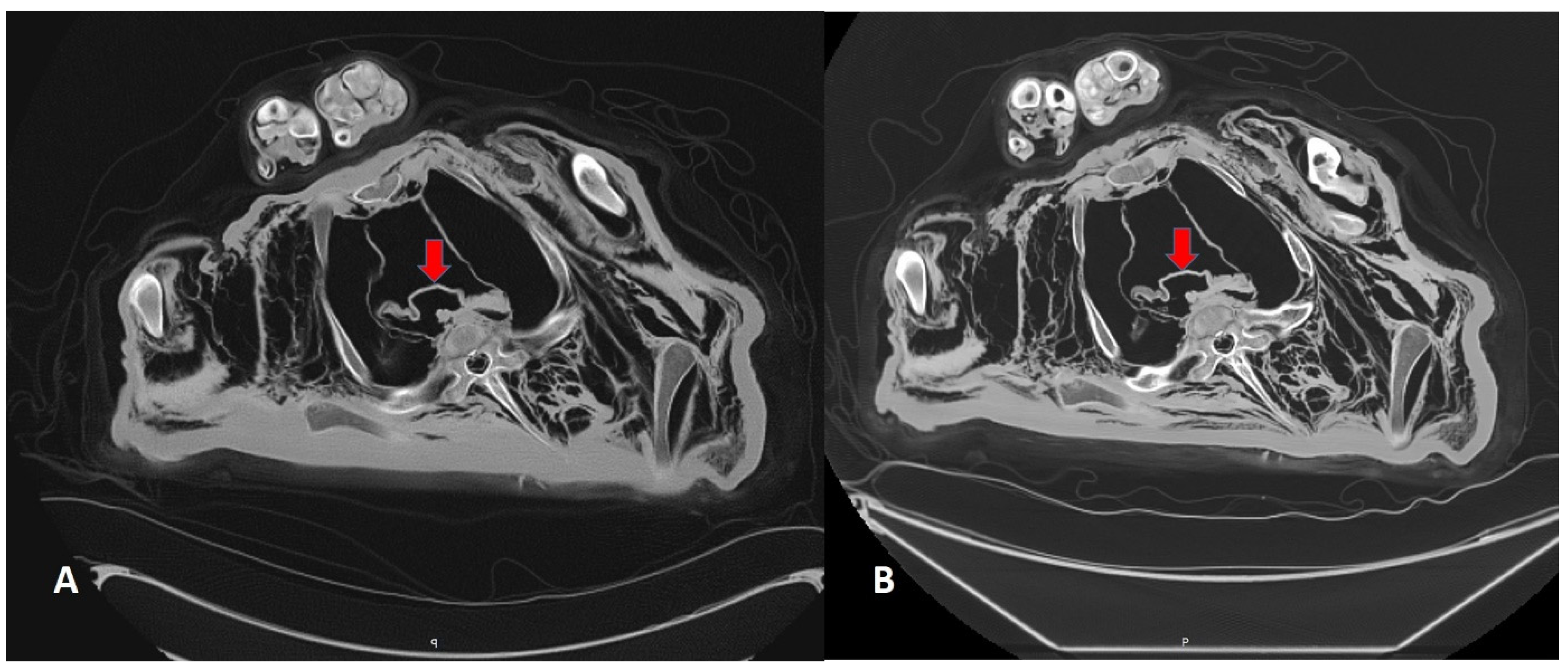
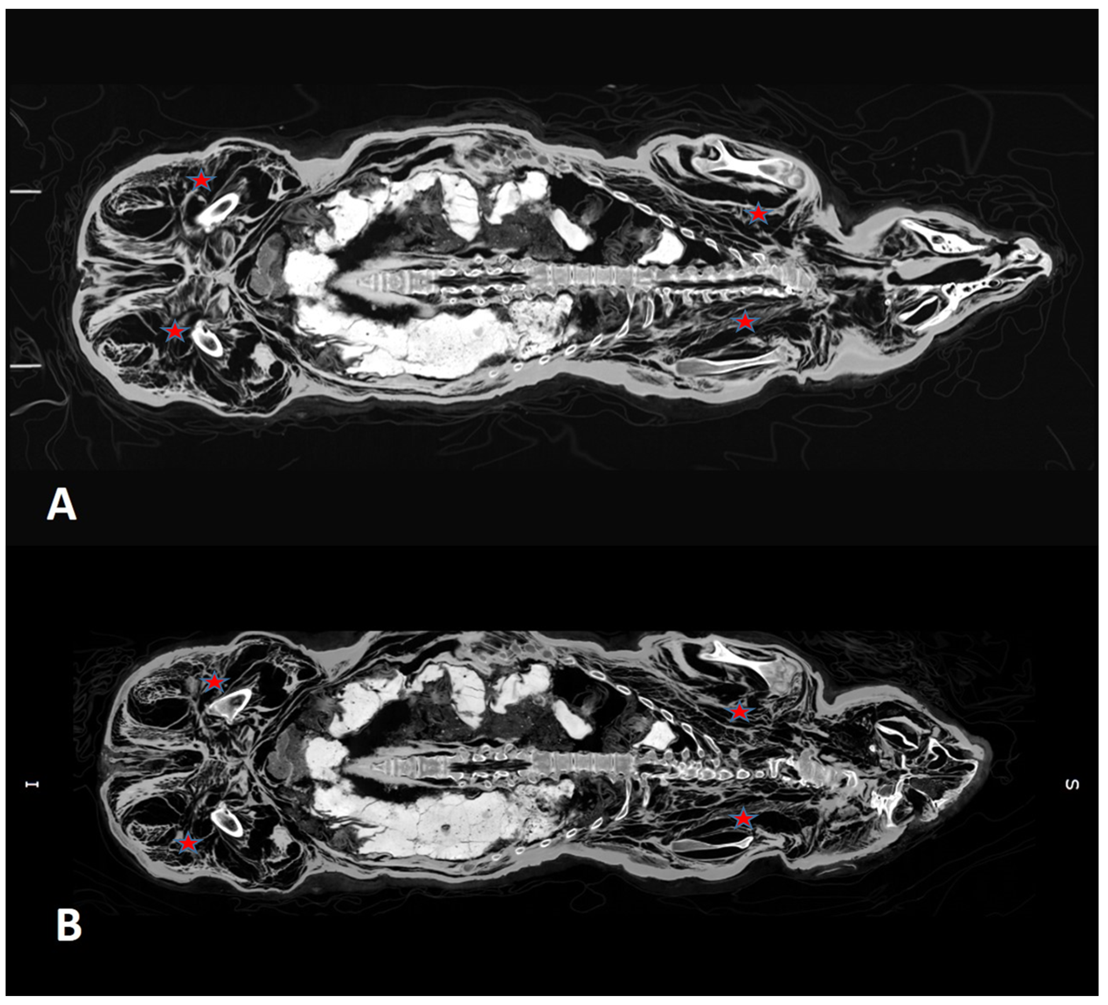
3.2. CT Scans After 7 and 13 Years

Both follow-up examinations began with a complete CT body scan performed under identical conditions. Both scans, despite being 6 years apart, showed fairly similar results with respect to the preservation of osseous and soft tissue structures, including some of the inner organs. The body was excellently preserved with correct anatomical positioning of all osseous structures. The remnant of the shrunken heart was seen in situ; the heart muscle was significantly shrunken but still retained its typical position within the pericardium (Figure 4). However, the other preserved internal organs, such as the kidneys and the internal genitalia, were no longer seen. The soft tissues of thoracic and lumbar muscles revealed spongy dehiscence of larger muscle bundles with interlacing air-filled spaces of unknown origin (Figure 4, Figure 5 and Figure 6). These were prominent in the lumbar musculature, where the muscles seemed to transform into a “spongy tissue” (Figure 5). The thoracic and abdominal cavities were filled with natron sachets and some amorphous material that was not discernible from the scans (possibly a conglomerate of herbs and spices) (Figure 6). Importantly, there were no differences from the scans taken at 7 and 13 years.

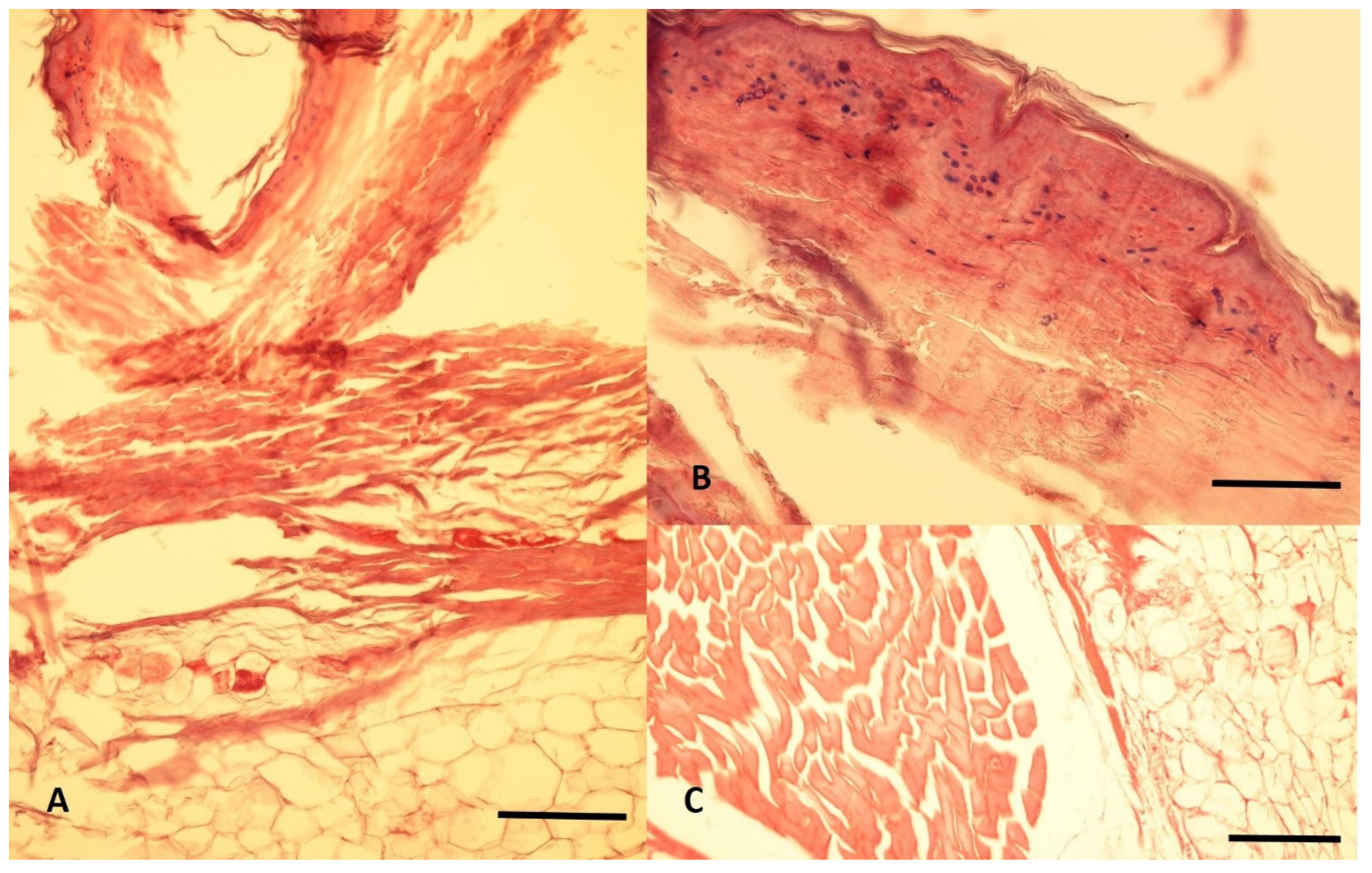
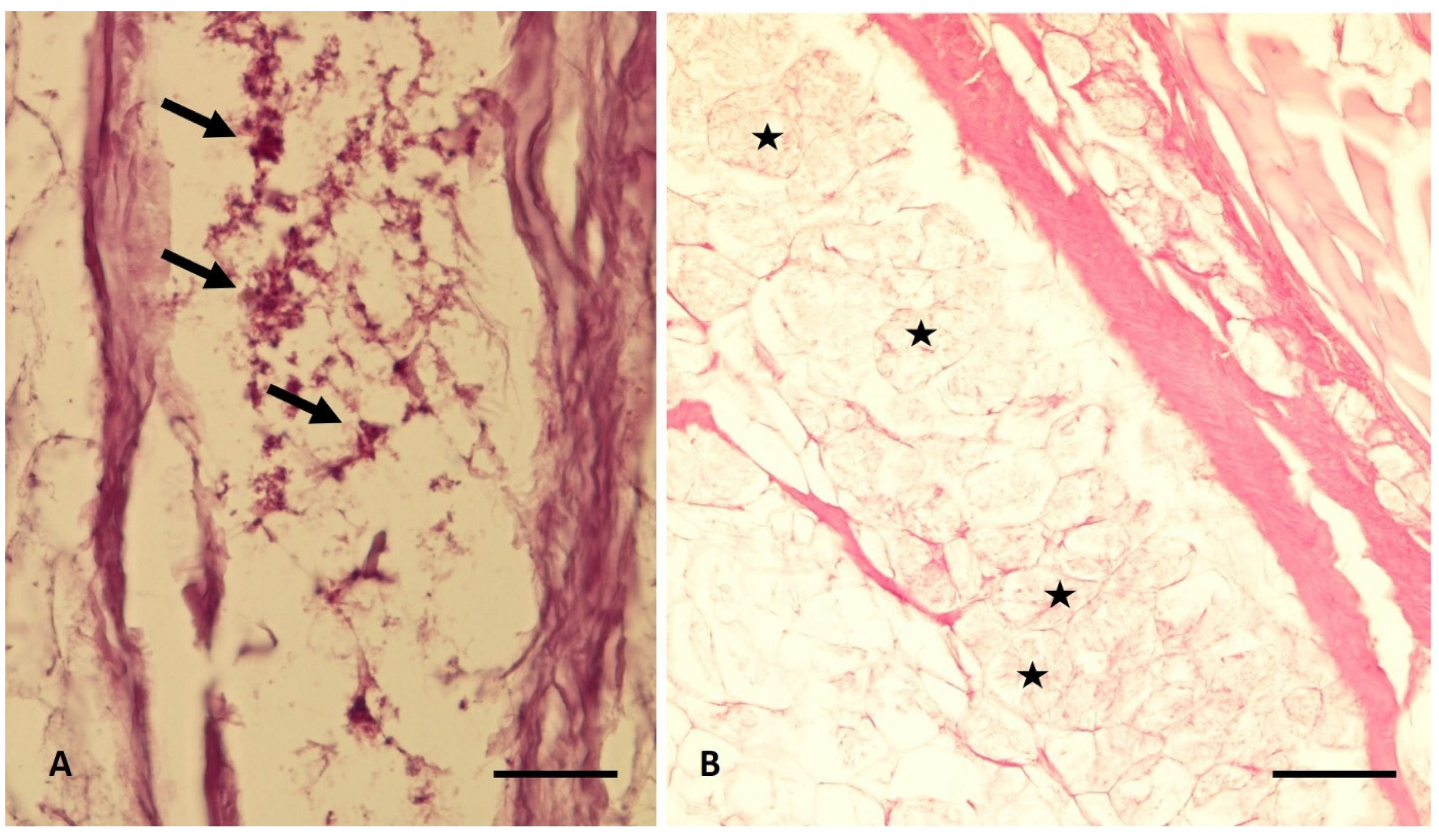
3.3. Histological Analyses

In order to identify possible changes in the microarchitecture of the tissue during the process of desiccation, skin, subcutaneous tissue and parts of the musculature were analyzed. At baseline, the histoarchitecture was completely preserved, including the superficial epidermal layer. Interestingly, fragmented residues of basal epidermal skin nuclei were still visible (Figure 7). The application of special stains, including May-Grünwald-Giemsa (MGG) and periodic acid-Schiff’s reagent (PAS) for the identification of bacteria, fungi or protozoa, all remained negative. There was no evidence of tissue destruction, and adipocire formation was not seen.
The drill biopsy material from 7 years included superficial skin, subcutaneous fat and also the musculature seen on the CT scans as irregular “air inclusions”. Microscopic analysis again identified almost intact structures of the epidermis and dermis (Figure 8A), even focally retained fragments of cell nuclei, and the dermis (Figure 8B), as well as excellently preserved muscle fibers with typical striation and complete conservation of myotube conformations (Figure 8C). Between these structures were seen occasional small roundish corpuscles that stained positively with May-Grünwald-Giemsa (MGG) but not Periodic-acid-Schiff’s stain (PAS). These were attributed to bacterial spores; meanwhile, fungal spores were morphologically excluded (Figure 9A). Furthermore, adipocytes of the subcutaneous tissue showed adipocire formation to a moderate extent, suggesting post-mortem transformation of the fat tissue before the desiccation process has completely halted (Figure 9B).
At 13 years, a third set of biopsies was obtained. The histological investigation again showed well-preserved histostructures with remnants of an otherwise intact epidermal layer. However, in the epidermis, no nuclear remnants were seen. In the deep part of the sample, typical skeletal musculature and regular adipose tissue were present (Figure 10). On microscopy, the tissue structures were fairly similar to the 7-year examination. The detailed analysis confirmed signs of adipocire formation as in the previous biopsy (Figure 11A). However, corpuscular inclusions, such as seen in the 7-year biopsy, were not present in the material retrieved. As further evidence for excellent preservation, even small blood vessels could be found (Figure 11B).

3.4. Microbiological Investigations

For the microbiological analyses, parallel samples as for the histology were used for all three-time points. At baseline and 7 years, the microscopy showed few corpuscular structures suggestive of bacterial spores and no fungal structures. The cultures on differential media remained sterile under aerobic conditions. The anaerobic cultures, in contrast, showed low-level growth of Clostridium haemolyticum, which was confirmed by a specific amplification product in 16S-rRNA sequencing. Additionally, a low-level signal for Bacillus spp. was detected on the PCR amplification, which was further confirmed by MALDI-TOF mass spectrometry. The latter further identified Bacillus cereus. Fungal cultures and PCR were negative. In the 13-year sample, no specific microscopic structures could be identified. Aerobic cultures again were negative, while anaerobic cultures and the 16S-rRNA sequencing again showed Clostridium haemolyticum; there were again no signs of fungal growth.

3.5. Overall Evaluation of the Experimental Outcome

Taking the criteria for the quality of preservation of the experimental mummy into account, excellent results were found in almost all specifications (see Table 1), probably with the only exception of internal organs (especially kidneys and inner genitalia), which dissolved during the experiment. Taking, however, the outer appearance, and thereby the integrity of the skin, as the principal consideration, the experiment’s outcome can be classed as excellent.

4. Discussion

This study showed that in a porcine model, using the Herodotus technique [25], excellent preservation was achieved over a 13-year period. This confirms that Sus domestica is a suitable animal model to test human adult cadaveric ancient embalming techniques, especially in terms of weight and body surface structure. However, there exist some important differences, such as the body’s anatomy. The advantage is that this model circumvents all ethical discussions about the use of a human corpse, sampling of tissues and declaration of the time for the end of the study as it was performed on a dead body which would otherwise have been subjected to carcass disposal.
The embalmed carcass can be intermittently studied, is not resource intensive, and can be easily stored in controlled conditions. There are no problems with the ethics of consent and the role of the bereaved family or with any requirement to set a date for formal burial. However, it must be noted that repeated sampling within a short period of time may change the surface milieu of the corpse and thereby may significantly influence the embalming effect, causing unwanted deterioration in the preservation.
The present experiment showed rapid and effective dehydration within the 40-day natron treatment, followed by further loss of weight, which gradually slowed down to a final rate of fluid loss of about 140 g every 6 months. The latter was probably due to the very slow evaporation of the remaining fluid within the body.
Slight focal adipocire formation in the subcutaneous fat tissue in both histological samples with 7 and 13 years was noted. This was absent in the initial skin histology after 41 days of natron dehydration. In consequence, despite the excellent dehydration and body preservation, chemical reactions such as adipocire formation were active (although minimally) during the desiccation period. Similarly, the preservation of fragmented cell nuclei in the basal epidermis after 41 days and even after 7 years, but no longer after 13 years, was also noted. Other cellular remnants, e.g., in stromal fibroblasts and muscle myotubes, were already absent after 41 days. Other cellular substructures, such as cross-striation of muscle myotubes, were present throughout the examination period.
The excellent histological preservation coincided with a lack of any major bacterial growth, as seen in the histological slides, with only a few foci of bacterial spores. The repeated microbiological analyses also detected only bacterial spores, mostly anaerobic Clostridial spp., but no active bacteria. In this regard, it is interesting that the CT scans after both 7 and 13 years showed irregular destruction of muscle and soft tissue with some air inclusions. Originally, on imaging, it was thought that these air-filled spaces might represent foci of putrid destruction, but histology showed this not to be the case.
Since the start of the project, important new information has been obtained on the ancient Egyptian embalming process through the chemical investigation of an embalmer’s workshop from Saqqara in the Late Period [5]. In this analysis, several pots with detailed inscriptions revealed the chemical composition of embalming ingredients that were used for specific body parts. This information was not available at the start of this experiment. With this new information, any further experimental planning of ancient Egyptian-style human body preservation might lead to an even more elaborate preservation technique. As yet, however, no information is available as to the concentration of the embalming substances or the exact timing of the procedures for the preservation of the internal organs.
This project used a porcine model to test Herodotus’ description of the ancient Egyptians’ embalming method and showed controlled desiccation over a 13-year period with excellent preservation of the carcass. Natron was the principal ingredient for desiccation. Of note, preservation of the soft tissue organs, here the internal genitalia and kidneys, was poor. Newer studies suggest a more tailored approach to embalming these organs could be tried. This model is suitable for testing this as it avoids the ethical considerations necessary when using human cadavers.

Author Contributions

Conceptualization, methodology, validation, resources, writing – original draft preparation, supervision: A.G.N. and O.K.P.; software, data curation: S.P. and F.F.; formal analysis, investigation, writing – review and editing: A.G.N., S.P., F.F., O.K.P.; project administration: A.G.N. All authors have read and agreed to the published version of the manuscript.

Funding

This research received no external funding.

Data Availability Statement

All data of this study are included in the present publication.

Acknowledgments

The authors’ thanks go to the Department of Animal Breeding, Faculty of Veterinary Medicine, Ludwig-Maximilians-University Munich, B. Kessler, for providing us with the dead animal. The technical help by A. Riepertinger and R. Gillich, Institute of Pathology, Academic Clinic Munich-Schwabing, is appreciated. The microbiological studies have kindly been performed by H. Freidank, Institute of Medical Microbiology, Academic Clinic Munich-Schwabing, Munich und S. Suerbaum, Max-von-Pettenkofer Institute for Microbiology, Ludwig-Maximilians University Munich. We are very grateful to Professor Simon T. Donell, Norwich Medical School, for advice and the English language polishing of this manuscript.

Conflicts of Interest

The authors declare no conflict of interest.

References

  1. David, R. Egyptian Mummies and Modern Science; Cambridge University Press: Cambridge, UK, 2008. [Google Scholar]
  2. Ikram, S.; Dodson, A. The Mummy in Ancient Egypt: Equipping the Dead for Eternity; Thames and Hudson: London, UK, 1998. [Google Scholar]
  3. Smith, E.; Dawson, W. Egyptian Mummies; Dial Press: New York, NY, USA, 1924. [Google Scholar]
  4. Aufderheide, A.C. The Scientific Study of Mummies; Cambridge University Press: Cambridge, UK, 2003. [Google Scholar]
  5. Rageot, M.; Hussein, R.B.; Beck, S.; Altmann-Wendling, V.; Ibrahim, M.I.M.; Bahgat, M.M.; Yousef, A.M.; Mittelstaedt, K.; Filippi, J.J.; Buckley, S.; et al. Biomolecular analyses enable new insights into ancient Egyptian embalming. Nature 2023, 614, 287–293. [Google Scholar] [CrossRef] [PubMed]
  6. Lucas, A. The use of natron in mummification. J. Egyptol. Archaeol. 1932, 18, 125–140. [Google Scholar] [CrossRef]
  7. Sandison, A.T. The Use of Natron in Mummification in Ancient Egypt. J. Near East. Stud. 1963, 22, 259–267. [Google Scholar] [CrossRef]
  8. Garner, R. Experimental mummification. In The Manchester Mummy Project; David, A.R., Ed.; Manchester University Press: Manchester, UK, 1979; pp. 19–24. [Google Scholar]
  9. Notman, D.N.H.; Aufderheide, A.C. Experimental mummification and computed imaging. In Actas del I Congreso Internacional de Estudios Sobre Momias; Museo Arqueológico de Tenerife: Tenerife, Spain, 1984; pp. 821–828. [Google Scholar]
  10. Aturaliya, S.; Wallgren, J.; Aufderheide, A.C. Studies in human taphonomy. I. an experimental animal model. In Actas del I Congreso Internacional de Estudios Sobre Momias; Museo Arqueológico de Tenerife: Tenerife, Spain, 1984; pp. 803–812. [Google Scholar]
  11. García-Jiménez, L. Application of oils and resins during the process of mummification: Experimental analysis and study problems. In Pharmacy and Medicine in Ancient Egypt; Dinarès Solà, R., Fernàndez Georges, M., Guasch-Jané, R.M., Eds.; Archaeopress: Oxford, UK, 2021; pp. 15–29. [Google Scholar]
  12. Ikram, S. Manufacturing Divinity: The Technology of Mummification. In Divine Creatures: Animal Mummies in Ancient Egypt; Ikram, S., Ed.; The American University in Cairo Press: Cairo, NY, USA, 2015; pp. 17–44. [Google Scholar]
  13. Brier, B.; Wade, R.S. The use of natron in human mummification: A modern experiment. Zschr. Ägyptologie 1997, 124, 89–100. [Google Scholar] [CrossRef]
  14. Zimmermann, M.; Brier, B.; Wade, R.S. Twentieth-century replication of an Egyptian mummy: Implications for paleopathology. Am. J. Phys. Anthropol. 1998, 107, 417–420. [Google Scholar] [CrossRef]
  15. Wade, A.D.; Beckett, R.G.; Conlogue, G.J.; Gonzalez, R.; Wade, R.; Brier, B. MUMAB: A conversation with the past. Anat. Rec. 2015, 298, 954–973. [Google Scholar] [CrossRef]
  16. Vanezis, P.; Buckley, S.; Fletcher, J.; Coe, M. Positive reaction to mummy show. The Scarborough News, 26 October 2011. [Google Scholar]
  17. Panzer, S.; Borumandi, F.; Wanek, J.; Papageorgopoulou, C.; Shved, N.; Colacicco, G.; Rühli, F.J. Modeling ancient Egyptian embalming: Radiological assessment of experimentally mummified human tissue by CT and MRI. Skeletal Radiol. 2013, 42, 1527–1535. [Google Scholar] [CrossRef]
  18. Shved, N.; Haas, C.; Papageorgopoulou, C.; Akguel, G.; Paulsen, K.; Bouwman, A.; Warinner, C.; Rühli, F.J. Post mortem DNA degradation of human tissue experimentally mummified in salt. PLoS ONE 2014, 9, e110753. [Google Scholar] [CrossRef]
  19. Papageorgopoulou, C.; Shved, N.; Wanek, J.; Rühli, F.J. Modeling ancient Egyptian mummification on fresh human tissue: Macroscopic and histological aspects. Anat. Rec. 2015, 298, 974–987. [Google Scholar] [CrossRef]
  20. Debeer, S.; Le Luduec, J.-B.; Kaiserlian, D.; Laurent, P.; Nicolas, J.-F.; Dubois, B.; Kanitakis, J. Comparative histology and immunohistochemistry of porcine versus human skin. Eur. J. Dermatol. 2013, 23, 456–466. [Google Scholar] [CrossRef]
  21. Sachs, D.H. The pig as a potential xenograft donor. Vet. Immunol. Immunopathol. 1994, 43, 185–191. [Google Scholar] [CrossRef] [PubMed]
  22. Yamamoto, T.; Iwase, H.; King, T.W.; Hara, H.; Cooper, D.K.C. Skin xenotransplantation: Historical review and clinical potential. Burns 2018, 44, 1738–1749. [Google Scholar] [CrossRef] [PubMed]
  23. Xi, J.; Zheng, W.; Chen, M.; Zou, Q.; Tang, C.; Zhou, X. Genetically engineered pigs for xenotransplantation: Hopes and challenges. Front. Cell. Dev. Biol. 2023, 10, 1093534. [Google Scholar] [CrossRef] [PubMed]
  24. Nerlich, A.G.; Saleh, A.; Röcker, P.; Freidank, H.; Fischer, J.; Bock, M.; Panzer, S.; Peschel, O. Experimental Mummification Using Herodotus Description—A 7-Year Longterm Experience of a Human-Sized Animal Model. In Pharmacy and Medicine in Ancient Egypt; Dinarès Solà, R., Fernàndez Georges, M., Guasch-Jané, R.M., Eds.; Archaeopress: Oxford, UK, 2021; pp. 68–77. [Google Scholar]
  25. Feix, J. Herodot: Historien. Buch II. Reihe Tusculum; Artemis & Winkler/Patmos Verlag: Düsseldorf, Germany, 2001. [Google Scholar]
  26. Nerlich, A.G.; Zink, A.; Hagedorn, H.G.; Szeimies, U.; Weyss, C. Anthropological and palaeopathological analysis of the human remains from three “Tombs of the Nobles” of the necropolis of Thebes-West, Upper Egypt. Anthropol. Anz. 2000, 58, 321–343. [Google Scholar] [CrossRef]
  27. Metwallya, S.M.; Abo-Elmagdb, M.; Salamaa, E. Modeling the dependency of radon concentration levels inside ancient Egyptian tombs on the ambient temperature variations. Arab. J. Nucl. Sci. Appl. 2007, 40 (Special Issue), 197–206. [Google Scholar]
  28. Gruber, E.; Salamaa, E.; Rühm, W. Real-time measurement of individual occupational radon exposures in tombs of the valley of the kings, Egypt. Radiat. Prot. Dosim. 2011, 144, 620–626. [Google Scholar] [CrossRef]
  29. Panzer, S.; Ketterl, S.; Bicker, R.; Schoske, S.; Nerlich, A.G. How to CT scan human mummies: Theoretical considerations and examples of use. Int. J. Paleopathol. 2019, 26, 122–124. [Google Scholar] [CrossRef]
  30. Özen, A.C.; Ludwig, U.; Öhrström, L.M.; Rühli, F.; Bock, M. Comparison of ultrashort echo time sequences for MRI of an ancient mummified human hand. Magn. Reson. Med. 2015, 75, 701–708. [Google Scholar] [CrossRef]
  31. Fischer, J.; Özen, A.C.; Kurzhunov, D.; Reisert, M.; Tesfai, A.; Rühli, F.; Ludwig, U.; Bock, M. Cross-modality MR image reconstruction: CT-constrained anisotropic diffusion to preserve edge information in MRI of an ancient mummified hand. In Proceedings of the 25th Annual Meeting of ISMRM, ISMRM 2017, Honolulu, HI, USA, 22–27 April 2017. [Google Scholar]
  32. Nerlich, A.G.; Panzer, S.; Wimmer, J.; Hamann, C.; Peschel, O.K. Adipositas and metabolic bone disorder in a 16th century Upper Austrian infant crypt mummy—An interdisciplinary palaeopathological insight into historical aristocratic life. Front. Med. 2022, 9, 979670. [Google Scholar] [CrossRef]
  33. Nerlich, A.G.; Kirchhoff, S.M.; Panzer, S.; Lehn, C.; Bachmeier, B.E.; Bayer, B.; Anslinger, K.; Röcker, P.; Peschel, O.K. Chronic active non-lethal human-type tuberculosis in a high royal Bavarian officer of Napoleonic times–a mummy study. PLoS ONE 2021, 16, e0249955. [Google Scholar] [CrossRef]
  34. Bast, E. Mikrobiologische Methoden, 3rd ed.; Springer: Heidelberg, Germany; New York, NY, USA, 2014. [Google Scholar]
  35. Hiergeist, A.; Reischl, U.; Gessner, A. Multicenter quality assessment of 16S ribosomal DNA-sequencing analysis reveals high inter-venter variability. Int. J. Med. Microbiol. 2016, 306, 334–342. [Google Scholar] [CrossRef] [PubMed]
  36. Singhal, N.; Kumar, M.; Kanaujia, P.K.; Virdi, J.S. MALDI-TOF mass spectrometry: An emerging technology for microbial identification and diagnosis. Front. Microbiol. 2015, 6, 00791. [Google Scholar] [CrossRef] [PubMed]
Figure 1. Preparation of the experimental mummy. The various steps of preparation are described in the text. Briefly, (A) The carcass after the initial cleansing; (B) Covering of the eviscerated body by synthetic natron; (C) The carcass after 40 days embedded in natron; (D) The body after cleansing with water and wine; (E) Application of bitumen to the body; (F) The “mummy” after complete bandaging.
Heritage 08 00194 g001
Figure 2. Natron sachets: (A) an experimentally prepared sachet; (B) two (opened) sachets from an ancient Egyptian excavation (Thebes-West, TT-85; New Kingdom to Late Period, [15]).
Heritage 08 00194 g002
Figure 3. Weight curve showing the most important data over the time period of 13 years (i.e., days 1 till 4.749 = x-axis; weight in kg = y-axis).
Heritage 08 00194 g003
Figure 4. Axial CT scans through the thorax showing the heart muscle in situ (red arrows) in identical positions after 7 years (A) and 13 years (B).
Heritage 08 00194 g004
Figure 5. Soft tissue changes in the experimental model: Both scans show the dissociation of the musculature of the upper body wall with spongy transformation (red arrows) between the 7-year (A) and 13-year analyses (B).
Heritage 08 00194 g005
Figure 6. CT scans through the abdomen show the natron sachets. Note the irregular structure of the muscle of the lumbar and thoracic regions (red asterisks). There are no significant differences between the investigation at 7 years (A) and 13 years (B).
Heritage 08 00194 g006
Figure 7. Histology of a soft tissue biopsy at baseline, i.e., following the natron desiccation with very well-preserved micromorphological structures, including remnants of cell nuclei. (A) The overview shows excellently preserved histoarchitecture, which is also evident more in detail (B). (C) the epidermal layer even contains fragments of nuclear debris (all stains: H&E; magnification bar: (A) 5 mm; (B) 750 µm, (C) 15 µm).
Heritage 08 00194 g007
Figure 8. Histological features of the embalmed tissue at 7 years (with some artifacts of biopsy removal). (A,B): The epidermal surface is still retained even with remnants of cell nuclei (B) and regular dermis and superficial subcutis (A). (C): In the deep zone, typical muscle and adipose tissue are seen (all stains: H&E; magnification bars: (A): 50 µm; (B): 20 µm; (C): 50 µm).
Heritage 08 00194 g008
Figure 9. Histology with special stains: (A) MGG-stain shows occasional small corpuscular inclusions in the deep fat tissue (arrows), strongly suggesting bacterial spores. (B) The van-Gieson connective tissue stain shows focal adipocire transformation of fat cells (asterisks) (stains: (A): MGG-stain, (B): van- Gieson stain; magnification bars: (A): 15 µm; (B): 20 µm).
Heritage 08 00194 g009
Figure 10. The histological architecture of the skin biopsy after 13 years. (A): The overview shows an intact epidermis; the dermal connective tissue is normal. (B): A detailed view on the epidermal surface shows loss of nuclear remnants; (C): the deep fat tissue and the musculature are typically arranged (stains: all H&E; magnification bars: (A): 50 µm; (B): 30 µm; (C): 50 µm).
Heritage 08 00194 g010
Figure 11. (A): Fat tissue shows typical adipocire formation (asterisks), such as seen at 7 years. (B): The excellent tissue preservation is also evidenced by the presence of a small blood vessel (arrows) (stains all H&E; magnification bars: (A) and (B):10 µm).
Heritage 08 00194 g011
Table 1. Evaluation of preservation quality—criteria and outcome.
CriteriumExcellent OutcomeGood OutcomePoor Outcome
Macroscopic appearanceFully intact body surfaceSmall defect of the body surfaceMajor/ large defects of the body surface
CT-resultsCompletely preserved soft tissue and remaining organsFocal defects of soft tissue and remaining organsStrong degradation of inner organs/ complete loss
Histological appearanceAlmost complete preservation of skin and musculaturePartial loss of tissue integrity of skin and musculatureExtensive defects of skin and muscular integrity
Microbiological investigationNo microbial growth or only bacterial sporesFocal bacterial growth in the tissueExtensive microbial growth in the tissue
Disclaimer/Publisher’s Note: The statements, opinions and data contained in all publications are solely those of the individual author(s) and contributor(s) and not of MDPI and/or the editor(s). MDPI and/or the editor(s) disclaim responsibility for any injury to people or property resulting from any ideas, methods, instructions or products referred to in the content.

