Abstract
Background: The arcuate foramen (AF) is a bony bridge formed by the ossification of the atlantooccipital ligament that covers the groove of the vertebral artery (VA). It contains the VA, the suboccipital nerve and the vertebral venous plexus. Objectives: We aimed to assess and compare the frequency of the complete AF in a Chalcolithic and a contemporary Iberian Peninsula sample. Methods: We analyzed in situ the presence or absence of the AF in 34 adult Iberian Peninsula Chalcolithic skeletons and in 120 Iberian Peninsula adult present-day subjects that underwent a computed tomography study. Results: The AF prevalence was 11.6% for the current subjects and 11.8% for Chalcolithic remains (p = 0.927). No significant difference was observed in AF presence between males and females in both the present-day (p = 0.757) and Chalcolithic samples (p = 0.580). Conclusions: There were no AF prevalence differences between the Iberian Peninsula Chalcolithic and the present-day Iberian Peninsula samples analyzed. This information will serve to provide pertinent knowledge regarding the presence of the AF in Iberian Peninsula Chalcolithic samples in comparison to Iberian Peninsula present-day subjects.
1. Introduction
The arcuate foramen (AF) is a bony bridge formed by the ossification of the atlantooccipital ligament that covers the groove for the vertebral artery (VA) [1,2] that contains the VA, suboccipital nerve and the vertebral venous plexus [3,4], and is considered an anatomical variation (Figure 1). Anatomical variations can be defined as deviations from the standard anatomy that are still within the range of normal human variation without causing the structure to lose its function or causing disruptions within the body.
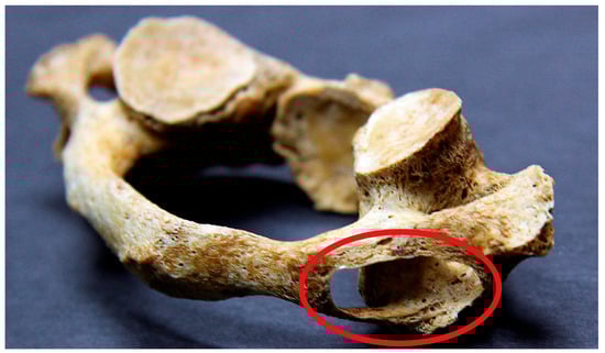
Figure 1.
Dry atlas (inferolateral view) presents with an arcuate foramen (highlighted by the red circle).
AF has many names in the literature, including ponticulus posticus, foramen sagittale, foramen atlantoideum posterior, Kimmerle variant, foramen arcuale, foramen retroarticulare, canalis vertebralis, retroarticular VA ring, and retrocondylar VA ring [5,6,7]. Currently, the term ponticulus posticus is most frequently used [8]. It was first reported by the Dutch anatomist Louis Bolk in 1906, but evidence of this anatomical variation in human skeletons dates back to the 12th century [9,10]. Macalister and Le Double described this structure in detail in the 19th century, using terms such as “posterior bridge” or “retroarticular canal” [11].
Archeological and anthropological evidence indicates that significant demographic changes began in the Early Neolithic period, approximately 9000 years ago. This era marks the gradual disappearance of hunting and gathering practices, particularly across Europe [12]. The transitional phase from the Chalcolithic to the Bronze Age is of particular interest due to increased contact between local communities and incoming groups from other parts of Europe. In Spain—especially in the southern regions—the Chalcolithic period spans from approximately 3300/3200 to 2250 BC. This period is marked by a notable rise in settlements and population driven by advancements in tool use, early urbanization, and the development of agricultural practices [13].
At present, there is little information about the possible differences in AF prevalence between Chalcolithic and present subjects. For our study, we aimed to compare the frequency of the complete AF between a Mediterranean Iberian Peninsula skeletal sample from the Chalcolithic burial of Camino del Molino and a cohort of present-day Mediterranean Iberian Peninsula subjects (Figure 2).
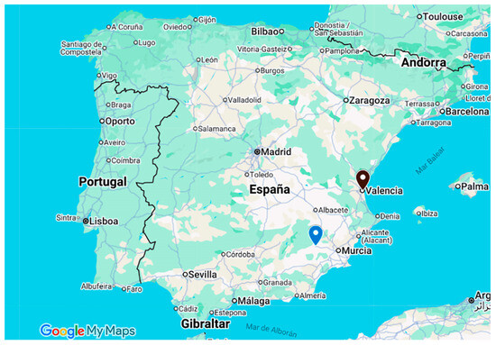
Figure 2.
Map of the Iberian Peninsula (Google, s.f.) that shows the location of the Chalcolithic Camino del Molino site (blue) and Valencia city (black).
2. Materials and Methods
To analyze the presence of the complete AF in an Iberian Peninsula Chalcolithic Mediterranean skeletal sample, we performed an observational transversal study. There were non-incomplete AF forms included in this research. The skeletal sample was located in Eastern Spain and its AF prevalence was compared to a living Iberian Peninsula Spanish Mediterranean sample also located in Eastern Spain. We only focused on the complete AF and no other anatomical variations of the transverse processes, anterior or posterior arches.
The Chalcolithic remains were recovered from the Chalcolithic burial site of Camino del Molino, Caravaca de la Cruz, Murcia, Spain. The Chalcolithic burial site of Camino del Molino was discovered accidentally in 2007. It consists of a circular funerary structure carved into travertine. Geomorphological study of the site suggests it may have been an artificial cave or hypogeum, with an entrance at the top [14]. This site was used for approximately 400 years as a burial site featuring different funerary phases with a minimum number of identified individuals of 1348. It is the largest known cemetery of its kind in European prehistory [15]. It contains remains from both sexes and all ages groups making it a key site for understanding the Chalcolithic population in southeast Iberia [14,15].
The Camino del Molino Chalcolithic remains were housed at the University of Murcia, Spain. The adult age and sex were determined by evaluation of the sternal extremities and rib traits, thyroid cartilage ossification, cranial bone synostosis, and pubic symphysis morphology [16,17,18]. For inclusion in our research, the Chalcolithic atlases needed to be perfectly preserved and from remains between 20 and 40 years of age at death. From an initial sample of 78 (100%), only 34 (43.6%) met the inclusion criteria. Presence or absence of a complete AF was determined by in situ visualization.
The present-day sample was composed of living Mediterranean Iberian Peninsula subjects born in Spain who live in Valencia (eastern coast of Spain near the Camino de Molino Chalcolithic burial site) with an age ranging from 20 to 40 years old. Each needed to have undergone a computed tomography (CT) scan after a neck trauma because CT scans in living subjects can only be performed to assess medical conditions (i.e., to detect neoplasm metastasis, after whiplash/neck traumas, etc.) and are not legally allowed in healthy subjects due to radiation exposure. CT scans were performed at the Ascires CT Unit of Valencia, Spain, in accordance with the World Medical Association’s Declaration of Helsinki and written consent was obtained. The study was approved by the Ethics Committee of the University of Valencia (reference number: H1393415855483 on 08/04/2014. For inclusion in the research, the atlases needed to be free of pathologies (i.e., fractures, osteoporosis, etc.) and the AF needed to be detected on the CT scans. As a result, after application of the above criteria, from an initial sample of 156 (100%) CT scans, only 120 (76.9%) were valid for the study. The z test was used for analyzing differences between samples. A p-value of <0.05 was considered significant.
3. Results
Table 1 details the presence of AF in both the Chalcolithic and present-day samples analyzed.

Table 1.
Arcuate foramen presence in the Chalcolithic and present-day samples.
No significant differences (p = 0.927) were found in AF presence between the Chalcolithic remains (11.6%) and the present-day subjects (11.6%).
The presence of AF in males and females is presented in Table 2 and Table 3. The living sample was composed of 36 males (30.0%) and 84 (70.0%) females (p < 0.001) while the Chalcolithic atlases were from 23 (67.6%) females and 11 (32.4%) males (p = 0.004).
Table 2.
Arcuate foramen presence in Chalcolithic and present-day males.
Table 3.
Arcuate foramen presence in Chalcolithic and present-day females.
No significant differences were observed in AF presence between males and females in both the present-day (p = 0.757) and Chalcolithic samples (p = 0.580).
4. Discussion
We have analyzed the presence of the complete AF in one of the most important Chalcolithic burial sites in Europe [14,15]. While AF is defined in the literature as a congenital structure, other authors state that AF may have a genetic basis. It is also among the claims, however, that it may result from ossification due to aging or external mechanical factors. A different approach suggests that this variation has developed to maintain the VA position during head and neck movements [19]. Studies in the literature show no relationship between the presence of AF and chronological age, which weakens the theory that AF is caused by ossification [20,21]. However, studies claiming the opposite are still available in the literature [22]. Tubbs and colleagues [4] state that the theory of ossification in ligaments is impossible due to the absence of ossification centers in this region. Although the cause of morphological variability of cervical vertebrae has not been fully clarified, it has been determined that there is a genetic predisposition. It has been reported that mutations in Hox genes in particular may play a role in the development of variations [23]. In particular, the family study conducted by Saunders & Popovich [24] showed that there were significant correlations between parents, offspring, and siblings in the occurrence of AF. These data support a genetic basis for the occurrence of AF. It should be noted, however, that anatomical variations result from a combination of genetic and environmental factors.
While many anatomical variants do not cause any symptoms, the AF has been associated with the presence of vertigo, diplopia, shoulder and neck pain, Barré–Liéou syndrome, retroorbital pain, headache, phonation disorders, vertigo, head and neck pain, epilepsy, Meniere syndrome, and acute hearing loss [4,6,9,25,26,27,28,29]. Thus, in theory, the Chalcolithic subjects with AF may have presented with some of the above symptoms during their lives.
In our study, no statistically significant difference was observed when comparing the AF prevalence of the Chalcolithic sample with the present-day cohort. The small sample size, however, may explain the lack of differences. In addition, the sample group did not show a homogeneous sex distribution.
Nevertheless, results obtained in our Iberian Chalcolithic and present-day samples are similar to those presented in the literature [29]. Both samples are from a European country, and a meta-analysis [29] revealed that the AF prevalence in Europe is 11.2%, which is almost identical to that presented in the Chalcolithic sample. In addition, when we examined in terms of sex, we found no significant differences between males and females, as other authors have observed [5,30,31], but we know it is important to note that our samples were unbalanced.
5. Limitations
Our research has certain limitations, such as low sample sizes with significant differences in the number of females and males but with results aligning with those presented in the scientific literature. In addition, we have not analyzed other atlas vertebra anatomical variations, such as congenital arch defects, unfused transverse foramina, lateral ponticles, and retrotransverse foramina. Another limitation of the study is the absence of genetic data but this is not a genetic study and we have no permission to obtain genetic data. We also have no data about AF in living healthy subjects because radiologic studies are not legally allowed to be performed without a medical reason that justifies the subject being irradiated. For our research, the medical reason was a neck trauma. Nevertheless, our study will serve as a catalyst for other anthropologic research groups to analyze the anatomical variations in past populations and compare those results with present-day populations.
6. Conclusions
We have observed that there are no AF prevalence differences between the Iberian Peninsula Mediterranean Chalcolithic sample of Camino del Molino and the present-day Iberian Peninsula Mediterranean sample. In addition, the AF prevalence of the Chalcolithic sample is quite similar to that presented in the literature for current European populations.
Author Contributions
Conceptualization, S.L. and J.S.-G.; methodology, N.T.-C., S.L., M.O.-D., J.J.V.-F., G.G., and J.S.-G.; software, F.M.-E. and E.B.-P.; validation, S.L., J.L.-M., M.H.-U., G.G. and J.E.L.-R.; formal analysis, N.T.-C., S.L., M.O.-D., J.J.V.-F. and J.S.-G.; investigation, S.L., J.L.-M., M.H.-U.; resources, M.O.-D., J.J.V.-F., G.G. and J.E.L.-R.; data curation, S.L., J.L.-M., M.H.-U., E.B.-P. and F.M.-E.; writing—original draft preparation, N.T.-C. and J.S.-G.; writing—review and editing, N.T.-C., S.L., J.L.-M., M.H.-U., F.M.-E., E.B.-P., J.J.V.-F., M.O.-D., G.G., J.E.L.-R. and J.S.-G.; visualization, J.J.V.-F.; su-pervision, J.S.-G.; project administration, J.S.-G. All authors have read and agreed to the published version of the manuscript.
Funding
This research received no external funding.
Institutional Review Board Statement
The study was approved by the Ethics Committee of the University of Valencia (reference number: H1393415855483 on 08/04/2014).
Informed Consent Statement
Informed consent was obtained from all living subjects involved in the study.
Data Availability Statement
The datasets generated and/or analyzed during this study are not publicly available, as CT data and DICOM headers contain patient information. Data can be obtained on reasonable request from the corresponding author.
Conflicts of Interest
The authors declare no conflicts of interest.
Abbreviations
The following abbreviations are used in this manuscript:
| AF | Arcuate foramen |
| VA | Vertebral artery |
| CT | Computed tomography |
References
- Ahn, J.; Duran, M.; Syldort, S.; Rizvi, A.; D’Antoni, A.V.; Johal, J.; Iwanaga, J.; Oskouian, R.J.; Tubbs, R.S. Arcuate Foramen: Anatomy, Embryology, Nomenclature, Pathology, and Surgical Considerations. World Neurosurg. 2018, 118, 197–202. [Google Scholar] [CrossRef]
- Cirpan, S.; Yonguc, G.N.; Edizer, M.; Mas NGi Magden, A.O. Foramen arcuale: A rare morphological variation located in atlas vertebrae. Surg. Radiol. Anat. 2017, 39, 877–884. [Google Scholar] [CrossRef] [PubMed]
- Rios, L.; Mata-Escolano, F.; Blanco-Perez, E.; Llido, S.; Bastir, M.; Sanchis-Gimeno, J.A. Acute headache at-tributed to whiplash in arcuate foramen and non-arcuate foramen subjects. Eur. Spine J. 2017, 26, 1262–1265. [Google Scholar] [CrossRef]
- Tubbs, R.S.; Johnson, P.C.; Shoja, M.M.; Loukas, M.; Oakes, W.J. Foramen arcuale: Anatomical study and review of the literature. J. Neurosurg. Spine 2007, 6, 31–34. [Google Scholar] [CrossRef]
- Elliott, R.E.; Tanweer, O. The Prevalence of the Ponticulus Posticus (Arcuate Foramen) and Its Importance in the Goel-Harms Procedure: Meta-Analysis and Review of the Literature. World Neurosurg. 2014, 82, E335–E343. [Google Scholar] [CrossRef]
- Krishnamurthy, A.; Nayak, S.R.; Khan, S.; Prabhu, L.V.; Ramanathan, L.A.; Kumar, C.G.; Sinha, A.P. Arcuate foramen of atlas: Incidence, phylogenetic and clinical significance. Rom. J. Morphol. Embryol. 2007, 48, 263–266. [Google Scholar]
- Simsek, S.; Yigitkanli, K.; Comert, A.; Acar, H.I.; Seckin, H.; Er, U.; Belen, D.; Tekdemir, I.; Elhan, A. Posterior osseous bridging of C1. J. Clin. Neurosci. 2008, 15, 686–688. [Google Scholar] [CrossRef] [PubMed]
- Huang, D.G.; Hao, D.J.; Fang, X.Y.; Zhang, X.L.; He, B.R.; Liu, T.J. Ponticulus posticus. Spine J. 2015, 15, E17–E19. [Google Scholar] [CrossRef] [PubMed]
- Nedelcu, A.H.; Hutanu, A.; Nedelcu, I.; Vicoleanu, S.P.; Statescu, G.; Gavril, L.; Haliciu, A.M.; Ursaru, M.; Tarniceriu, C.C. The Prevalence and Morphology-Wise Demographic Distribution of Ponticulus Posticus on CT Scans-A Retrospective Observational Study. Medicina 2023, 59, 650. [Google Scholar] [CrossRef]
- Wysocki, J.; Bubrowski, M.; Reymond, J.; Kwiatkowski, J. Anatomical variants of the cervical vertebrae and the first thoracic vertebra in man. Folia Morphol. 2003, 62, 357–363. [Google Scholar]
- Monika, L.; Anupama, M.; Sanjay, P.; Jagdev, S.K. An Anatomical Study of the Morphometric Differences between Complete Arcuate Foramina and Ipsilateral Foramina Transversaria in Human Atlas Vertebrae Could These Be Responsible for Vaso-occlusive Symptoms? Nat. J. Clin. Anat. 2019, 8, 106–111. [Google Scholar]
- Pinhasi, R.; Thomas, M.G.; Hofreiter, M.; Currat, M.; Burger, J. The genetic history of Europeans. Trends Genet. 2012, 28, 496–505. [Google Scholar] [CrossRef]
- Kibaroglu, M.; Schuhmacher, T.X.; Mederos, A.; Falkenstein, F.; Vargas, J.M.; Mertz-Kraus, R.; Hacıosmanoğlu, S. Investigating the Late Chalcolithic pottery production and consumption at Valencina de la Concepciton (Seville, SW-Spain): An archaeometric analysis using petrographic and LA-ICP-MS techniques. J. Archaeol. Sci. Rep. 2024, 53, 104299. [Google Scholar]
- Díaz-Navarro, S.; García-González, R.; Cirotto, N.; Haber-Uriarte, M. New insight into prehistoric craft specialisation. Tooth-tool use in the Chalcolithic burial site of Camino del Molino, Murcia, SE Spain. J. Archaeol. Sci. Rep. 2023, 50, 104066. [Google Scholar] [CrossRef]
- Díaz-Navarro, S.; Haber Uriarte, M.; Tejedor-Rodríguez, C.; Lomba Maurandi, J. Emphasising the community: Demographic composition of an exceptional tomb—The Chalcolithic burial site of Camino del Molino, Caravaca de la Cruz, Murcia. Archaeol. Anthropol. Sci. 2023, 15, 140. [Google Scholar]
- Loth, S.R.; Iscan, M.Y. Morphological assessment of age in the adult: The thoracic region. In Age Markers in the Human Skeleton; Iscan, M.Y., Ed.; Charles C Thomas: Springfield, IL, USA, 1989; pp. 105–135. [Google Scholar]
- Reverte-Coma, J.M. Antropología Forense [Forensic Anthropology]; Ministerio de Justicia, Secretaria General Técnica, Centro de Publicaciones: Madrid, Spain, 1991. [Google Scholar]
- Burns, K.R. Manual de Antropología Forense [Manual of Forensic Anthropology]; Edicions Bellaterra SL: Barcelona, Spain, 2008. [Google Scholar]
- Macri, M.; Perrella, G.; Varvara, G.; Murmura, G.; Traini, T.; Rendina, F.; Festa, F. Assessments of Prevalence of Ponticulus Posticus, Atlas Posterior Arch Deficiency, Sella Turcica Bridging, Maxillary Canine Impaction, and Associations Among Them in 500 CBCTs of Italian Orthodontic Patients. Front. Dent. Med. 2021, 2, 708169. [Google Scholar] [CrossRef]
- Geist, J.; Geist, S.M.R.Y.; Lin, L.M. A cone beam CT investigation of ponticulus posticus and lateralis in children and adolescents. Dentomaxillofacial Radiol. 2014, 43, 20130451. [Google Scholar] [CrossRef]
- Schilling, J.; Schilling, A.; Galdames, I.S. Ponticulus posticus on the Posterior Arch of Atlas, Prevalence Analysis in Asymptomatic Patients. Int. J. Morphol. 2010, 28, 317–322. [Google Scholar] [CrossRef]
- Erdem, S.; Gündüz, K.; Kasap, P. Evaluation of the Ponticulus Posticus with Cone-beam Computed To-mography in a Turkish Population. J. Anat. Soc. Ind. 2023, 72, 140–144. [Google Scholar]
- Karapetian, M.K. Discrete morphological variants of human cervical vertebrae: Exploring pattern of distribution and biological significance. HOMO 2017, 68, 176–198. [Google Scholar] [CrossRef]
- Saunders, S.R.; Popovich, F. A family study of two skeletal variants: Atlas bridging and clinoid bridging. Am. J. Phys. Anthropol. 1978, 49, 193–203. [Google Scholar] [CrossRef]
- Cakmak, O.; Gurdal, E.; Ekinci, G.; Yildiz, E.; Cavdar, S. Arcuate foramen and its clinical significance. Saudi Med. J. 2005, 26, 1409–1413. [Google Scholar]
- Chitroda, P.K.; Katti, G.; Baba, I.A.; Najmudin, M.; Ghali, S.R.; Kalmath, B.; G, V. Ponticulus Posticus on the Posterior Arch of Atlas, Prevalence Analysis in Symptomatic and Asymptomatic Patients of Gulbarga Population. J. Clin. Diagn. Res. 2013, 7, 3044–3047. [Google Scholar] [CrossRef]
- Koutsouraki, E.; Avdelidi, E.; Michmizos, D.; Kapsal, S.E.; Costa, V.; Baloyannis, S. Kimmerle’s Anomaly as a Possible Causative Factor of Chronic Tension-Type Headaches and Neurosensory Hearing Loss: Case Re-port and Literature Review. Int. J. Neurosci. 2010, 120, 236–239. [Google Scholar] [CrossRef]
- Pekala, P.A.; Henry, B.M.; Pekala, J.R.; Hsieh, W.C.; Vikse, J.; Sanna, B.; Walocha, J.A.; Tubbs, R.S.; Tomaszewski, K.A. Prevalence of foramen arcuale and its clinical significance: A meta-analysis of 55,985 subjects. J. Neurosurg. Spine 2017, 27, 276–290. [Google Scholar] [CrossRef]
- Pekala, P.A.; Henry, B.M.; Phan, K.; Pekala, J.R.; Taterra, D.; Walocha, J.A.; Tubbs, R.S.; Tomaszewski, K.A. Presence of a foramen arcuale as a possible cause for headaches and migraine: Systematic review and meta-analysis. J. Clin. Neurosci. 2018, 54, 113–118. [Google Scholar] [CrossRef]
- Natsis, K.; Piperaki, E.T.; Fratzoglou, M.; Lazaridis, N.; Tsitsopoulos, P.P.; Samolis, A.; Kostares, M.; Piagkou, M. Atlas posterior arch and vertebral artery’s groove variants: A classification, morphometric study, clinical and surgical implications. Surg. Radiol. Anat. 2019, 41, 985–1001. [Google Scholar] [CrossRef]
- Paschopoulos, I.; Piagkou, M.; Triantafyllou, G.; Papadopoulos-Manolarakis, P.; Duparc, F.; Demetriou, F.; Tsakotos, G.; Tudose, R.C.; Rusu, M.C.; Toader, O.D. The Potential Morphological Stenosis Pattern of the Arcuate Foramen. Diagnostics 2025, 15, 1203. [Google Scholar] [CrossRef]


Disclaimer/Publisher’s Note: The statements, opinions and data contained in all publications are solely those of the individual author(s) and contributor(s) and not of MDPI and/or the editor(s). MDPI and/or the editor(s) disclaim responsibility for any injury to people or property resulting from any ideas, methods, instructions or products referred to in the content. |
© 2025 by the authors. Licensee MDPI, Basel, Switzerland. This article is an open access article distributed under the terms and conditions of the Creative Commons Attribution (CC BY) license (https://creativecommons.org/licenses/by/4.0/).