Article

An Ultradolichocephaly in a Knight of the Order of Calatrava from the Castle of Zorita de los Canes (Guadalajara, Spain) Dated Between the 13th and 15th Centuries

1
Department of Basic Medical Sciences, Faculty of Medicine, and Health Sciences, University Rovira i Virgili, Carrer Sant Llorenç, 21, 43201 Reus, Spain
2
ArchaeoSpain, Juan Gavala 2, Carrascosa del Campo, 16555 Cuenca, Spain
3
Independent Researcher (Medieval History), Universitat de Barcelona, Montalegre 6, 08001 Barcelona, Spain
4
Departament d’Història i Arqueologia, Universitat de Barcelona, Montalegre 6, 08001 Barcelona, Spain
*
Author to whom correspondence should be addressed.
Heritage 2025, 8(10), 414; https://doi.org/10.3390/heritage8100414
Submission received: 19 August 2025 / Revised: 20 September 2025 / Accepted: 1 October 2025 / Published: 3 October 2025

Abstract

This study presents a paleopathological analysis of individual T4.2 from the archaeological site of the castle of Zorita de los Canes (Guadalajara, Spain). The individual exhibits ultradolichocephaly and was dated between the 13th and 15th centuries. Based on the context, the individual could have been a member of the military–religious Order of Calatrava. Standard bioanthropological and paleopathological methods were applied. The results indicate a male aged between 45 and 49 years with a maximum cranial length and width of 230 mm and 122 mm, respectively. The cranial index (53%) allows him to be classified as ultradolichocephalic. The remains present craniosynostosis at the sagittal, squamosal and sphenofrontal sutures, together with underdevelopment of the skull base width and a dolichognathic mandible with the possible presence of type III prognathism. The left hemimandible and hemimaxilla display a large amount of tartar covering the occlusal, labial and lingual areas of the teeth completely. The right hemimandible and hemimaxilla exhibit almost no tartar, and some teeth were lost in vivo. In addition, two stab wounds—to the external occipital protuberance and to the left temple—as well as a contused injury in the left tibia, can be observed without evidence of bone remodeling. Differential diagnosis indicates a case consistent with Crouzon syndrome. This individual is of particular significance because he was a possibly knight of the Order of Calatrava who presents a syndromic craniosynostosis consistent with Crouzon syndrome and exhibits lesions, which may suggest that he could have died in battle.

1. Introduction

The archaeological site of the castle of Zorita de los Canes (Guadalajara, Spain) dates from the 13th to 15th centuries and was occupied by the Knights of Calatrava [1]. This was a military–religious order associated with the Cistercian Order [1]. The Order of Calatrava was founded in the Kingdom of Castile in 1158 to replace the order of the Temple and to respond to the growing threat of Almohad attacks [2,3]. Its primary mission was to defend the borders of Castile against invading Muslim forces in that period [1]. Over time, their protective efforts extended to the frontier regions between the kingdoms of Castile and Aragon. One of the strategic fortifications under their control was the castle of Zorita de los Canes (Figure 1), located on the left bank of the Tagus River and in the southernmost region of Castile on the frontier with al-Andalus [4]. The Order itself comprised both religious and lay members who shouldered military responsibilities [2]. Starting in the 13th century CE, this armed force experienced a process of aristocratization, leading to increasing secularization [2]. At the highest levels of the Order, power was concentrated in the hands of the realm’s most prominent noble families [2]. The nobility contributed human resources, established familial agreements, and made substantial property donations. As a result, the Order of Calatrava evolved into a powerful institution, endowed with substantial material and financial resources, and gradually gained political influence within the kingdom over the centuries [2,3]. Despite the vow of poverty taken by its members, this commitment was often broken. Members saw their affiliation as both a religious journey toward salvation and a means of economic gain and social advancement [4]. This was the consequence of the Order’s practice of primarily recruiting its members from the lower nobility or urban oligarchies, groups that constituted the majority of its knights [2]. The military forces of the Order encompassed a spectrum ranging from Freire Knights (heavy cavalry with their own horses and entourage), Freire Sergeants (fighting on horseback but with more simple weapons and directed by the highest lay and ecclesiastical hierarchies), Associated Fighters (temporary volunteers, primarily knights), Mercenaries, and Vassals [4]. Despite substantial historical knowledge about the social origins and composition of this order, very few studies have examined the skeletal remains of its members from this period [4]. Between 2014 and 2019, archaeological excavations conducted in the Christian cemetery of the Counts’ Courtyard of Zorita de los Canes castle, under the direction of Catalina Urquijo and Dionisio Urbina (ArchaeoSpain) with the collaboration of Davidson Day School of North Carolina uncovered many human skeletal remains [1]. The few osteological studies on these individuals [4,5] revealed that most of them were primarily adult men of varying ages. Significantly, signs of trauma (stab wounds and contused injuries) were identified on different areas of the skeleton (cranium, ilium, rib cage, femur, among others) of these individuals, suggesting violent incidents, probably battle wounds [4,5], in keeping with its primary use as a cemetery for the Order of Calatrava. These individuals were archaeologically dated between the 13th and 15th centuries CE [1].
The aim of the present study is to describe and paleopathologically analyze one of these individuals exhumed from the Counts’ Courtyard cemetery of Zorita de los Canes castle (individual T4.2). The particularity of this individual is a malformation of the skull, which can be defined as ultradolichocephalic. This is a morphological malformation caused by the premature closure of several cranial sutures.

2. Materials and Methods

The material analyzed consists of the skeletal remains corresponding to individual T4.2 exhumed in the Counts’ Courtyard of the castle of Zorita de los Canes (Corral de los Condes del Castillo de Zorita de los Canes) site in Zorita de los Canes (Guadalajara, Spain), during the excavation seasons between 2014 and 2019. This individual had originally been interred in a wooden coffin, which had already decayed by the time of excavation; only a few wooden fragments were recovered. The skeletal material arrived individually packed in a box at the Laboratory of the Human Anatomy Unit of the Department of Basic Medical Sciences of the Faculty of Medicine and Health Sciences of the Universitat Rovira i Virgili in Reus (Tarragona, Spain).
In the laboratory, the study began with a dry cleaning of the remains, using fine brushes and wooden awls to avoid damaging the bones. In cases where it was difficult to remove the excess sediment deposited on the bones, they were cleaned under water and then dried at room temperature in the shade. Once cleaned, they were reconstructed with adhesive tape which can be removed manually.
Bioanthropological analysis consisted of the calculation of the preservation index, sex and age estimation, anthropometric study, and paleopathological analysis of the individual. The preservation status of the individual was calculated according to the preservation index of Alesan [6], which shows the percentage of preserved elements [IP3 = (Number of present bones/22) × 100]. Twenty-two bones are considered: long bones plus both girdles and the cranium and mandible. The presence of a part of a bone is considered as entire.
Regarding sex and age estimation, these estimations have to depend on unaffected postcranial traits, because of the limitations imposed by craniosynostosis on cranial indicators. Therefore, in order to estimate sex, the qualitative characteristics of the hip bone [7,8,9] were considered, and discriminant functions based on the long bones and pelvis [7,8,9,10] were applied.
The age was estimated on the bases of the pubic symphysis as proposed by Todd, McKern and Stewart, and Gilbert and McKern [11], the auricular surface of the ilium [12], the acetabulum [13], and the sternal end of the fourth rib [14]. These methods were selected due to its popularity in forensic and bioarchaeological Spanish contexts, which is highly recommended in Spanish anthropological manuals [15].
Cranial and postcranial anthropometric analysis was based on the standard anthropological measurements of Martin and Saller [16] and Olivier [17]. Skeletal indices were calculated to describe bone shape and size. Given the Mediterranean origin of the remains of the Calatrava Knights, height was calculated using Pearson’s formulas [18], as these are based on the French population of the late 19th century.
The presence or absence of physical activity markers on bone was also assessed, following the guidelines of Fernández-Suarez [19]. These markers provide information on the individual’s potential physical activities [20,21].
The evaluation of oral paleopathology on the dental elements of the maxilla and mandible was conducted through macroscopic analysis, following the criteria outlined by Hillson [22] and Cobo [23]. The assessment focused on the presence of caries, bone loss, and fistulas. Additionally, markers of environmental stress—such as enamel hypoplasia, dental calculus, and tooth wear—were systematically examined.
To assess skeletal pathologies or injuries, a macroscopic analysis of the remains was performed following the methodology of Ortner [24]. In addition, a radiological study of the teeth was also conducted at the Gnation Clinic in Barcelona—intraoral X-ray equipment X-Mind AC/DC by Acteon (Satelec) and CS Imaging (Carestream Dental).

3. Results

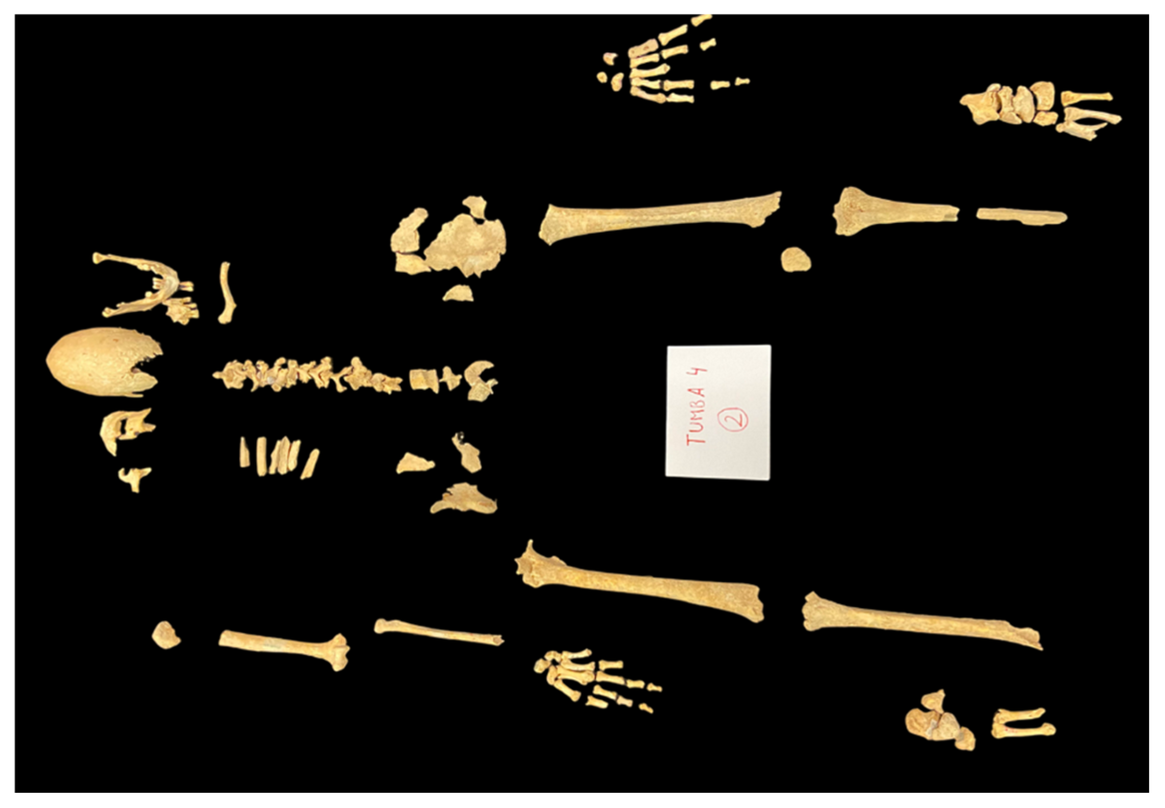
Results indicate that the individual was a male, aged between 45 and 49 years, whose height cannot be estimated due to the long bone epiphyses were not fully preserved. The index of preservation indicates an IP3 = 0.545, with the neurocranium, maxilla, mandible, clavicle, hip bones, and diaphysis of long bones (right humerus and radius, and both femurs and tibiae) preserved (Figure 2). In addition, the specimen included vertebrae, ribs, metacarpal and metatarsal bones, and phalanges (Figure 2).
Table A1 and Table A2 in the Appendix A show the data from the postcranial (Table A1) and cranial (Table A2) skeleton. This individual exhibited eurybrachia and eurycnemia in the humerus. This means that the humerus had a rounded shaft (shaft index greater than 76.5%) (Table A1). However, the insertion of the deltoid muscle was very marked, suggesting intense physical activity of this muscle. Furthermore, the insertion of the conoid ligament was very marked in the clavicle. This ligament limits the protraction and retraction movements of the shoulder. The presence of this marker is common in people who carry bags on their shoulders [19]. It was not possible to determine whether the individual was right-handed or left-handed because only the right upper limb was recovered (Figure 2).
The tibiae were eurycnemic, indicating little physical activity with the leg. However, the femur exhibited a prominent linea aspera, suggesting significant muscular activity in the thigh, particularly involving the adductors and the vastus lateralis and vastus medialis heads of the quadriceps femoris. This last marker is commonly observed in individuals who ride horses [19].
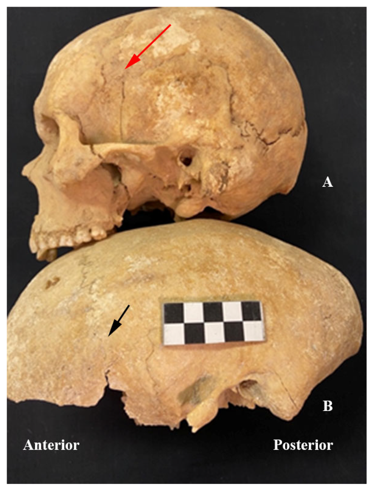
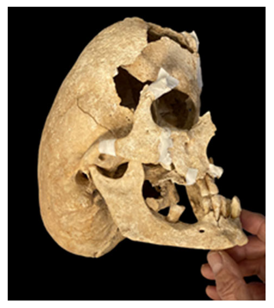
Regarding the skull (Table A2), the results showed an extremely elongated skull with a maximum length of 230 mm and a maximum width of 122 mm (Figure 3), resulting in a cranial index of 53%, while the normal range in the Spanish population is 75.37 ± 2.99% [25]. These values allowed this skull to be classified as ultradolichocephalic (cranial index < 65%) [17]. Craniosynostosis was observed in the sagittal, squamosal, and sphenofrontal sutures, visible both endocranially and ectocranially, along with a reduced width of the skull base (Figure 3 and Figure 4). The perimeter of the foramen magnum was larger than expected, probably due to the malformation of the skull base (Figure 4). The frontal bone was slightly prominent in relation to the facial mass (Figure 5). Furthermore, when reconstructing the splanchnocranium and placing it in anatomical position with the neurocranium, it was observed that the latter is markedly oriented posteriorly and inferiorly (Figure 6).
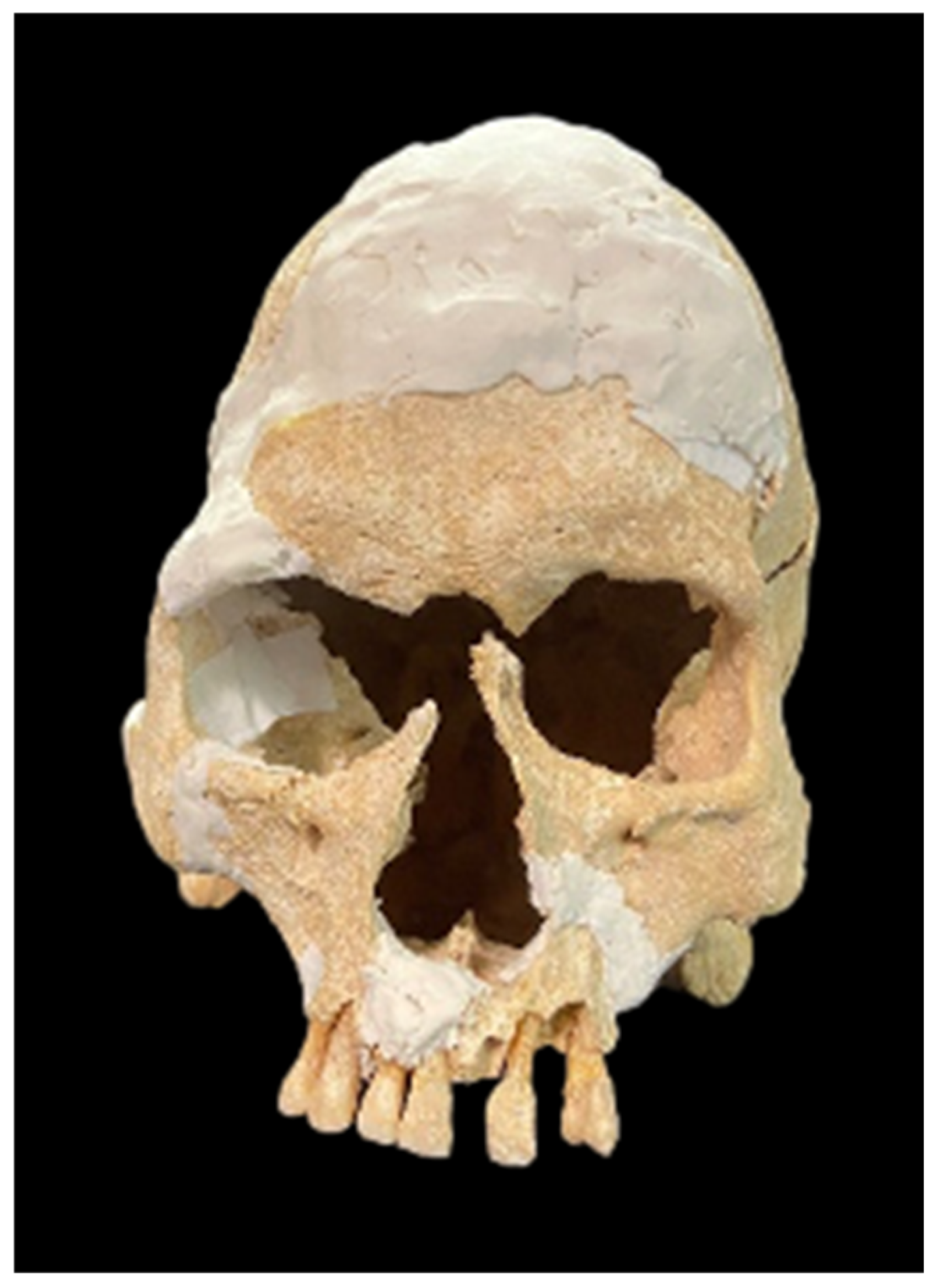
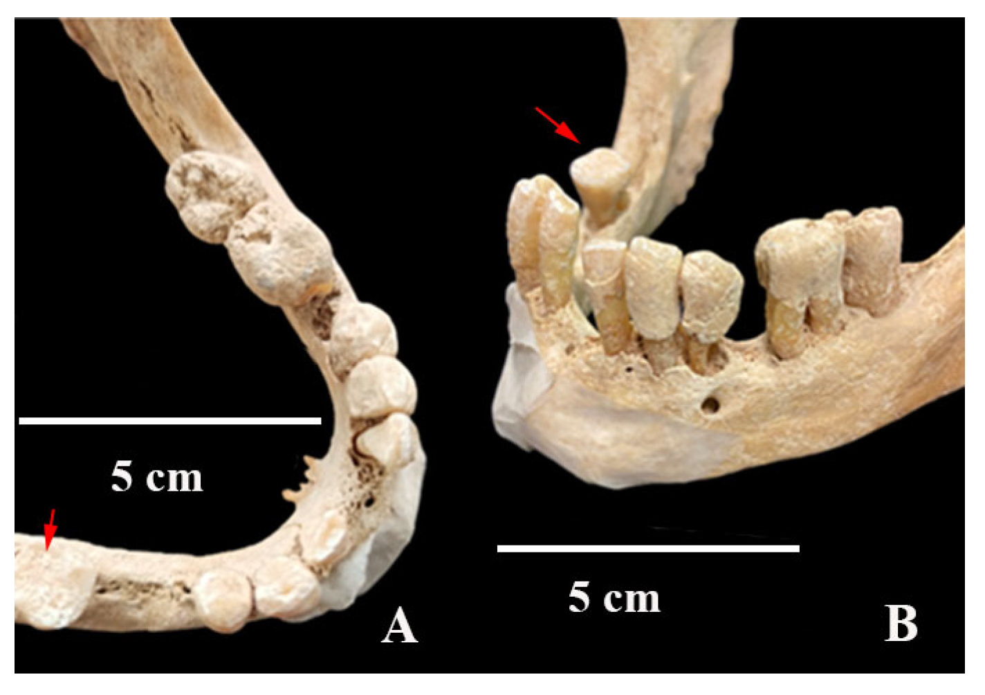
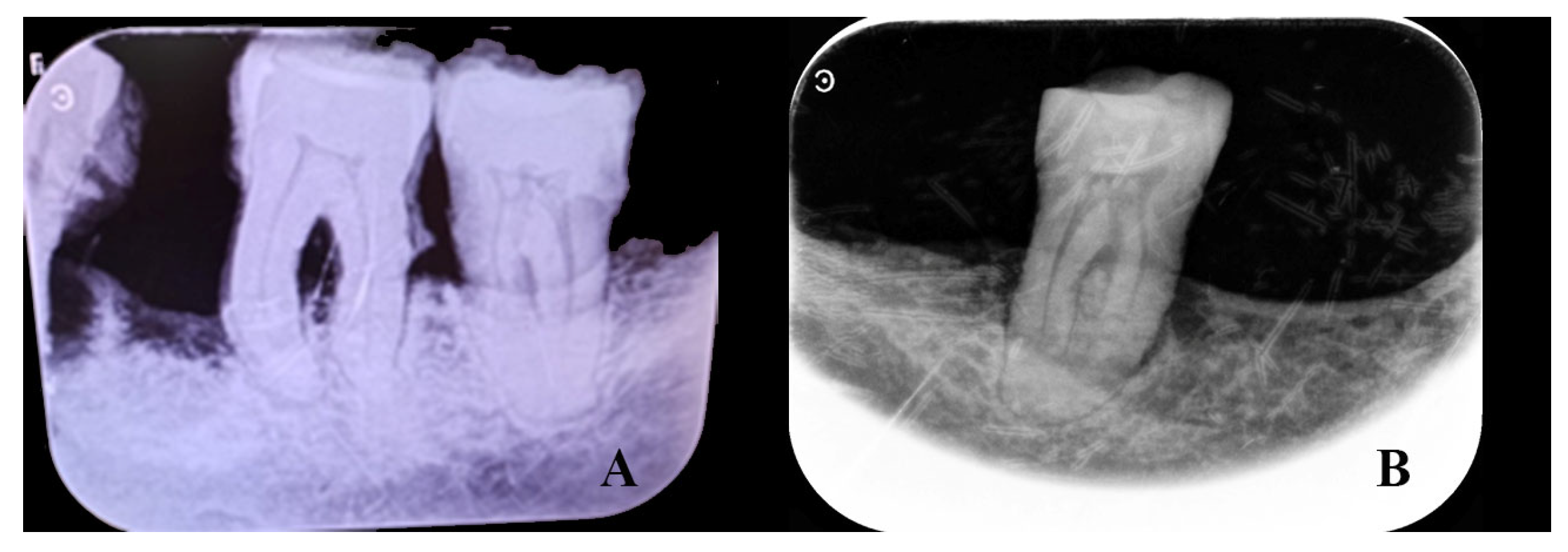
Examination of the splanchnocranium (Table A2), along with the skull, revealed an outward inclination of the incisors (Figure 7) and normal orbital dimensions, with a height of 31 mm and a breadth of 32 mm (Figure 7). According to Testut and Latarjet [26], the normal orbital range is 26–48 mm in height and 32–48 mm in breadth, with the lower end of this range being more typical among Spaniards. The mandible was very narrow (dolichognathous), with a mandibular index of 98.3 (Table A2). Absolute values indicate a morphological alteration with an increase in its anteroposterior measurement and a decrease in the laterolateral diameter, as is the case with the cranial measurements. These measurements suggest the presence of type III prognathism. The left hemimaxilla contained the second incisor, canine, and first premolar (the remaining dental elements were lost postmortem). The left hemimandible consisted of the second incisor, canine, first premolar, and first and second molars. The first incisor and second premolar had been lost during life (Figure 8). Examination of the dental elements on the left side highlights the large amount of tartar, which completely covers the lingual, labial, and occlusal surfaces of the preserved elements (Figure 8) and the presence of very little wear. On the right side, the hemimaxilla had retained the two incisors, the canine, and the first premolar (Figure 8). The right hemimandible had preserved the first and second incisors, the canine, and the second molar. The remaining elements of the right hemimandible were lost during life (Figure 8). In contrast to the left side, the right side showed tooth wear and very little tartar (Figure 8 and Figure 9). Significant resorption of the mandibular body was observed (Figure 9). Radiographic images confirm alveolar bone resorption in the mandible, with no evidence of periapical changes (Figure 10). In general, on the left side, the presence of minimal tooth wear and an extreme amount of tartar is notable. This could indicate the left dentition had not been used, probably because of unilateral mastication due to occlusal dysfunction; however, periodontal disease and hygiene differences cannot be excluded.
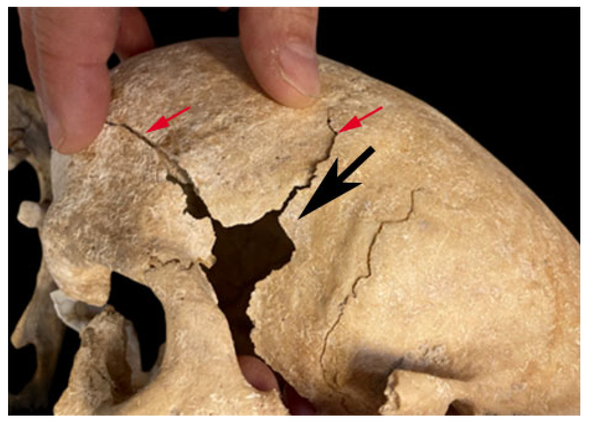
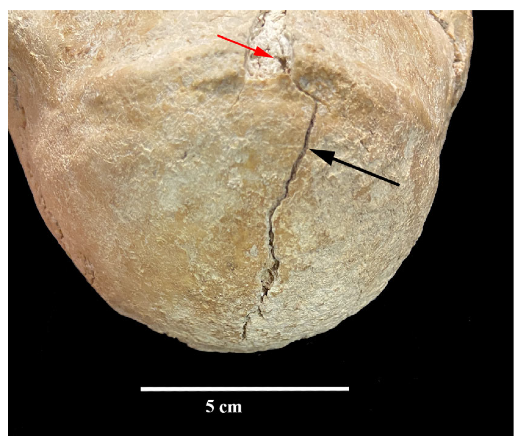
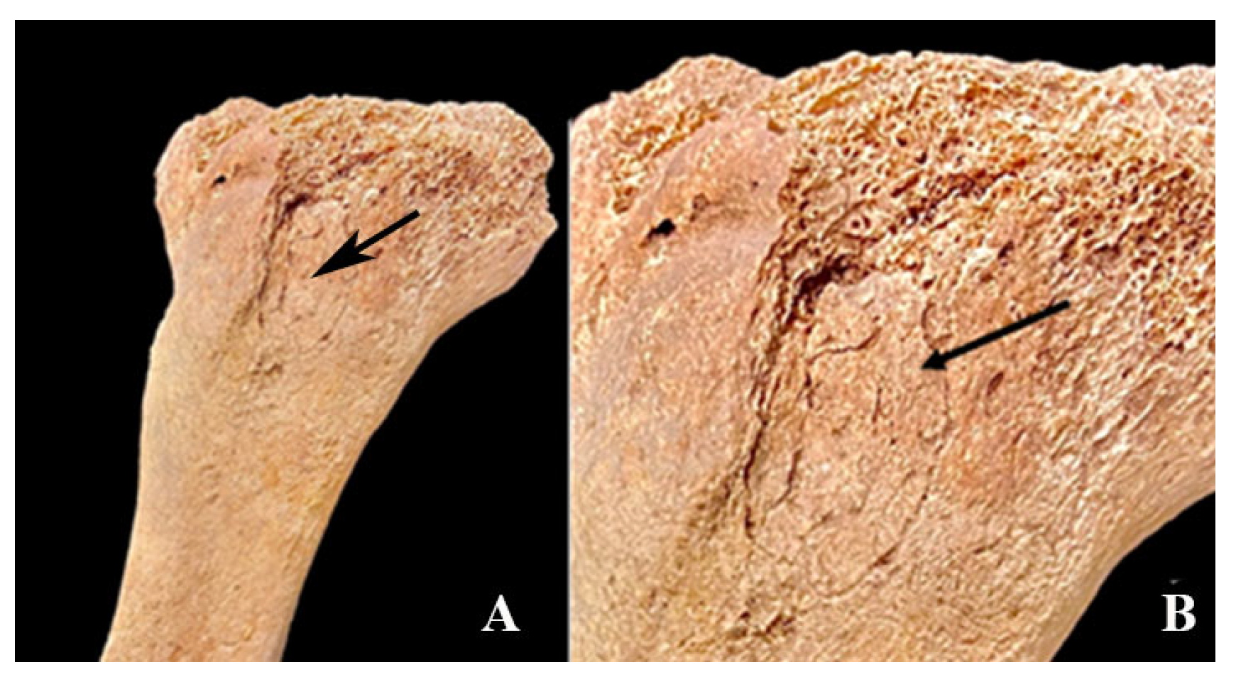
Detailed examination of skeleton of T4.2 revealed two stab wounds: one to the left temple (Figure 5) and the other to the external occipital protuberance (Figure 11). The first caused penetration of the weapon into the cranium and radiating bone fractures (Figure 5). The margins of this lesion were sharp and well-defined, with smooth edges consistent with penetration by a pointed implement into fresh bone of this cranial region. The second, although it did not penetrate into the cranium, caused a radiating fracture along the occipital squama (Figure 11). The external margin of the lesion was sharp, while the external surface exhibited slight beveling along the point of impact. In addition, this individual also exhibited a blunt force injury, resulting in cortical collapse and concentric fractures around the impact site, in the left anterolateral region of the left tibia, just below the proximal epiphysis (Figure 12). These features, together with the radiating fracture patterns, confirm that the injuries were produced when the bone was fresh (perimortem) [8,23,27]. No bone remodeling could be observed in either injury.

4. Discussion

Individual T4.2, analyzed in this study, is a male aged between 45 and 49 years. He exhibited unhealed penetrating stab wounds to the temple and occipital regions, as well as a blunt force injury to the tibia. All of these lesions are consistent with perimortem trauma. While distinguishing trauma from minor anatomical variants can sometimes be challenging [28], the characteristics of the lesions analyzed in this case clearly indicate a traumatic origin. In addition, he is part of a group of male individuals buried in the Counts’ Courtyard cemetery of the Zorita de los Canes castle, which primarily served as a burial site for members of the Order of Calatrava. All the individuals in this group exhibited multiple stab wounds on various parts of the body—including the cranium, ilium, ribs, and femur—indicating episodes of violence, probably battle wounds [4,5]. These findings are consistent with the interpretation that these individuals were warriors affiliated with the Order of Calatrava, and that they possibly died as a result of armed conflict.
These described lesions are independent of his cranial malformation. Therefore, the differential diagnosis should focus on the craniosynostosis, involving the sagittal, squamosal, and sphenofrontal sutures. This condition results in a slightly prominent frontal bone relative to the facial skeleton, a neurocranium that is markedly oriented posteriorly and inferiorly, and an outward inclination of the incisors. Overall, the dental elements on the left side exhibit significantly less tooth wear and a substantial accumulation of calculus compared to those on the right. This asymmetry likely reflects unilateral mastication, possibly resulting from occlusal dysfunction. However, the potential influence of periodontal disease or differences in oral hygiene practices cannot be discarded.

4.1. Differential Diagnosis

Craniosynostosis is defined as the premature fusion of one or more of the cranial sutures. Its prevalence has been estimated at 1 case in 2500 worldwide [29]. It can be of secondary or primary etiology. Secondary craniosynostosis is caused by a non-genetic origin, for example, vitamin deficiencies, exposure to toxins, drugs, or mechanical causes. Primary craniosynostosis can be caused by an isolated genetic mutation or by a set of phenotypic alterations as a result of genetic mutations associated with developmental abnormalities, which also usually affect the extremities, heart, and central nervous system [30]. The latter are known as syndromic craniosynostoses. They account for 30% of all primary synostoses, and most are due to autosomal dominant mutations [29]. However, cases of autosomal recessive mutations, incomplete penetrance mutations, or variable gene expression, in addition to mosaicism, can also be observed. Therefore, the differential diagnosis must first assess the etiology.

4.1.1. Secondary or Primary Craniosynostosis?

Environmental factors etiologically associated with secondary craniosynostosis include rickets (caused by vitamin D deficiency, vitamin D resistance, chronic renal failure, or hypophosphatemia), hyperparathyroidism, the use of teratogens, or mechanical causes. Rickets is a childhood disease (called osteomalacia in adults), which results in the inability to incorporate calcium into the bones. In children, this causes the long bones to bend, growth is delayed, and cribra orbitalia develops [24,31]. In adults, multiple stress fractures (small, linear fractures, within the external bone cortex) can be observed with different stage of healing [24,32]. Healed evidence of osteomalacia may comprise severe deformities in the axial skeleton caused by bone softening [24,32,33]. These characteristics are not consistent with this individual’s morphology, and thus this pathology can be ruled out as a cause.
Thyroid deficiency during early development produces dysmorphic dwarfism due to the body’s inability to absorb Ca and therefore convert cartilage into bone. Long bones normally grow in width, but not in length [34]. Growth of the cranial cap is not inhibited, while that of the base is reduced. The overall effect is a normal-sized skull (albeit with a narrow base) on a very short postcranial skeleton. These findings differ from the morphology of our subject, and this pathology can also be ruled out as a cause.
Overactive parathyroid glands produce hyperactive osteoclasts that rapidly destroy bone, while underactive parathyroid glands produce short stature and structurally deficient bone [35]. Both characteristics are incompatible with our individual and can therefore be dismissed.
Teratogens associated with craniosynostosis include phenytoin, tretinoin, valproate, aminopterin, methotrexate, fluconazole, and cyclophosphamide [36]. These chemical causal factors could also be excluded since individual T4.2 was dated to the 13th–15th centuries (1201–1401), when these drugs were not in use.
Regarding mechanically induced morphological changes in the skull, the neurocranium would be left with marks from bandages or boards which caused the morphological change [37]. In the individual analyzed no-marks or alterations suggestive of mechanical changes were observed, allowing us to exclude environmental factors and, consequently, the possibility of secondary craniosynostosis.

4.1.2. What Type of Primary Craniosynostosis: Syndromic or Non-Syndromic?

Primary craniosynostoses are due to a developmental error during embryogenesis and are divided into non-syndromic and syndromic. Non-syndromic craniosynostoses involve the premature fusion of one cranial suture (70–85% are simple) or more (20–25% are multisutural), without affecting the rest of the skeleton [30]. Syndromic craniosynostoses involve more than one suture, leading to variable malformations in the limbs, heart, and central nervous system. Eighty-five percent of primary craniosynostoses are non-syndromic (isolated in normal individuals), while 15% are syndromic (belong to polyformative syndromes) [30].
Autosomal dominant mutations in a single gene are detected in one-third of primary craniosynostosis cases [38,39], and half of these cases are the result of recent mutations [40]. Although the most common forms of syndromic craniosynostosis are inherited as autosomal dominant inheritance, recessive inheritance, incomplete penetrance, and variable expressivity can also be observed in patients [36,41]. On the other hand, mosaicism can also be the cause of craniosynostosis [30]. The most common types of craniosynostosis are (Table 1) Crouzon syndrome, Treacher Collins syndrome, Apert syndrome, Pfeiffer syndrome, Saethre-Chotzen syndrome, Craniofrontonasal syndrome, Noonan syndrome, Muenke syndrome, and Neurofibromatosis. They can be differentiated by the presence or absence of malformations in the hand, foot, lip, and palate, as well as nasal malformations (Table 1). Brain growth restriction can lead to severe complications such as seizures, brain damage, vision loss, cognitive and mental impairment, and respiratory problems in infants. These complications are primarily associated with syndromic cases [30]. Therefore, the differential diagnosis of primary craniosynostosis should focus on assessing the possible existence of syndromic craniosynostosis, since cranial morphology (skull vault and base) and mouth opening are affected.
Crouzon syndrome is a congenital craniofacial synostosis characterized by premature closure of the sagittal, coronal, and lambdoid sutures, with the sagittal suture being most common (40–60% of cases), followed by the coronal suture (25%), and the metopic suture in less than 10%. In this genetic disorder, the coronal suture is affected most severely [42], resulting in a brachycephalic individual. Hypertelorism, maxillary hypoplasia with varying degrees of choanal atrophy, inversion of the dental articulation due to a receding maxilla, a sunken palate, and hearing loss due to atresia of the auditory meatus and cervical vertebral fusion are also observed [43,44,45]. However, a high phenotypic diversity is observed [44] and the individuals affected for this condition have a normal cognitive level [44].
In this study, the greatest degree of involvement falls, in order, on the sagittal, squamous, and sphenofrontal sutures, as well as the pterion to a lesser extent, resulting in an ultradolichocephaly, with involvement of the temporomandibular joint. Furthermore, the morphology of the maxilla and mandible differs from a micrognathic phenotype due to their marked length. The EAC was formed, with patency and absence of any stenosis or obliteration (Figure 3 and Figure 4). There is no clear evidence of significant cognitive impairment in this individual. His contextual association with the Order of Calatrava, along with the presence of multiple injuries consistent with battle-related trauma—similar to those observed in previous osteological studies [4,5] of other male individuals buried in the Counts’ Courtyard cemetery—suggests that he was cognitively functional, possibly within the borderline range of intellectual ability.
Due to the phenotypic variability of this condition, the most frequent involvement of the sagittal suture, the cognitive functional possibility of the individual examined, and the absence of involvement in the extremities, Crouzon syndrome could be reasonably compatible with the pathological description of the individual analyzed.
b.
Treacher Collins Syndrome (Table 1)
Treacher Collins syndrome or mandibulofacial dysostosis is an autosomal dominant disorder characterized by an alteration in the development of the structures derived from the first and second branchial arches due to a delay or failure in the differentiation of the maxillary mesoderm. The main anatomical alteration is a defect in the stapedial artery during embryogenesis, which causes morphological alterations in the incus, stapes, and vessels of the first branchial arch that irrigate the maxilla. The diagnosis of this syndrome is based on alterations in the facial bones, digestive and respiratory systems, visual and hearing problems, language and dental disorders, as well as malformations of the bones of the hands, among others [46]. Regarding the facial bones, the underdevelopment of the zygomatic bones stands out, causing the so-called sunken facial bone characteristic of this syndrome. Further characteristic is the agenesis of teeth, and the absence or underdevelopment of the first finger of the hand.
In the individual analyzed (T4.2), neither zygomatic arches displayed any alterations (Figure 7), nor was dental agenesis observed, nor was the absence or poor development of the first finger of the hand (Figure 2). There were also no signs of alterations in the palate or palatine bones, and the characteristic depressions of this syndrome (facial sunken bone) did not exist. On the other hand, the metacarpals of individual T4.2 did not show morphological or numerical alterations (Figure 2). Accordingly, the phenotype exhibited by individual T4.2 does not align with the diagnostic features of Treacher Collins Syndrome, thereby supporting the exclusion of this condition.
c.
Apert Syndrome (Table 1)
Apert syndrome is an autosomal dominant disorder that has also been observed in individuals with no family history of the condition. It has a low incidence of 1 in every 160,000 births, with no significant sexual differences [47]. This condition is characterized by malformations, particularly in the skull, midface, hands, and feet. Hand and foot abnormalities are usually syndactyly, which may affect only soft tissue or extend to bone [47].
The diagnostic features of Apert syndrome are acrocephaly (accelerated cranial growth of the head, giving it a narrow, elongated, or conical appearance) and brachytrurricephaly (the skull grows upward, resembling a tower in its final shape), causing intellectual disability and visual disturbances due to shortening of the optic nerve [48]. Furthermore, facial hypoplasia of the middle third and orbital rim is present, along with hypertelorism and proptosis of the eyeballs. At the jaw level, Apert syndrome commonly presents with an ogival upper jaw, dental crowding, and a Class III mandible [49]. Apert syndrome may also present with fusion of the index, middle, and ring fingers. Shortening of the upper limbs has also been described, as well as aplasia or ankylosis, generally of the shoulders, elbows, and hips [48,49]. In general, these individuals also present cognitive and hearing deficits [43,50]. Furthermore, individuals affected by Apert syndrome also present many important soft tissue alterations such as cardiovascular defects, pulmonary atresia, permanent arterial duct, pyloric stenosis or polycystic kidneys [48].
The morphological alterations of the individual analyzed do not coincide with the diagnostic features of Apert syndrome, because of this, Apert syndrome can be excluded
d.
Pfeiffer Syndrome (Table 1)
Pfeiffer syndrome was first described in 1964, when a series of patients with the same clinical characteristics were found. These patients presented with broad thumbs, large phalanges, fusion of the proximal and distal phalanges of the thumb, polydactyly, craniosynostosis, symmetrical syndactyly, facial hypoplasia, proptosis, varying degrees of mental retardation, exophthalmos, and other ocular abnormalities. This condition is divided into three subtypes based on the severity of their phenotype. First, there is classic Pfeiffer syndrome or type I, which shows a subtle phenotype with adequate neurological development and acceptable social functioning. Second, there is type II, which manifests as severe craniosynostosis (trilobate skull), mental retardation, and significant physical impairment. Occasionally, the cloverleaf skull is accompanied by hypertelorism [43]. Finally, type III is very similar to type II, but does not present trilobate craniosynostosis. Of the three types of Pfeiffer syndrome, types II and III have the worst prognosis.
Therefore, all three subtypes of Pfeiffer syndrome can be excluded as a potential diagnosis for individual T4.2, as no phalangeal abnormalities, facial hypoplasia, or trilobate cranial morphology were observed. Thus, Pfeiffer syndrome can be ruled out.
e.
Saethre–Chotzen Syndrome (Table 1)
Saethre–Chotzen syndrome, or acrocephalosyndactyly type III, was first described in 1931. Its prevalence is 1 in every 25,000–50,000 live births, with no differences between the sexes [51]. In addition to unilateral or bilateral coronal craniosynostosis, which causes acrocephaly and brachycephaly, this condition is characterized by facial asymmetry, a low forehead line, ptosis, strabismus, and tear duct stenosis. Brachydactyly (increased width of the first toes due to duplication of the first phalanx) and cutaneous syndactyly of the index and middle digits, both in the upper and lower limbs, are frequently present [52]. In the individual analyzed in this study, there were no abnormalities in the hands and feet (Figure 2) that would indicate brachydactyly, as these appeared normal. Furthermore, the cranial morphology was not compatible with acrocephaly or brachycephaly. The individual analyzed presented marked synostosis of the sagittal suture, with the coronal suture visible, creating a cranial morphology completely different from that presented in Saethre-Chotzen syndrome. Based on all of the above, Seathre–Chozen syndrome can be discarded.
f.
Craniofrontonasal Syndrome (Table 1)
Craniofrontonasal syndrome is caused by a series of mutations in the EFNB1 gene [52], inherited through the X chromosome, with a higher incidence in men. When it occurs in women, the phenotypic abnormalities are higher. Its overall incidence is estimated at approximately 1 in 100,000 live births. There are no clear characteristics in craniofrontonasal syndrome that link genotype and phenotype, since the same genetic alteration can cause various alterations at the craniofacial level [53]. In women, the morphological alterations are more severe, characterized by the presence of hypertelorism, cranial synostosis, drooping shoulders, clavicular dysplasia, cleft palate, and duplication of the first finger [54]. In men, they present few manifestations, among which only marked hypertelorism stands out [53].
Given that these characteristic features of this condition differ from the morphology of our subject, it is unlikely that the T4.2 individual suffered from craniofrontonasal syndrome. Therefore, craniofrontonasal syndrome can be discarded.
g.
Noonan Syndrome (Table 1)
Noonan syndrome is characterized by craniofacial abnormalities associated with short stature due to growth retardation. Craniofacial abnormalities include hypertelorism, palpebral fissures, palpebral ptosis, ocular deviation, epicanthus, low posterior hairline, low-set auricles with slight rotations, and a thick helix [55]. All of these phenotypic characteristics contribute to their softening over the years, becoming less visible once the individual reaches adulthood [56].
Individuals affected by Noonan syndrome commonly present with systemic abnormalities, including pulmonary valve stenosis, hypertrophic cardiomyopathy (HCM), and other cardiac malformations. Additional features may include lymphatic disorders, cryptorchidism, breast deformities, pectus excavatum or carinatum, and a broad chest. Approximately 15% of patients develop scoliosis, along with other skeletal abnormalities such as cubitus valgus, brachydactyly, radioulnar synostosis, and joint hyperextensibility [56]. Notably, the malformations associated with this syndrome predominantly affect soft tissue structures [57].
The musculoskeletal data from individual T4.2 allow us to exclude Noonan syndrome, as the bone abnormalities observed are inconsistent with those typically associated with this condition.
h.
Muenke Syndrome (Table 1)
Muenke syndrome is a syndromic craniosynostosis due to a single point mutation in the fibroblast growth factor receptor (FGFR3) gene. Although significant phenotypic variability may occur, this condition is characterized by coronal synostosis (most commonly bilateral), midfacial retrusion, strabismus, hearing loss, developmental delay, clinodactyly, and larger toes relative to the tarsal bones. Furthermore, fusion of the carpal and/or tarsal bones is very common. This tarsal fusion, if accompanied by other bone abnormalities, is a particularly relevant criterion for the diagnosis of this syndrome [58].
The individual analyzed in this study appears to have normal metacarpal and metatarsal morphology. Additionally, the absence of tarsal bone fusion—a pathognomonic feature of Muenke syndrome—supports the exclusion of this pathology.
i.
Neurofibromatosis Syndrome (Table 1)
Neurofibromatosis type 1 is a nervous system disorder that also causes bone deformities. This pathology is caused by a mutation in the NF1 gene, which encodes a protein responsible for tumor suppression. This mutation causes the loss of neurofibromin function, increasing levels of activated RAS with the resulting uncontrolled cell growth [59]. This pathology is included within a subtype called RASopathies, due to its alteration in the RAS/MAPK signaling pathway. Clinical manifestation of neurofibromatosis can include hearing loss, learning disabilities, cardiovascular problems, vision loss, and severe pain. Skeletal features of this condition include osteomalacia sebera, reduced bone mass, and skeletal malformations in specific areas (e.g., scoliosis, bowing of limbs) [59]. A relationship has also been observed between alterations in the RAS/MAPK signaling pathway and the development of sagittal craniosynostosis, as well as various syndromes causing cranial synostosis [60].
Given that the morphological characteristics described in the individual T4.2 do not coincide with those described in Neurofibromatosis Syndrome, this condition can be excluded as a diagnosis for this individual.
Therefore, the craniosynostosis observed in T4.2 without no affectation in the post-cranial skeleton are more consistent with a diagnosis of Crouzon syndrome. Beyond the skeletal differences, one could argue that if the individual T4.2 had suffered from any of the other syndromic primary craniosynostosis considered in the differential diagnosis, particularly those involving significant soft tissue abnormalities, he likely would not have survived, given that he lived during the Middle Ages, a period without access to clinical treatment. However, current clinical evidence indicates that individuals with mild forms of these primary craniosynostosis may survive into adulthood even in the absence of medical intervention [61,62,63]. On the other hand, currently clinical diagnosis of syndromic primary craniosynostoses is carried out by analyzing the corresponding genes of each syndrome [30,38,39]. Thus, a definitive diagnosis remains challenging. The present study is only morphological and, as such, the diagnosis must be considered as tentative. Future research, including genetic analysis, would be necessary to complement the morphological assessment and, if possible, confirm the diagnosis.

5. Limitations

Establishing a differential diagnosis based solely on morphological features in a non-very good skeleton presents significant challenges. Nevertheless, analysis of such remains is scientifically valuable [64].
A key limitation of the present study is that it is based solely on morphological analysis, and as such, the diagnosis must be regarded as tentative. In clinical settings, a definitive diagnosis of syndromic primary craniosynostosis—such as Crouzon syndrome—typically requires genetic testing, particularly targeting the FGFR2 and FGFR3 genes located on chromosome 10q25–10q26 [27,33,34]. As this study relies exclusively on skeletal morphology, any diagnostic conclusion remains provisional. Future research incorporating genetic analysis would be essential to support or confirm the morphological interpretation.

6. Conclusions

This study has presented one of the rare paleopathological cases of an adult (45 to 49 years) ultradolichocephalic individual, most likely related to syndromic craniosynostosis—specifically, possible Crouzon syndrome—identified in the context of the Calatrava knight (monk-warrior) from medieval central Spain.
Primary craniosynostosis is rare (affecting approximately 1 in 2500 newborns), and most documented cases—particularly in the medieval period—are pediatric [65,66]. The survival of this individual into adulthood without surgical intervention is especially noteworthy, given the potential complications associated with syndromic craniosynostosis.
While craniosynostosis has been previously described in the paleopathological literature, these cases typically involve simpler forms, such as isolated sagittal synostosis (scaphocephaly) [67]. Notably, Campillo [67] hypothesized a possible diagnosis of Crouzon syndrome in a pediatric case, one of the few references to this syndrome in archaeological contexts.
This study contributes one of the few differential diagnoses of craniosynostosis in an adult, focusing on primary rather than secondary etiologies. The morphological features of the individual T4.2—such as skull shape, cranial base underdevelopment, and mandibular morphology—are consistent with the craniofacial phenotype observed in Crouzon syndrome. Although a definitive diagnosis cannot be confirmed without genetic testing, the observed features align with syndromic craniosynostosis. The diagnosis of Crouzon syndrome must be considered tentative. Future research, including genetic analysis, would be necessary to complement the morphological assessment and, if possible, confirm the diagnosis.
Importantly, despite the craniofacial abnormalities, there is no skeletal evidence of severe cognitive impairment. The individual’s postcranial remains exhibit signs of an active lifestyle, which could be consistent with that of a warrior. Unhealed perimortem trauma caused by a bladed weapon (penetrating the left temple and occipital region) and a blunt force injury to the left tibia suggest that he could have died in battle. Although this interpretation should be considered tentative, similar traumas have been described in other male individuals buried in the Counts’ Courtyard cemetery at the Zorita de los Canes castle, likely linked to medieval warriors of the Order of Calatrava [4,5].
This case stands out for its rarity and significance. It documents a possible adult case of Crouzon syndrome in a possible medieval knight, who not only survived into middle age but also could have served actively in warfare. The remains were recovered from the castle of Zorita de los Canes (Guadalajara, Spain) and are dated between the 13th and 15th centuries. The skull displays extreme elongation (ultradolichocephaly), cranial base narrowing, and a dolichognathic mandible with possible type III prognathism. Dental pathology is also notable, with extensive calculus covering most of the left dental arcade, in contrast to tooth loss and minimal calculus on the right side.
Taken together, this case represents a rare intersection of congenital pathology, long-term survival, and martial activity. It provides valuable insight into the lived experience of individuals with craniofacial syndromes in medieval Europe.

Author Contributions

C.R., O.C. and B.R. wrote the main manuscript text and prepared the figures. The Rx were undertaken by S.C. Text revision, corrections and improvements were made by C.R., P.B. and L.L. However, all authors contributed ideas and reviewed the final version of the text. The excavation and collection of the materials analyzed in this study and the archaeological context have been provided by D.U. and C.U. All authors have read and agreed to the published version of the manuscript.

Funding

This research was funded by: MONBONES, Ministerio de Ciencia e Innovación (MICINN) Ref: PID2020-118194RJ-I00, SGR Evolució Social, Cultural i Biològica al Pleistocè (StEP) Ref: 2021 SGR.

Data Availability Statement

The data that support the findings of this study are available from the corresponding author upon reasonable request.

Acknowledgments

The authors wish to express their gratitude to the Museo de Guadalajara (Guadalajara, Spain) for granting access to the osteological collection of the Knights of Calatrava, and to Clínica Gnation (Barcelona) for facilitating the radiographic examinations.

Conflicts of Interest

The authors declare no conflicts of interest.

Appendix A

Table A1. Measurements in mm and indices of the postcranial skeleton T4.2. R-right. L-left.
HumerusRLFemurRLRadiusRL
Minimum perim64-Midshaft Perim8991Minimum perim-43
Midshaft perím76-Subtrochanteric perim93118Midshaft perim.-48
Midshaft minimum diam22-Head transvers diam3036Midshaft minimum. perim.-11
Midshaft. maximum diam25-Subtrochanteric antero-pos diam2836Midshaft minimum. diam.-16
TibiaRLClavicula-34Radial tuberosity. perim-58
Minimum perim84-Midshaft perim.-19Head perim-72
Midshfta perim.88-Acromium maximum width-34
Nut-foram A-P Ø-33
Nut-foram T Ø-29
Midshaft A-P Ø.24-
Midshaft T Ø.30-
Postcranial indices
Diaphyseal index at R humeral midshaft 88 Cnemic index of the Tibia                               88
Diaphyseal index at L radial midshaft  69 Diaphyseal index at R tibial midshaft                         125
Table A2. Measurements in mm and indices of the craniofacial skeleton T4.2. R-right. L-left.
Neurocranium Cranial Indices
Maximum length 230Cranial index53.04
Maximum width122Cranial height indices
Biasterionic breadth100Auricular height-width index (R)106.36
Porion-bregmatic height (R)117Auricular height-width index (L)107.27
Porion-bregmatic height (L)118Longitudinal auricular index (R)50.87
Minimum frontal breadth102Longitudinal auricular index (L)51.30
Maximum frontal breadth101Mean height index (R)68.82
Total facial breadth100Mean height index (L)69.41
Parietal sagittal chord 168Facial indices
Occipital sagittal chord44Frontal transversal index100.99
Occipital squama sagittal chord68Frontoparietal transversal index83.61
Parietal sagittal arc83Orbital index (L) 103.23
Occipital sagittal arc43Mandibular indices
Occipital squama sagittal arc160Yugomandibular index73.02
Facial skeleton Mandibular ramus index (R)46.48
Facial breadth126Mandibular ramus index (L)47.83
Bigonial breadth 99Mandibular index (Thompson) 98.30
Orbital height (L)32Mandibular robus index (symphysis) 80.95
Orbital breadth (L)31Mandibular robus index (mentalia foramina) (R)55.00
Mandibular ramus breadth (R)33Mandibular Robus index (mentalia foramina) (L) 47.62
Mandibular ramus breadth (L)33Mandibular symphysis breadth17
Mandibular ramus height (R)71Mandibular symphsys length21
Mandibular ramus height (L)69Breadth at the mentalia foramina (R)11
Mandibular length116Breadth at the mentalia foramina (L)10
Bicondylar breadth118Height at the mentalia foramina (R)20
Height at the mentalia foramina (L)21

References

  1. Urbina, D.; Urquijo, C. El Castillo de Zorita (Guadalajara). Historia y Arqueología; El Tercer Sello: Madrid, Spain, 2022. [Google Scholar]
  2. Rodriguez-Picavea, E. Los Monjes Guerreros en Los Reinos Hispánicos; La Esfera de Los Libros: Madrid, Spain, 2008. [Google Scholar]
  3. Ayala Martinez, C. Las Órdenes Militares Hispánicas en La Edad Media (Siglos XII–XV); Latorre Literaria: Madrid, Spain, 2003. [Google Scholar]
  4. Perez-Ramallo, P.; Rissech, C.; Lloveras, L.; Lucas, M.; Urbina, D.; Urquijo, C.; Roberts, P. Unravelling social status in the first medieval military order of the Iberian Peninsula using isotope analysis. Sci. Rep. 2024, 14, 11074. [Google Scholar] [CrossRef] [PubMed]
  5. Rissech, C.; Creo, O.; Revuelta, B. Estudio preliminar de los restos humanos encontrados en el yacimiento Castillo de Zorita (Guadalajara, España). In El Castillo de Zorita (Guadalajara). Historia y Arqueología; Martínez, D.U., de Toledo, C.U.Á., Eds.; El Tercer Sello: Madrid, Spain, 2022; pp. 265–333. [Google Scholar]
  6. Alesan, A. Estudi d’una Població Subadulta de l’Edat del Ferro: Demografia, Antropometria i Creixement. Master’s Thesis, Universitat Autònoma de Barcelona, Barcelona, Spain, 1990. [Google Scholar]
  7. Ferembach, D.; Schwidetzky, I.; Stloukal, M. Recommendations for Age and Sex Diagnoses of Skeletons. J. Hum. Evol. 1980, 9, 517–549. [Google Scholar] [CrossRef]
  8. Ubelaker, D.H. Enterramientos Humanos: Excavación, Análisis, Interpretación; Sociedad de Ciencias Aranzadi: San Sebastián, Spain, 2007. [Google Scholar]
  9. Krenzer, U. Compendio de Métodos Antropológico-Forenses; CAFCA, Centro de Análisis Forense y Ciencias Aplicadas: Guate, Guatemala, 2006. [Google Scholar]
  10. Aleman, I.; Botella, M.C.; Ruiz, L. Determinación del sexo en el esqueleto postcraneal. Estudio de una población mediterránea actual. Arch. Esp. Morfol. 1997, 2, 7–17. [Google Scholar]
  11. Krogman, W.M.; Icşan, M.Y. The Human Skeleton in Forensic Medicine; Charles C Thomas Publisher: Springfield, IL, USA, 1986. [Google Scholar]
  12. Lovejoy, C.O.; Meindl, R.S.; Pryzbeck, T.R.; Mensforth, R.P. Chronical Metamorphosis of the Auricular Surface of the Ilium: A New Method for the Determination of Adult Skeletal Age at Death. Am. J. Phys. Anthr. 1985, 68, 15–28. [Google Scholar] [CrossRef]
  13. Rissech, C.; Winburn, A.P.; San-Millán, M.; Sastre, J.; Rocha, J. The acetabulum as an adult age marker and the new IDADE2 (the IDADE2 web page). Am. J. Phys. Anthr. 2019, 169, 757–764. [Google Scholar] [CrossRef]
  14. Isçan, Y.M.; Loth, S.R.; Wright, R.K. Metamorphosis at the sternal rib end. A new method to estimate age at death in white males. Am. J. Phys. Anthr. 1984, 65, 147–156. [Google Scholar] [CrossRef]
  15. Márquez-Grant, N.; Rissech, C.; López-Costas, O.; Caro-Dobón, L. Spain/España. In The Routledge Handbook of Archaeological Human Remains and Legislation: An International Guide to Laws and Practice in the Excavation, Study and Treatment of Archaeological Human Remains; Márquez-Grant, N., Fibiger, L., Eds.; Routledge: London, UK, 2010; pp. 423–438. [Google Scholar]
  16. Martin, R.; Saller, K. Lehrbuch der Anthropologie; Georg Fischer: Stuttgart, Germany, 1957; pp. 429–597. [Google Scholar]
  17. Olivier, G. Pratique Antropologique; Vigot Frères: Paris, France, 1960; pp. 202–245. [Google Scholar]
  18. Pearson, K. Mathematical contributions to the Theory of evolution. V. On the reconstruction of the stature of prehistoric races. Phil. Trans. R. Soc. 1899, 192, 169–244. [Google Scholar]
  19. Fernández-Suárez, M.E. Marcadores Óseos de Actividad Ocupacional en las Poblaciones Históricas de Palat de Rey (León) y de Gormaz (Soria). Ph.D. Thesis, Universidad de León, León, Spain, 2008. [Google Scholar]
  20. Capasso, L. Atlas of Occupational Markers on Human Remains; Edigrafital, S.P.A.: Teramo, Italy, 1998. [Google Scholar]
  21. Kennedy, K.A.R. Skeletal markers of occupational stress. In Reconstruction of Life from the Skeleton; Isçan, M.Y., Kennedy, K.A.R., Eds.; Alan, R. Liss: New York, NY, USA, 1989; pp. 129–160. [Google Scholar]
  22. Hillson, S. Dental Anthropology; Cambridge University Press: Cambridge, UK, 1996. [Google Scholar]
  23. Cobo, S. Estudio Dental de los Individuos del Hipogeo Neolítico Final-Calcolítico del Carrer París de Cerdanyola del Vallès (Barcelona). Master’s Thesis, Universitat de Barcelona, Barcelona, Spain, 2022. [Google Scholar]
  24. Ortner, D.J. Identification of Pathological Conditions in Human Skeletal Remains, 2nd ed.; Academic Press: Cambridge, MA, USA, 2003; pp. 1–645. [Google Scholar]
  25. Del Río Múñoz, P.A. Estudio Antropológico-Forense, Antropométrico y Morfológico, de la Colección de la Escuela de Medicina Legal de Madrid. Ph.D. Thesis, Universidad Complutense de Madrid, Madrid, Spain, 2000. [Google Scholar]
  26. Testut, L.; Latarjet, A. Tratado de Anatomía Humana, Vol. 1, Osteología, Artrología, Miología; Salvat: Barcelona, Spain, 1975. [Google Scholar]
  27. Villa, P.; Mahieu, E. Breakage patterns of human long bones. J. Hum. Evol. 1991, 21, 27–48. [Google Scholar] [CrossRef]
  28. Mann, R.W.; Hunt, D. Non-metric traits and anatomical variants that can mimic trauma in the human skeleton. Forensic Sci. Int. 2019, 301, 202–224. [Google Scholar] [CrossRef]
  29. Yilmaz, E.; Mihci, E.; Nur, B.; Alper, Ö.; Taçoy, Ş. Recent Advances in Craniosynostosis. Pediatr. Neurol. 2019, 99, 7–15. [Google Scholar]
  30. Coussens, A.K.; Wilkinson, C.R.; Hughes, I.P.; Morris, C.P.; van Daal, A.; Anderson, P.J.; Powell, B.C. Unravelling the molecular control of calvarial suture fusion in children with craniosynostosis. BMC Genom. 2007, 8, 458. [Google Scholar] [CrossRef] [PubMed]
  31. Martín, A.G.; Martín, M.C.; Rodríguez, F.R.; Abascal, I.P. Evidencias paleopatológicas de raquitismo en España. In Sistematización Metodológica en Paleopatología; Actas V Congreso Nacional Asociación Española de Paleopatología: Alcalá la Real, Spain, 1999; pp. 139–145. [Google Scholar]
  32. Brickley, M.; Ives, R. The Bioarchaeology of Metabolic Bone Disease; Elsevier: London, UK, 2008; pp. 114–150. [Google Scholar]
  33. Kranzler, J.H.; Rosenbloom, A.L.; Proctor, B.; Diamond, F.B.; Watson, M. Is short stature a handicap? A comparison of the psychosocial functioning of referred and nonreferred children with normal short stature and children with normal stature. J. Pediatr. 2000, 136, 96–102. [Google Scholar] [CrossRef] [PubMed]
  34. Darbà, J.; Marsà, A. Epidemiology and management of parathyroid gland disorders in Spain over 15 years: A retrospective multicentre analysis. PLoS ONE 2020, 15, e0230130. [Google Scholar] [CrossRef] [PubMed]
  35. Tirado-Pérez, I.S.; Sequeda-Monterroza, J.F.; Zarate-Vergara, A.C. Craneosinostosis: Revisión de literatura. Univ. Salud 2016, 18, 182–189. [Google Scholar] [CrossRef]
  36. Reina, F.; Carrera, A.; Rissech, C.; Coromina, N.; Burch, J.; Sagrera, J.; Vivo, J. Descripció morfològica de vuit cranis procedents del sector de l’antic cementiri parroquial del jaciment arqueològic de Sant Julià de Ramis (Girona). In What Bones Tell Us. El que ens Expliquen els Ossos; Lloveras, L., Rissech, C., Nadal, J., Fullola, J.M., Eds.; Monografies del SERP 12; Universitat de Barcelona: Barcelona, Spain, 2016; pp. 105–111. [Google Scholar]
  37. Kimonis, V.; Gold, J.A.; Hoffman, T.L.; Panchal, J.; Boyadjiev, S.A. Genetics of Craniosynostosis. Semin. Pediatr. Neurol. 2007, 14, 150–161. [Google Scholar] [CrossRef]
  38. Wilkie, A.O.; Byren, J.C.; Hurst, J.A.; Jayamohan, J.; Johnson, D.; Knight, S.J.; Wall, S.A. Prevalence and complications of single-gene and chromosomal disorders in craniosynostosis. Pediatrics 2010, 126, e391–e400. [Google Scholar] [CrossRef]
  39. Ko, J.M. Genetic syndromes associated with craniosynostosis. J. Korean Neurosur. Soc. 2016, 59, 187–191. [Google Scholar] [CrossRef]
  40. Governale, L.S. Craniosynostosis. Pediatr. Neurol. 2025, 53, 394–401. [Google Scholar] [CrossRef]
  41. Díaz, P.A.; Hernández, J.A. Síndrome de Crouzon: Revisión de tema y reporte de caso. Rev. Estomatol. 2016, 24, 26–32. [Google Scholar] [CrossRef]
  42. de León, C.P.F. Craneoestenosis. II. Análisis de las craneoestenosis sindromáticas y diferentes tipos de tratamiento. Med. Hosp. Infant. Mex. 2011, 68, 409–418. [Google Scholar]
  43. Ibarra, L.; Pérez, J.I.; Baltar, F.; Lucas, L.; Costa, G.; Borbonet, D.; González, G. Guía clínica: Alteraciones de la forma del cráneo. Arch. Pediatría Urug. 2022, 93, e804. [Google Scholar] [CrossRef]
  44. Orfila, D.; Tiberti, L. Atresia congénita del oído y su manejo. Med. Clínica Condes 2016, 27, 880–891. [Google Scholar] [CrossRef]
  45. Mollinedo, M.; Quisbert, I.J. Síndrome de Treacher Collins (STC). Rev. Actual. Clín. UMSA 2014, 46, 2347–2441. [Google Scholar]
  46. Papp, E. Síndrome de Apert (Acrocefalosindactilia): Presentación de dos casos clínicos. Acta Odontológica Venez. 1999, 37, 163–167. [Google Scholar]
  47. Kaplan, L.C. Clinical assessment and multispecialty management of Apert syndrome. Clin. Plast. Surg. 1991, 18, 217–225. [Google Scholar] [CrossRef]
  48. Kaloust, S.; Ishii, K.; Vargervik, K. Dental development in Apert syndrome. Cleft Palate-Craniofacial J. 1997, 34, 117–121. [Google Scholar] [CrossRef]
  49. Reardon, C. Craniosynostosis. Diagnosis, evaluation and management. J. Med. Genet. 2000, 37, 727. [Google Scholar] [CrossRef]
  50. De Los Ríos Quintanero, B.D.; Gracia Rojas, E.; Ortiz-Movilla, R.; Cabrejas-Núñez, M.J.; Marín-Gabriel, M. Saethre-Chotzen syndrome: A case report. Arch. Argent. Pediatr. 2021, 119, E129–E132. [Google Scholar] [CrossRef]
  51. Santana-Hernández, E.E.; Llauradó-Robles, R.A. Síndrome de Saethre Chotzen. Rev. Cienc. Médicas 2017, 21, 271–276. [Google Scholar]
  52. Marzin, P.; Cormier-Daire, V. Enfermedades óseas constitucionales. EMC Pediatría 2021, 56, 1–13. [Google Scholar] [CrossRef]
  53. Ortube, M.C.; Vélez, F. Estrabismo en craneosinostosis. Acta Estrabológica 2022, 2, 64–78. [Google Scholar]
  54. Carcavilla, A.; Suárez, L.; Rodríguez, A.; Gonzalez, I.; Ramón, M.; Labarta, J.; Quinteiro, S.; Riaño Galán, I.; Ezquieta Zubicaray, B.; López, J.P. Noonan syndrome: Genetic and clinical update and treatment options. An. Pediatr. 2020, 93, 61.e1–61.e14. [Google Scholar] [CrossRef] [PubMed]
  55. Carcavilla, A.; Santomé, J.; Galbis, L.; Ezquieta, B. Mesa redonda ¿de la clínica al gen, o del gen a la clínica? Síndrome de Noonan. Rev. Esp. Endocrinol. Pediatr. 2013, 4 (Suppl. S1), 71–86. [Google Scholar] [CrossRef]
  56. Domínguez-Carrillo, L.G. Sinostosis Radiocubital Proximal Congénita en Femenino. Rev. Med. Clin. 2020, 9, 57–58. [Google Scholar] [CrossRef]
  57. Doherty, E.S.; Lacbawan, F.; Hadley, D.W.; Brewer, C.; Zalewski, C.; Kim, H.J.; Muenke, M. Muenke syndrome (FGFR3-related craniosynostosis): Expansion of the phenotype and review of the literature. Am. J. Med. Gene. Part A 2007, 143, 3204–3215. [Google Scholar] [CrossRef]
  58. Sharafi, P.; Ayter, S. Possible modifier genes in the variation of neurofibromatosis type 1 clinical phenotypes. J. Neurogenet. 2018, 32, 65–77. [Google Scholar] [CrossRef]
  59. Ueda, K.; Yaoita, M.; Niihori, T.; Aoki, Y.; Okamoto, N. Craniosynostosis in patients with RASopathies: Accumulating clinical evidence for expanding the 30 phenotype. Am. J. Med. Gene. Part A 2017, 173, 2346–2352. [Google Scholar] [CrossRef]
  60. Gabilondo, F.E. Paleopatología de los restos humanos de San Andrés de Astigarribia (Motrico, Guipuzcoa). Zainak 1987, 4, 279–288. [Google Scholar]
  61. Amar, T.; Krishna, V.; Sona, K. Apert syndrome: A rarepresentation. J. Indian Acad. Clin. Med. 2007, 8, 245–246. [Google Scholar]
  62. Rathore, E.; Rathore, A.H. Apert Syndrome: Report of a rare congenital malformation. Pak. J. Med. Sci. 2017, 33, 773–775. [Google Scholar] [CrossRef]
  63. Ruggiere, M.; Gentile, A.E.; Ferrara, V.; Papi, M.; Praticò, A.D.; Mudry, A.; Taruscio, D.; Micali, G.; Polizzi, A. Neurocutaneous syndromes in art and antiquities. Am. J. Med. Genet. 2021, 187, 224–234. [Google Scholar] [CrossRef]
  64. Cevallos, A.; Tomàs, X.; Lloveras, L.; Rissech, C. Reconstructing a Lay Individual’s Elbow Fracture at Santa Caterina Friary, Barcelona (15th–16th Century): The Contribution of Paleopathology to the Valorization of Bioarcheological Heritage. Heritage 2024, 7, 4182–4192. [Google Scholar] [CrossRef]
  65. Argote, N.; Botella, M.; Etxebarría, F. Necrópolis medievales del País Vasco: Estado actual de la investigación antropológica. Sautuola Rev. Inst. Prehist. Arqueol. Sautuola 2013, 18, 275–289. [Google Scholar]
  66. Campillo, D. Craneoencephalic pathology in paleopathology. Int. Congr. Ser. 2006, 1296, 41–54. [Google Scholar] [CrossRef]
  67. Lloret, F.R.; Díaz, J.A.S.; de Togores-Muñoz, C.R.; Rodrigo, J.C.; Rodrigo, B.C.; Lloret, J.B.M. Paleopatología traumática en dos cráneos encontrados en el nivel III de la Cova d’En Pardo (Planes, Alicante). MARQ Arqueol. Mus. 2006, 1, 9–24. [Google Scholar]
Figure 1. Left, Spain in yellow and with the location of Guadalajara in red. The asterisk shows the location of the castle of Zorita de los Canes in the province of Guadalajara. Right, the castle with the Tagus River.
Heritage 08 00414 g001
Figure 2. Surviving parts of the skeleton T4.2 from the Counts’ Courtyard cemetery in Zorita de los Canes castle.
Heritage 08 00414 g002
Figure 3. Left lateral views of the crania of another individual from the Counts’ Courtyard cemetery (A) and individual T4.2 (B), highlighting the marked elongation of T4.2’s neurocranium in comparison to that of the normal cranium. Black and red arrows point to the pterion; when the crania are compared, the sutural fusion of this anatomical point in individual T4.2 is clearly highlighted.
Heritage 08 00414 g003
Figure 4. Superior (A), left lateral (B) and inferior (C) views of the neurocranium of individual T4.2, showing craniosynostosis of the sagittal, squamosal, and sphenofrontal sutures, visible both endocranially and ectocranially, along with a reduced width of the skull base.
Heritage 08 00414 g004
Figure 5. Left lateral view of the skull of individual T4.2, showing a slight prominence of the frontal bone. The black arrow indicates a puncture wound in the temple region, accompanied by radiating fractures (red arrows).
Heritage 08 00414 g005
Figure 6. Right frontolateral view of the skull of individual T4.2, highlighting the pronounced posterior and inferior orientation of the neurocranium.
Heritage 08 00414 g006
Figure 7. Anterior view of the reconstructed skull of T4.2.
Heritage 08 00414 g007
Figure 8. Superior (A) and antero-lateral (B) views of the T4.2 mandible, highlighting the presence of tartar on the occlusal, labial, and lingual surfaces of the teeth. The lower right second molar shows wear (red arrow).
Heritage 08 00414 g008
Figure 9. Left lateral views of the mandible of individual T4.2 (A) and another knight of Calatrava (B). Note the alveolar and body resorption of T4.2.
Heritage 08 00414 g009
Figure 10. Radiographic images of the lower left first and second molars (A), and the lower right second molar (B) of individual T4.2, showing evidence of alveolar bone resorption. The lower right second molar (B) also shows wear.
Heritage 08 00414 g010
Figure 11. Inferior external view of the occipital bone of individual T4.2, highlighting a puncture wound (red arrow) at the external occipital protuberance, accompanied by radiating fracture (black arrow).
Heritage 08 00414 g011
Figure 12. Left anterolateral view of the left tibia of individual T4.2 (A) and detailed view of a blunt force injury (arrow), which caused cortical collapse and concentric fractures around the impact site (B).
Heritage 08 00414 g012
Table 1. Summary characteristics on the most common syndromic primary craniosinostoses.
SyndromeGenGen
Dominance
Affected SuturesCraniofacial Malformations Associated Malformations
CrouzonFGFR2/FGFR3 (10q25–10q26)Autosomal
Dominant.
Sagittal (the most prevalent), Coronal, and/or lambdoidea.
Variability
High phenotypic variability
Hypertelorism, maxillary hypoplasia, choanal atrophy, receding maxilla, a sunken palate, and atresia of the auditory meatus.
Normal cognitive level, possible cervical vertebral fusion.
Treacher CollinsTCOF1
POLR1C
POLR1D
Depending on the gene can be autosomal dominant or recessive.Underdevelopment of the zygomatic bones.
The facial sunken bone is characteristic
Midface hypoplasia, micrognathia as well as sporadically cleft palate and choanal atresia or stenosis.Digestive and respiratory systems, visual and hearing problems, language and dental disorders, as well as malformations of the hands bones and absence of the first finger.
Apert FGFR2 (10q25–10q26)Autosomal
Dominant.
Mostly coronal. However, it can affect any cranial suture.Acrocephaly,
brachytrurrycephaly,
facial hypoplasia, ogival upper jaw, dental crowding.
Syndactyly in hands, cognitive and hearing deficits. Aplasia of the shoulders, elbows, and hips.
PfeifferFGFR1/FGFR2
(8p11.22-p12 y 10q26-q26)
Autosomal
Dominant.
Coronal and/or sagittal.
Possible cloverleaf skull
Maxillary hypoplasia, choanal atresiaPartial syndactyly of the hands and feet. Normal cognitive level. Patients with cloverleaf skull have a high mortality rate.
Saethre–ChotzenTWIST1/FGFR2 (7p 21–22)Autosomal
Dominant.
Coronal, lambdoid and/or metopic.Acrocephaly and brachycephaly, maxillary hypoplasia.Brachydactyly in hands and feet. They do not present cognitive deficit.
CraniofrontonasalEFNB1
in X chromosome
Autosomal
dominant
Cranial synostosisHypertelorismDrooping shoulders, clavicular dysplasia, cleft palate, and duplication of the first finger
NoonanPTPN11
SOS1, RAF1, KRAS
Autosomal
dominant
Craniofacial abnormalitiesPtosis, low-set ears, wide neck, and broad forehead.Short stature, growth retardation, cardiovascular problems.
Syndactyly.
MuenkeFGFR3 (4p)Autosomal
dominant
Coronal (unilateral or bilateral)Exophthalmia, midfacial hypoplasiaClinodactyly, tarsal and carpal fusion. Hearing loss
NeurofibromatosisGen NF1Autosomal
dominant
SagittalIncreased size and prominence of the frontal and occipital bonesCardiovascular problems
Frontal hypertrichosis, café-au-lait spots on the dermis
Disclaimer/Publisher’s Note: The statements, opinions and data contained in all publications are solely those of the individual author(s) and contributor(s) and not of MDPI and/or the editor(s). MDPI and/or the editor(s) disclaim responsibility for any injury to people or property resulting from any ideas, methods, instructions or products referred to in the content.