

Revealing the Materials, Painting Techniques, and State of Preservation of a Heavily Altered Early 19th Century Greek Icon through MA-XRF

 ,
, 
Abstract
:1. Introduction
2. Materials and Methods
2.1. Spot XRF
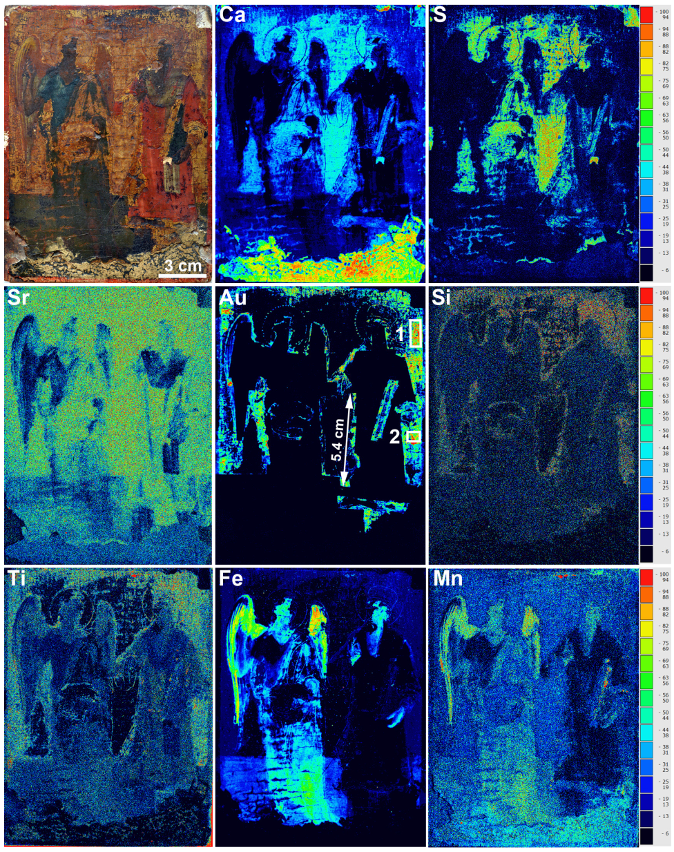
2.2. MA-XRF
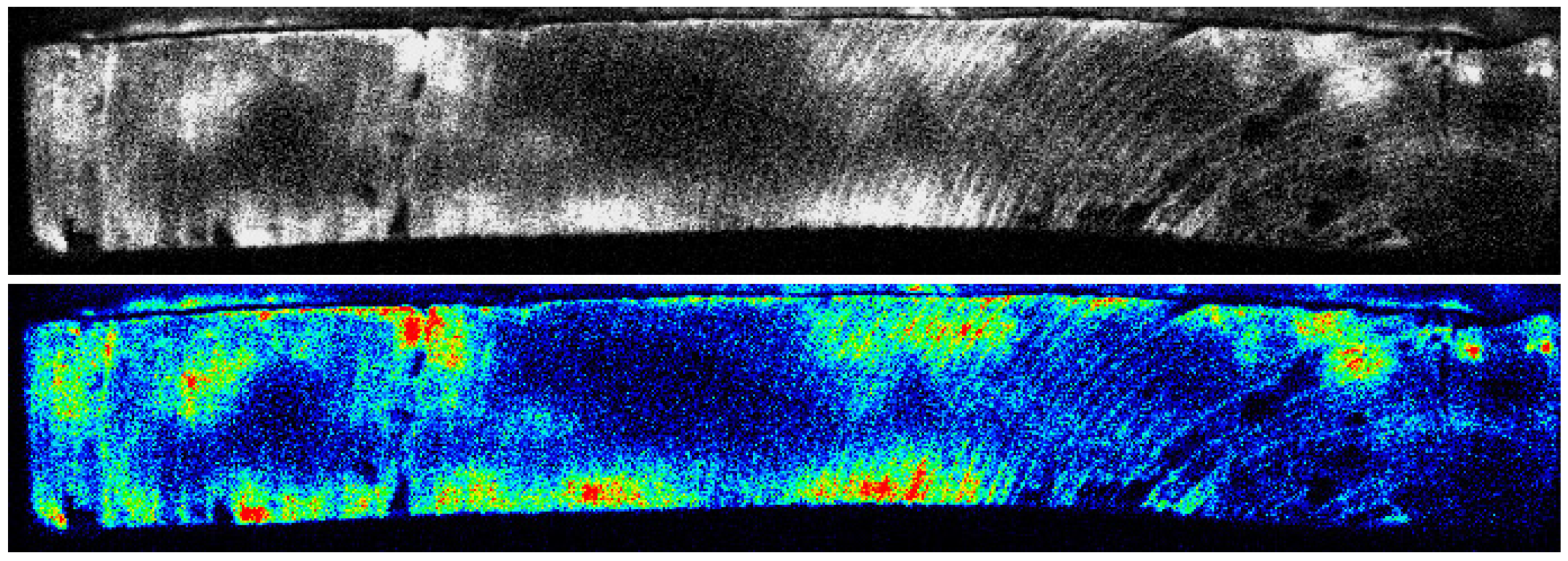
2.3. XRR
2.4. Stereoscope
3. Results and Discussion
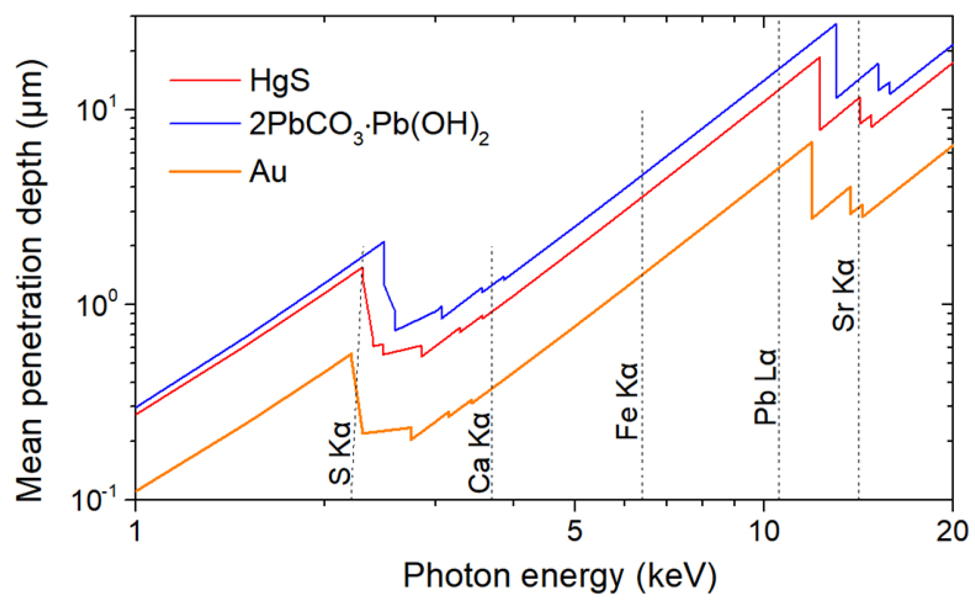
3.1. Preliminary Considerations
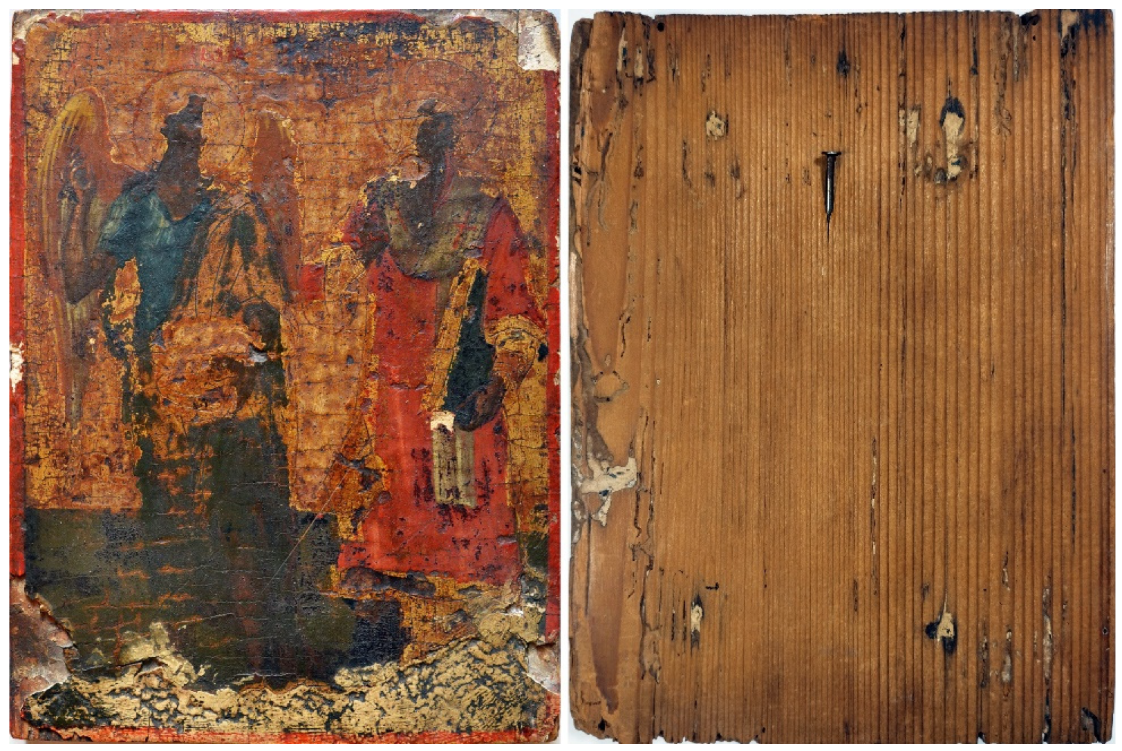
3.2. The Recto of the Icon (Painting)
3.2.1. The Ground/Preparatory Layer
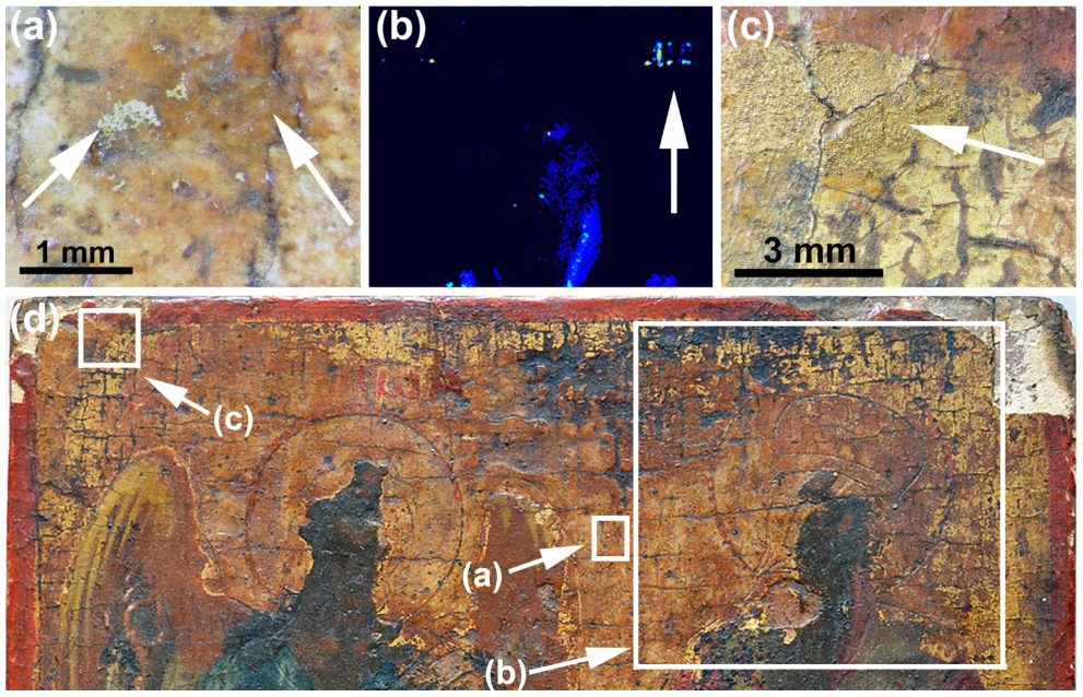
3.2.2. The Gilded Decorations
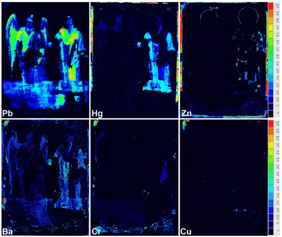
3.2.3. The Paint Layers
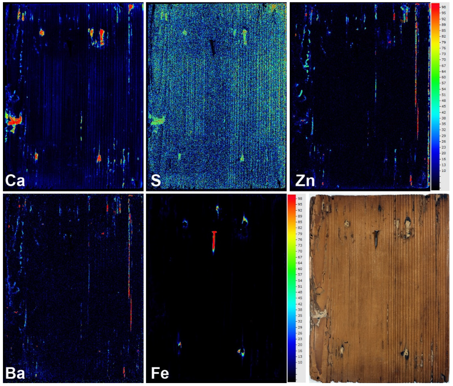
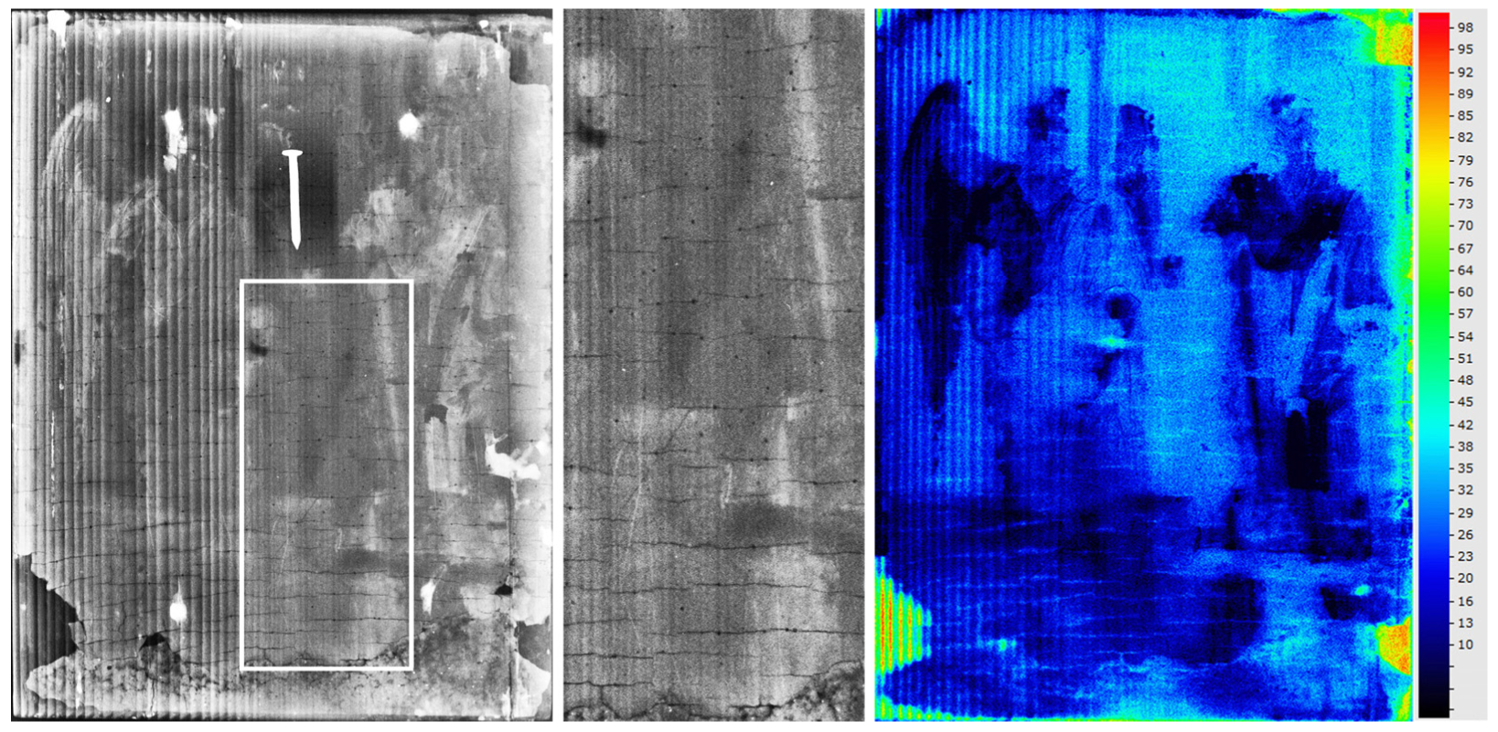
3.3. The Verso of the Icon (Wooden Panel)
4. Discussion
5. Conclusions
Author Contributions
Funding
Institutional Review Board Statement
Informed Consent Statement
Data Availability Statement
Acknowledgments
Conflicts of Interest
References
- Mantler, M.; Schreiner, M. X-ray fluorescence spectrometry in art and archaeology. X-ray Spectrom. 2000, 29, 3–17. [Google Scholar] [CrossRef]
- Thompson, G. Penetration of radiation into old paint films. Natl. Gallery Tech. Bull. 1979, 3, 25–33. [Google Scholar]
- Janssens, K.; Van der Snickt, G.; Vanmeert, F.; Legrand, S.; Nuyts, G.; Alfeld, M.; Monico, L.; Anaf, W.; De Nolf, W.; Vermeulen, M.; et al. Non-Invasive and Non-Destructive Examination of Artistic Pigments, Paints, and Paintings by Means of X-Ray Methods. Top. Curr. Chem. 2016, 374, 1–52. [Google Scholar] [CrossRef]
- Romano, F.P.; Caliri, C.; Nicotra, P.; Di Martino, S.; Pappalardo, L.; Rizzo, F.; Santos, H.C. Real-time elemental imaging of large dimension paintings with a novel mobile macro X-ray fluorescence (MA-XRF) scanning technique. J. Anal. At. Spectrom. 2017, 32, 773–781. [Google Scholar] [CrossRef]
- Van der Snickt, G.; Legrand, S.; Slama, I.; Van Zuien, E.; Gruber, G.; Van der Stighelen, K.; Klaassen, L.; Oberthaler, E.; Janssens, K. In situ macro X-ray fluorescence (MA-XRF) scanning as a non-invasive tool to probe for subsurface modifications in paintings by P.P. Rubens. Microchem. J. 2018, 138, 238–245. [Google Scholar] [CrossRef]
- Walczak, M.; Tarsińska-Petruk, D.; Płotek, M.; Goryl, M.; Kruk, M.P. MA-XRF study of 15th–17th century icons from the collection of the National Museum in Krakow, Poland. X-Ray Spectrom. 2019, 48, 303–310. [Google Scholar] [CrossRef]
- Kokiasmenou, E.; Caliri, C.; Kantarelou, V.; Karydas, G.A.; Romano, F.P.; Brecoulaki, H. Macroscopic XRF imaging in unravelling polychromy on Mycenaean wall-paintings from the Palace of Nestor at Pylos. J. Archaeol. Sci. Rep. 2020, 29, 102079. [Google Scholar] [CrossRef]
- Alfeld, M.; Mösl, K.; Reiche, I. Sunset and moonshine: Variable blue and yellow pigments used by Caspar David Friedrich in different creative periods revealed by in situ XRF imaging. X-ray Spectrom. 2021, 50, 341–350. [Google Scholar] [CrossRef]
- Mazzinghi, A.; Ruberto, C.; Castelli, L.; Czelusniak, C.; Giuntini, L.; Mandò, P.A.; Taccetti, F. MA-XRF for the Characterisation of the Painting Materials and Technique of the Entombment of Christ by Rogier van der Weyden. Appl. Sci. 2021, 11, 6151. [Google Scholar] [CrossRef]
- Janssens, K.; Dik, J.; Cotte, M.; Susini, J. Photon-based techniques for nondestructive subsurface analysis of painted cultural heritage artifacts. Acc. Chem. Res. 2010, 43, 814–825. [Google Scholar] [CrossRef]
- Ghigo, T.; Bone, D.; Howell, D.; Domoney, K.; Gironda, M.; Beedy, A. Material Characterisation of William Burges’ Great Bookcase within the Disruption of a Global Pandemic. Stud. Conserv. 2022, 1–16. [Google Scholar] [CrossRef]
- Alfeld, M.; Broekaert, J.A.C. Mobile depth profiling and sub-surface imaging techniques for historical paintings—A review. Spectrochim. Acta Part B At. Spectrosc. 2013, 88, 211–230. [Google Scholar] [CrossRef]
- Lizun, D.; Kurkiewicz, T.; Szczupak, B. Exploring liu kang’s paris practice (1929–1932): Insight into painting materials and technique. Heritage 2021, 4, 828–863. [Google Scholar] [CrossRef]
- Martins, A.; Davis, E.; Kwartler, T. Max Ernst’s woman, old man, and flower (1923–1924): Four paintings in one revealed by technical imaging. Heritage 2021, 4, 2224–2236. [Google Scholar] [CrossRef]
- Miguel, C.; Bottura-Scardina, S.; Bottaini, C.; Valadas, S.; Candeias, A.; Bilou, F. The Power of Combining MA-XRF, Infrared Reflectography and Digital Microscopy to Unveil the Production of the 16th Century Illuminated Charter of Évora: What May Be Hidden under a Painted Surface? Heritage 2022, 5, 286–296. [Google Scholar] [CrossRef]
- Gerodimos, T.; Asvestas, A.; Mastrotheodoros, G.P.; Chantas, G.; Liougos, I.; Likas, A.; Anagnostopoulos, D.F. Scanning X-ray Fluorescence Data Analysis for the Identification of Byzantine Icons’ Materials, Techniques, and State of Preservation: A Case Study. J. Imaging 2022, 8, 147. [Google Scholar] [CrossRef]
- Sotiropoulou, S.; Daniilia, S. Materials aspects of icons. A review on physicochemical studies of Greek icons. Acc. Chem. Res. 2010, 43, 877–887. [Google Scholar] [CrossRef]
- Mastrotheodoros, G.P.; Beltsios, K.G.; Bassiakos, Y.; Papadopoulou, V. On the Grounds of Post-Byzantine Greek Icons. Archaeometry 2016, 58, 830–847. [Google Scholar] [CrossRef]
- Mastrotheodoros, G.P.; Beltsios, K.G.; Bassiakos, Y.; Papadopoulou, V. On the Metal-Leaf Decorations of Post-Byzantine Greek Icons. Archaeometry 2018, 60, 269–289. [Google Scholar] [CrossRef]
- Dionysios; Hetherington, P. Dionysius of Fourna, The ‘Painter’s Manual’ of Dionysius of Fourna; Hetherington, P., Translator; Oakwood Publications: Torrans, CA, USA, 1996. [Google Scholar]
- Βοκοτόπουλος, Π. Βυζαντινές Εικόνες [Byzantine Icons]; Ekdotike Athenon: Athens, Greece, 1995. [Google Scholar]
- Newman, N.; Harrison, L.; Thomas, D.; Dyer, J.; Taylor, J. The study and conservation of four ancient Egyptian funerary portraits: Provenance, conservation history and structural treatment. Br. Mus. Tech. Res. Bull. 2013, 7, 1–14. [Google Scholar]
- Κόρδης, Γ. Oι προσωπογραφίες του Φαγιούμ και η βυζαντινή εικόνα [The Fayum Portraits and the Byzantine Icon]; Armos Publications: Athens, Greece, 2001. [Google Scholar]
- Evseeva, L.M. A History of Icon Painting: Sources, Traditions, Present Day; Evseeva, L.M., Ed.; Cook, K., Translator; Grand-Holding Publishers: Moscow, Russia, 2007. [Google Scholar]
- Χατζηδάκης, Μ. Έλληνες ζωγράφοι μετά την Άλωση (1450–1830) [Greek Painters after the Fall of Constantinople (1450–1830)]; Center for Neo-Hellenic Studies: Athens, Greece, 1987. [Google Scholar]
- Mastrotheodoros, G.P.; Beltsios, K.G.; Bassiakos, Y. On the blue and green pigments of post-byzantine Greek icons. Archaeometry 2020, 62, 774–795. [Google Scholar] [CrossRef]
- Mastrotheodoros, G.P.; Beltsios, K.G.; Bassiakos, Y. On the red and yellow pigments of post-byzantine Greek icons. Archaeometry 2021, 63, 753–778. [Google Scholar] [CrossRef]
- Mastrotheodoros, G.P.; Beltsios, K.G. Sound practice and practical conservation recipes as described in Greek post-byzantine painters’ manuals. Stud. Conserv. Stud. Conserv. 2019, 64, 42–53. [Google Scholar] [CrossRef]
- Serafima, S.; Duliu, O.G.; Manea, M.M.; Vasilica, S.; Radulescu, C.; Constantinescu, B.; Stan, D.; Culicov, O.-A.; Zincovscaia, I. Complex investigation of the five 19th century Russian-Lipovan icons. Microchem. J. 2019, 150, 104126. [Google Scholar] [CrossRef]
- Klisińska-Kopacz, A.; Obarzanowski, M.; Frączek, P.; Moskal-del Hoyo, M.; Gargano, M.; Goslar, T.; Chmielewski, F.; Dudała, J.; del Hoyo-Meléndez, J.M. An analytical investigation of a wooden panel painting attributed to the workshop of Lucas Cranach the Elder. J. Cult. Herit. 2022, 55, 185–194. [Google Scholar] [CrossRef]
- Βοκοτόπουλος, Π. Εικόνες της Κέρκυρας [Icons of Corfu]; National Bank of Greece Cultural Foundation: Athens, Greece, 1990. [Google Scholar]
- Βασιλάκη, Μ. Oι Εικόνες του Aρχοντικού Τοσίτσα. H Συλλογή του Ευάγγελου Aβέρωφ [The Icons of the Tositsa Mansion. The Collection of Evangelos Averof]; Baron Michael Tossizza Foundation: Athens, Greece, 2012. [Google Scholar]
- Chatzidakis, M.; Djurić, V.; Lazović, M. Les Icons Dans les Collections Suisses; Benteli Publications: Berne, Switzerland, 1968. [Google Scholar]
- Borboudakis, M. (Ed.) Εικόνες Της Κρητικής Τέχνης [Icons of the Cretan Art]; Vikelaia Library and Editions of the University of Crete: Heraklion, Greece, 1993. [Google Scholar]
- Wolff, T.; Tagle, R.; Reinhardt, F.; Waldschlägerand, U.; Schwager, T. On the Interaction of X-rays and Samples by Quantification of Energy Deposition. In Proceedings of the Oral presentation in “European Conference on X-ray Spectrometry 2022”/EXRS-2022, Bruges, Belgium, 26 June–1 July 2022. [Google Scholar]
- NIST XCOM. Photon Cross Sections Database. Available online: http:/physics.nist.gov/xcom (accessed on 20 January 2023).
- Genestar, C. Characterization of grounds used in canvas and sculpture. Mater. Lett. 2002, 54, 382–388. [Google Scholar] [CrossRef]
- Franceschi, E.; Locardi, F. Strontium, a new marker of the origin of gypsum in cultural heritage? J. Cult. Herit. 2014, 15, 522–527. [Google Scholar] [CrossRef]
- Brunetti, A.; Golosio, B.; Schoonjans, T.; Oliva, P. Use of Monte Carlo simulations for cultural heritage X-ray fluorescence analysis. Spectrochim. Acta Part B At. Spectrosc. 2015, 108, 15–20. [Google Scholar] [CrossRef]
- Schoonjans, T.; Solé, V.A.; Vincze, L.; Sanchez del Rio, M.; Appel, K.; Ferrero, C. A general Monte Carlo simulation of energy-dispersive X-ray fluorescence spectrometers—Part 6. Quantification through iterative simulations. Spectrochim. Acta Part B At. Spectrosc. 2013, 82, 36–41. [Google Scholar] [CrossRef]
- Bomford, D.; Dunkerton, J.; Gordon, D.; Roy, A. Art in the Making: Italian Painting before 1400; National Gallery: London, UK, 1990. [Google Scholar]
- Milanou, K.; Vourvopoulou, C.; Vranopoulou, L.; Kalliga, A. Angelo’s Painting Technique: A description of panel painting construction, materials and painting method based on a study of seven signed icons. In Icons by the Hand of Angelos; Benaki Museum: Athens, Greece, 2008; pp. 19–113. [Google Scholar]
- Μαστροθεόδωρος, Γ.Π. Χρωστικές Κονίες και άλλα Υλικά Μεταβυζαντινή Ζωγραφικής [Pigments and Other Materials of Post-Byzantine Painting]. Ph.D. Thesis, University of Ioannina, Ioannina, Greece, 2016. [Google Scholar]
- Mastrotheodoros, G.P.; Anagnostopoulos, D.F.; Filippaki, E.; Beltsios, K.G.; Bassiakos, Y. Probing the birthplace of the “Epi-rus/NW Greece School” of painting: Analytical investigation of the Filanthropinon monastery murals. Part IΙ: Non-pigment materials and painting technique. Archaeol. Anthropol. Sci. 2019, 11, 5781–5798. [Google Scholar] [CrossRef]
- Grygar, T.; Hradilová, J.; Hradil, D.; Bezdička, P.; Bakardjieva, S. Analysis of earthy pigments in grounds of Baroque paintings. Anal. Bioanal. Chem. 2003, 375, 1154–1160. [Google Scholar] [CrossRef] [PubMed]
- Mastrotheodoros, G.P.; Beltsios, K.G. Pigments—Iron-based red, yellow, and brown ochres. Archaeol. Anthropol. Sci. 2022, 14, 35. [Google Scholar] [CrossRef]
- Eastaugh, N.; Walsh, V.; Chaplin, T.; Siddall, R. Pigment Compendium: A Dictionary and Optical Microscopy of Historical Pigments; Butterworth-Heinemann: London, UK, 2008. [Google Scholar]
- Gettens, R.J.; Kühn, H.; Chase, W.T. Lead white. In Artist’s Pigments: A Handbook of Their History and Characteristics; Roy, A., Ed.; National Gallery of Art: Washington, DC, USA, 1993; Volume 2, pp. 67–82. [Google Scholar]
- Gliozzo, E.; Ionescu, C. Pigments—Lead-based whites, reds, yellows and oranges and their alteration phases. Archaeol. Anthropol. Sci. 2022, 14, 17. [Google Scholar] [CrossRef]
- Mastrotheodoros, G.P.; Beltsios, K.G. Recipes for pigment manufacturing in Greek post-byzantine painting manuals. Sci. Cult. 2022, 8, 147–159. [Google Scholar] [CrossRef]
- Kühn, H. Zinc white. In Artist’s Pigments: A Handbook of Their History and Characteristics; Feller, R.L., Ed.; National Gallery of Art; Cambridge University Press: Cambridge, UK, 1986; pp. 169–186. [Google Scholar]
- Feller, R.L. Barium Sulfate—Natural and Synthetic. In Artist’s Pigments: A Handbook of Their History and Characteristics; Feller, R.L., Ed.; National Gallery of Art; Cambridge University Press: Cambridge, UK, 1986; pp. 47–64. [Google Scholar]
- Gabrieli, F.; Doherty, B.; Miliani, C.; Degano, I.; Modugno, F.; Uldank, D.; Kunzelman, D.; Buzzegoli, E.; Pattie, M.; Rosi, F. Micro-Raman and SER spectroscopy to unfold Lefranc’s early organic pigment formulations. J. Raman Spectrosc. 2016, 47, 1505–1513. [Google Scholar] [CrossRef]
- Karapanagiotis, I.; Minopoulou, E.; Valianou, L.; Daniilia, S.; Chryssoulakis, Y. Investigation of the colourants used in icons of the Cretan School of iconography. Anal. Chim. Acta 2009, 647, 231–242. [Google Scholar] [CrossRef]
- Kühn, H.; Curran, M. Chrome Yellow and Other Chromate Pigments. In Artist’s Pigments: A Handbook of Their History and Characteristics; Feller, R.L., Ed.; National Gallery of Art; Cambridge University Press: Cambridge, UK, 1986; pp. 186–217. [Google Scholar]
- Gulotta, D.; Goidanich, S.; Bertoldi, M.; Bortolotto, S.; Toniolo, L. Gildings and false gildings of the baroque age: Characterization and conservation problems. Archaeometry 2012, 54, 940–954. [Google Scholar] [CrossRef]
- Bisacca, G.; de la Fuente Martínez, J. The Treatment of Dürer’s Adam and Eve Panels at the Prado Museum. In Facing the Challenges of Panel Paintings Conservation: Trends, Treatments, and Training; Phenix, A., Chui, S.A., Eds.; The Getty Conservation Institute: Los Angeles, CA, USA, 2011; pp. 10–24. [Google Scholar]
- Knut, N. The restoration of Paintings; Könemann: Cologne, Germany, 1999. [Google Scholar]
- Vrebos, B.A.R. Compensation Methods in Quantitative Analysis, Handbook of Practical X-ray Fluorescence Analysis; Kawahara, N., Shoji, T., Beckhoff, B., Kanngießer, B., Langhoff, N., Wedell, R., Wolff, H., Eds.; Springer: Berlin/Heidelberg, Germany, 2006; p. 364. [Google Scholar]
- Alexopoulou, A.; Kaminari, A.A.; Moutsatsou, A. Multispectral and hyperspectral studies on Greek monuments, archaeological objects and paintings on different substrates. Achievements and limitations. Commun. Comput. Inf. Sci. 2019, 962, 443–461. [Google Scholar]


Disclaimer/Publisher’s Note: The statements, opinions and data contained in all publications are solely those of the individual author(s) and contributor(s) and not of MDPI and/or the editor(s). MDPI and/or the editor(s) disclaim responsibility for any injury to people or property resulting from any ideas, methods, instructions or products referred to in the content. |
© 2023 by the authors. Licensee MDPI, Basel, Switzerland. This article is an open access article distributed under the terms and conditions of the Creative Commons Attribution (CC BY) license (https://creativecommons.org/licenses/by/4.0/).
Share and Cite
Mastrotheodoros, G.P.; Asvestas, A.; Gerodimos, T.; Anagnostopoulos, D.F. Revealing the Materials, Painting Techniques, and State of Preservation of a Heavily Altered Early 19th Century Greek Icon through MA-XRF. Heritage 2023, 6, 1903-1920. https://doi.org/10.3390/heritage6020102
Mastrotheodoros GP, Asvestas A, Gerodimos T, Anagnostopoulos DF. Revealing the Materials, Painting Techniques, and State of Preservation of a Heavily Altered Early 19th Century Greek Icon through MA-XRF. Heritage. 2023; 6(2):1903-1920. https://doi.org/10.3390/heritage6020102
Chicago/Turabian StyleMastrotheodoros, Georgios P., Anastasios Asvestas, Theofanis Gerodimos, and Dimitrios F. Anagnostopoulos. 2023. "Revealing the Materials, Painting Techniques, and State of Preservation of a Heavily Altered Early 19th Century Greek Icon through MA-XRF" Heritage 6, no. 2: 1903-1920. https://doi.org/10.3390/heritage6020102
APA StyleMastrotheodoros, G. P., Asvestas, A., Gerodimos, T., & Anagnostopoulos, D. F. (2023). Revealing the Materials, Painting Techniques, and State of Preservation of a Heavily Altered Early 19th Century Greek Icon through MA-XRF. Heritage, 6(2), 1903-1920. https://doi.org/10.3390/heritage6020102