Abstract
Among the different forms of art, the puppet theatre constitutes a long-standing and often little-known tradition. The use of puppets as support for acting dates back to the Greek age, and it was mainly developed during the modern period. The reason for such a large diffusion was due to the possibility of using affordable materials, such as papier-mâché, for the puppets’ manufacture. In this paper, a method based on the combined use of pulsed thermography (PT) and mid-wave infrared reflectography (MIR) is, for the first time, proposed for the characterization of papier-mâché artworks. In particular, some puppets belonging to the collection of the Museo delle Civiltà in Rome and made by Olga Lampe Minelli, a 20th-century puppet master, were investigated in order to detect damaged areas, such as those affected by insect attacks, and, consequently, to specifically plan suitable restoration works. Finally, the investigations were also carried out after the restoration to evaluate the effectiveness of the adopted treatments.
1. Introduction
Puppetry is a form of theatre that involves the use of puppets resembling animals and/or different kinds of human figures, often associated with political satire. This type of theatre has been developed since the Greek age, reaching its maximum popularity between the 16th and 18th centuries, when it was also used as a means of communicating popular ideologies. The reason for its great diffusion across different geographical areas is mainly due to the possibility of organizing less expensive performances and, in addition, the opportunity to employ cost-effective puppets for the representation of different characters. The use of puppets in the theatre began to decline in the 20th century because of the development of new methods of entertainment [1,2,3,4].
Over the centuries, puppets have been built according to different manufacturing methods using several materials. While the clothes were made of recycled fabric, eventually enriched with different accessories, the head could be made of painted wood or papier-mâché, the latter being chosen mainly for economic reasons. In this regard, the variety of materials and structures adopted for puppet’ manufacture makes the planning of restoration treatments suitable for effective preservation of this kind of artwork difficult [5,6,7]. In fact, while papier-mâché can be affected by the sorts of damage typical of cellulose-based materials, i.e., hydrolysis [8], foxing [9,10], oxidation [11] and/or attack by insects [12,13], the overlaying paint layers can present additional deterioration issues, often consisting of detachments, cracks and lacunas [14]. Moreover, papier-mâché puppets may also have undergone a series of changes over time—i.e., restorations, retouching, etc.—for stylistic adjustment and conservation purposes. Due to the above-mentioned reasons, papier-mâché artefacts can be considered as complex and multi-layer objects that require different investigation methods to identify the possible presence of damage and, consequently, to plan specifically designed repair works.
In the literature, some studies report on investigations into polychrome papier-mâché sculptures to obtain information on both the support and the paint layer. The latter, in particular, has been analysed by micro-X-ray fluorescence and Raman microscopy techniques, with the aim of identifying the pigment compositions [15]. Moreover, UV fluorescence has been used to characterize the varnish layer of a 17th-century bust and to evaluate the composition of the layers underlying the papier-mâché support [16]. It was not until 2017 that a systematic study was proposed for the analysis of a contemporary papier-mâché sculpture. In particular, the use of both X-ray radiography and UV fluorescence has been adopted for the visualization of the internal iron armature and for the analysis of the synthetic varnish on the surface, respectively. Moreover, some coating layers have been analysed by means of Fourier transform infrared spectroscopy (FT-IR) for the characterization of the acrylic resin on the surface layer [17].
Damage typical of papier-mâché artefacts has been studied in other kinds of cultural heritage items by means of infrared imaging techniques, such as pulsed thermography (PT) and MWIR reflectography (MIR). PT, one of the photo-thermal techniques used for the evaluation of different thermal parameters in several materials [18,19], has been employed to investigate different kinds of artefacts [20,21,22,23,24,25], enabling the study of both optically opaque samples, such as bronzes [26,27,28,29,30], and semi-transparent materials [31,32], such as paintings [33,34,35,36] and books [37]. In particular, it has been proven to be very effective for the study of ancient codices, enabling the non-destructive characterization of the main features of both book bindings [37,38,39] and illuminations [40,41]; in addition, it is used for the evaluation of the state of conservation of the books. Moreover, PT has been used to reveal subsurface graphical elements underlying illuminations, such as underdrawings and pentimenti [40]. In recent years, the combined use of PT and MIR has been proposed for the investigation of painted artefacts. The main idea behind such an approach is to employ techniques able to provide complementary results, in an effort to try and obtain in-depth analysis of the painting structure. In particular, this approach has been adopted for a number of different paintings on both wood and canvas to detect pentimenti and also to visualize the changes that have been made over time, such as restorations, overpainting and retouching [42,43].
In this study, the combined use of PT and MIR is proposed, for the first time, to study papier-maché artefacts. Such an approach allowed us to obtain valuable information concerning the layered structure of the puppets by revealing structural features (i.e., the insect galleries) buried beneath the visible pictorial layer and by mapping the restorations. It is worth noting that these aspects can be separately investigated by using different kinds of techniques, such as, for instance, X-ray radiography and UV fluorescence for the detection of the insect galleries and the restored areas, respectively. X-ray radiography enables probing the object to a depth greater than that detectable by PT, but it has limitations in performing depth-resolved investigations of the artefact, since it provides a depth-integrated image. Moreover, the complexity of the X-ray equipment and the safety issues involved often limit its in situ application. Concerning UV fluorescence, it can be successfully used to investigate the outermost layers of the artefacts to detect and characterize surface features such as pictorial retouching and restored areas, but not features located deeper beneath the surface layers. In that regard, the use of MWIR techniques can be very useful since they enable the detection of both sub-surface and invisible surface features.
Some puppets belonging to the Lampe Minelli collection, donated in 2013 to the Italian Museum of Folk Arts and Traditions by Elisabetta and Corrado Lampe, descendants of the famous Italian puppeteer Olga Lampe Minelli, and today preserved at the Museo delle Civiltà in Rome, were investigated. As shown later on, structural defects, such as detachment of the pictorial layer, and other kinds of damage, such as exit holes due to the attack of insects, were detected by PT and MIR. The obtained results were then used to define the necessary restoration steps. Finally, PT investigations were also carried out after the restoration in order to assess the effectiveness of the adopted treatments and, in addition, to plan the most suitable conditions of conservation for the museum’s exhibition.
2. The Puppets
As regards the structure of the investigated items, puppets are mainly composed of three parts: the head, body, and accessories. The body is anchored to the head through metal nails hidden by the collar, and it is typically made of a cloth dress with arms and hands. The head can be made of wood, carved so as to obtain the desired shape, or papier-mâché, whose manufacture is described below.
2.1. Papier-Mâché Support
Papier-mâché has been used to create different types of cultural heritage objects, such as bas-reliefs, statues, globes and masks, with the aim of mimicking more precious and expensive materials, such as gold, silver and marble. [44,45]. The raw material is paper, with which other components, such as gypsum, additives and colouring substances, are mixed in order to obtain a solid consistency after drying. More specifically, the manufacturing process of papier-mâché artefacts begins with the maceration of the paper, to which glues, fillers and plaster are then added to strengthen the final structure. After maceration, the resulting mixture is placed in moulds, typically made of plaster or clay, for the drying phase. After drying, the surface is painted, thus reproducing in the puppets the typical layered structure of paintings: a ground layer, a pictorial layer and a varnish coat on top. In some cases, gold and silver features may be added to decorate the surface [46,47].
2.2. The Studied Puppets
The puppets were handmade by the artist Olga Lampe Minelli, who used materials typical of these kinds of artefacts: painted papier-mâché for the head and reused clothes for the body [48].
Four puppets were selected among the 122 items comprising the collection. The first two puppets represent the Man with the tie (Figure 1) and the clown called Padella (Figure 2), respectively.
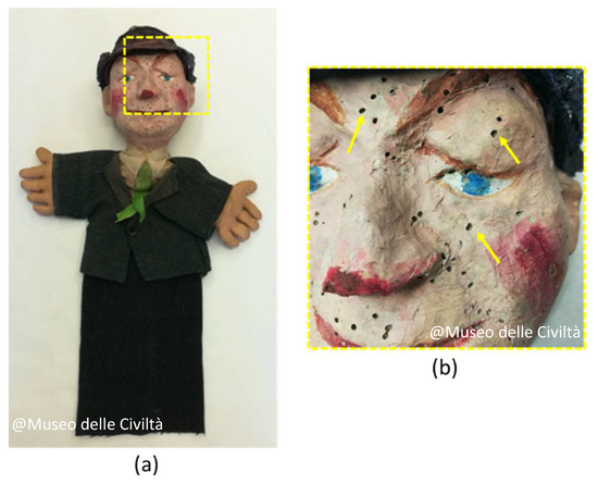
Figure 1.
The Man with the tie puppet before the restoration: (a) photo; (b) magnifications of the areas highlighted in (a). The yellow arrows indicate some of the exit holes due to previous entomological attacks. @ Museo delle Civiltà.
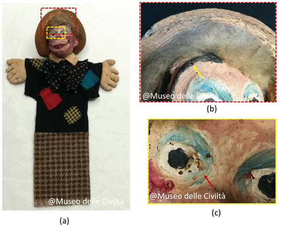
Figure 2.
The Padella puppet before the restoration: (a) photo; (b,c) magnifications of the sample areas highlighted in (a). The yellow arrow in (b) indicates one of the exit holes due to the insect attack; the red arrow (c) indicates a hole in the pictorial layer.@ Museo delle Civiltà.
The puppets exhibited similar damage, such as widespread abrasions and stains, fractures in the papier-mâché and exit holes due to entomological attacks (Figure 1b and Figure 2b). In addition, fading and holes due to the weakening of the pictorial layer can be observed in Figure 2c.
The Seal with the ball (Figure 3) and the Horse (Figure 4) show similar damages. In particular, those concerning the fading of the colour and the detachment of the pictorial layer, as in the case of the horse’s eye, where it is possible to observe the preparatory layer under the surface (red arrows in Figure 4b).
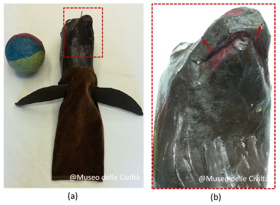
Figure 3.
The Seal with the ball before the restoration: (a) photo; (b) magnification of the sample areas highlighted in (a). The red arrows in (b) indicate some of the many exit holes due to previous entomological attacks. @ Museo delle Civiltà.
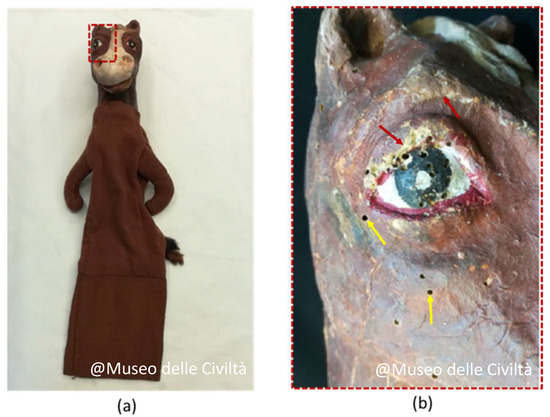
Figure 4.
The Horse before the restoration: (a) photo; (b) magnifications of the sample areas highlighted in (a) corresponding to the horse’s eye. The yellow arrows indicate some of the exit holes due to previous entomological attacks and the red arrows the detachments of the pictorial layer. @ Museo delle Civiltà.
3. Mid-Wave Infrared Imaging Techniques
Nowadays, imaging techniques operating in the mid-wave infrared (MWIR) range (3–5 μm), such as PT and MIR, are considered very effective tools for the characterization of cultural heritage.
PT is based on the monitoring of the time dependence of the IR radiation emitted in the MWIR spectral range following the sample heating induced by the absorption of a short visible-light pulse. The emitted radiation is then recorded during the cooling stage by means of an IR camera, which provides a sequence of images, called thermograms, each corresponding to a different delay time with respect to the onset of the heating pulse, that give information about the evolution of the temperature distribution on the sample’ surface. In particular, the possible presence of buried elements is detected through their influence on the local heat diffusion that gives rise to contrasting features in the recorded thermograms. Information about eventual subsurface features, such as their size or position within the sample volume, can also be evaluated [49,50].
In the present study, the thermal stimulus was induced by two flash lamps (Bowens Estime 3000, maximum power 650 W) delivering 5 ms long light pulses. The lamps were positioned at a distance of 0.4 m with respect to the investigated puppet with their axes at 45° (Figure 5). Thermographic sequences were recorded by a Cedip JADE camera characterized by a noise equivalent temperature difference (NETD) < 25 mK at 30 °C (320 × 240 pixel, InSb focal plane array, 30 μm pitch, 3.6–5.1 μm wavelength range) for 1 s in full-frame mode with a frame rate of 150 Hz and, thereafter, processed by the Altair 5.50 software. To evaluate the time-dependent change in the thermographic signal for all the image pixels, the corresponding signal levels pertaining to the frame obtained just before the flash pulse were subtracted from each pixel in all the recorded thermograms. This gave the possibility of displaying the induced changes in the thermograms with a larger dynamic range [51,52].
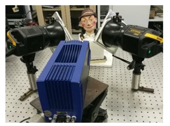
Figure 5.
Laboratory set-up for thermographic acquisitions on the puppets.
MIR has been recently introduced into cultural heritage investigations, thus extending the exploited spectral range as compared to the one employed in near-infrared (NIR) reflectography (0.75–1.4 μm). In the MWIR range, pigments usually show different absorption and scattering properties than in the NIR range so as to provide complementary information. In comparison with PT, MIR is characterized by a larger sensitivity to features located close to the artefact surface. On the other hand, PT is more effective in providing information about features located at a greater depth, and, unlike MIR, it may also allow for the quantitative evaluation of their depth position through the analysis of the time dependence of the recorded thermogram sequence. In particular, the larger the time delay of the thermogram from the thermal perturbation, the deeper the features shown by the thermogram.
In the reflectographic setup, the illumination of the sample was carried out by means of two carbon filament IR sources, characterized by an effective emission in the MWIR range and positioned at about 2 m from the artefact. Special care was taken to minimize the exposure time (~0.1 s) and incident IR power (<100 W) so as to reduce the sample heating. By so doing, the contrast in the recorded MIR images mainly originated from local differences in the reflectivity of the incident MWIR radiation and was not associated with local emissions due to the sample heating. The same camera and software were used for both PT and MIR investigations, thus ensuring the correspondence of the same sample areas in the images recorded by the two techniques.
4. Results and Discussion
4.1. Pre-Restoration Survey
Before discussing the obtained results, it is worth mentioning that both the MIR and PT images will be presented according to a greyscale palette where the lighter colours correspond to a larger amount of the emitted IR radiation. Moreover, the images obtained with both the employed techniques and from different depths were recorded using the same device with an unchanged viewpoint and configuration.
In the Man with the tie puppet (Figure 6a), the MWIR images revealed a number of details of interest. In particular, the reflectogram in Figure 6b shows the presence of lighter areas corresponding to previous restorations (yellow arrows). The lighter appearance is related to the greater IR reflectivity of the superficial paint layer employed for the restoration.
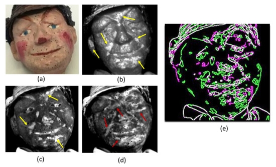
Figure 6.
The Man with the tie puppet before the restoration: (a) photo; (b) reflectogram where the yellow arrows indicate the restored areas; (c) thermogram recorded 0.01 s after the pulsed heating also showing the restored areas; (d) thermogram recorded 2 s after the pulsed heating where the red arrows indicate the extent of the insect galleries; (e) image obtained by the Sobel edge detection algorithm where the contours of the contrasting areas detected by PT at larger delay (green) and by MIR (purple) can be simultaneously viewed in a single image overlapping the geometry of the puppet (white contours).
The same features can also be detected in the thermogram of Figure 6c, recorded immediately after the perturbation. In this case, the lighter appearance is related to the larger degree of emission of IR radiation associated with the materials used for the restoration, at the level of the most superficial layers. The thermogram obtained at a later delay time (Figure 6d) shows additional light features originating from within the vicinity of the exit holes left by the insect galleries (red arrows).
Such voids appear lighter because the subsurface voids hinder the in-depth heat diffusion, thus causing such areas to remain hotter longer than in the surrounding areas, with a consequent larger emission of IR radiation. So, the combined analysis in the MWIR region by reflectography and thermography enable one to detect features that are not so evident to the naked eye. In order to better highlight the capability of the MWIR techniques to detect features at different depths, the Sobel edge detection algorithm [53] was applied in the MATLAB2020b suite to the reflectogram (Figure 6b) and the thermogram recorded at a larger delay (Figure 6d). Figure 6e shows the obtained contours that outline the main features as detected by the two techniques with the respect to the geometry of the puppet. Such elaboration enables the pixel-by-pixel correspondence between the thermogram and the reflectogram, making it possible to highlight the different nature or location of the detected features. In particular, the purple contours correspond to the superficial features obtained from the reflectogram related to the previous restorations, while the green contours correspond to the thermogram recorded 2 s after the light pulse, and they are referred to as elements that evolve in shape and depth within the puppet’s volume. As can be seen from the figure, the presented results allow for the comparison of elements detected at different depths with different techniques without any elaboration on the images coming directly from the IR camera, ensuring the correspondence of the matrices in the algorithm elaborations [54,55]. Features similar to those observed in the Man with the tie have also been detected in the puppet called Padella (Figure 7a). The reflectogram reveals a number of restored areas that are barely visible to the naked eye, corresponding to some of the lighter areas in Figure 7b (yellow arrows in Figure 7b), which also appear in the thermogram recorded right after the perturbation (Figure 7c). Additionally, in this case the extent of the insect galleries located at a greater depth can be detected in the thermogram recorded at a larger delay (red arrows in Figure 7d).
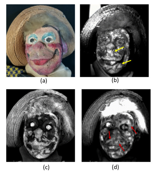
Figure 7.
The Padella puppet before the restoration: (a) photo; (b) reflectogram where the yellow arrows indicate the restored areas; (c) thermogram recorded 0.01 s after the perturbation showing the restored areas detected in (b); (d) thermogram recorded 2 s after the perturbation where the red arrows indicate the galleries of the insects below the surface.
For the Seal with the ball puppet (Figure 8a), the reflectogram (Figure 8b) displays the presence of a shiny layer of either paint or adhesive on the surface producing a very bright aspect which made it difficult to detect other features. The thermograms obtained at both short and longer delays (red arrows in Figure 8c,d, respectively) showed the presence of the subsurface voids due to entomological galleries, indicating that they were also located at shallow depths. Moreover, Figure 8c also shows the joints of some overlapping paper flaps (green arrows) that were formed during the manufacturing process.
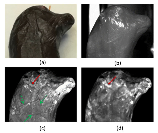
Figure 8.
The Seal with the ball puppet before the restoration: (a) photo; (b) reflectogram; (c) thermogram recorded 0.01 s after the perturbation where the green arrows indicate the joints of the overlapped paper flaps and the red arrow the extent of the gallery below the surface; (d) thermogram recorded 2 s after the perturbation where the red arrows indicate the gallery detected in (c).
Finally, Figure 9 shows the results obtained for the horse puppet, which reveal features of a similar nature to those observed previously, displayed, in this case, only in the obtained thermograms. The thermogram in Figure 9b, recorded 0.03 s after the perturbation, clearly displays the restored areas (lighter area), whose presence can also be seen by the naked eye.
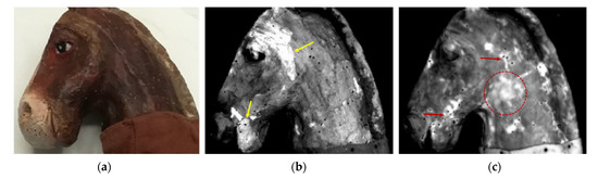
Figure 9.
The Horse puppet before the restoration: (a) photo; (b) thermogram recorded 0.03 s after the perturbation where the yellow arrows indicate the restored areas; (c) thermogram recorded 1.5 s after the perturbation where the red arrows indicate the galleries and the red dashed circle an extended detachment.
The thermogram recorded 1.5 s after the perturbation displays the subsurface insect galleries (red arrows in Figure 7c). In this case, an additional lighter area appears (red dashed circle), most likely corresponding to an extended subsurface detachment that acts as a barrier to the in-depth heat diffusion process, like in the case of the insect galleries.
4.2. Restoration and Evaluation of the Conservation Treatments
In this section, as an example, the results of the analysis by the thermographic imaging technique in evaluating the outcome of some of the applied restoration treatments, particularly those involving the subsurface features, are reported for the case of the Man with the tie.
The first approach to the intervention required a disinfestation procedure, which was essential for the next operations to be carried out. In particular, the anoxic procedure (without oxygen) was fundamental for the elimination of the insects and larvae inside the artefacts. After this treatment, a dry-cleaning operation was performed for the removal of various superficial residues. In addition, in different parts of the fabric, a wet-cleaning operation was necessary for the removal of stains of various natures—particularly on the hands and along the shirt of the Man with the tie, the most yellowish part of the object.
The tears in the fabric were filled with linen fabric and sewn up with transparent silk thread. The metal elements were mechanically cleaned with a glass-fibre pen for rust removal.
The numerous exit holes caused by entomological damage to the head’s surface were filled with a mixture of microcrystalline cellulose mixed with a hydroxypropylcellulose-based adhesive [56] in a 1:1 ratio, applied with the help of a syringe, allowing for complete penetration of the mixture into the exit holes, as highlighted by the thermographic investigations. The grouted areas were then retouched using watercolour.
At the end of the treatments, the puppets were placed inside a specially made box for storage, as required by the museum.
After the restoration procedures, an additional thermographic survey was carried out to verify the effectiveness of the consolidation practices.
The visual examination of the puppet immediately confirmed the effectiveness of the restoration of the surface of the puppet, where the flicker holes appeared to be repaired (arrows in Figure 10a,b). Moreover, from the comparison between the thermograms before (Figure 11a) and after (Figure 11b) the restoration, further information denoting the effectiveness of the adopted procedure can be gathered. In particular, the post-restoration thermogram (Figure 11b) shows in general a smaller number of insect exit holes (circled area) and galleries (red arrows) visible with respect to the pre-restoration situation. It must nevertheless be pointed out that even in the repaired areas, some residual faint white tracks where the branched tracks existed can still be detected. This is due to the fact that the tracks were not fully filled and/or the thermal properties of the used filler may be different from those of the papier-mâché, thus producing a residual small thermal barrier effect, giving rise to the contrasting features. It is worth noting that the possibility of detecting not fully filled areas is of fundamental importance for conservators and restorers in assessing the effectiveness of the adopted treatments and planning further conservation strategies.
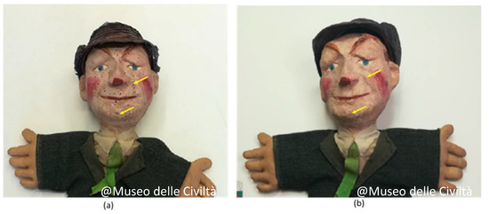
Figure 10.
The Man with the tie puppet: photo (a) before and (b) after the restoration. The arrows indicate some of the many exit holes repaired. @ Museo delle Civiltà.
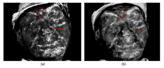
Figure 11.
The Man with the tie puppet: thermograms (a) before and (b) after the restoration. The dashed red circle and the arrow mark some of the many restored areas.
5. Conclusions
A non-destructive procedure combining the use of thermographic and reflectographic imaging techniques in the mid-wave infrared range has been presented for the investigation of some papier-mâché puppets dating back to the 20th century and belonging to the collection of the Museo delle Civiltà in Rome. In particular, a pre-restoration survey was carried out to assess their preservation state so as to determine the most suitable restoration procedures. The combined use of both imaging techniques enabled the detection and characterization of inhomogeneities in the structure of the puppets, identifying restored areas and damages related to entomological attacks. The results have been used to plan specifically designed consolidation procedures in the areas identified by the diagnostic survey. Finally, the effectiveness of the adopted treatments could be verified by additional thermographic investigations carried out after the restoration.
Author Contributions
Conceptualization, S.C., N.O. and E.C.; Methodology, S.C., F.M., S.P.; N.O., U.Z., investigation, E.C., S.C., F.M., S.P. and A.T.; data elaboration, S.C., E.C. and F.M.; writing—original draft preparation, S.C., N.O. and C.C.; writing—review and editing, N.O., F.M., C.C., S.P. and U.Z.; supervision, F.M. and U.Z. All authors have read and agreed to the published version of the manuscript.
Funding
No funding.
Institutional Review Board Statement
Not applicable.
Informed Consent Statement
Not applicable.
Data Availability Statement
Not applicable.
Acknowledgments
The authors want to thank the Museo delle Civiltà in Rome, especially Serena Francone and Maria Francesca Quarato of the Laboratory of Conservation and Restoration, for their contribution.
Conflicts of Interest
The authors declare no conflict of interest.
References
- Schiavetti, A. Burattini & Marionette. II Meraviglioso Mondo del Teatro di Figura; Bandecchi & Vivaldi: Pontedera, Italy, 2011. [Google Scholar]
- Allegri, L. Per una Storia del Teatro Come Spettacolo: Il Teatro di Burattini e di Marionette; Centro Studi e Archivio della Comunicazione: Parma, Italy, 1978. [Google Scholar]
- Giufrè, M. Maria Signorelli: Teatro, Marionette e Burattini; Joshua Libri: Sestri Levante, Italy, 2000. [Google Scholar]
- Currell, D. The Complete Book of Puppetry; Plays: Boston, MA, USA, 1976. [Google Scholar]
- Van der Reyden, D.; Williams, D. The Technology and Conservation Treatment of a 19th Century “Papier-Mâché Chair. In Preprints of the American Institute for Conservation, 14th Annual Meeting, Chicago; The American Institute for Conservation of Historic and Artistic Works: Washington, DC, USA, 1986; pp. 125–142. [Google Scholar]
- Copedè, M. La Carta e Il Suo Degrado; Nardini Editore: Firenze, Italy, 2003. [Google Scholar]
- Thornton, J. The History, Technology, and Conservation of Architectural Papier Mâch. J. Am. Inst. Conserv. 1993, 32, 165–176. [Google Scholar] [CrossRef]
- Jablonsky, M.; Jozef, Š. Oxidative degradation of paper–A minireview. J. Cul. Herit. 2021, 48, 269–276. [Google Scholar] [CrossRef]
- Peters, D. An alternative to foxing. Pap. Restaur. 2000, 1, 801–806. [Google Scholar]
- Krstić, D.; Zdravko, S. Characterization of Foxing Stains on Eighteenth Century Books. HDKBR Info Mag. 2013, 3, 32–39. [Google Scholar]
- Daniels, V. Oxidative damage and the preservation of organic artefacts. Free. Radic. Res. Commun. 1989, 5, 213–220. [Google Scholar] [CrossRef]
- Trematerra, P.; David, P. Museum Pests—Cultural Heritage Pests. In Recent Advances in Stored Product Protection; Springer: Berlin, Germany, 2018; pp. 229–260. [Google Scholar] [CrossRef]
- Moşneagu, M.; Ardelean, E.; Mustaţă, M. Low temperature treatment of pest infected paper documents. Eur. J. Sci. Theol. 2011, 7, 35–46. [Google Scholar]
- Mecklenburg, M.F. Micro climates and moisture induced damage to paintings. In Proceedings of the Conference on Micro Climates in Museums, Copenhagen, Denmark, 19–23 November 2007. [Google Scholar]
- Manso, M.; Bidarra, A.; Longelin, S.; Pessanha, S.; Ferreira, A.; Guerra, M.; Coroado, J.; Carvalho, L. Micro-Analytical study of a rare papier-mâché sculpture. Microsc. Microanal. 2015, 21, 56–62. [Google Scholar] [CrossRef]
- Lanteri, L.; Agresti, G. Ultraviolet fluorescence 3D models for diagnostics of cultural heritage. Eur. J. Sci. Theol 2017, 13, 35–40. [Google Scholar]
- Rocco, F. A multisciplinary approach to the study and conservation of the contemporary Sarah Lucas’ papier-maché sculpture Love Me. CeROArt Conserv. Expo. Restaur. D’objets D’art 2017, EGG 6. [Google Scholar] [CrossRef]
- Zammit, U.; Marinelli, M.; Mercuri, F.; Paoloni, S. Effect of confinement and strain on the specific heat and latent heat over the nematic-isotropic phase transition of 8CB liquid crystal. J. Physic. Chem. B 2009, 113, 14315–14322. [Google Scholar] [CrossRef]
- Paoloni, S.; Mercuri, F.; Marinelli, M.; Zammit, U.; Neamtu, C.; Dadarlat, D. Simultaneous characterization of optical and thermal parameters of liquid-crystal nanocolloids with high-temperature resolution. Phys. Rev. E 2008, 78, 042701. [Google Scholar] [CrossRef] [PubMed]
- Blessley, K.; Young Christina, R.T.; Nunn, J.; Coddington, J.; Shepard, S.M. The feasibility of flash thermography for the examination and conservation of works of art. Stud. Conserv. 2014, 55, 107–120. [Google Scholar] [CrossRef]
- Gavrilov, D.; Maev, R.G.; Almond, D.P. A review of imaging methods in analysis of works of art: Thermographic imaging method in art analysis. Can. J. Phys. 2014, 92, 341–364. [Google Scholar] [CrossRef]
- Sfarra, S.; Regi, M. Wavelet analysis applied to thermographic data for the detection of sub-superficial flaws in mosaics. Eur. Phys. J. Appl. Phys. 2016, 73, 31001. [Google Scholar] [CrossRef]
- Krankenhagen, R.; Maierhofer, C. Pulse phase thermography for characterising large historical building façades after solar heating and shadow cast—A case study. Quant. InfraRed Thermogr. J. 2014, 11, 10–28. [Google Scholar] [CrossRef]
- Carlomagno, G.M.; Di Maio, R.; Meola, C.; Roberti, N. Infrared thermography and geophysical techniques in cultural heritage conservation. Quant. InfraRed Thermogr. J. 2005, 2, 5–24. [Google Scholar] [CrossRef]
- Bodnar, J.L.; Nicolas, J.L.; Mouhoubi, K.; Detalle, V. Stimulated infrared thermography applied to thermophysical characterization of cultural heritage mural paintings. Eur. Phys. J. Appl. Phys. 2012, 60, 21003. [Google Scholar] [CrossRef]
- Mercuri, F.; Caruso, G.; Orazi, N.; Zammit, U.; Cicero, C.; Colacicchi Alessandri, O.; Ferretti, M.; Paoloni, S. Interface thermal conductance characterization by infrared thermography: A tool for the study of insertions in bronze ancient Statuary. Infrared Phys. Technol. 2018, 90, 31–39. [Google Scholar] [CrossRef]
- Mercuri, F.; Paoloni, S.; Orazi, N.; Cicero, C.; Zammit, U. Pulsed infrared thermography applied to quantitative characterization of the structure and the casting faults of the Capitoline She Wolf. Appl. Phys. A 2017, 123, 327. [Google Scholar] [CrossRef]
- Mercuri, F.; Orazi, N.; Zammit, U.; Giuffredi, A.; Salerno, C.S.; Cicero, C.; Paoloni, S. The manufacturing process of the Capitoline She Wolf: A thermographic method for the investigation of repairs and casting faults. J. Archaeol. Sci. Rep. 2017, 14, 199–207. [Google Scholar] [CrossRef]
- Orazi, N.; Mercuri, F.; Zammit, U.; Paoloni, S.; Marinelli, M.; Giuffredi, A.; Salerno, C.S. Thermographic analysis of bronze sculptures. Stud. Conserv. 2016, 61, 236–244. [Google Scholar] [CrossRef]
- Tao, N.; Wang, C.; Cunlin Zhang, C.; Sun, J. Quantitative measurement of cast metal relics by pulsed thermal imaging. Quant. InfraRed Thermogr. J. 2022, 19. [Google Scholar] [CrossRef]
- Mercuri, F.; Paoloni, S.; Cicero, C.; Zammit, U.; Orazi, N. Infrared emission contrast for the visualization of subsurface graphical features in artworks. Infrared Phys. Technol. 2018, 89, 223–230. [Google Scholar] [CrossRef]
- Caruso, G.; Paoloni, S.; Orazi, N.; Cicero, C.; Zammit, U.; Mercuri, F. Quantitative evaluations by infrared thermography in optically semi-transparent paper-based artefacts. Measurement 2019, 143, 258–266. [Google Scholar] [CrossRef]
- Laureti, S.; Sfarra, S.; Malekmohammadi, H.; Burrascano, P.; Hutchins, D.A.; Senni, L.; Ricci, M. The use of pulse-compression thermography for detecting defects in paintings. NDT E Int. 2018, 98, 147–154. [Google Scholar] [CrossRef]
- Daffara, C.; Ambrosini, D.; Pezzati, L.; Paoletti, D. Thermal quasi-reflectography: A new imaging tool in art conservation. Opt. Express 2012, 20, 14746–14753. [Google Scholar] [CrossRef]
- Ambrosini, D.; Daffara, C.; Di Biase, R.; Paoletti, D.; Pezzati, L.; Bellucci, R.; Bettini, F. Integrated reflectography and thermography for wooden paintings diagnostics. J. Cult. Herit. 2010, 11, 196–204. [Google Scholar] [CrossRef]
- Liu, K.; Huang, K.; Sfarra, S.; Yang, J.; Liu, Y.; Yao, Y. Factor analysis thermography for defect detection of panel paintings. Quant. InfraRed Thermogr. J. 2021. [Google Scholar] [CrossRef]
- Orazi, N. Mid-wave Infrared Reflectography and Thermography for the Study of Ancient Books: A Review. Stud. Conserv. 2020, 65, 437–449. [Google Scholar] [CrossRef]
- Tortora, M.; Sfarra, S.; Casieri, C. NMR Relaxometry and IR Thermography to Study Ancient Cotton Paper Bookbinding. Appl. Sci. 2019, 9, 3406. [Google Scholar] [CrossRef]
- Sfarra, S.; Regi, M.; Tortora, M.; Casieri, C.; Perilli, S.; Paoletti, D. A multi-technique nondestructive approach for characterizing the state of conservation of ancient bookbindings. J. Therm. Anal. Calorim. 2018, 132, 1367–1387. [Google Scholar] [CrossRef]
- Mercuri, F.; Bonora, P.b.; Cicero, C.; Helas, P.; Manzari, F.; Marinelli, M.; Paoloni, S.; Pasqualucci, A.; Pinzari, F.; Romani, M.; et al. Metastructure of illuminations by infrared thermography. J. Cult. Herit. 2018, 31, 53–62. [Google Scholar] [CrossRef]
- Doni, G.; Orazi, N.; Mercuri, F.; Cicero, C.; Zammit, U.; Paoloni, S.; Marinelli, M. Thermographic study of the illuminations of a 15th century antiphonary. J. Cult. Herit. 2014, 15, 692–697. [Google Scholar] [CrossRef]
- Mercuri, F.; Ceccarelli, S.; Orazi, N.; Cicero, C.; Paoloni, S.; Felici, A.C.; Zammit, U. Combined use of infrared imaging techniques for the study of underlying features in the Santa Maria in Cosmedin altarpiece. Archaeometry 2021, 63, 1009–1023. [Google Scholar] [CrossRef]
- Ceccarelli, S.; Guarneri, M.; Orazi, N.; Francucci, M.; Ciaffi, M.; Mercuri, F.; Petrucci, F. Remote and contactless infrared imaging techniques for stratigraphical investigations in paintings on canvas. Appl. Phys. B 2021, 127, 106. [Google Scholar] [CrossRef]
- Basanoff, A. Itinerario Della Carta—Dall’oriente All’occidente e la Sua Diffusione in Europa; II Polifilo: Milan, Italy, 1965. [Google Scholar]
- Casciaro, R. La Scultura in Cartapesta—Sansovino, Bernini e i Maestri Leccesi Tra Tecnica e Artificio; Editore Silvana: Milan, Italy, 2008. [Google Scholar]
- Flammia, E. Storia Dell’arte Della Cartapesta; Dino Audino Editore: Roma, Italy, 2017. [Google Scholar]
- Casciaro, R. Cartapesta e Scultura Polimaterica; Congedo Editore: Lecce, Italy, 2012. [Google Scholar]
- Flammia, E. Fare Cartapesta e Scultura di Stoffa; Dino Audino Editore: Roma, Italy, 2014. [Google Scholar]
- Maldague, X. Theory and Practice of Infrared Technology for Nondestructive Testing; Wiley: New York, NY, USA, 2001. [Google Scholar]
- Balageas, D.L. Defense and illustration of time-resolved pulsedthermography for NDE. Quant. InfRared Therm. J. 2012, 9, 3–32. [Google Scholar] [CrossRef]
- Ibarra-Castanedo, C.; González, D.; Klein, M.; Pilla, M.; Vallerand, S.; Maldague, X. Infrared image processing and data analysis. Infrared Phys. Technol. 2004, 46, 75–83. [Google Scholar] [CrossRef]
- Khodayar, F.; Sojasi, S.; Maldague, X. Infrared thermography and NDT: 2050 horizon. Quant. InfraRed Thermogr. J. 2016, 13, 210–231. [Google Scholar] [CrossRef]
- Parker, J.R. Algorithms for Image Processing and Computer Vision; John Wiley & Sons, Inc.: New York, NY, USA, 1997; pp. 23–29. [Google Scholar]
- Shoa, P.; Hemmat, A.; Amirfattahi, R.; Gheysari, M. Automatic extraction of canopy and artificial reference temperatures for determination of crop water stress indices by using thermal imaging technique and a fuzzy-based image-processing algorithm. Quant. InfraRed Thermogr. J. 2022, 19, 85–96. [Google Scholar] [CrossRef]
- Selle, J.J.; Shenbagavalli, A.; Sriraam, N.; Venkatraman, B.; Jayashree, M.; Menaka, M. Automated recognition of ROIs for breast thermograms of lateral view-a pilot study. Quant. InfraRed Thermogr. J. 2018, 15, 194–213. [Google Scholar] [CrossRef]
- Picker-Freyer, K.M.; Dürig, T. Physical mechanical and tablet formation properties of hydroxypropylcellulose: In pure form and in mixtures. AAPS PharmSciTech 2007, 8, 82–90. [Google Scholar] [CrossRef] [PubMed]


Publisher’s Note: MDPI stays neutral with regard to jurisdictional claims in published maps and institutional affiliations. |
© 2022 by the authors. Licensee MDPI, Basel, Switzerland. This article is an open access article distributed under the terms and conditions of the Creative Commons Attribution (CC BY) license (https://creativecommons.org/licenses/by/4.0/).