1. Introduction
Imaging spectroscopy (IS) is a well-established analytical method in heritage science [
1,
2]. Based on the literature review done in 2014 [
3] and a more recent one in 2020, it was possible to identify a deficiency about standards and experimental studies for the quantification of the impact and monitoring sensitivity of IS on heritage materials. With the intent of partly filling this knowledge gap we have been using the “SEPIA” hyperspectral imager of the
Nationaal Archief (National Archives of The Netherlands) as a case study instrument. The described method can be transferred to validating other IS instruments and applications.
Assessing the impact of the environment and potential changes on cultural heritage during exhibitions is part of the professional standards of conservators [
4] and their code of ethics [
5]. The environmental parameters for long-term preservation of historical documents in storage are recommended to be 16 °C to 20 °C, 35 to 50% RH, and complete darkness [
6,
7,
8]. When a historical document is not in storage but exhibited or otherwise exposed to light, it could undergo unwanted degradation. In conservation assessment, qualitative methods such as visual examination or photographic documentation are usually employed. More advanced and objective methods for monitoring changes are point-based measurements done with non-destructive methods such as single beam spectrometry or with micro-invasive chemical analysis by micro-sampling the monitored object. Both methods are effective in giving information about the analysed area, but they preclude the assessment of the overall condition of those artefacts that are very inhomogeneous. Moreover, the use of micro-invasive tests is in most of the cases not allowed and it cannot be repeated multiple times on the same spot. IS represents for this reason the most promising method for monitoring the optical properties of heritage materials in a non-invasive way and in the long term [
9,
10,
11]. The spatial and spectral characteristics of IS instruments can in fact allow for the identification of entire areas on the monitored document where changes occur rather than single spots.
There is a clear distinction between using IS as an analytical tool and as a monitoring instrument. Monitoring involves technical challenges such as the alignment and comparison of images taken of the same area at different times, repositioning of the camera and illumination and choice of calibration standards. A project on the digitization of paintings using multispectral imaging at the National Gallery of London in 1988 was one of the first works in the field of heritage science that addressed these issues [
12]. Following to that, the VASARI project was the first attempt to standardize monitoring of paintings [
13,
14]. This project was then followed by the MARC and the CRISATEL projects [
15,
16,
17]. In book and paper conservation, only a few research projects have so far addressed the use of IS for monitoring purposes [
18,
19,
20,
21,
22,
23,
24].
From a conservation point of view, ideally repeated measurements should not damage the monitored objects at all. It is of course essential that the monitored condition indicators are not changed more significantly by the monitoring instrument itself than by the exhibition conditions. The cumulative light dose depends on the measurement frequency, monitoring period (typically years), and the construction of the IS instrument itself. The IS instrument is deemed to be suitable for the intended monitoring applications, if any cumulative colour changes induced by the measurement itself remain well below the threshold of visual perception for even the most light-sensitive material. Note that different criteria and thresholds for the acceptable impact of the instrument may be chosen, depending on the range of materials, the measurement schedule, and the intended monitoring period.
Ideally, the IS instrument has to enable the detection of changes before such changes become visibly detectable so to prevent the occurrence of damages rather than documenting them. The repeatability of measurements, which determines the sensitivity limit for detecting changes in the object, depends on the construction of the instrument and the measurement method, spectral calibration, and methods and software algorithms applied to the comparison of measurements of the same object taken at different times.
2. Materials and Methods
In this case study, the “SEPIA” Hyperspectral Imager (Art Innovation BV, now DEMCON based in Enschede, The Netherlands) was used. The instrument features two identical wavelength-tunable light sources (70 spectral bands in the range 365 nm–1100 nm) and a monochrome CCD camera (4 megapixel) mounted at a 45°/0° geometry with respect to the surface of the recorded document. This is a fully enclosed system, meaning that no external light can enter the recording area.
Figure 1 shows a schematic drawing of the setup and an image of the instrument installed at the Nationaal Archief.
During a measurement, the document is consecutively illuminated for pre-programmed exposure time periods by monochrome light from the light sources at each band. The instrument and the recording parameters are described in more detail in a previous publication [
25].
For measuring the monitoring impact of repeated IS recordings, a set of Blue Wool Standard (BWS) acquired from Preservation Equipment Ltd. (based in Norfolk, UK) was used. BWS are dyed textile references with well-defined fading characteristics and they are available in a range of light-fastness grades [
26].
These references are very well-known in the field of conservation as they are generally used to assess the impact of illumination during exhibitions in a qualitative way [
27] or to calculate the cumulative light dose through calibration [
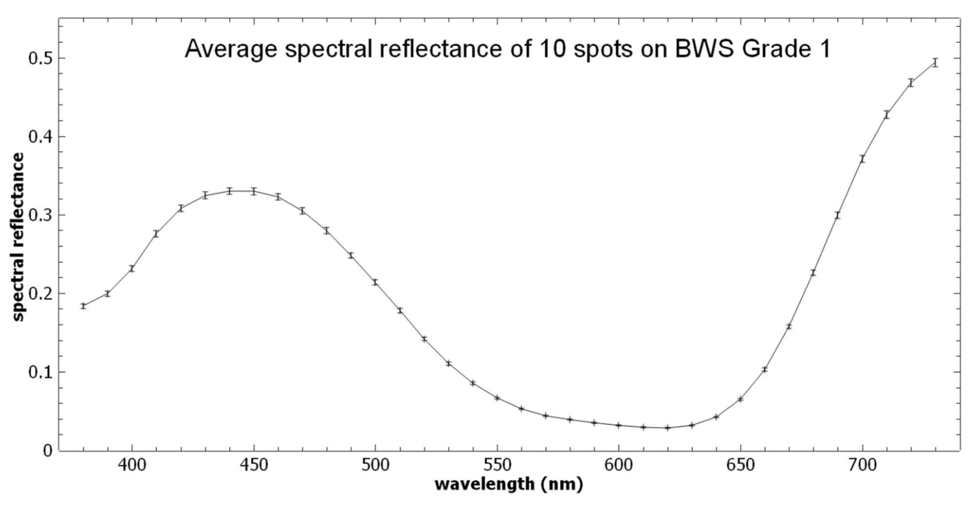
28]. In this study, samples of grades 1 to 8 were mounted on a flat black-painted sample holder. To provide a reference, one half of the area of each sample was shielded with a paper board for most of the measurements, while the other half was exposed to the lights of the instrument at every IS measurement. The samples were exposed to 45 IS measurements, corresponding to a monitoring period of about 20 years. The monitoring period of 20 years was chosen as the best representation for the operational lifetime of such an instrument and because two recordings per year were considered for the exhibited documents at the Nationaal Archief as they follow a rotation schedule every six months. The effect of the IS measurements on the BWS samples was determined by repeated measurements of their reflectance spectra with a spectrophotometer (model Xrite i1pro-1) that provides reflectance values at every 10 nm over the spectral range of 380 to 730 nm, using a measurement spot diameter of 4.5 mm in a 45°/0° illumination/detection geometry. After calibration with the white standard provided with the device (calibration software “i1 Diagnostic v 4.0.0.127”), 10 different spots were measured on each BWS sample. The reproducibility of these spot measurements is shown in
Figure 2, with the spectrum of BWS Grade 1 before any exposure to IS as an example. The average spectral reflectance values over the 10 spot measurements are connected by straight lines. The error bars around each average value indicate ±1 standard deviation (SD) of the spectral reflectance values of the 10 individual measurements. For further analysis, the average spectra of the 10 spots were used.
To enable the assessment of the impact of IS, the reproducibility of the photospectrometer for measuring any spectral changes must be high enough so that the corresponding error on the measured colour change is below the visual detection threshold. This was verified by comparing any two photometer spectra (each averaged over 10 spots) that were measured on the same BWS reference area when it had been exposed to the same number of IS measurements. For each BWS sample, 51 unique pairs of such measurements are available. For each pair, the difference spectrum of the later minus the earlier one of both measurements and their colour difference ΔE2000 [
29] were calculated. The visual threshold was set at a value of ΔE2000 = 0.7, which based on the work of Pretzel [
30] corresponds to the 30% probability that a human observer is able to detect a colour difference.
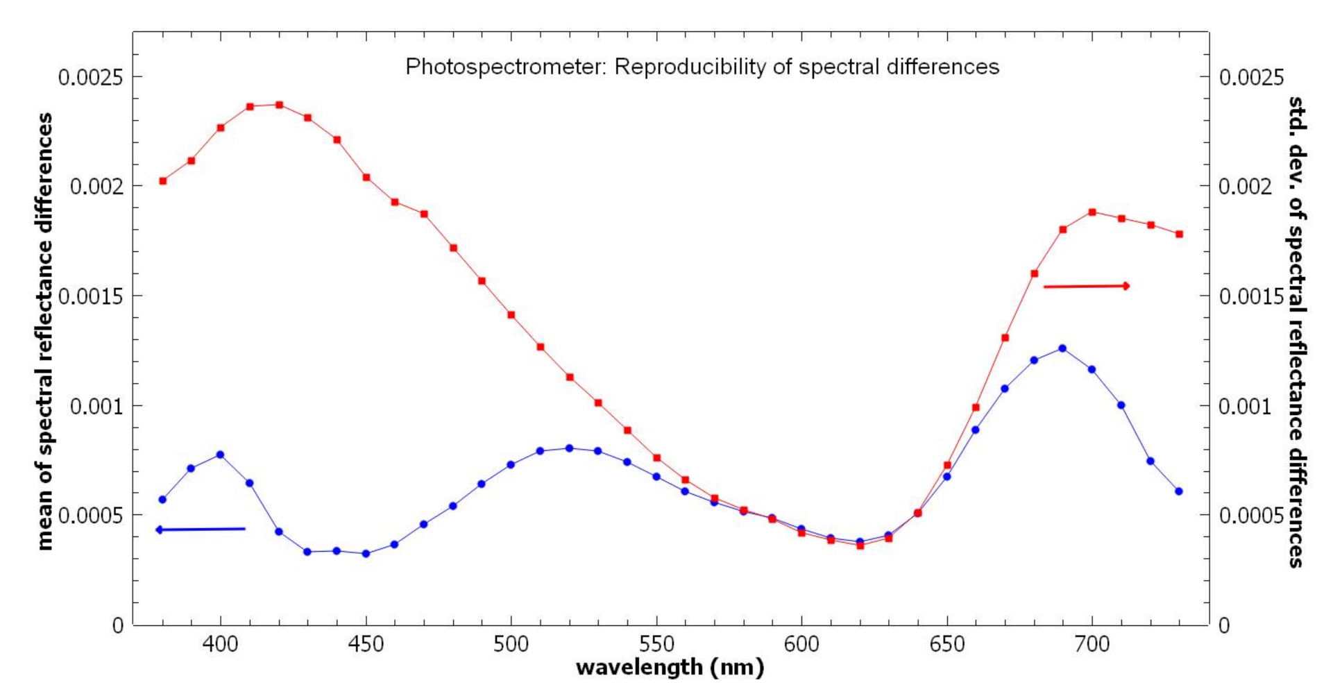
As seen in
Figure 3, the mean value of the differences does not exceed the SD, and for most wavelengths it even remains considerably below the SD. This means that there is no significant bias (drift) of the photospectrometer measurements and no significant change of the spectra of the reference area in the course of the experiment. The SD of the photospectrometer measurements on the reference areas can therefore be used to estimate the measurement error: measured spectral changes on the exposed BWS areas can be assumed to be significant (i.e., real changes) if they exceed ±2 SD.
For each of the 51 pairs of reference area measurements, the colour difference (ΔE2000) was calculated. The average colour difference is 0.14 (SD = 0.08), and for 95% (i.e., for 49 pairs) of the measured colour difference values were less than 0.27. The latter value can be used as a conservative estimate of the measurement error for the colour difference: if a colour difference measured on an exposed area exceeds this value, it very likely indicates a true colour change rather than a measurement error.
Spectrophotometer measurements themselves required irradiation of the BWS samples, and the overall results therefore represent an overestimation of both the measurement error and of the impact of long-term monitoring.
For testing the monitoring sensitivity, a second set of samples containing the BWS as well as common archival materials were exposed to photodegradation using a range of light doses.
Table 1 reports the six test materials that were selected.
The main light sources used in the exhibition
De Verdieping van Nederland at the Nationaal Archief at the time of the experiments are quartz-tungsten halogen reflector lamps without infrared suppression, which are mounted on the room ceiling for spot illumination of the objects in their glass cases. The dedicated setup that was built for accelerated light ageing [
33,
34] was designed to approximate the light spectrum of the illumination of the objects in the exhibition. To achieve this, the same type of halogen reflector lamps were used and the light was transmitted through the top glass plate of an exhibition case. As opposed to the situation in the exhibition, a homogeneous distribution of the light intensity on the samples at the intended level was required. Therefore, the light transmitted through the glass was diffused by scattering it from white cardboard in two steps before it reached the sample areas. The homogeneity with each area was verified with a lux meter.
Figure 4 shows the spectral power distribution of the homogenized halogen light with which the samples were irradiated in this research.
The setup was used to induce accelerated light ageing at five intensity levels, by placing a set of samples at a suitable distance from the diffused light source. The light intensity to which each sample set was exposed was measured with a lux meter and monitored after each exposition time.
To prevent excessive heating of the samples, which could impact on their ageing behaviour in an uncontrolled way, forced air cooling was applied to the confined space of the sample chamber. The air temperature and relative humidity were monitored inside and just outside of the sample chamber. During the light ageing experiments the temperature ranged from 20 °C to 26 °C and the relative humidity from 35% to 62% RH.
The illumination in the De Verdieping van Nederland exhibition at the time of the experiments were 50 lux for 7 h/day over a period of three months.
The samples in this research were exposed to light doses ranging from ca. 1.5 to 500 klx·h, which corresponds to a range of 4–1400 days of exhibition at the above conditions.
Table 2 reports the range of light doses obtained using different combinations of irradiation intensities and exposure times. In addition, 5 subsamples of each material underwent the same preparation but remained covered (i.e., 0 lx) during the accelerated light ageing process to serve as reference samples, having experienced only a small light dose during sample preparation.
Table 2 also indicates the number of days in the exhibition that would result in the same accumulative light dose. The row with 0 lx indicates the reference subsamples that remained covered during the accelerated aging.
The number of 6 materials and the number of light intensities were chosen to optimize the recording surface of the IS instrument. For each of the six materials, 5 × 6 = 30 subsamples were cut out, to be used in the experiments. The samples were arranged to fit into the field-of-view (FOV) of the tested IS instrument (120 mm × 120 mm) in order to optimize the duration of measurements and improve the quality of the results.
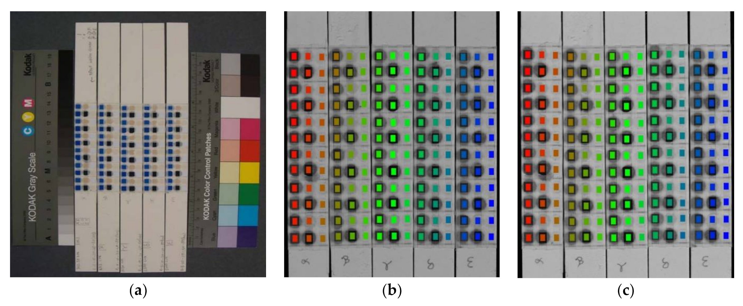
Round samples (Ø 5 mm) were cut taking care they were taken from a homogeneous area of each test material. The pieces were mounted on five buffered RagMat Museum Board 4 ply Natural, without liquid adhesive but instead using Filmoplast
® by Neschen that covered only small margins of the samples as shown in
Figure 5a.
The resulting set of 180 sub-samples was measured with the IS instrument twice: before and after the accelerated light-ageing experiment. Each measurement results in a calibrated hyperspectral datacube (a stack of calibrated spectral images) that contains a spectral reflectance curve for each image pixel. For each of the 180 material samples a region-of-interest (ROI) consisting of 38 × 52 = 1976 pixels was defined, which corresponds to an area = 2.3 × 3.2 mm. Any slight shift of the samples in the field-of-view of the instrument, visually detected in the second measurement with respect to the first measurement, was compensated by a corresponding shift of the ROI areas, see
Figure 5b,c.
For each ROI, the mean spectral curve and the standard deviations (SD) over all 1976 pixels were calculated for both measurements.
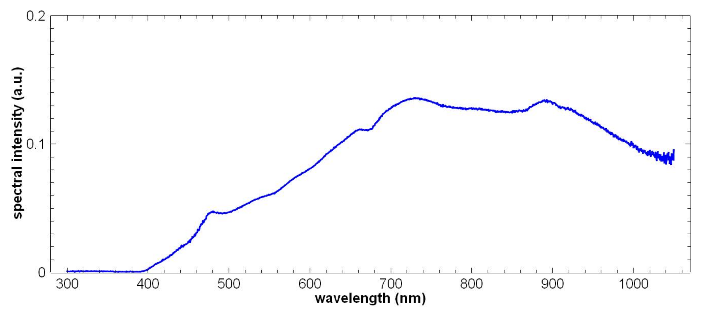
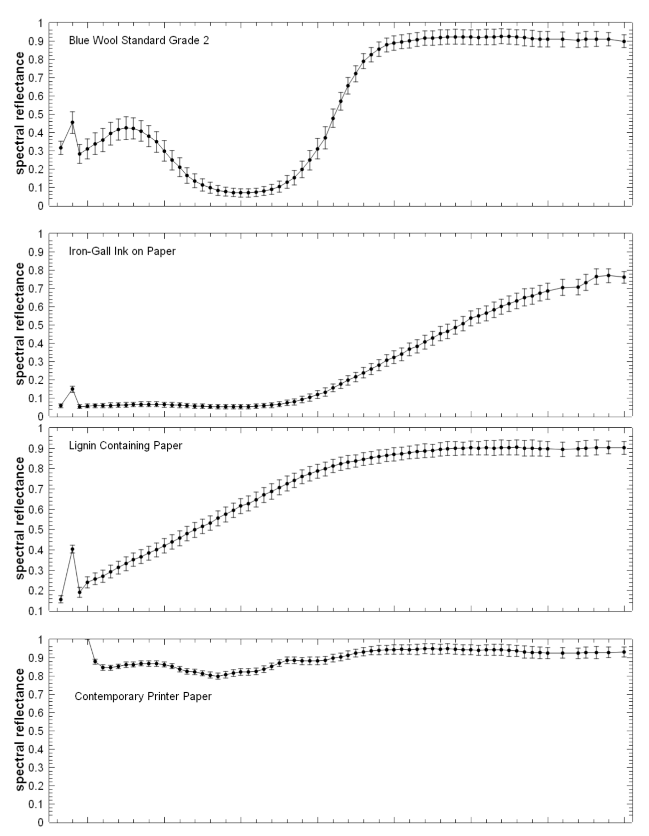
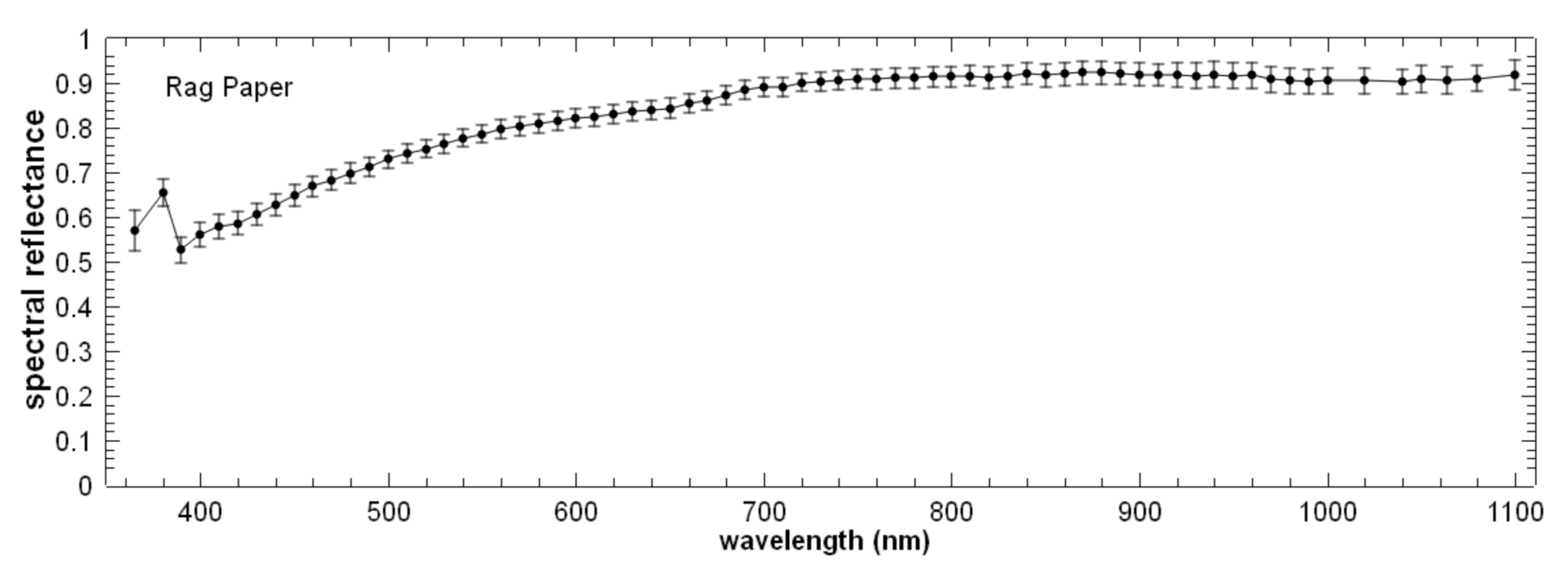
Figure 6 shows the mean spectral curves of one particular ROI for each test material before artificial ageing. The standard deviation reflects the combination of measurement noise of individual pixel values and the inhomogeneity of the material within the ROI area. Note that the peak in the curves of all samples at 380 nm is most likely caused by an imperfection of the corresponding spectral filter of the instrument, i.e., it is a measurement artefact rather than a reflectance characteristic of the samples.
For each sample, the difference spectrum was calculated by subtracting the corresponding ROI spectral curve before light ageing from the spectral curve of the same ROI after ageing. These difference spectra correspond to the spectral changes over the wavelength range of 365 to 1100 nm induced by ageing.
An estimate of the measurement error at each spectral band was obtained from the difference spectra as follows. For each spectral band, the mean value and standard deviation for all 30 difference values (5 reference subsamples of all 6 materials) were calculated. The mean value can be interpreted as a systematic error, whereas the standard deviation reflects a random contribution to the error. Measured difference values outside the range of the mean value ± 2 SD are considered to be statistically significant, i.e., the object has changed. However, since the wavelength-dependent measurement errors vary for the different materials, more conservative (i.e., larger) error limits can be derived by extending the ±2 SD limits by the minimum and the maximum difference values for the particular material.
When considering degradation of archival materials, an important criterion is whether such degradation can be detected visually (e.g., by comparison with colour charts). Therefore, in addition to the spectral differences at all wavelengths, the colour difference values ΔE2000 were calculated for samples before and after ageing. These results are reported in
Table 3.
As an estimate for the measurement error ε(ΔE2000), for each material the maximum colour difference measured between the exposed and unexposed samples was used. It therefore takes into account the repeatability of the IS measurement itself, including any residual ROI positioning error and also any change in the reference samples not exposed to ageing. Any detected colour change >ε(ΔE2000) can thus be assumed to be a true change rather than measurement error. This means that ε(ΔE2000) defines the detection limit of the method, for the respective material. Note that for all materials, the measurement error ε(ΔE2000) is well below the threshold value of ΔE2000 = 0.7, above which there is a 30% chance that a colour change is visually noticeable for a human observer.
The ΔE2000 value indicates whether a colour change induced by the ageing could be visibly detectable. Taking into account the particulars of human colour perception in combination with standardized illumination condition, reflectance changes in different wavelength regions are weighted differently and by definition, reflectance changes outside the visible range are not taken into account for colour measurements.
An alternative measure of spectral change that takes into account all wavelengths in the measured spectral range is the so-called standardized Euclidian distance Δ
Euclid. It is not based on (and limited by) human colour perception, and it uses all spectral values over the entire spectral range provided by the used instrument. The contribution from each wavelength to Δ
Euclid is weighted according to the estimated error. It is defined as:
where
while the other symbols used are defined in
Table 4. Note that all spectral measurements were carried out at the same spectral bands, which means that the sums in Equations (1) and (2) have the same number of terms for all samples. The standardized Euclidian distances are therefore comparable for all samples without the need to normalize for the number of spectral bands.
As an estimate for the measurement error ε(Δ
Euclid) the maximum Δ
Euclid value determined for the 5 unexposed samples was used for each material reported in
Table 1. Since the standardized Euclidian distance includes measurement data from additional spectral bands outside the visible range, there is the chance that allows the detection of changes of the monitored material before they can be detected by measurements in the visible range only.
3. Results
The eight different BWS samples were subjected to a number of IS measurements that correspond to 20 years of regular monitoring, in order to assess the impact. Before any IS measurements and after certain numbers of IS measurements, the samples were measured with the photospectrometer in order to quantify the impact of the multiple IS measurements on the materials with an independent instrument.
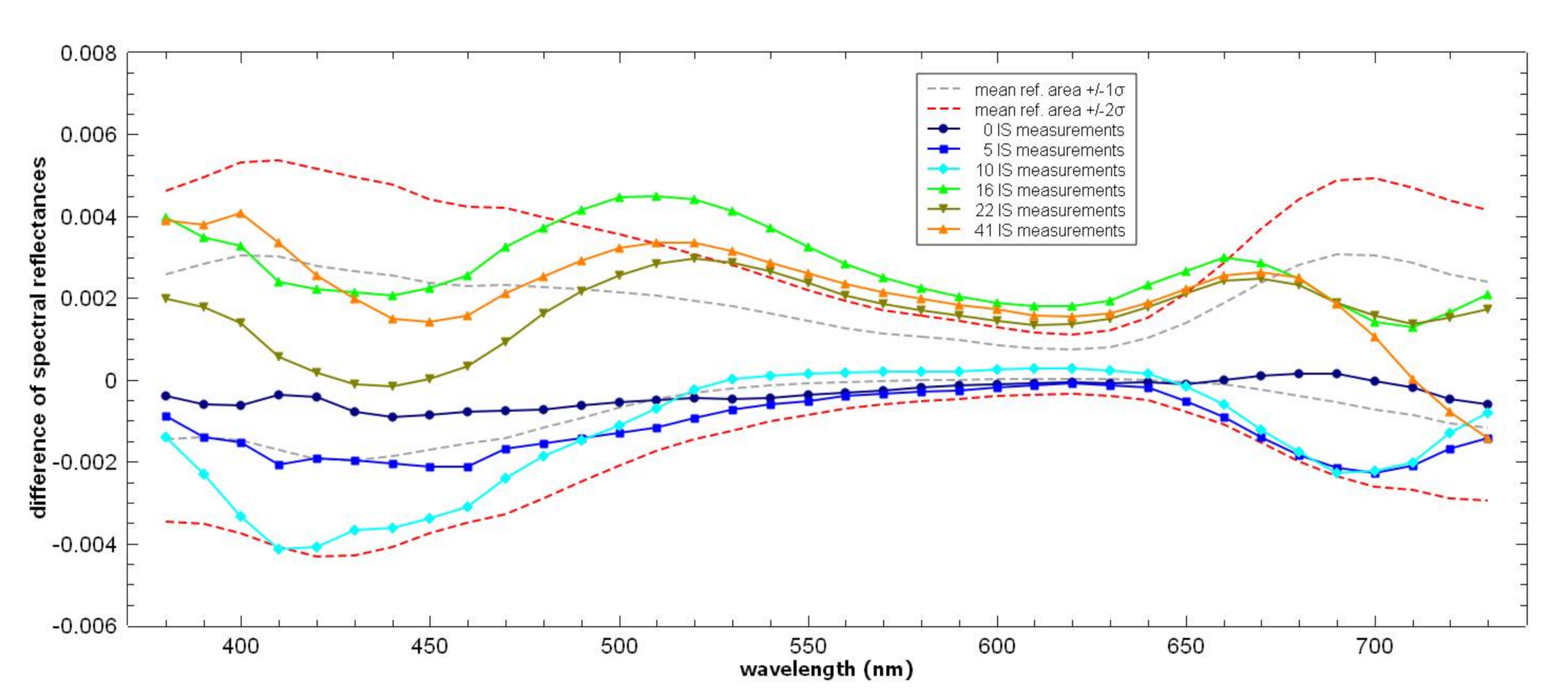
Figure 7 shows for BWS1 the differences in the photospectrometer spectra recorded after different numbers of IS measurements with respect to the initial spectrum before any IS measurement. BWS grade 1 with the lowest light-fastness was chosen among the eight samples as it is deemed to be representative for the most light sensitive archival materials such as newspapers, photographs and watercolours. The grey and the red dashed curves indicate the estimated error of the photometer measurements with, respectively, ±1
σ(
λ) and ±2
σ(
λ) ranges around the mean of the spectral differences measured for the reference area where both measurements were exposed to the same number of IS measurement. After 16, 22, and 41 IS measurements, the differences in measured reflectance for the wavelength range of 530 to 650 nm are consistently outside the ±2
σ(
λ) range. The photospectrometer is thus capable of detecting the corresponding small spectral differences in this wavelength range, which occur on the exposed area of the BWS1 sample after multiple IS measurements.
While comparing measurements at individual wavelengths is useful, it is possible that small, correlated changes in the spectra give rise to statistically significant differences before they can be detected at any of the individual measurements. In the present case the main question was whether any changes caused by repeated IS measurements could become visible to the human eye.
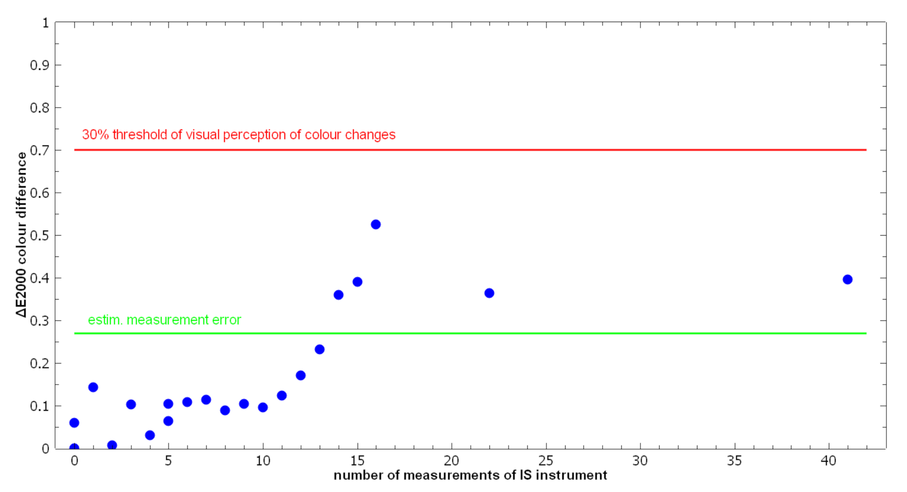
Figure 8 shows ΔE2000 for BWS Grade 1 as a function of the number of repeated IS instrument measurements. As described in the Materials and Methods section of this article, the ΔE2000 = 0.27 is the estimated error of the spectrophotometer measurement and values higher than this indicate statistically significant colour changes. The limit value of ΔE2000 = 1 is per definition considered to be the smallest colour difference a trained human observer can detect. The ΔE2000 = 0.7 level has been shown to be the threshold value above which the statistically more than 30% of the human observers are able to detect a difference in colour. All the measured colour differences remain below this more stringent threshold value for the 41 repeated IS measurements.
Results show that even very light-sensitive documents can be monitored with the SEPIA over periods of more than 20 years with the frequency of measurement of twice a year, before light exposure inherent to the measurement itself starts to effect any visible changes. These results are specific for the SEPIA device but other IS measurement systems could lead to similar results depending on their design. One of the key design features of the SEPIA instrument is that the wavelength selection for each spectral image is done inside the light sources. For each spectral image the measured object is irradiated only by the relatively low-intensity monochromatic light required to record this particular image. Other IS instrument concepts that rely on broadband illumination and sequential image recording with spectral filters at the camera expose the monitored objects to unnecessary high light doses and are therefore much less suitable for long-term monitoring. The camera lens and image sensor (CCD or CMOS) of the IS instrument has to be efficient in collecting the light reflected from the object, so that for a given light intensity the camera exposure time can be as short as possible. After the camera exposure has finished, the light source has to be switched off, so that the overall light dose received by the object during the entire measurement is minimized. At the time of the experiment, the Nationaal Archief was monitoring a range of circa 30 documents.
The Xrite spectrophotometer proved to be sensitive enough for control measurements as the colour changes induced in the most light-sensitive BWS became just measurable at the 15th repeated measurement, although still well below the visibility threshold. However, any further data analysis for the less light-sensitive BWS became obsolete with this, as even Grade 1 BWS was not affected significantly.
The second set of experiments addressed the sensitivity of IS measurements for detecting light-induced changes of six different materials.
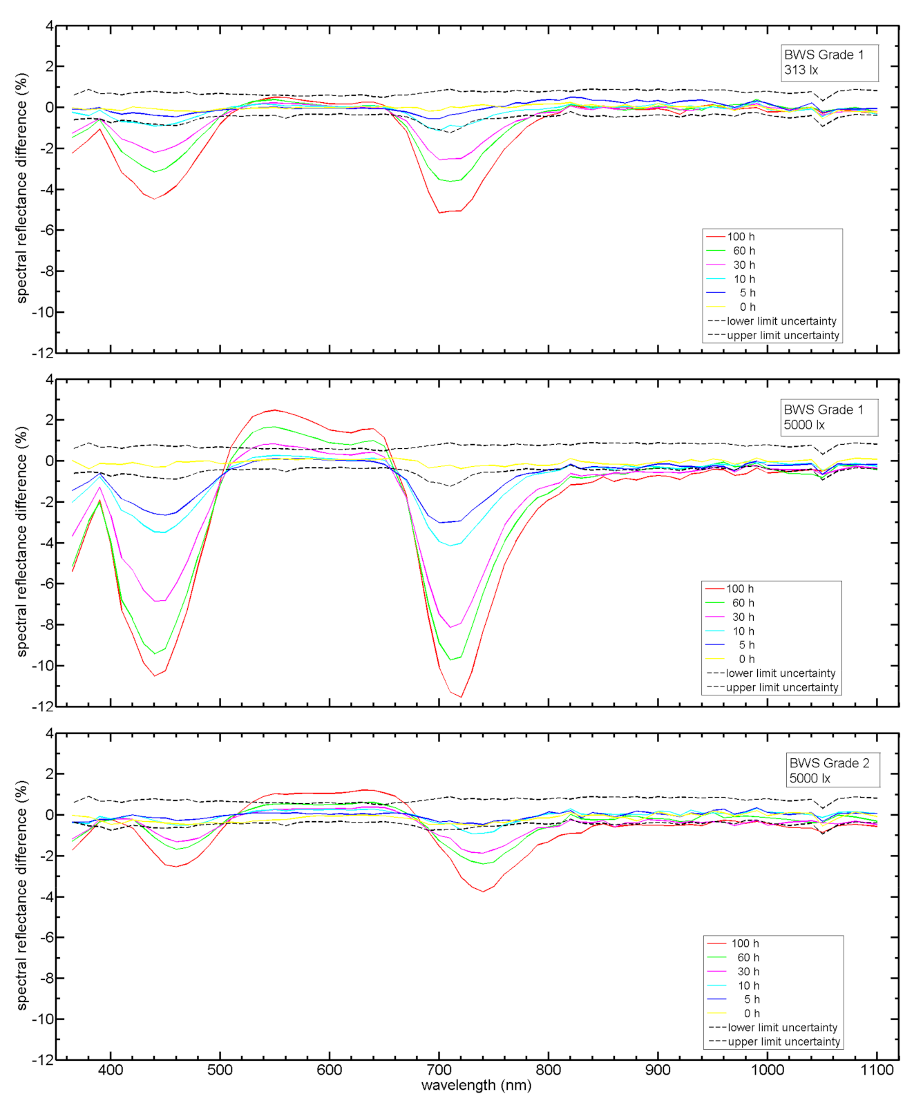
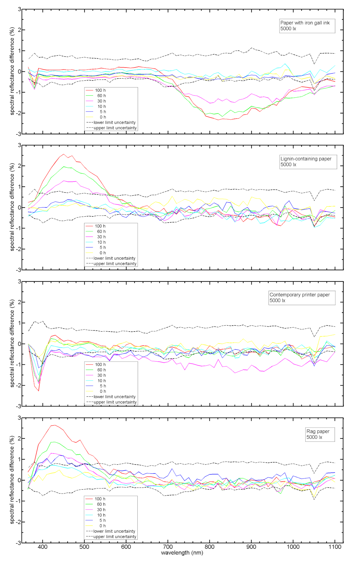
Figure 9 and
Figure 10 show the measured spectral changes for the six materials as induced by exposure to the maximum intensity of 5000 lux and a range of exposure periods. Please note the different y-axis scales in the diagrams of both figures.
Figure 9 also includes the spectral changes at the minimum light intensity of 313 lux for BWS Grade 1. At the maximum light dose (i.e., at the maximum exposure time at the given intensity), and typically already at much lower light doses, all materials exhibit statistically significant spectral changes at several wavelengths.
For each material, the particular wavelength was determined where the maximum absolute spectral change was measured. The capability of the IS instrument to measure spectral differences in a given material can be expressed as the minimum light dose required to induce a spectral change that exceeds the measurement error at the material-specific wavelength with maximum change. The wavelengths and values of maximum change, the error limits at these wavelengths and the minimum detectable light doses of all materials are given in
Table 5.
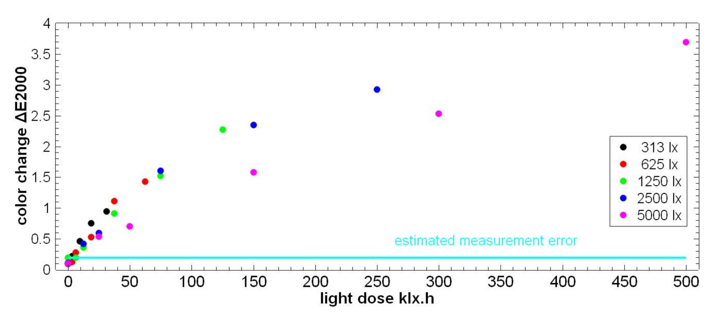
From the spectral reflectance curves measured with the IS instrument before and after accelerated ageing the function of induced colour change ΔE2000 vs. total light dose was calculated.
Figure 11 shows the data for BWS Grade 1.
In line with the reciprocity principle [
35], it would be expected that for a given light dose the same colour change is obtained regardless of the actual combination of light intensity and exposure duration. However, the data points corresponding to 5000 lx clearly show a slower increase than the rest. This could indicate deviation from reciprocity at high intensities, however, it might also reflect an uncertainty of the intensity levels in the light ageing setup. In any case, the dependence of colour change on light intensity is sufficiently low for the purpose of this experiment, i.e., to quantify the lowest light dose that induces colour change detectable by the IS instrument.
For the BWS1 sample, the ΔE2000 = 0.7 visibility threshold is obtained at light doses ~20–30 klx·h, corresponding to ~60–90 exhibition days at 50 lx, 7 h/day, from the halogen light sources as used in the exhibition De Verdieping van Nederland. Furthermore, the conservative estimation of ΔE2000 = 0.5 as induced by IS measurements over 20 years can be estimated as equivalent to ~40–60 days of exhibition under the same conditions.
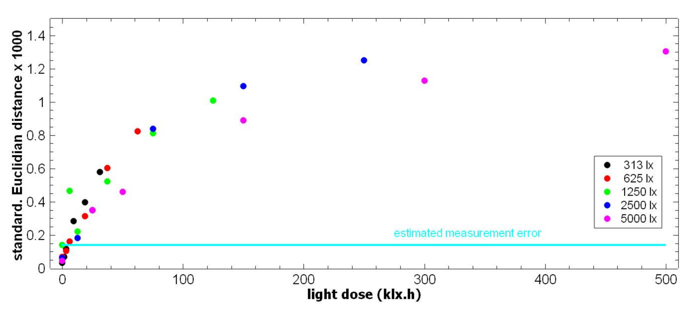
In addition to the ΔE2000, the standardized Euclidian distance Δ
Euclid was calculated for the spectral reflectance curves measured after and before accelerated light ageing, as shown in
Figure 12 for the BWS Grade 1.
Table 6 and
Table 7 list ΔE2000 and Δ
Euclid, respectively, at the maximum light dose of 500 klx·h, for all the sample materials. As expected on the basis of
Figure 9 and
Figure 10, the biggest change is measured for BWS Grade 1 and the smallest change is measured for contemporary printer paper.
Based on the detection limits ε(ΔE2000) and ε(Δ
Euclid) an estimate of the light dose required to induce the minimum detectable change was made for each material. The corresponding values are listed in the last column of
Table 6 and
Table 7, respectively. The required light dose of 3.5 klx·h to achieve a detectable colour change in BWS Grade 1 samples corresponds to ~10 days of exhibition under the stated conditions.
For contemporary printer paper with its much higher light-fastness, the colour and the spectral changes at the maximum applied light dose of 500 klx·h is at the same level as the corresponding detection limits given by the estimated measurement errors. Note that the fact that the maximum colour and spectral difference were measured at lower light doses is also an effect of the measurement uncertainty. For the iron-gall ink on paper sample, the ΔE2000 colour change measured at 500 klx·h is also just at the level of the measurement error so that this light dose can only be used as a lower limit. The ΔEuclid value at 500 klx·h is about twice the estimated measurement error, allowing an estimate of 135 klx·h for the minimum detectable light dose.
Both ΔE2000 and ΔEuclid are generic measures for spectral change. It can be expected that by using material-specific measures that give higher weights to those spectral regions where the strongest changes are induced, the sensitivity of the IS measurements for detecting light-induced changes can be further improved.
4. Discussion
This article describes a generally applicable approach to evaluation whether an imaging spectroscopic instrument is suitable as a tool for monitoring of archival documents. Depending on the object type to be investigated, instruments are typically selected based on their spectral range and resolution, their field-of-view and spatial resolution. However, in order to be useful for monitoring, an IS instrument has to fulfil two additional requirements:
We have shown that Requirement 1 can be tested by subjecting Blue Wool Standards to repeated measurements, corresponding to the intended duration of the monitoring schedule. The induced spectral change needs to be determined with an independent, sufficiently sensitive method, such as a spectrophotometer.
From a conservation point of view, the decision whether the predicted spectral changes induced by IS measurements themselves are acceptable or not must become part of the overall risk assessment of an institution’s exhibition and monitoring schedule. A reasonable guideline would be that the spectral changes induced by measurements must be significantly less than those expected from exposure during exhibitions.
In the case study discussed here, the “SEPIA” hyperspectral imaging system was tested for an intended monitoring period of 20 years with an average of two measurements per year. The spectral changes induced by the tested IS instrument in Blue Wool Standard Grade 1 remained below the threshold for visually perceptible colour changes of ΔE2000 = 0.7, which fulfils Requirement 1.
In order to test Requirement 2, sample materials representative of the monitored objects should be used in addition to standard reference materials. This is because the wavelength range and degree of spectral change depend on the material composition. Furthermore, the strength and spectral characteristics of light-induced change typically depend significantly on the irradiation spectrum. It is therefore advisable that the sample materials are exposed to accelerated degradation under conditions similar to those they are exposed to during exhibitions, in order to induce the same type of spectral change.
In our case study, four archival materials were subjected to accelerated ageing by 30 different combinations of light intensity and duration, corresponding to light doses of up to 500 klx·h. The deployed lamp type was the same as that used in the exhibition room
De Verdieping van Nederland at the Nationaal Archief, to ensure that the same type of spectral change is induced as expected for exhibitions. The samples were irradiated with only moderate light intensities of up to 5000 lx and forced-air cooling was applied to minimize their temperature increase. The induced spectral changes should not be distorted significantly by any non-linearity of the material response with respect to the intensity (reciprocity-failure [
36]) or by very high local sample temperature and low humidity. The spectral changes in the experiment are therefore expected to be comparable to those incurred by the materials over many years due to the normal exhibition schedule.
The changes were evaluated by calculating colour differences ΔE2000 and standardized Euclidian distances ΔEuclid between the sample spectra measured before and after accelerated light ageing. The minimum light dose required to induce a detectable spectral change varied considerably from material to material. For the most light-sensitive Blue Wool Standard Grade 1, the detection limit for light-induced colour change corresponded to an exhibition period of ~10 days.
The instrument was thus shown to have an acceptably small impact on the materials, fulfilling Requirement 1, and acceptable high detection sensitivity for changes induced by exhibition lighting, i.e., also fulfilling Requirement 2.

 ,
, 


