Article

Simultaneous Visual Detection of 12 Pathogenic Bacteria Based on Multiplex PCR-Gene Membrane Chip Technology

1
Yangzhou Center for Food and Drug Control, Yangzhou 225000, China
2
College of Food Science and Engineering, Yangzhou University, Yangzhou 225009, China
*
Author to whom correspondence should be addressed.
Submission received: 2 June 2026 / Revised: 24 July 2026 / Accepted: 31 July 2026 / Published: 4 August 2026
(This article belongs to the Section Biology & Life Sciences)

Abstract

Rapid and accurate detection of pathogenic bacteria is of critical concern in the food and pharmaceutical sectors. In this study, a gene membrane chip detection method combining multiplex PCR with reverse dot blot hybridization was developed to enable visual, high-throughput simultaneous identification of 12 common pathogenic bacteria. Specific primers and probes were designed targeting Klebsiella pneumoniae, Proteus mirabilis, Vibrio parahaemolyticus, Enterobacter cloacae, Pseudomonas putida, Listeria monocytogenes, Salmonella spp., Pseudomonas aeruginosa, Escherichia coli, Acinetobacter baumannii, Clostridium perfringens, and Staphylococcus aureus. The multiplex PCR reaction system, hybridization temperature, and color development conditions were systematically optimized to construct the gene membrane chip detection platform. The specificity, limit of detection, and stability of the method were rigorously evaluated. Results demonstrated that the 12 selected specific primer pairs effectively amplified the corresponding targets, yielding amplicon lengths ranging from 81 to 298 bp, with no nonspecific amplification observed. Under the optimized detection system, each probe produced clear, visually distinct color spots exclusively for its target, with no cross-reactivity. The method achieved a limit of detection as low as 0.01 ng/μL for mixed templates, and exhibited excellent intra- and inter-assay reproducibility. The established visual gene membrane chip assay successfully enables simultaneous differentiation of 12 control bacteria in a single reaction, providing a robust technical foundation and promising potential for rapid screening in food and drug safety, but its practical application requires further validation through authentic or spiked food samples.

1. Introduction

Pathogenic bacterial contamination in food products remains a significant cause of foodborne illness worldwide, manifesting clinically as diarrhea, nausea, and vomiting. In severe cases, such infections can escalate to life-threatening conditions, posing substantial risks to human health and imposing a considerable burden on healthcare systems [1]. Projections indicate that by 2050, annual mortality attributable to pathogenic bacteria may exceed 10 million individuals, underscoring the urgent need for robust detection and intervention strategies [2]. A diverse array of pathogenic species—including Staphylococcus aureus, Pseudomonas species, and Escherichia coli—is frequently isolated from food specimens and complex environmental matrices such as water, soil, and processing surfaces. The rapid and accurate detection and identification of these pathogens are therefore critical for protecting public health and maintaining societal stability. Conventional microbiological detection methods, which predominantly rely on microbial cultivation techniques, offer notable advantages including low operational cost and high accuracy when target organisms are present in sufficient numbers. However, these approaches exhibit several significant limitations that hinder their efficacy in contemporary settings. They require prolonged analysis time, often extending over multiple days, thereby preventing rapid pathogen identification during outbreaks. Detection is confined to viable microorganisms, excluding non-viable bacterial cells that may still pose a risk of toxin release or genetic transfer. Sensitivity is often reduced, hampering the detection of low-abundance microorganisms that are present below the threshold of culture-based methods. Additionally, these techniques are dependent on cultivability, precluding the detection of non-culturable species or viable but non-culturable states. These constraints collectively limit their applicability in modern pathogen detection scenarios, where speed, sensitivity, and broad-spectrum coverage are paramount [3].
Molecular diagnostic techniques have become extensively employed in pathogen detection due to their high efficiency, specificity, and precision. Polymerase chain reaction (PCR) represents one of the most widely adopted molecular detection methodologies. PCR employs primers targeting highly conserved microbial gene sequences—such as 16S rRNA or species-specific virulence genes—and utilizes a thermostable DNA polymerase to amplify target DNA fragments through repeated cycles of denaturation, annealing, and extension. The resulting amplicons are then analyzed via gel electrophoresis, real-time fluorescence, or sequencing to achieve microbial detection. Furthermore, since Chamberlain’s introduction of multiplex PCR, this technology has enabled the simultaneous amplification of multiple pathogen target sequences within a single reaction. By incorporating multiple primer pairs, multiplex PCR facilitates the concurrent detection of several pathogens, substantially enhancing detection efficiency and reducing both reagent costs and turnaround time. Consequently, multiplex PCR has found extensive application in pathogen detection across various fields, with demonstrated efficacy in clinical, environmental, and food safety settings [4,5,6].
Reverse dot blot hybridization (RDBH) is a typical visualization-based detection method. It utilizes capture probes that are immobilized on a solid support—such as a nylon membrane or a microfluidic chip—and hybridized with homologous amplified target DNA that has been labeled with fluorescent or chemiluminescent dyes. The resulting signals are detectable by visual inspection or laser scanning, enabling straightforward interpretation [7]. RDBH offers relatively low cost and high throughput compared to other molecular approaches, and it is widely applied for detecting or identifying pathogens from diverse sources, including food, animal, plant, and clinical specimens [8,9]. For example, Yun et al. [10] previously designed a microfluidic thin-film chip combined with an automated RDBH instrument for detecting five genetically modified corn varieties. They immobilized specific DNA probes on square nylon membranes, which were subsequently placed into microreaction chambers of the developed auto-microfluidic thin-film chip (AMTC). Biotin-labeled PCR products containing target DNA fragments from template amplification were then introduced into the microreaction chambers using the microfluidic system. As the PCR products passed through the square nylon membrane, target DNA fragments were captured through complementary base pairing, and signals were visualized using streptavidin-conjugated alkaline phosphatase colorimetric detection kits. In a subsequent study, they further developed an AMTC instrument integrating one-step multiplex PCR and RDBH, enabling simultaneous high-throughput detection of 14 diarrheagenic Escherichia coli strains [11].
Building upon the promising outcomes of prior research, we designed specific primers and probes for 12 key microorganisms: Klebsiella pneumoniae, Proteus mirabilis, Vibrio parahaemolyticus, Enterobacter cloacae, Pseudomonas putida, Listeria monocytogenes, Salmonella spp., Pseudomonas aeruginosa, Escherichia coli, Acinetobacter baumannii, Clostridium perfringens, and Staphylococcus aureus. These 12 bacterial strains are among the microorganisms requiring focused monitoring in the domain of food and pharmaceutical safety. Salmonella spp., Listeria monocytogenes, Staphylococcus aureus, Escherichia coli, and Vibrio parahaemolyticus are internationally recognized foodborne pathogens and common causative agents of foodborne disease outbreaks. Klebsiella pneumoniae and Pseudomonas aeruginosa, as opportunistic pathogens, represent significant agents of healthcare-associated infections and may also contaminate food products and processing environments, with their presence indicating poor hygiene conditions or potential contamination risks. Pseudomonas putida serves as an indicator organism for assessing environmental cleanliness and the hygienic status of production processes. Through the selection of specific genetic targets, the design and validation of highly specific primer and probe combinations, and the optimization of multiplex PCR reaction systems and hybridization conditions, a gene membrane chip-based detection platform was constructed. The method’s specificity, reproducibility, and other key performance indicators were systematically evaluated, aiming to provide reliable novel technical tools for the rapid screening and precise traceability of quality control for these 12 bacterial strains. Through this study, we successfully screened and optimized a multiplex PCR–membrane chip system capable of simultaneously and specifically amplifying 12 pathogenic bacteria closely associated with food and drug safety. This system provides a reference for simultaneous multi-bacterial screening and holds clear industrial application potential.

2. Materials and Methods

2.1. Bacterial Strain

The 12 strains used in this study were purchased from the American Type Culture Collection (ATCC) and the China Industrial Culture Collection (CICC). These strains were Klebsiella pneumoniae (ATCC 700603), Proteus mirabilis (ATCC 25933), Vibrio parahaemolyticus (CICC 21617), Enterobacter cloacae (CICC 21539), Pseudomonas putida (ATCC 49128), Listeria monocytogenes (CICC 21633), Salmonella spp. (CICC 21513), Pseudomonas aeruginosa (ATCC 9721), Escherichia coli (CICC 21530), Acinetobacter baumannii (ATCC 17978), Clostridium perfringens (CICC 22949), and Staphylococcus aureus (ATCC 25923).

2.2. PCR Primer and Probe Design

Conserved sequences and specific genes of target pathogens were analyzed and screened based on information sequences from the GenBank database, with primers designed using Primer 5.0. Reverse primers were labeled with biotin modification at the 5′ end and modified with NH2C6. The sequences of specific primers and probes are shown in Table 1. The bacterial conserved sequence 16S rDNA gene was selected as the internal reference gene. Positive oligonucleotide single-strand DNA sequences, positive control nucleic acid probes (PCs), and negative control nucleic acid probes (NCs) were used for quality control during membrane chip hybridization.

2.3. DNA Extraction and Amplification

Sample DNA was extracted using the Tianamp Bacteria DNA Kit (DP302, Tiangen Biochemical Technology (Beijing) Co. Ltd., Beijing, China). The concentration of extracted genomic DNA was determined using an ultra-micro spectrophotometer (NanoDrop ONE/C, Thermo Fisher Scientific, Waltham, MA, USA). When nucleic acid sample concentrations exceeded 20 ng/μL and the A260/A280 ratio was between 1.8 and 2.0, samples were used for subsequent studies. Nucleic acids were diluted to 10 ng/μL, 1 ng/μL, 0.1 ng/μL, 10−2 ng/μL, 10−4 ng/μL, and 10−6 ng/μL using DNase-free water.

2.4. Primer Specificity Testing

Using 10−2 ng/μL DNA template, PCR amplification was performed according to the PCR Amplification Kit (R011, Takara Co., Ltd. Tokyo, Japan) instructions to screen and confirm primer specificity.

2.5. Primer Detection Limit Testing

DNA templates at concentrations of 1 ng/μL, 10−2 ng/μL, 10−4 ng/μL, and 10−6 ng/μL were used for PCR amplification according to the PCR Amplification Kit (R011, Takara Co., Ltd. Tokyo, Japan) instructions.

2.6. Primer Optimal Annealing Temperature Testing

Using 10−2 ng/μL DNA template, each primer pair was tested using gradient annealing temperatures. PCR amplification was performed according to the PCR Amplification Kit (R011, Takara Co., Ltd. Tokyo, Japan) instructions, with annealing temperature gradients of 50 °C, 55 °C, and 60 °C.

2.7. Specific Probe Layout and Probe Hybridization Efficiency Testing

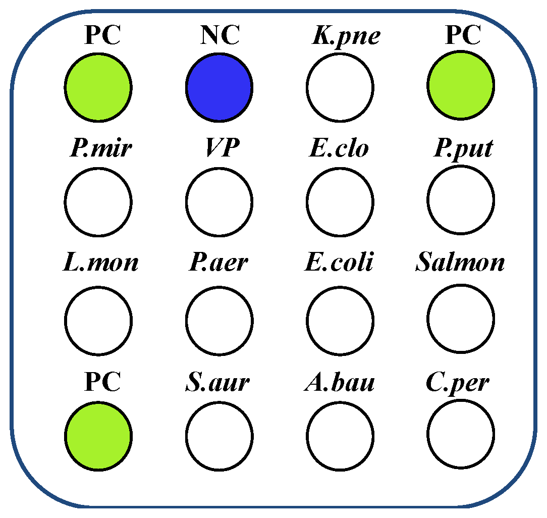
Probes were diluted to 10 μM, and 0.1 μL aliquots were manually spotted onto nylon membranes according to the probe layout shown in Figure 1. After probe spotting was completed, nylon membranes were baked in an 80 °C oven for 1.5 h, cooled to room temperature, trimmed, and placed in hybridization cassettes for hybridization testing. Negative control (NC) template was not added, while hybridization quality controls (PCs) were labeled with biotin to monitor normal color development reactions.
PCR amplification products were denatured at 95 °C for 5 min, then stored at 4 °C for later use. Automated hybridization was performed using a gene membrane chip detection workstation (MFS-8, Sichuan Huahan Sanchuang Biotechnology Co., Ltd., Chengdu, China). According to instrument instructions, denatured PCR products were added to 500 μL hybridization solution (R2) to prepare the hybridization system. Alkaline phosphatase-labeled streptavidin (AP-Streptavidin) was added to enzyme labeling solution (R4) at a 1:250 ratio to prepare the enzyme labeling system, prepared fresh for immediate use. The hybridization program was loaded and automatically executed: hybridization (45 °C, 10 min); hybridization washing (52 °C, 3 min) twice; enzyme labeling (42 °C, 10 min); enzyme labeling wash 1 (42 °C, 3 min) twice; enzyme labeling wash 2 (37 °C, 3 min) once; and color development (37 °C, 8 min). All test templates were amplified using the same primers. After hybridization completion, results were analyzed against the membrane chip layout diagram, selecting probes that showed strong color development for target probes while showing no color development for non-target probes as specific probes.

2.8. Multiplex PCR System Specificity Testing

Multiplex PCR Mix was configured using selected specific primers, and multiplex PCR amplification was performed according to Taq Pro Multiplex DNA Polymerase (high sensitivity) (PM202-1, Vazyme Biotechnology Co., Ltd., Nanjing, China) instructions. The multiplex PCR reaction mixture consisted of 10 µL of 2 × KAPA2G Fast Multiplex Mix (KAPA Biosystems, Wilmington, MA, USA), 1 µL of PCR primer mix, 2 µL of bacterial extract, and 7 µL of ddH2O. The mixture was pre-heated at 37 °C for 5 min, followed by initial denaturation at 95 °C for 3 min, and then 35 cycles. Each cycle comprised denaturation at 95 °C for 15 s, annealing at 55 °C for 30 s, and extension at 68 °C for 15 s. A final extension step was performed at 68 °C for 3 min, after which the tubes were rapidly cooled to 4 °C. Amplification products were subjected to membrane chip hybridization according to Section 2.7.

2.9. Multiplex PCR System Detection Limit and Stability Testing

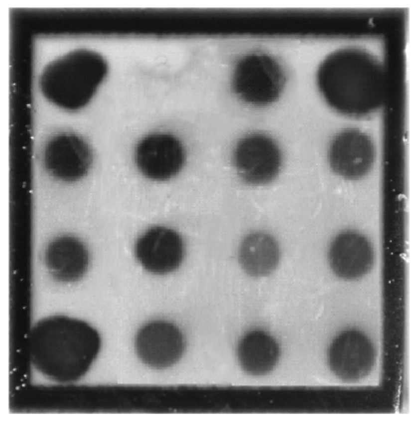
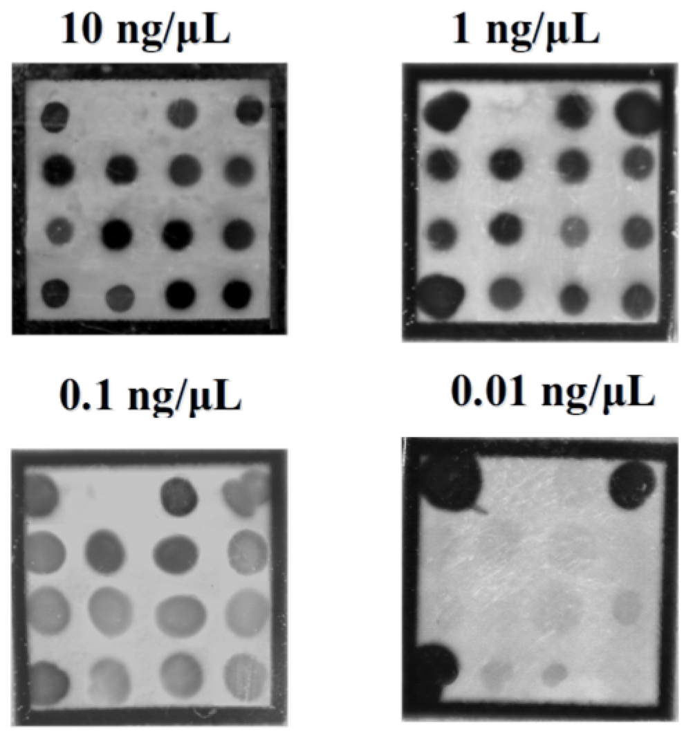
Mixed DNA templates were diluted to 10 ng/μL, 1 ng/μL, 0.1 ng/μL, and 0.01 ng/μL, respectively, and tested according to Section 2.8. Stability testing was performed using repeated measurements with the Mixed DNA template at 1 ng/μL.

2.10. Data Analysis

Gene membrane chip results were automatically photographed by the membrane chip hybridization instrument and processed using MFS-24 automatic analysis software (MFS24.V.1.0.5.21.03, Sichuan Huahan Sanchuang Biotechnology Co., Ltd., China). All key experiments (specificity, limit of detection, multiplex PCR) were independently repeated three times (n = 3).

3. Results

3.1. DNA Extraction

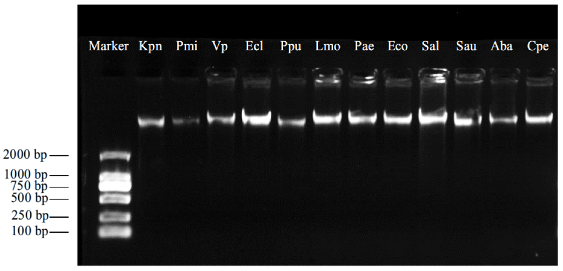
As shown in Figure 2, the extracted DNA bands from the six target strains were single and clear. Quality concentration was detected using an ultra-micro nucleic acid protein analyzer (Nanodrop, Thermo), with A260 nm/A280 nm ratios between 1.8 and 2.0, indicating that the corresponding DNA was suitable for subsequent studies.

3.2. Specific Primer Screening

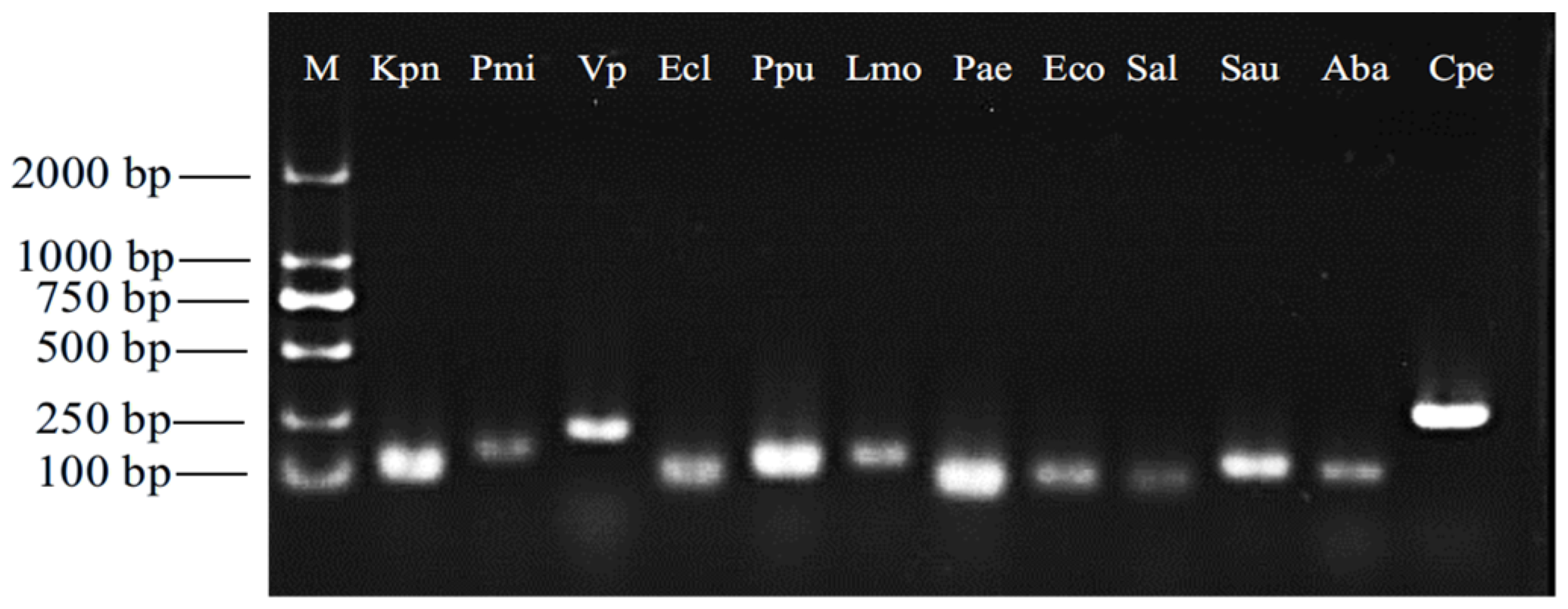
The synthesized primers were used to amplify the corresponding target strains. As shown in Figure 3, each primer successfully amplified the expected bands in their respective strains. The amplification product sizes obtained with each specific primer exactly matched the expected amplicon lengths of the target genes, and the migration positions of the bands corresponded to the theoretical molecular weights of the fragments, indicating that the selected specific primers are suitable for subsequent studies.

3.3. Results of Primer Specificity Testing

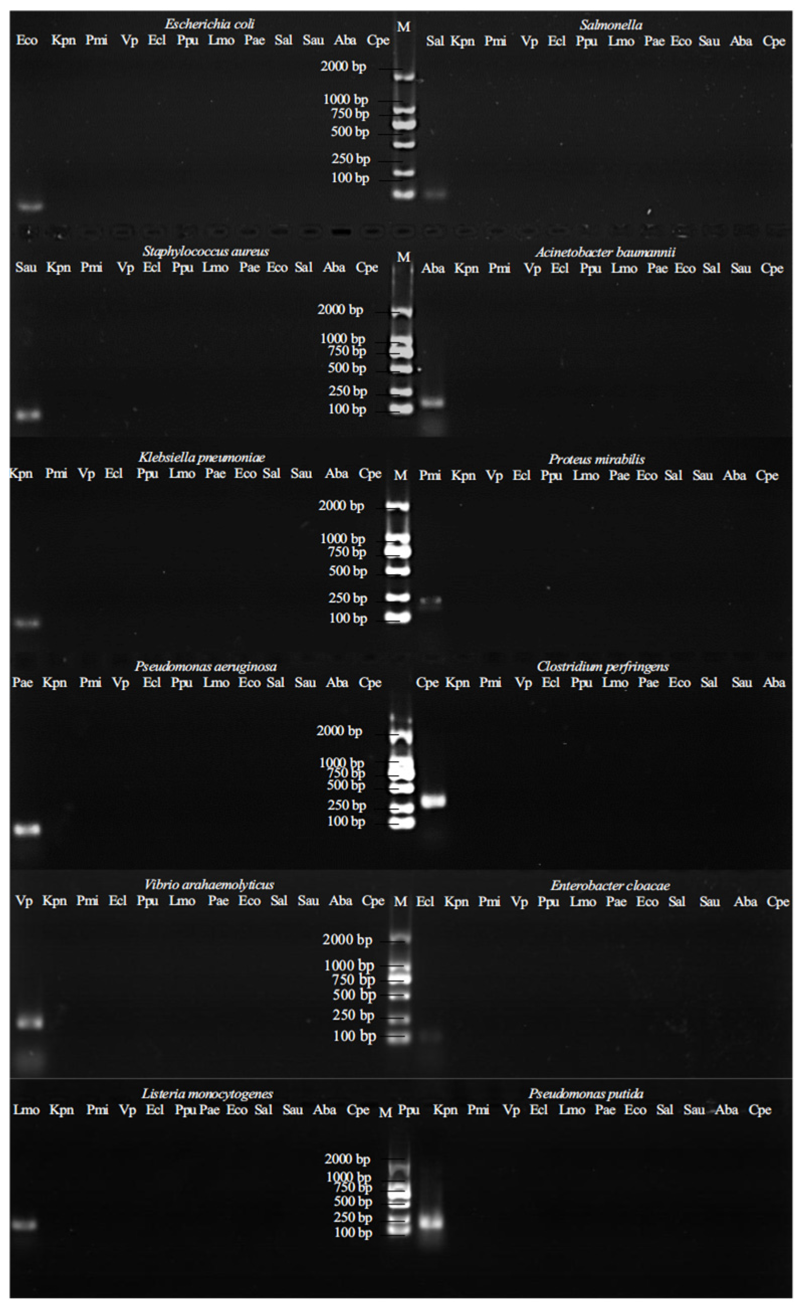
As shown in Figure 4, each primer amplified corresponding bands in their respective target strains, while no identical bands appeared in other control strains, demonstrating excellent specificity of all primers.

3.4. Results of Primer Detection Limit Testing

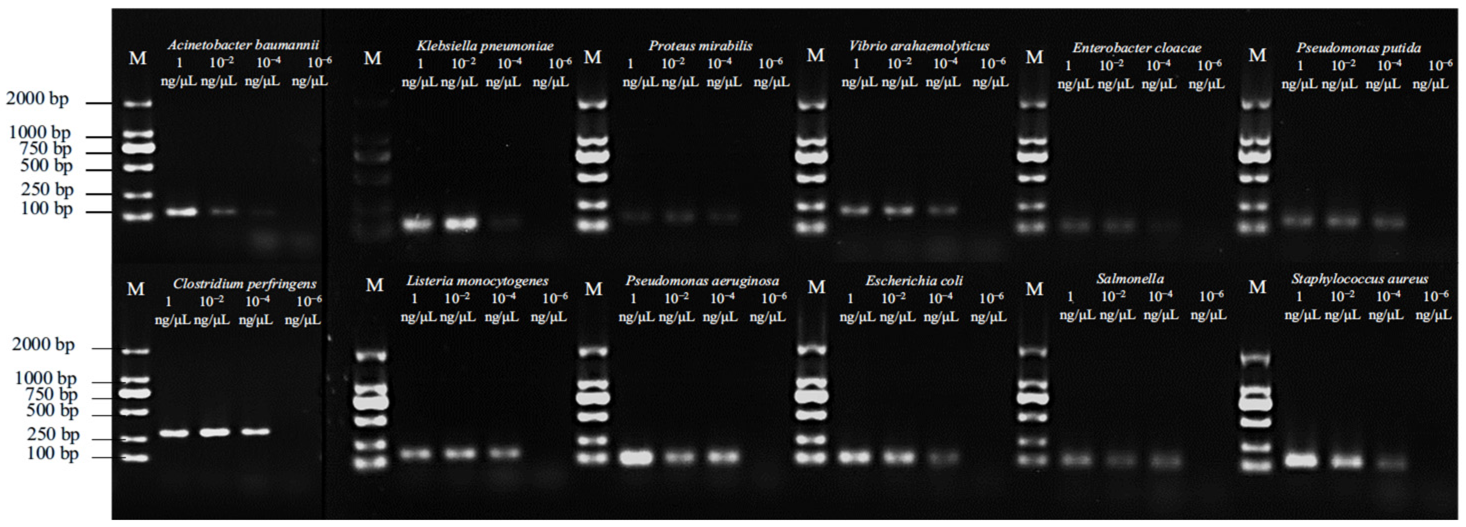
As shown in Figure 5, amplification bands were still obtained with 10−2 ng/μL template, indicating that the selected specific primers met the amplification efficiency requirements.

3.5. Optimal Primer Annealing Temperature Testing

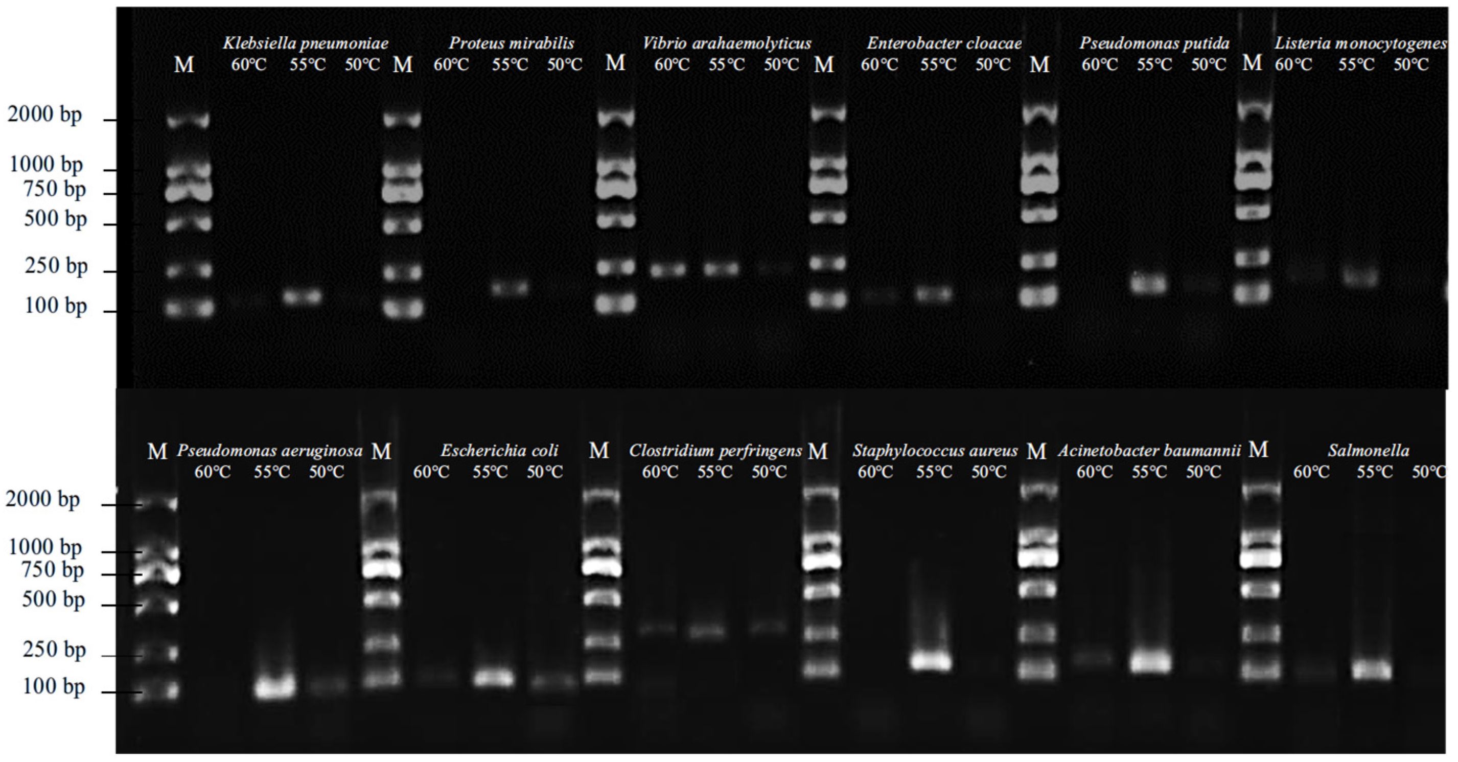
As shown in Figure 6, annealing temperature significantly affected primer amplification. All specific primers exhibited highest band intensity at an annealing temperature of 55 °C. Therefore, 55 °C was selected as the optimal annealing temperature.

3.6. Probe Hybridization Efficiency Testing

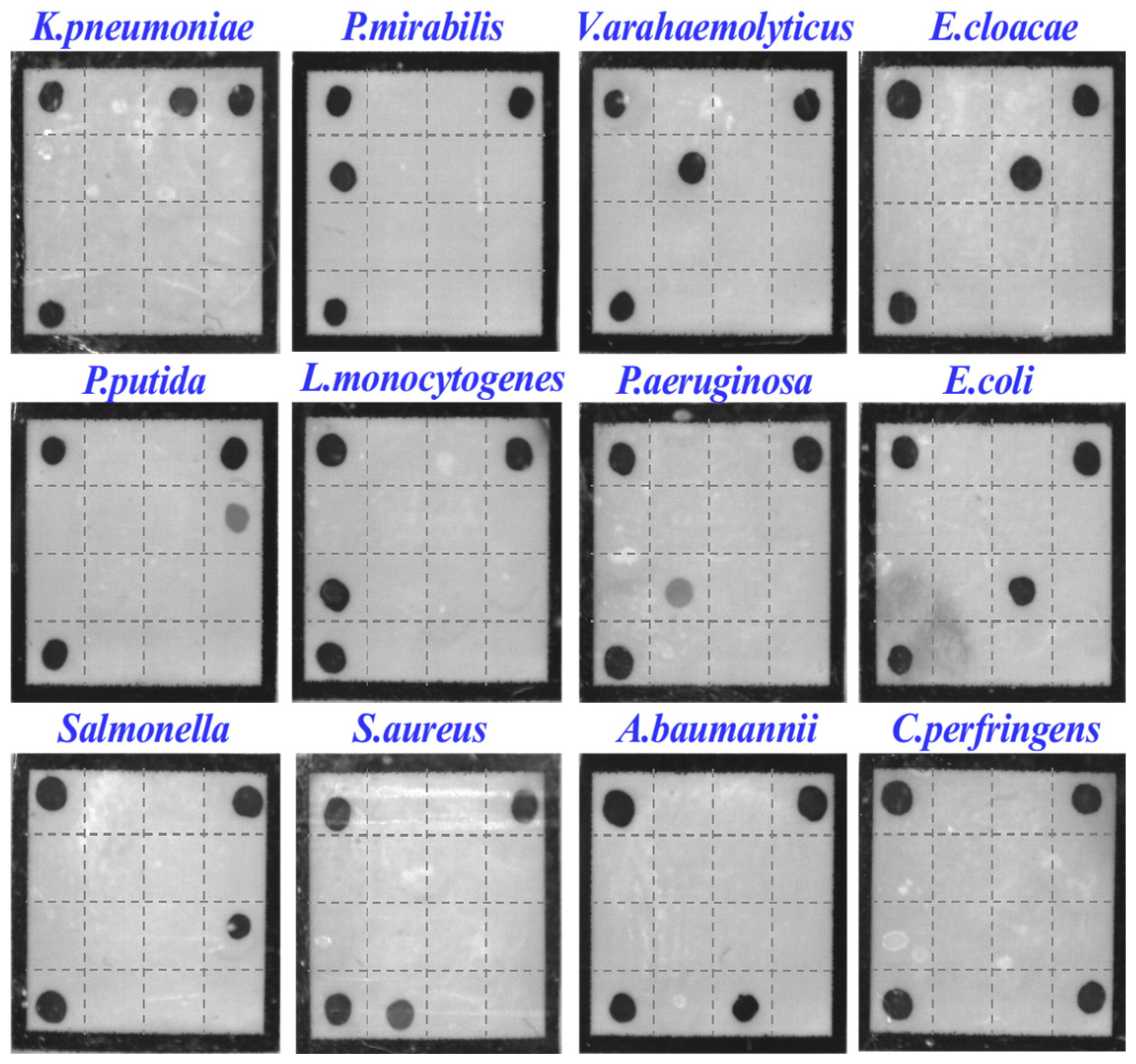
As shown in Figure 7, the finally selected specific probes demonstrated excellent hybridization efficiency. The target probes showed strong colorimetric signals while non-target probes remained colorless.

3.7. Multiplex System Specificity Testing

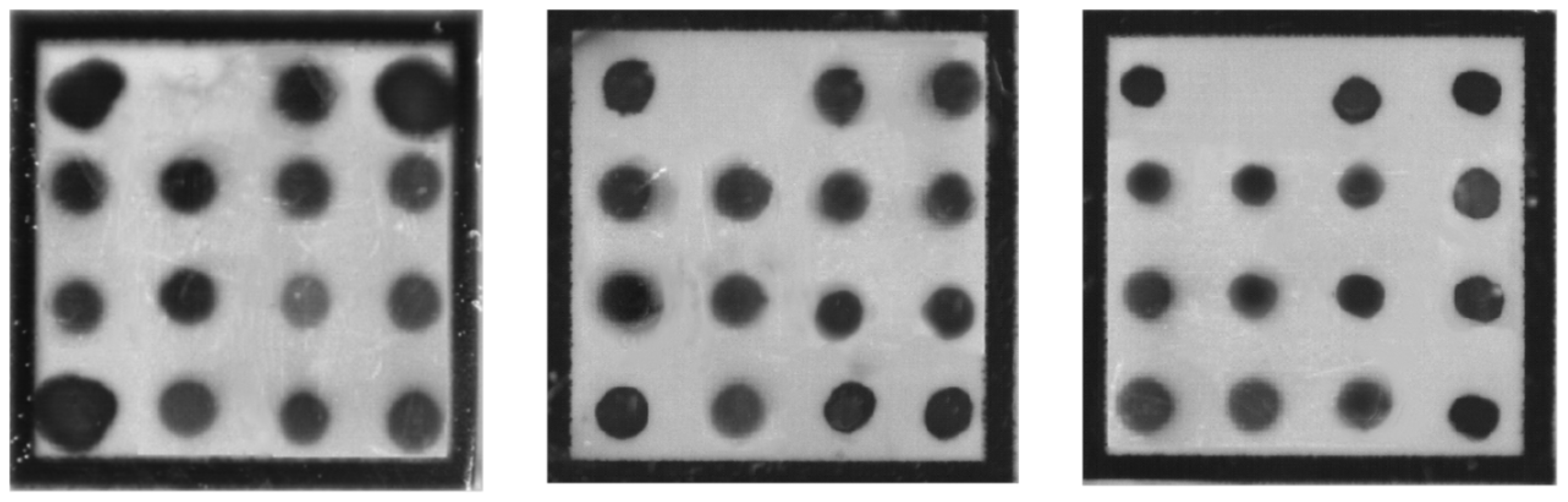
As shown in Figure 8, under mixed template conditions, specific chromogenic signals were observed for each target, with variations in signal intensity; nonetheless, all signals were clearly interpretable.

3.8. Multiplex System Detection Limit Testing

As shown in Figure 9, target coloration decreased with declining template concentration. At a mixed template concentration of 0.01 ng/μL, all targets remained detectable, demonstrating that the system achieves a detection limit of 0.01 ng/μL.

3.9. Multi System Stability Testing

As shown in Figure 10, all targets were able to develop color and exhibited good stability, indicating that the system has high stability.

4. Discussion

The simultaneous detection of multiple pathogenic bacteria represents a core requirement for ensuring food and drug safety control. Comprehensive identification, risk assessment, and characterization of microorganisms throughout all stages of food and drug production play an indispensable role in guaranteeing product quality and consumer safety. PCR technology serves as the fundamental amplification method in this context. Operating on the well-established principles of DNA denaturation and renaturation, PCR employs specific primers to cyclically amplify conserved gene fragments derived from target pathogens. This technique is characterized by its high sensitivity, high specificity, and rapid efficiency, enabling the amplification of trace amounts of target sequences by millions of times within a relatively short period. Such amplification capacity lays a critical foundation for subsequent detection steps, as noted in research [12,13]. However, conventional PCR technology is inherently limited in the number of pathogen types it can detect within a single reaction system, which poses a significant challenge when attempting to meet the growing demand for simultaneous multi-target recognition in complex samples. RDHB technology effectively compensates for this limitation. This approach immobilizes specific probes designed for different pathogens onto a solid-phase membrane. Following hybridization with PCR amplification products, the technique generates what is commonly referred to as a “gene membrane chip” [14,15]. By orderly fixing specific probes for multiple pathogens on the membrane and hybridizing them with multiplex PCR products, RDHB enables multi-target synchronous identification through colorimetric or fluorescent signal readout. This method uniquely combines the advantages of high throughput, intuitive results, and automated interpretation. When employed in combination, multiplex PCR first amplifies the target sequences of various pathogens present in the sample, and then RDHB achieves simultaneous detection by leveraging the multi-probe recognition capability of the gene membrane chip [16]. This integrated approach harnesses the amplification efficiency of multiplex PCR while utilizing the gene membrane chip for high-throughput and precise multi-target screening, thereby significantly improving both detection efficiency and accuracy.
RDHB technology has demonstrated excellent performance in numerous applications, including the simultaneous detection of fungal pathogens [17], intestinal pathogens (such as co-infections involving Salmonella typhi, Shigella, and Vibrio cholerae) [8], Mycobacterium tuberculosis [18,19] and potyviruses [20]. This method not only rapidly clarifies the type of infection but also enables pathogen typing, providing critical support for clinical precision diagnosis and epidemiological traceability. In the present study, a novel method combining multiplex PCR with RDHB for the simultaneous detection of 12 microorganisms was successfully established. The reaction parameters of the multiplex PCR system are crucial for effective target gene amplification and for ensuring subsequent hybridization efficiency. In this investigation, by comparing the effects of three annealing temperatures (50 °C, 55 °C, and 60 °C) on primer amplification efficiency, we confirmed that an annealing temperature of 55 °C optimally enhances amplification efficiency. Moreover, when multiple primer pairs are introduced into a single reaction system, interactions or interference between primers can significantly reduce the amplification efficiency of certain primers relative to their performance in singleplex reactions. By carefully adjusting the concentrations of all 12 primer pairs within the multiplex PCR reaction system, we obtained signals of clear intensity suitable for reliable interpretation, thereby successfully establishing the multiplex PCR–reverse dot blot hybridization system. Farajnia et al. [21] simultaneously detected four indicator pathogenic bacteria—Escherichia coli, Staphylococcus aureus, Pseudomonas aeruginosa, and Salmonella—in a single reaction using multiplex PCR, achieving a detection limit below 10 CFU/g of product, with the entire assay completed within 8 h. Huang et al. [22] developed a photoinduced PMAxx-coupled multiplex PCR method for the simultaneous detection of five foodborne pathogens (E. coli, S. aureus, Shigella, P. aeruginosa, and Klebsiella pneumoniae) in fruit juice; importantly, this method effectively discriminated between viable and dead bacteria, thereby resolving the issue of false positives arising from non-viable cells.
The established method achieved an overall detection sensitivity of 0.01 ng/μL. In related work, Yun et al. [10,11] compared the LOD for transgenic plant loci using both RDHB and PCR methods. Their results indicated that the LOD of RDHB could reach 0.1%, whereas the LOD for qPCR and conventional PCR was only 1% for some loci. These findings demonstrate that the detection performance of the reverse dot blot method is at least comparable to, and in some cases surpasses, that of qPCR and conventional PCR, offering a viable and sensitive alternative for multi-target detection. The current sensitivity result of this study is at the DNA concentration level (0.01 ng/μL). Expressing the detection limit in CFU/mL or CFU/g would offer greater practical relevance. Future research should establish a standard curve between DNA concentration and bacterial colony-forming units to determine the method’s detection limit for actual bacterial samples (CFU/mL or CFU/g).
Compared to gene chips that utilize slides, optical fibers, or electrodes as probe carriers, the nylon membrane carrier chip employed in this study offers notable advantages, including relatively simple preparation procedures and lower overall cost. Furthermore, the positive reaction spots on the nylon membrane developed in this study can be directly identified by the naked eye, whereas DNA chips based on slides, optical fibers, electrochemistry, and suspension beads require specialized instruments such as fluorescence scanners, optical signal detectors, electrical signal detectors, and flow cytometers [23,24,25]. Naturally, this detection method has inherent limitations. Since the target analyte is microbial DNA, DNA originating from dead bacteria present in the sample will also be extracted and amplified. Consequently, the results may include detection of non-viable bacteria that lack infectivity, potentially leading to overestimation of viable pathogen load. In future research, pretreatment methods capable of distinguishing between dead and live bacteria will require further investigation. For example, propidium monoazide, a high-affinity photoreactive dye for DNA, can intercalate into double-stranded DNA and form irreversible chemical modifications upon exposure to strong light, thereby preventing PCR amplification of DNA from dead cells while leaving DNA from intact viable cells available for detection [26,27]. The present study remains in the phase of method development and preliminary validation, employing only pure cultures of standard strains. This work primarily focused on the initial verification of the method’s feasibility, specificity, sensitivity, and stability. Since no validation has been performed using real or spiked samples, additional verification studies employing artificially contaminated food samples (e.g., milk, meat, vegetables) and actual environmental samples are necessary. Meanwhile, the objective of this study was the rapid identification of bacterial species rather than pathogenicity subtyping; therefore, detection of virulence genes was not pursued. In practical production and quality control settings, species-level identification alone is sufficient to trigger early warning and traceability measures. The detection of these bacteria, irrespective of the presence of specific virulence genes, indicates microbial contamination of the product or environment and necessitates immediate control actions. As a preliminary screening tool, this method offers the advantages of high throughput and rapid batch risk assessment, substantially reducing the confirmation workload for negative samples while meeting the frontline regulatory demand for fast screening of large sample volumes. Future studies could incorporate virulence gene probes into the current chip to enable simultaneous subtyping of pathogenic strains.

5. Conclusions

Based on multiplex PCR–reverse dot blot hybridization technology, a novel method for the simultaneous detection of 12 microorganisms (Klebsiella pneumoniae, Proteus mirabilis, Vibrio parahaemolyticus, Enterobacter cloacae, Pseudomonas putida, Listeria monocytogenes, Salmonella spp., Pseudomonas aeruginosa, Escherichia coli, Acinetobacter baumannii, Clostridium perfringens, and Staphylococcus aureus) was successfully established. This was achieved through the screening of specific primers and probes, along with the optimization of the reaction system. The method possesses the advantages of a low limit of detection, strong specificity, and good reproducibility, meeting the requirements for inspection and testing methodologies. It provides technical reserves and promising potential for further enhancing microbial risk prevention and control in food and pharmaceutical production. However, its practical application still requires confirmation through additional validation using authentic or spiked food samples.

Author Contributions

Writing—original draft preparation, J.Y.; writing—review and editing, Y.Y.; supervision, W.F. All authors have read and agreed to the published version of the manuscript.

Funding

This research was supported by the Jiangsu Provincial Administration for Market Regulation Science and Technology Program (KJ2024096).

Institutional Review Board Statement

Not applicable.

Informed Consent Statement

Not applicable.

Data Availability Statement

The original contributions presented in the study are included in the article; further inquiries can be directed to the corresponding author.

Conflicts of Interest

The authors declare no conflicts of interest.

References

  1. Mebrahtu, A.R.; Likulunga, L.E.; Chauwa, A.; Zulu, M.; Malama, S. A Systematic Review and Meta-analysis of Antibiotic Resistance of Foodborne Pathogenic Bacteria. BMC Infect. Dis. 2025, 25, 535. [Google Scholar] [CrossRef] [PubMed]
  2. United Nations Environment Programme. Bracing for Superbugs: Strengthening Environmental Action in the One Health Response to Antimicrobial Resistance; United Nations Environment Programme: Geneva, Switzerland, 2023. [Google Scholar]
  3. Srisamran, J.; Atwill, E.R.; Chuanchuen, R.; Jeamsripong, S. Detection and Analysis of Indicator and Pathogenic Bacteria in Conventional and Organic Fruits and Vegetables Sold in Retail Markets. Food Qual. Saf. 2022, 6, fyac013. [Google Scholar] [CrossRef]
  4. Li, P.; Feng, X.; Chen, B.; Wang, X.; Liang, Z.; Wang, L. The Detection of Foodborne Pathogenic Bacteria in Seafood Using A Multiplex Polymerase Chain Reaction System. Foods 2022, 11, 3909. [Google Scholar] [CrossRef] [PubMed]
  5. Cruz, P.; Mehretu, A.M.; Buttner, M.P.; Trice, T.; Howard, K.M. Development of A Polymerase Chain Reaction Assay for the Rapid Detection of the Oral Pathogenic Bacterium, Selenomonas noxia. BMC Oral Health 2015, 15, 95. [Google Scholar] [CrossRef] [PubMed]
  6. Soleimani, M.; Eini, F.; Fallah Raufi, M.; Azari, F.; Farzampour, S.; Jamshidian, E.; Khoshdel, A.; Majidzadeh, K. Design of Multiplex Polymerase Chain Reaction (PCR) Method for Molecular Detection of Yersinia pestis Bacterium. Cell J. 2010, 12, 363–370. [Google Scholar] [CrossRef] [PubMed]
  7. Zhang, C.Y.; Chen, G.F.; Liu, Y.; Wang, Y.Y.; Xu, Z.; Zhang, B.Y.; Wang, G.C. Simultaneous Detection of Harmful Algae by Multiple Polymerase Chain Reaction Coupled with Reverse Dot Blot Hybridization. Harmful Algae 2014, 35, 9–19. [Google Scholar] [CrossRef]
  8. Hu, X.; Lin, C.; Li, G.; Jiang, T.; Shen, J. A Microfluidic Chip-Based Multiplex PCR-Reverse Dot Blot Hybridization Technique for Rapid Detection of Enteropathogenic Bacteria. J. Microbiol. Methods 2023, 211, 106785. [Google Scholar] [CrossRef] [PubMed]
  9. Wan, L.; Guo, Q.; Wei, J.-H.; Liu, H.-C.; Li, M.-C.; Jiang, Y.; Zhao, L.-L.; Zhao, X.-Q.; Liu, Z.-G.; Wan, K.-L.; et al. Accuracy of A Reverse Dot Blot Hybridization Assay for Simultaneous Detection of The Resistance of Four Anti-Tuberculosis Drugs in Mycobacterium Tuberculosis Isolated From China. Infect. Dis. Poverty 2020, 9, 38. [Google Scholar] [CrossRef] [PubMed]
  10. Yun, Z.; Peng, L.; Huo, S.; Wu, X.; Zhou, J.; Zhang, H.; Li, W.; Qi, C.; Huang, G. Auto-Microfluidic Thin-Film Chip for Genetically Modified Maize Detection. Food Control. 2017, 80, 360–365. [Google Scholar] [CrossRef]
  11. Yun, Z.; Zeng, L.; Huang, W.; Wu, Q.; Fan, Y.; Zheng, S.; Peng, L.; Han, J.; Huang, Y.; Zhou, H.; et al. Detection and Categorization of Diarrheagenic Escherichia Coli with Auto-Microfluidic Thin-Film Chip Method. Sci. Rep. 2018, 8, 12926. [Google Scholar] [CrossRef] [PubMed]
  12. Li, Z.; Yang, X.; Yang, B.; Yang, J.; Tao, C.; Zhang, D.; Yamaguchi, Y. A Comprehensive Review of Methodological and Technological Advancement in PCR During the Last 15 Years. Biotechnol. Adv. 2025, 85, 108719. [Google Scholar] [CrossRef] [PubMed]
  13. Konrad, H.; Beyer, A. How reliable is PCR? A mini review. Q. Rev. Biophys. 2025, 58, e15. [Google Scholar] [CrossRef] [PubMed]
  14. Böhme, K.; Cremonesi, P.; Severgnini, M.; Villa, T.G.; Fernández-No, I.C.; Barros-Velázquez, J.; Castiglioni, B.; Calo-Mata, P. Detection of Food Spoilage and Pathogenic Bacteria Based on Ligation Detection Reaction Coupled to Flow-Through Hybridization on Membranes. BioMed Res. Int. 2014, 1, 156323. [Google Scholar] [CrossRef] [PubMed]
  15. Bernal, D.F.; Mosqueda, J.; Pérez-Sánchez, G.; Chávez, J.A.C.; Martínez, M.N.; Rodríguez, A.; Carvajal-Gamez, B. Loop-Mediated Isothermal Amplification Coupled with Reverse Line Blot Hybridization for the Detection of Pseudomonas aeruginosa. Microorganisms 2024, 12, 2316. [Google Scholar] [CrossRef] [PubMed]
  16. Chen, G.F.; Zhang, C.Y.; Wang, Y.Y.; Chen, W. Application of Reverse Dot Blot Hybridization to Simultaneous Detection and Identification of Harmful Algae. Environ. Sci. Pollut. Res. 2015, 22, 10516–10528. [Google Scholar] [CrossRef] [PubMed]
  17. Lin, C.; Tang, H.; Hu, X.; Li, G.; Jiang, T.; Yang, W.; Xia, Z.; Zhu, Y.; Xu, H.; Zhou, J.; et al. A PCR-reverse Dot Blot Hybridization Based Microfluidics Detection System for the Rapid Identification of 13 Fungal Pathogens Directly After Blood Cultures Over A Period of Time. Infect. Drug Resist. 2023, 16, 5347–5357. [Google Scholar] [CrossRef] [PubMed]
  18. Qian, G.; Yan, Y.; Ling, Z.Y.; Qin, Z.X.; Guang, L.Z.; Yuan, Z.Y.; Lian, L.G.; Hao, W.J.; Mou, W.Y.; Lin, W.K. Rapid Detection of Rifampin-Resistant Clinical Isolates of Mycobacterium Tuberculosis by Reverse Dot Blot Hybridization. Biomed. Environ. Sci. 2015, 28, 25–35. [Google Scholar] [CrossRef] [PubMed]
  19. Wu, X.; Zhang, J.; Chao, L.; Liang, J.; Lu, Y.; Li, H.; Yang, Y.; Liang, Y.; Li, C. Identification of Rifampin-Resistant Genotypes in Mycobacterium tuberculosis by PCR-reverse Dot Blot Hybridization. Mol. Biotechnol. 2009, 41, 1–7. [Google Scholar] [CrossRef] [PubMed]
  20. Hsu, Y.-C.; Yeh, T.-J.; Chang, Y.-C. A new combination of RT-PCR and reverse dot blot hybridization for rapid detection and identification of potyviruses. J. Virol. Methods 2005, 128, 54–60. [Google Scholar] [CrossRef] [PubMed]
  21. Farajnia, S.; Hassan, M.; Nezhadi, S.H.; Mohammadnejad, L.; Milani, M.; Lotfipour, F. Determination of Indicator Bacteria in Pharmaceutical Samples by Multiplex PCR. J. Rapid Methods Autom. Microbiol. 2009, 17, 328–338. [Google Scholar] [CrossRef]
  22. Huang, T.; Shi, Y.; Zhang, J.; Han, Q.; Xia, X.-S.; Zhang, A.-M.; Song, Y. Rapid and Simultaneous Detection of Five, Viable, Foodborne Pathogenic Bacteria by Photoinduced Pmaxx-coupled Multiplex PCR in Fresh Juice. Foodborne Pathog. Dis. 2021, 18, 640–646. [Google Scholar] [CrossRef] [PubMed]
  23. Gabig, M.; Wegrzyn, G. An introduction to DNA chips: Principles, technology, applications and analysis. Acta Biochim. Pol. 2001, 48, 615–622. [Google Scholar] [CrossRef] [PubMed]
  24. Ahn, S.; Kulis, D.M.; Erdner, D.L.; Anderson, D.M.; Walt, D.R. Fiber-Optic Microarray for Simultaneous Detection of Multiple Harmful Algal Bloom Species. Appl. Environ. Microbiol. 2006, 72, 5742–5749. [Google Scholar] [CrossRef] [PubMed]
  25. Noppakuadrittidej, P.; Charlermroj, R.; Makornwattana, M.; Kaew-amdee, S.; Waditee-Sirisattha, R.; Vilaivan, T.; Praneenararat, T.; Karoonuthaisiri, N. Development of Peptide Nucleic Acid-Based Bead Array Technology for Bacillus cereus Detection. Sci. Rep. 2023, 13, 12482. [Google Scholar] [CrossRef] [PubMed]
  26. Malayil, L.; Chattopadhyay, S.; Sripathi, N.; Mongodin, E.F.; Sapkota, A.R. Comparison of Two DNA Labeling Dyes Commonly Used to Detect Metabolically Active Bacteria. Microorganisms 2025, 13, 1015. [Google Scholar] [CrossRef] [PubMed]
  27. Bairoliya, S.; Goel, A.; Mukherjee, M.; Xiang, J.K.Z.; Cao, B. Monochloramine Induces Release of DNA and RNA from Bacterial Cells: Quantification, Sequencing Analyses, and Implications. Environ. Sci. Technol. 2022, 56, 15791–15804. [Google Scholar] [CrossRef] [PubMed]
Figure 1. Layout of Specific Probes K.pne: Klebsiella pneumoniae, P.mir: Proteus mirabilis, VP: Vibrio arahaemolyticus, E.clo: Enterobacter Cloacae, P.put: Pseudomonas putida, L.mon: Listeria monocytogenes, P.aer: Pseudomonas aeruginosa, E.coli: Escherichia coli, Salmon: Salmonella spp., S.aur: Staphylococcus aureus, A.bau: Acinetobacter baumannii, C.per: Clostridium perfringens, PC: positive control, NC: negative control.
J 09 00024 g001
Figure 2. DNA agarose gel electrophoresis of standard target strain. Kpn: Klebsiella pneumoniae, Pmi: Proteus mirabilis, Vp: Vibrio arahaemolyticus, Ecl: Enterobacter Cloacae, Ppu: Pseudomonas putida, Lmo: Listeria monocytogenes, Pae: Pseudomonas aeruginosa, Eco: Escherichia coli, Sal: Salmonella spp., Sau: Staphylococcus aureus, Aba: Acinetobacter baumannii, Cpe: Clostridium perfringens.
J 09 00024 g002
Figure 3. Agarose gel electrophoresis pattern amplified by specific primers. M: Marker, Kpn: Klebsiella pneumoniae, Pmi: Proteus mirabilis, Vp: Vibrio arahaemolyticus, Ecl: Enterobacter Cloacae, Ppu: Pseudomonas putida, Lmo: Listeria monocytogenes, Pae: Pseudomonas aeruginosa, Eco: Escherichia coli, Sal: Salmonella spp., Sau: Staphylococcus aureus, Aba: Acinetobacter baumannii, Cpe: Clostridium perfringens.
J 09 00024 g003
Figure 4. Agarose gel electrophoresis of single primer specific detection. M: Marker, Kpn: Klebsiella pneumoniae, Pmi: Proteus mirabilis, Vp: Vibrio arahaemolyticus, Ecl: Enterobacter Cloacae, Ppu: Pseudomonas putida, Lmo: Listeria monocytogenes, Pae: Pseudomonas aeruginosa, Eco: Escherichia coli, Sal: Salmonella spp., Sau: Staphylococcus aureus, Aba: Acinetobacter baumannii, Cpe: Clostridium perfringens.
J 09 00024 g004
Figure 5. Agarose gel electrophoresis of single primer detection limit. M: Marker.
J 09 00024 g005
Figure 6. Agarose gel electrophoresis at single primer annealing temperature. M: Marker.
J 09 00024 g006
Figure 7. Selected specific probe amplification efficiency. The specificity of the probe layout is identical to that shown in Figure 1.
J 09 00024 g007
Figure 8. Mixed template amplification results. The specificity of the probe layout is identical to that shown in Figure 1.
J 09 00024 g008
Figure 9. Multi system detection limit. Four target concentrations (0.01 ng/μL–10 ng/μL) were employed for detection. The specificity of the probe layout is identical to that shown in Figure 1.
J 09 00024 g009
Figure 10. Multi system stability. Target concentrations (1 ng/μL) were employed for detection. The specificity of the probe layout is identical to that shown in Figure 1.
J 09 00024 g010
Table 1. Specific primer and probe sequence. 1, F: Forward primer. 2, R: Reverse primer. 3, P: Probe.
Target NameSequence (5′ → 3′)Target Gene
Klebsiella pneumoniaeK.pne-F 1: CGAGGTATTGGTGACTGGAATGC
K.pne-R 2: Biotin-GCTGTCGGCTATCGTCATTGAA
K.pne-P 3: NH2C6-TGCGACGTCAGCGCCCCGC
aqpZ (CP146087.1)
Proteus mirabilisP.mir-F: TAATGAATGCCGAAGAAG
P.mir-R: Biotin-TCACTAATCAAATCCGCTA
P.mir-P: NH2C6-TCGCGAAACGCATTGCCCGTGCCATT
rsmH (CP046048.1)
Vibrio arahaemolyticusVP-F: TGCGAATTCGATAGGGTGTTAACC
VP-R: Biotin-TTGTACACGGCAGATCAAGTGAT
VP-P: NH2C6-ACATATCTCACCCTGAAAGCCCCCACACAC
DLM1799 (CP064035.1)
Enterobacter cloacaeE.clo-F: TCGGGTACGGCAGAAACG
E.clo-R: Biotin-AAACCAGCCTTGAAGCAGC
E.clo-P: NH2C6-CGTTGGCGACGATCCTCAGTTCACCC
rsmC (CP126341.1)
Pseudomonas putidaP.put-F: TACCTGAAGGCCAACAACGTC
P.put-R: Biotin-CGGCTTCTTCGGAAATGTTGTC
P.put-P: NH2C6-GTCGCCATCGCCGGTATCGACACTCG
carA (EF363547.1)
Listeria monocytogenesL.mon-F: TAAGTGAATACCAGCGAATATTGC
L.mon-R: Biotin-TCGTCAAATTAATGGCGACATC
L.mon-P: NH2C6-TGCGAACTGTTCAGTCAAAATGTCAAGT
recF (CP139336.1)
Pseudomonas aeruginosaP.aer-F: AAAGGTATCCATGCCGCGGCG
P.aer-R: Biotin-CGGCAGCTTGTCGTCCGGTACGT
P.aer-P: NH2C6-ATGGCCTTGCTCGCCGAGTCGG
V7S61_18840 (CP146220.1)
Escherichia coliE.coli-F: GGTGCTCGCCGCTTAACG
E.coli-R: Biotin-ATCCCATCGCCAATCAGCA
E.coli-P: NH2C6-GTCGCCATCGCCGGTATCGACACTCG
phoA (CP145946.1)
SalmonellaSalmon-F: TCACCGAAAGACCAACAGAA
Salmon-R: Biotin-GGTGGAACTCGCTGAAATGA
Salmon-P: NH2C6-GGCCTGTGGGTACTTCTCCTGCCA
iclR (CP145806.1)
Staphylococcus aureusS.aur-F: GATACACCTGAAACAAAGCATCC
S.aur-R: Biotin-GACCTTTGTCAAACTCGACTTC
S.aur-P: NH2C6-AGGTGTAGAGAAATATGGTCCTGAAGC
sasA (CP086121.1)
Acinetobacter baumanniiA.bau-F: GCCCTCTTAATTGGTCCCG
A.bau-R: Biotin-GCTACAACAGGTGCAGTTTCAGTA
A.bau-P: NH2C6-CGTTGGCGACGATCCTCAGTTCACCC
VP418_10200 (CP145430.1)
Clostridium perfringensC.per-F: TGGGAGATTCTCACGTTGGTC
C.per-R: Biotin-GCTTGCTTAAATGGAGGAGCA
C.per-P: NH2C6-GCTCTACCACCTGGTGTTGTCTCTGGAT
recA (CP148657.1)
Internal
reference
16S-F: GAAGCCGGAATCGCTAGTAATCG
16S-R: Biotin-ACCTTGTTACGACTTCACCCC
16S-P: NH2C6-TTGTACACACCGCCCGTCACACC
Positive controlOligo: Biotin-CTGGTACTTTGGACACTCGTTCTTCTCGCACTGCTCATT ATTGCTTCTGATCTGGATGC
PC-P: NH2C6-GCATCCAGATCAGAAGCAATAATGAGCAGTGCGAGAAG AACGAGTGTCCAAAGTACCAG
Negative controlNC-P: NH2C6-GGTTCCTTGAGAAATGTTTTACGGGGATTACTTCCATTGTT
TGTTGGATGATCCTATTTTC
Disclaimer/Publisher’s Note: The statements, opinions and data contained in all publications are solely those of the individual author(s) and contributor(s) and not of MDPI and/or the editor(s). MDPI and/or the editor(s) disclaim responsibility for any injury to people or property resulting from any ideas, methods, instructions or products referred to in the content.