1. Introduction and Clinical Significance
Solitary fibrous tumor (SFT) is a rare fibroblastic mesenchymal neoplasm that may arise at virtually any anatomical site. The term “hemangiopericytoma” (HPC), initially used for tumors with a characteristic perivascular growth pattern, has historical significance; however, current classifications incorporate soft-tissue tumors previously designated as HPC within the SFT spectrum [
1,
2,
3]. In this report, the term HPC is retained only when referring to the historical terminology used in the available pathology records. SFT exhibits heterogeneous biological behavior. Although many tumors remain localized, local recurrence and distant metastasis, including delayed relapse, may occur. Risk assessment incorporates clinicopathological variables such as patient age, tumor size, mitotic activity, and necrosis; therefore, long-term surveillance is warranted [
2,
3,
4]. The rarity of this entity is reflected in population-based data, with a reported incidence of 0.023 per 100,000 [
5]. Thoracic involvement is uncommon and may represent either a primary thoracic SFT or metastatic disease from an extrapulmonary primary. Malignant thoracic tumors may show local invasion and distant dissemination, emphasizing the importance of complete staging and prolonged follow-up [
6]. Imaging is essential for assessing tumor extent and planning treatment but is not diagnostic; histopathological examination, supported by immunohistochemistry when tissue is available, remains central to diagnosis [
2,
3]. Complete surgical resection is the principal treatment when technically feasible. The management of locally advanced or metastatic disease should be individualized through multidisciplinary discussion because evidence supporting systemic treatment remains limited [
3]. In patients presenting with unusual pulmonary masses and a history of recurrent soft-tissue tumors, active retrieval and review of prior pathology are essential to establish the origin of the pulmonary lesions. Here, we report a patient with bilateral pulmonary metastases from a previously poorly documented malignant thigh tumor, historically diagnosed as HPC, who presented with hemoptysis.
2. Case Presentation
A 33-year-old woman presented to the emergency department with sudden-onset hemoptysis. She had no relevant personal medical history, was a non-smoker, and reported no occupational exposure to respiratory toxins. Her family history was notable for lung cancer in her father. Emergency bronchoscopy revealed a small amount of blood within the bronchial tree bilaterally, more prominent on the left side. The left upper lobe bronchus was obstructed by a blood clot that could not be removed by bronchial toileting. Bronchoscopic biopsy was not attempted because no endobronchial lesion suitable for sampling was identified. In addition, the left upper lobe bronchus was obstructed by a blood clot, and the suspected vascular nature of the tumor raised concern regarding procedure-related bleeding. On admission, the patient was conscious and hemodynamically stable. Physical examination did not reveal signs of acute respiratory failure. Pulmonary auscultation was without focal abnormalities, and no peripheral lymphadenopathy or clinically evident soft tissue mass was identified at the time of presentation. The principal events in the patient’s clinical course, from the initial resection of the thigh tumor to the final outcome, are summarized in
Table 1.
2.1. Diagnostic Assessment
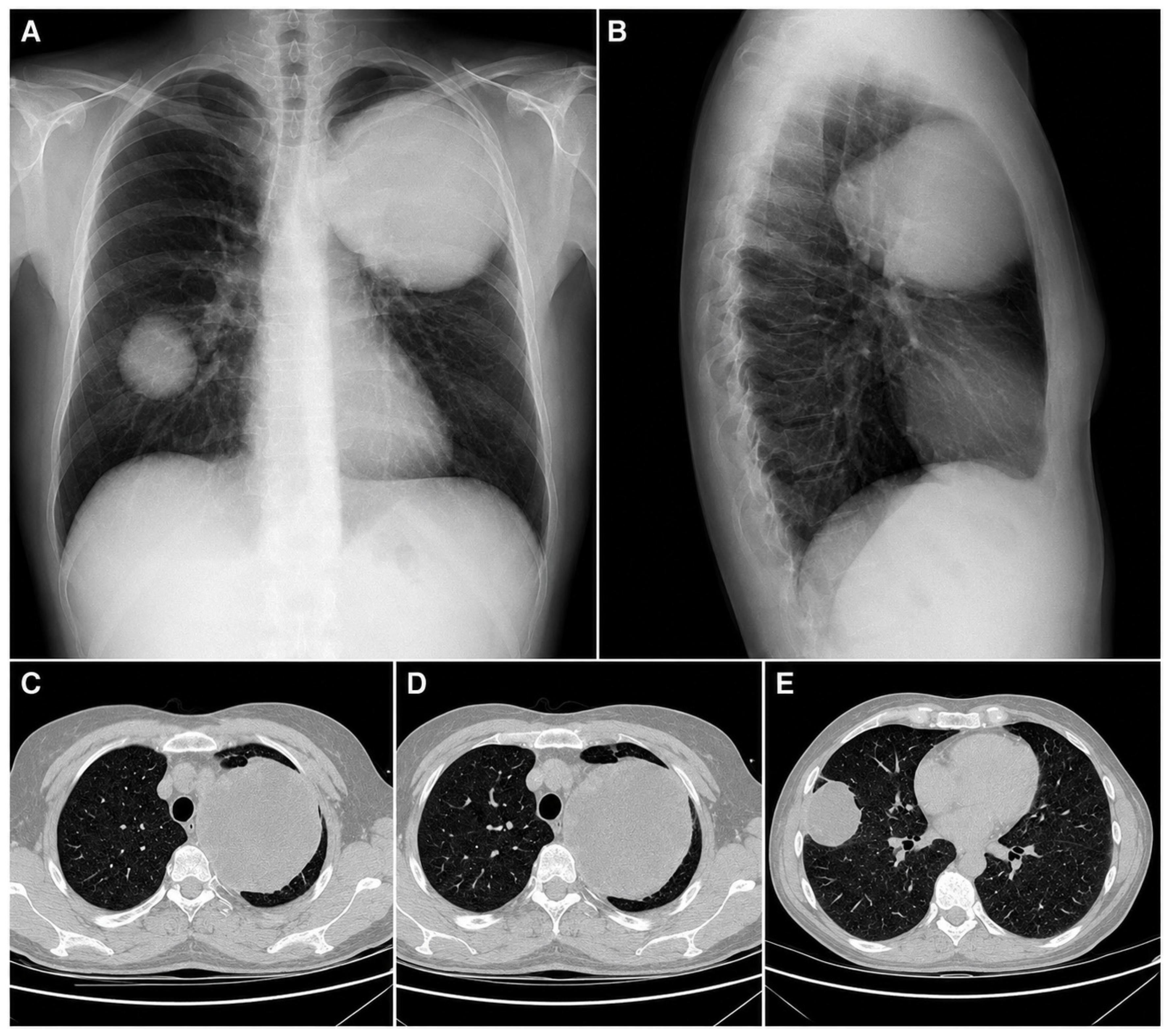
Chest radiography, including posteroanterior and lateral views, showed two well-defined rounded pulmonary masses: a large lesion of approximately 10 cm projected over the left upper lung field and a smaller lesion of approximately 4 cm projected over the right middle lung field (
Figure 1A,B). Routine laboratory tests were within normal limits, and serum anti-Echinococcus antibodies were negative. The initial differential diagnosis included benign pulmonary tumors, hydatid disease, synchronous primary pulmonary tumors, and, less likely, pulmonary metastases from an undiagnosed extrapulmonary malignancy.
Chest computed tomography revealed two solid pulmonary masses: one measuring 9.1 × 9.5 × 11 cm, located in the apical segment of the left upper lobe, with invasion of the mediastinal pleura, and a second lesion measuring 5.8 × 3.5 × 5 cm, located in the right middle lobe, with lateral pleural infiltration (
Figure 1C–E). No distant metastases were identified at the initial staging evaluation. Although histopathological confirmation was pending, the imaging characteristics raised strong suspicion of malignancy. The main diagnostic challenge was the absence of complete documentation regarding the patient’s previous recurrent thigh tumor at the time of pulmonary presentation. Because the prior lesion had reportedly been considered benign, metastatic hemangiopericytoma was not initially suspected. This delayed recognition of the extrapulmonary primary tumor and contributed to a broad initial differential diagnosis. At initial pulmonary staging, no distant metastases were identified; however, the presence of bilateral large pulmonary lesions and subsequent confirmation of a prior malignant thigh hemangiopericytoma supported metastatic disease with poor prognostic implications.
2.2. Therapeutic Intervention and Histopathological Diagnosis
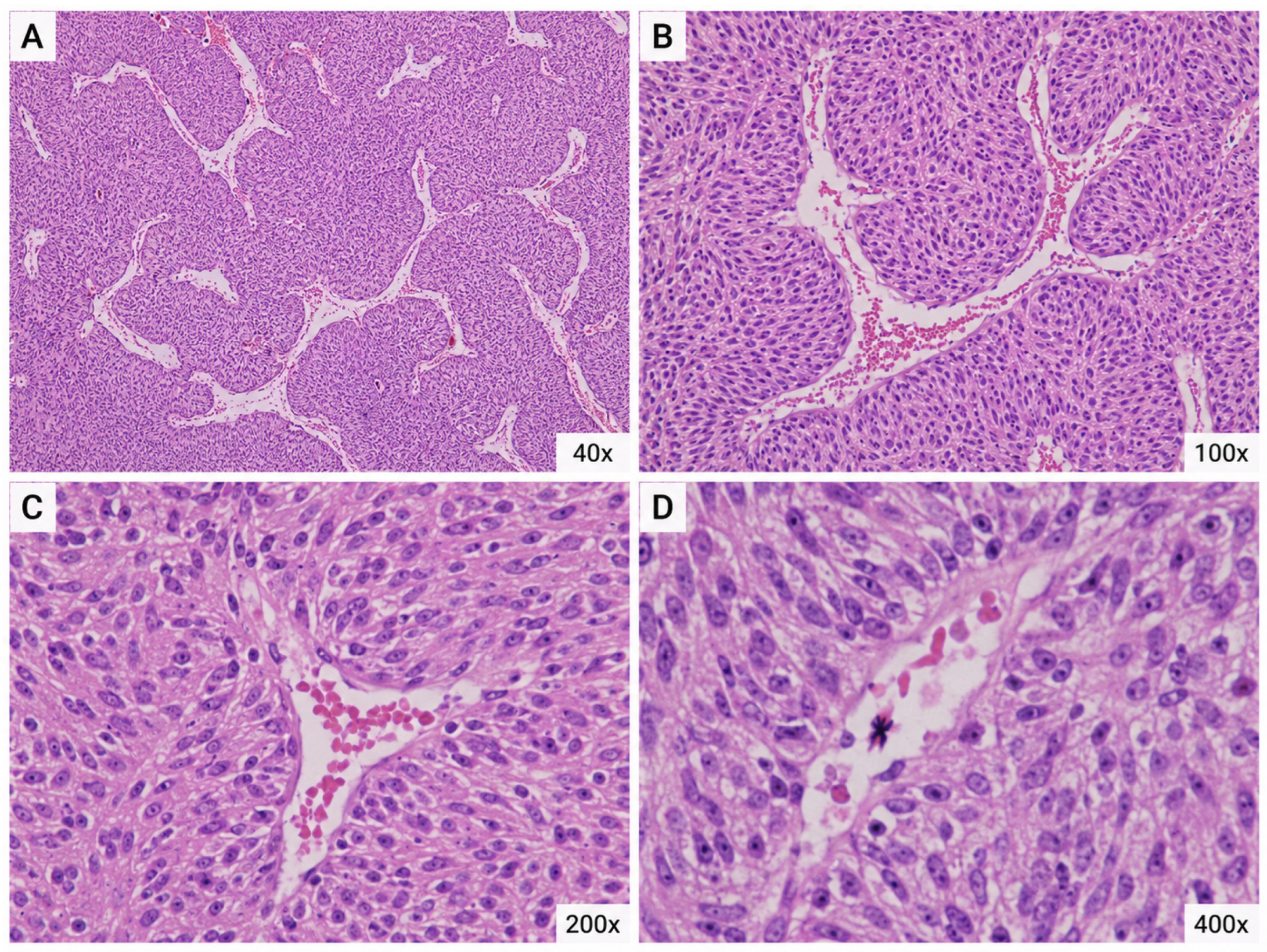
Because both pulmonary lesions were considered resectable, staged pulmonary resections were performed. The first intervention consisted of left upper lobectomy. The available operative records did not specify the surgical approach, resection-margin status, lymph-node evaluation, or the extent of macroscopic pleural or mediastinal invasion. Accordingly, no inference regarding complete resection or nodal status is made. Intraoperative frozen-section examination suggested a sarcomatous tumor, and final histopathological evaluation confirmed a malignant hemangiopericytoma/solitary fibrous tumor spectrum neoplasm (
Figure 2).
The histopathological findings were consistent with a malignant hemangiopericytoma/solitary fibrous tumor spectrum neoplasm. The available pathology report did not document intratumoral hemorrhage or a complete immunohistochemical profile. In particular, STAT6 immunohistochemistry was unavailable; therefore, confirmatory immunohistochemical characterization and definitive retrospective reclassification could not be performed. No specific pathological correlate for the presenting hemoptysis could be established retrospectively. Following the first pulmonary resection, the patient received six cycles of cisplatin-based systemic chemotherapy. Detailed information regarding the complete regimen, dose, treatment schedule, and rationale for treatment selection was not available in the records reviewed for this case report. An interim radiological assessment between completion of chemotherapy and right middle lobectomy was also unavailable; therefore, the radiological response of the right-sided pulmonary lesion could not be retrospectively categorized as response, stable disease, or progression. Subsequently, a right middle lobectomy was performed for the contralateral pulmonary lesion. Histopathological examination of the resected right-sided lesion also confirmed malignant hemangiopericytoma.
Because of the known recurrence and metastatic potential of this tumor type, the patient’s medical history was reassessed in detail. At that point, she disclosed two previous surgical interventions for a right thigh tumor. The first procedure, performed five years earlier, had reportedly resulted in a diagnosis of “myoma.” The second intervention, performed two years earlier for local recurrence, had no available histopathological report at the time of pulmonary diagnosis, as the patient and her family had not pursued further documentation, assuming the lesion was benign. After the second pulmonary surgery, the previous pathology report was retrieved and confirmed that the thigh lesion had been a malignant hemangiopericytoma. The bilateral pulmonary tumors were therefore interpreted as metastatic lesions originating from the previously treated soft tissue tumor.
2.3. Follow-Up and Outcomes
Despite aggressive surgical and oncological management, the disease progressed rapidly. Within one year, the patient developed frontal and occipital cerebral metastases, multiple bilateral pulmonary and mediastinal lesions, and a subcutaneous metastasis. Further oncological management was attempted; however, the disease followed an aggressive course with rapid systemic dissemination. The patient died within 18 months of the initial pulmonary diagnosis.
3. Discussion
Solitary fibrous tumor (SFT), historically termed hemangiopericytoma (HPC), is a rare fibroblastic mesenchymal neoplasm with heterogeneous biological behavior. Current WHO classification incorporates soft-tissue tumors historically designated as HPC within the SFT spectrum. The risk of local or distant recurrence is variable and may persist over a prolonged period; risk assessment is based on clinicopathological factors including patient age, tumor size, mitotic activity, and necrosis [
2,
3,
4].
Pulmonary involvement may represent a primary thoracic tumor or metastatic disease from an extrapulmonary SFT. In the present patient, retrieval of the previous thigh pathology report was decisive in establishing the metastatic origin of the bilateral pulmonary lesions. This diagnostic step is particularly important when imaging is non-specific and the initial history of a recurrent soft-tissue tumor is incomplete. In a study of 213 patients with cerebral hemangiopericytoma followed for up to 372 months, the most frequent metastatic sites were bone, reported in 19.6% of cases; pleura and lung, in 18.4%; liver, in 17.6%; and vertebrae, in 14.1% [
7]. Primary pulmonary hemangiopericytoma is even rarer, with only a limited number of cases described in the literature. These tumors are often minimally symptomatic; however, when present, hemoptysis and pain have been considered unfavorable prognostic indicators. Radiologically, endobronchial growth, reported in approximately 10% of cases, may be associated with a more favorable prognosis [
8,
9].
In Romania, only one case of primary pulmonary hemangiopericytoma with fatal outcome has previously been reported in the medical literature [
6]. To our knowledge, the present case represents the first documented Romanian case of metastatic pulmonary hemangiopericytoma. The diagnostic difficulty was mainly related to the absence of initial clinical suspicion, as the patient did not initially report the full history of her previous recurrent thigh tumor, and the prior histopathological documentation was not available at the time of pulmonary presentation.
The initial presentation with hemoptysis in a young patient from a tuberculosis-endemic country first raised the possibility of pulmonary tuberculosis. However, chest radiography revealed two well-defined pulmonary masses, shifting the differential diagnosis toward hydatid disease, benign pulmonary tumors, or synchronous primary lung tumors. The CT findings, particularly the presence of large bilateral solid lesions with pleural involvement, further supported the suspicion of malignancy. Given the patient’s young age, non-smoking status, and absence of occupational exposure, synchronous primary lung adenocarcinomas were also considered in the differential diagnosis.
Definitive diagnosis was established only after histopathological examination of the resected pulmonary lesions. This finding prompted a more detailed reassessment of the patient’s medical history and led to retrieval of the previous thigh tumor pathology report. The final interpretation was metastatic malignant hemangiopericytoma originating from a previously undertreated and insufficiently documented soft tissue tumor.
The role of systemic therapy in advanced hemangiopericytoma/solitary fibrous tumor spectrum neoplasms remains uncertain and should be individualized. In the present case, six cycles of cisplatin-based systemic chemotherapy were administered; however, detailed regimen and rationale data were unavailable, and the subsequent clinical course remained aggressive despite multimodal treatment. The absence of interim radiological assessment precluded retrospective categorization of treatment response. The principal contribution of this case is the diagnostic delay caused by incomplete documentation of a recurrent soft tissue tumor. In patients presenting with unexplained bilateral pulmonary masses and a history of recurrent soft tissue lesions, even when previously considered benign, earlier histopathological reports should be actively retrieved and reviewed. This case also emphasizes that imaging alone may not establish the diagnosis, that long-term surveillance is essential after recurrent soft tissue tumors, and that management of rare aggressive tumors requires individualized multidisciplinary decision-making.
The strength of this case report lies in the histopathological confirmation of both pulmonary lesions and the subsequent retrieval of the previous thigh tumor pathology report, which clarified the metastatic origin of the disease. The case also illustrates a clinically relevant diagnostic pitfall in patients with recurrent soft tissue tumors and unusual pulmonary masses. The main limitations are its retrospective nature, incomplete initial documentation of the primary thigh tumor, limited availability of detailed bronchoscopy and operative records, lack of an interim radiological assessment after chemotherapy, unavailability of a complete immunohistochemical profile including STAT6, incomplete details regarding chemotherapy regimen, dosing, treatment rationale, and tolerability, and the inability to obtain the patient’s perspective because of the fatal outcome.
4. Conclusions
This case highlights the diagnostic and therapeutic complexity of malignant hemangiopericytoma with pulmonary metastases, a clinical entity so rare that it may not be considered during the initial diagnostic work-up. In the present case, delayed recognition of a previous recurrent soft tissue tumor, together with the non-specific presentation of hemoptysis in a tuberculosis-endemic region, contributed to a challenging diagnostic pathway.
Despite staged pulmonary resections and systemic chemotherapy, the disease followed an aggressive course characterized by early recurrence and widespread dissemination, including cerebral, bilateral pulmonary, mediastinal, and subcutaneous metastases. This evolution underlines the unpredictable biological behavior of malignant hemangiopericytoma and the limited efficacy of currently available systemic treatment options in advanced disease.
Our experience emphasizes the importance of detailed medical history, systematic retrieval of previous histopathological reports, especially in patients with recurrent soft tissue tumors, and long-term surveillance for metastatic disease. International collaboration, dedicated case registries, and prospective studies are needed to better define evidence-based diagnostic and therapeutic algorithms for this rare and challenging tumor.




 ,
,