Heerfordt Syndrome Complicated by Bilateral Simultaneous Facial Palsy, PTH-Independent Hypercalcemia, and Bilateral Obstructive Acute Kidney Injury in the Absence of Thoracic Disease: A Case Report and Narrative Review

 ,
,
Abstract
1. Introduction and Clinical Significance
2. Case Presentation
2.1. History
2.2. Physical Examination
2.3. Laboratory Findings
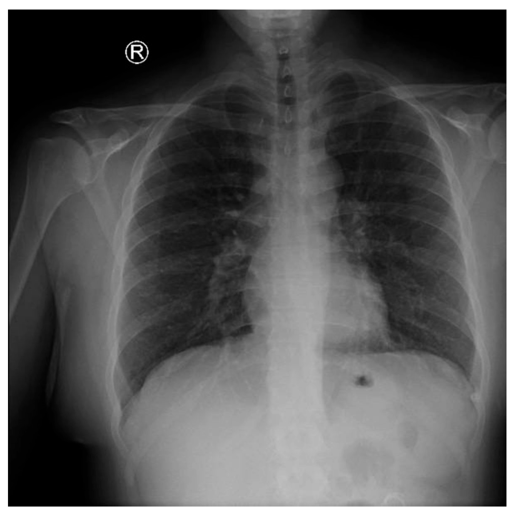
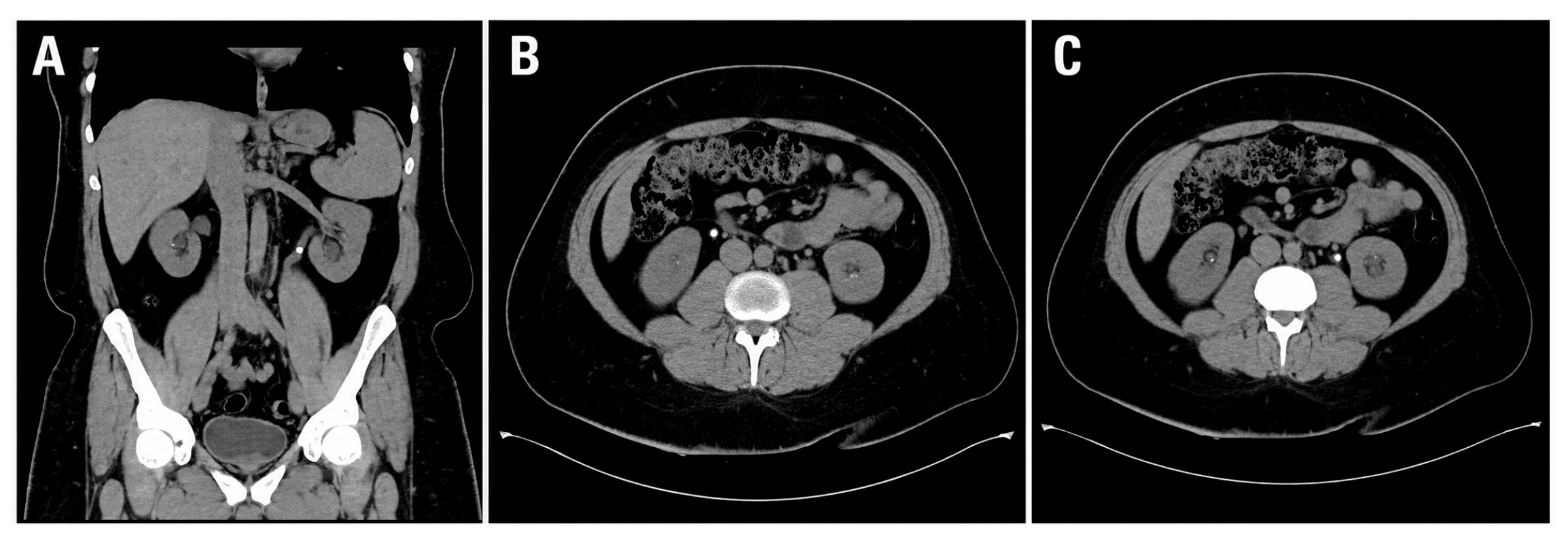
2.4. Imaging Studies
2.5. Histopathology
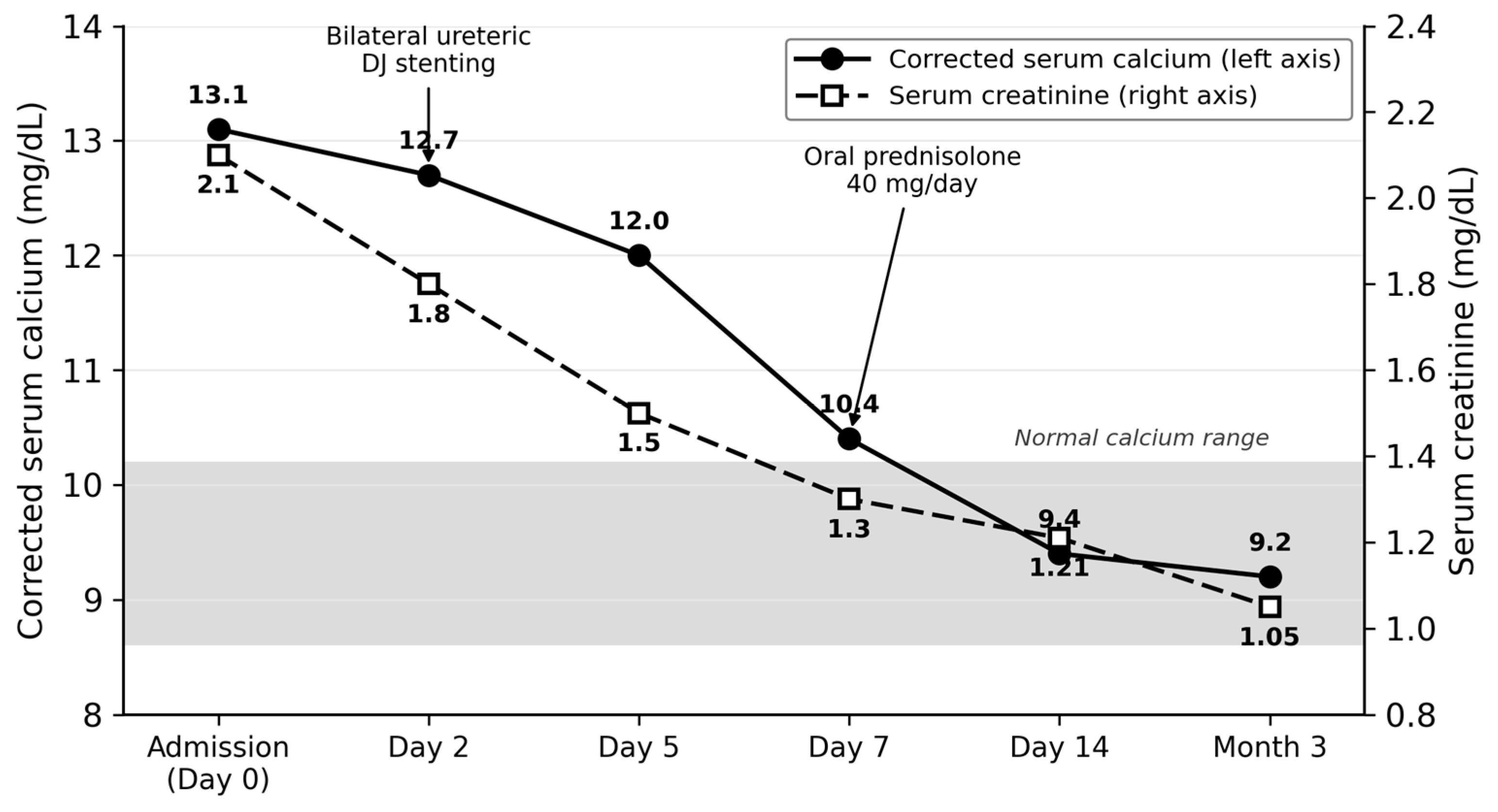
2.6. Management and Outcome
3. Discussion
3.1. Diagnostic Criteria and Phenotypic Variants of Heerfordt Syndrome
3.2. Bilateral Simultaneous Facial Palsy: A Clinical Red Flag
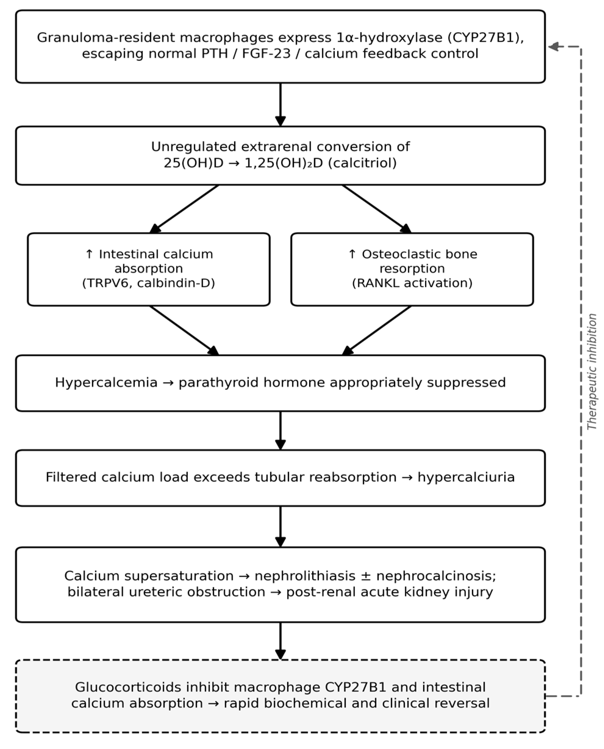
3.3. Pathophysiology of Hypercalcemia in Sarcoidosis
3.4. The Renal Phenotype of Sarcoidosis
3.5. Comparison with Published Cases
3.6. Therapeutic Considerations
3.7. Diagnostic Pitfalls and Learning Points
- PTH-independent hypercalcemia with elevated ACE in a young adult should prompt active investigation for sarcoidosis, even when the chest film is normal.
- Bilateral simultaneous peripheral facial palsy is a clinical red flag with a narrow differential; neurosarcoidosis should always be on the list.
- Posterior uveitis can occur in Heerfordt syndrome and does not exclude the diagnosis; anterior involvement is more common but not obligatory.
- Parotid biopsy is high-yield when accessible disease is present and avoids more invasive bronchoscopic sampling in radiographically silent chests.
- Sarcoidosis-related AKI is largely reversible when both the mechanical (stenting) and granulomatous (corticosteroid) substrates are addressed in parallel.
- Calcitriol-mediated hypercalcemia responds to glucocorticoids, which inhibit macrophage 1α-hydroxylase activity; bisphosphonates are seldom required.
3.8. Limitations
4. Conclusions
Author Contributions
Funding
Institutional Review Board Statement
Informed Consent Statement
Data Availability Statement
Acknowledgments
Conflicts of Interest
References
- Sève, P.; Pacheco, Y.; Durupt, F.; Jamilloux, Y.; Gerfaud-Valentin, M.; Isaac, S.; Boussel, L.; Calender, A.; Androdias, G.; Valeyre, D.; et al. Sarcoidosis: A Clinical Overview from Symptoms to Diagnosis. Cells 2021, 10, 766. [Google Scholar] [CrossRef] [PubMed]
- Arkema, E.V.; Cozier, Y.C. Epidemiology of sarcoidosis: Current findings and future directions. Ther. Adv. Chronic Dis. 2018, 9, 227–240. [Google Scholar] [CrossRef] [PubMed]
- Kharoubi, S. Syndrome de Heerfordt: à propos d’une observation et revue de la littérature [Heerfordt’s syndrome: About a case and literature review]. Pan Afr. Med. J. 2020, 37, 117. [Google Scholar] [CrossRef] [PubMed]
- Fraga, R.C.; Fernandes, T.A.; Takay, M.; Silva, L.C. Do you know this syndrome? Heerfordt–Waldenström syndrome. An. Bras. Dermatol. 2017, 92, 571–572. [Google Scholar] [CrossRef] [PubMed]
- Stern, B.J.; Royal, W., 3rd; Gelfand, J.M.; Clifford, D.B.; Tavee, J.; Pawate, S.; Berger, J.R.; Aksamit, A.J.; Krumholz, A.; Pardo, C.A.; et al. Definition and Consensus Diagnostic Criteria for Neurosarcoidosis: From the Neurosarcoidosis Consortium Consensus Group. JAMA Neurol. 2018, 75, 1546–1553. [Google Scholar] [CrossRef] [PubMed]
- Mahévas, M.; Lescure, F.X.; Boffa, J.J.; Delastour, V.; Belenfant, X.; Chapelon, C.; Cordonnier, C.; Makdassi, R.; Piette, J.C.; Naccache, J.M.; et al. Renal sarcoidosis: Clinical, laboratory, and histologic presentation and outcome in 47 patients. Medicine 2009, 88, 98–106. [Google Scholar] [CrossRef] [PubMed]
- Bergner, R.; Hoffmann, M.; Waldherr, R.; Uppenkamp, M. Frequency of kidney disease in chronic sarcoidosis. Sarcoidosis Vasc. Diffus. Lung Dis. Off. J. WASOG 2003, 20, 126–132. [Google Scholar]
- Adams, J.S.; Hewison, M. Extrarenal expression of the 25-hydroxyvitamin D-1-hydroxylase. Arch. Biochem. Biophys. 2012, 523, 95–102. [Google Scholar] [CrossRef] [PubMed]
- Tebben, P.J.; Singh, R.J.; Kumar, R. Vitamin D-Mediated Hypercalcemia: Mechanisms, Diagnosis, and Treatment. Endocr. Rev. 2016, 37, 521–547. [Google Scholar] [CrossRef] [PubMed]
- Chappity, P.; Kumar, R.; Sahoo, A.K. Heerfordt’s Syndrome Presenting with Recurrent Facial Nerve Palsy: Case Report and 10-year Literature Review. Sultan Qaboos Univ. Med. J. 2015, 15, e124–128. [Google Scholar] [CrossRef]
- Gallo, C.; Mazzini, L.; Varrasi, C.; Vecchio, D.; Virgilio, E.; Cantello, R. Bilateral Facial Palsy as the Onset of Neurosarcoidosis: A Case Report and a Revision of the Literature. NeuroSci 2022, 3, 321–331. [Google Scholar] [CrossRef] [PubMed]
- Keane, J.R. Bilateral seventh nerve palsy: Analysis of 43 cases and review of the literature. Neurology 1994, 44, 1198–1202. [Google Scholar] [CrossRef] [PubMed]
- Jha, G.; Azhar, S.; Kuttuva, S.; Elahi, S.; Baseer, A. Bilateral Facial Palsy: A Case Study of an Exceedingly Rare and Difficult Diagnosis. Cureus 2021, 13, e18900. [Google Scholar] [CrossRef] [PubMed]
- Sharma, O.P. Vitamin D, calcium, and sarcoidosis. Chest 1996, 109, 535–539. [Google Scholar] [CrossRef] [PubMed]
- Iannuzzi, M.C.; Rybicki, B.A.; Teirstein, A.S. Sarcoidosis. N. Engl. J. Med. 2007, 357, 2153–2165. [Google Scholar] [CrossRef] [PubMed]
- Sharma, O.P.; Vucinic, V.; James, D.G. Hypercalcemia, Anemia, and Acute Kidney Injury: A Rare Presentation of Sarcoidosis. Case Rep. Med. 2015, 2015, 565243. [Google Scholar] [CrossRef] [PubMed]
- Sharmeen, S.; Engel, M.E. Granulomatous Interstitial Nephritis Presenting as Hypercalcemia and Nephrolithiasis. Case Rep. Nephrol. 2016, 2016, 4186086. [Google Scholar] [CrossRef] [PubMed]
- Papalia, T.; Greco, R.; Mollica, A.; Bonofiglio, R. Acute Renal Failure in Sarcoidosis: A Review of the Literature. Arch. Clin. Nephrol. 2016, 2, 11–16. [Google Scholar] [CrossRef]
- Zakharova, E.V.; Stolyarevich, E.S. Sarcoidosis with combined kidney damage—Kidney stones, granulomatous interstitial nephritis, and acute kidney injury. Clin. Case Rep. Rev. 2016, 1, 412–416. [Google Scholar] [CrossRef]
- Denny, M.C.; Fotino, A.D. The Heerfordt–Waldenström syndrome as an initial presentation of sarcoidosis. In Baylor University Medical Center Proceedings; Taylor & Francis: London, UK, 2013; Volume 26, pp. 390–392. [Google Scholar]
- Fujiwara, K.; Furuta, Y.; Fukuda, S. Two Cases of Heerfordt’s Syndrome: A Rare Manifestation of Sarcoidosis. Case Rep. Otolaryngol. 2016, 2016, 3642735. [Google Scholar] [CrossRef] [PubMed]
- Baughman, R.P.; Valeyre, D.; Korsten, P.; Mathioudakis, A.G.; Wuyts, W.A.; Wells, A.; Rottoli, P.; Nunes, H.; Lower, E.E.; Judson, M.A.; et al. ERS clinical practice guidelines on treatment of sarcoidosis. Eur. Respir. J. 2021, 58, 2004079. [Google Scholar] [CrossRef] [PubMed]
- Crouser, E.D.; Maier, L.A.; Wilson, K.C.; Bonham, C.A.; Morgenthau, A.S.; Patterson, K.C.; Abston, E.; Bernstein, R.C.; Blankstein, R.; Chen, E.S.; et al. Diagnosis and Detection of Sarcoidosis. An Official American Thoracic Society Clinical Practice Guideline. Am. J. Respir. Crit. Care Med. 2020, 201, e26–e51. [Google Scholar] [CrossRef] [PubMed]
- Hilderson, I.; Van Laecke, S.; Wauters, A.; Donck, J. Treatment of renal sarcoidosis: Is there a guideline? Overview of the different treatment options. Nephrol. Dial. Transplant. 2014, 29, 1841–1847. [Google Scholar] [PubMed]
- Ungprasert, P.; Carmona, E.M.; Crowson, C.S.; Matteson, E.L. Diagnostic Utility of Angiotensin-Converting Enzyme in Sarcoidosis: A Population-Based Study. Lung 2016, 194, 91–95. [Google Scholar] [PubMed]



| Parameter | Result | Reference Range | Interpretation |
|---|---|---|---|
| Creatinine (admission) | 2.1 mg/dL | 0.67–1.17 mg/dL | Elevated (AKI) |
| Creatinine (post-treatment) | 1.21 mg/dL | 0.67–1.17 mg/dL | Improving |
| Urea | 64 mg/dL | 19–44 mg/dL | Elevated |
| Uric acid | 9.9 mg/dL | 3.4–7.0 mg/dL | Elevated |
| Total calcium (admission) | 13.1 mg/dL | 8.6–10.2 mg/dL | Severely elevated |
| Total calcium (post-treatment) | 9.4 mg/dL | 8.6–10.2 mg/dL | Normal |
| Phosphate | 4.40 mg/dL | 2.5–4.5 mg/dL | Normal |
| Magnesium | 1.90 mg/dL | 1.7–2.2 mg/dL | Normal |
| Intact PTH | 3.4 pg/mL | 15–65 pg/mL | Suppressed |
| 25-hydroxyvitamin D | 22 ng/mL | 30–100 ng/mL | Insufficient |
| 1,25-dihydroxyvitamin D | Not assayed | — | Unavailable at site |
| Angiotensin-converting enzyme | >100 U/L | 8–52 U/L | Elevated |
| TSH/free T4 | Within range | — | Euthyroid |
| Albumin | 4.7 g/dL | 3.5–5.2 g/dL | Normal |
| Total protein | 7.4 g/dL | 6.0–8.0 g/dL | Normal |
| AST/ALT/ALP/GGT | Within range | — | Normal liver panel |
| Sodium | 138 mmol/L | 136–145 mmol/L | Normal |
| Potassium | 4.63 mmol/L | 3.5–5.0 mmol/L | Normal |
| Chloride | 108 mmol/L | 98–106 mmol/L | Mildly elevated |
| Fasting glucose/HbA1c | 76 mg/dL/4.97% | — | Normal |
| Lipid panel | LDL 112, HDL 29 | — | Low HDL |
| LDH | 172 U/L | 140–280 U/L | Normal |
| Parameter | Result | Interpretation |
|---|---|---|
| Hemoglobin/Hematocrit | 14.2 g/dL/43.6% | Normal |
| WBC count (differential) | 8.7 × 10⁹/L; lymphocytes 12.8% | Lymphopenia |
| Platelets | 418 × 109/L | Mild thrombocytosis |
| PT/INR/APTT | Within range | Normal |
| ESR/CRP | 19 mm/h/10 mg/L | Mildly elevated |
| C3/C4 | 1.61 g/L/0.332 g/L | Normal |
| IgG | 12.20 g/L | Normal |
| ANA/c-ANCA/p-ANCA | Negative | Negative |
| Anti-Ro (SSA)/Anti-La (SSB) | Negative | Negative |
| HIV/HBsAg/HCV antibody | Non-reactive | Negative |
| IGRA (M. tuberculosis) | Non-reactive | Negative |
| 24 h urinary protein | 595.9 mg/24 h | Sub-nephrotic proteinuria |
| Urine leukocytes | >100/HPF | Sterile pyuria |
| Urine red blood cells | 3+ | Microscopic hematuria |
| Urine protein (dipstick) | 2+ | Proteinuria |
| Hyaline casts | 3+ | Present |
| Urine culture (×4) | No growth | Sterile |
| Feature | Classical Description | Reported Variants | Frequency in Published Series |
|---|---|---|---|
| Parotid enlargement | Bilateral, painful, persistent | Unilateral; submandibular involvement | ~80–95% |
| Uveitis | Anterior, granulomatous | Posterior uveitis; panuveitis; vitritis | ~70–80% |
| Facial nerve palsy | Unilateral, peripheral | Bilateral simultaneous; alternating; recurrent | ~50–60% |
| Fever | Low-grade, intermittent | Absent in up to 40% of contemporary reports | ~50–60% |
| Complete tetrad | All four features | Two of four with biopsy support is accepted | ~10–15% |
| Category | Specific Entity | Key Clues/Investigations |
|---|---|---|
| Infectious | Lyme borreliosis | Erythema migrans; tick exposure; serology |
| Infectious | HIV (acute or advanced) | Risk factors; ELISA/Western blot |
| Infectious | Tuberculous or fungal meningitis | Imaging; CSF analysis; cultures |
| Infectious | Otitis/parotitis/Ramsay Hunt | Local examination; vesicular rash |
| Inflammatory | Sarcoidosis (Heerfordt or pure neurosarcoidosis) | ACE; calcium; chest imaging; biopsy |
| Inflammatory | Sjögren syndrome | Sicca symptoms; anti-Ro/anti-La |
| Neurological | Guillain–Barré (incl. Miller Fisher) | Areflexia; albumin–cytological dissociation |
| Neoplastic | Leukemic/lymphomatous infiltration | Smear; LDH; bone marrow; CSF cytology |
| Neoplastic | Meningeal carcinomatosis | MRI with contrast; CSF cytology |
| Vascular/structural | Brainstem ischemia/pontine lesion | MRI brain |
| Congenital | Möbius syndrome | Onset in infancy; abducens involvement |
| Idiopathic | Bilateral Bell palsy (diagnosis of exclusion) | <25% of bilateral cases; exhaustive exclusion |
| Feature | Present Case | Chappity et al. [10] | Sharma et al. [16] | Sharmeen et al. [17] | Fujiwara et al. [21] | Denny et al. [20] | Zakharova et al. [19] | Gallo et al. [11] |
|---|---|---|---|---|---|---|---|---|
| Age/sex | 30/M | 52/F | 61/F | 55/M | 53/M + 55/F | 59/F | Not specified | 47/F |
| Heerfordt syndrome | Complete (3/4) | Complete | Absent | Absent | Incomplete + complete | Complete | Absent | Partial |
| Parotid involvement | Bilateral | Bilateral | No | No | Unilateral/bilateral | Bilateral | No | No |
| Facial palsy | Bilateral simultaneous | Bilateral alternating | No | No | Unilateral | Unilateral | No | Bilateral simultaneous |
| Uveitis | Posterior, bilateral | Anterior | No | No | Absent/present | Anterior | No | Absent |
| Hypercalcemia | Severe (13.1) | No | Severe | Severe | No | No | Severe | No |
| PTH | Suppressed (3.4) | Normal | Suppressed | Suppressed | — | — | Suppressed | — |
| ACE | Elevated (>100) | Elevated (129) | Elevated | Elevated | Elevated | Elevated | Elevated | Elevated |
| Renal involvement | Obstructive AKI | None | AKI | GIN + AKI | None | None | GIN + stones + AKI | None |
| Nephrolithiasis | Bilateral ureteric | No | No | Yes (history) | No | No | Yes | No |
| Thoracic imaging | Normal | Mediastinal LAD | Apical bronchiectasis | Mediastinal LAD | Mediastinal LAD | Hilar markings | Not detailed | BHL |
| Biopsy site | Parotid | Parotid | Marrow + lung | Kidney (GIN) | Eyelid + node | Lymph node | Kidney | Lymph node |
| Dialysis required | No | No | No | No | No | No | Yes (transient) | No |
| Steroid response | Excellent | Excellent | Good | Good | Excellent | Excellent | Good | Good |
Disclaimer/Publisher’s Note: The statements, opinions and data contained in all publications are solely those of the individual author(s) and contributor(s) and not of MDPI and/or the editor(s). MDPI and/or the editor(s) disclaim responsibility for any injury to people or property resulting from any ideas, methods, instructions or products referred to in the content. |
© 2026 by the authors. Licensee MDPI, Basel, Switzerland. This article is an open access article distributed under the terms and conditions of the Creative Commons Attribution (CC BY) license.
Share and Cite
Amer, K.A.; Al Shuqayfah, N.I.; Alhakamy, M.A.; Alasiri, A.J. Heerfordt Syndrome Complicated by Bilateral Simultaneous Facial Palsy, PTH-Independent Hypercalcemia, and Bilateral Obstructive Acute Kidney Injury in the Absence of Thoracic Disease: A Case Report and Narrative Review. Reports 2026, 9, 225. https://doi.org/10.3390/reports9030225
Amer KA, Al Shuqayfah NI, Alhakamy MA, Alasiri AJ. Heerfordt Syndrome Complicated by Bilateral Simultaneous Facial Palsy, PTH-Independent Hypercalcemia, and Bilateral Obstructive Acute Kidney Injury in the Absence of Thoracic Disease: A Case Report and Narrative Review. Reports. 2026; 9(3):225. https://doi.org/10.3390/reports9030225
Chicago/Turabian StyleAmer, Khaled Abdulwahab, Nawaf Ibrahim Al Shuqayfah, Mohammad Abdallah Alhakamy, and Abdullah Jaber Alasiri. 2026. "Heerfordt Syndrome Complicated by Bilateral Simultaneous Facial Palsy, PTH-Independent Hypercalcemia, and Bilateral Obstructive Acute Kidney Injury in the Absence of Thoracic Disease: A Case Report and Narrative Review" Reports 9, no. 3: 225. https://doi.org/10.3390/reports9030225
APA StyleAmer, K. A., Al Shuqayfah, N. I., Alhakamy, M. A., & Alasiri, A. J. (2026). Heerfordt Syndrome Complicated by Bilateral Simultaneous Facial Palsy, PTH-Independent Hypercalcemia, and Bilateral Obstructive Acute Kidney Injury in the Absence of Thoracic Disease: A Case Report and Narrative Review. Reports, 9(3), 225. https://doi.org/10.3390/reports9030225