
Review

From Combustion Emissions to Neurotoxicity: Brain Health Risks of Military Burn Pits Exposure

by
Katherine M. Eggers
1,2,
Zoe A. Keller
1,2,
Paul Barach
3,4,
Julie M. Tomáška
5,
Joshua P. Nixon
1,6,
Janeen H. Trembley
1,7,8 and
Tammy A. Butterick
1,9,10,*
1
Minneapolis Veterans Affairs Health Care System, Minneapolis, MN 55417, USA
2
Center for Veterans Research and Education, Minneapolis, MN 55417, USA
3
College of Population Health, Thomas Jefferson University, Philadelphia, PA 19107, USA
4
Sheps Health Services Research Center, University of North Carolina, Chapel Hill, NC 27516, USA
5
Burn Pits 360 Veterans Organization, Robstown, TX 78380, USA
6
Department of Surgery, University of Minnesota, Minneapolis, MN 55455, USA
7
Department of Laboratory Medicine and Pathology, University of Minnesota, Minneapolis, MN 55455, USA
8
Masonic Cancer Center, University of Minnesota, Minneapolis, MN 55108, USA
9
Department of Neuroscience, University of Minnesota, Minneapolis, MN 55455, USA
10
Department of Food Science and Nutrition, University of Minnesota, St. Paul, MN 55108, USA
*
Author to whom correspondence should be addressed.
Fire 2026, 9(6), 249; https://doi.org/10.3390/fire9060249
Submission received: 31 March 2026 / Revised: 5 June 2026 / Accepted: 7 June 2026 / Published: 11 June 2026

Abstract

Military burn pits used during post-9/11 U.S. military deployments functioned as uncontrolled combustion systems and were widely utilized to dispose of large volumes of outdoor waste by burning. Burn pits involved heterogeneous waste materials burned under variable temperature and oxygen conditions. These combustion environments generated complex, toxic, multipollutant airborne emission mixtures that included particulate matter (PM2.5), polycyclic aromatic hydrocarbons (PAHs), and volatile organic compounds (VOCs). This narrative review synthesizes epidemiologic, experimental, and mechanistic evidence linking burn pit emissions to disruption of the lung–brain axis and adverse neurological outcomes. We specifically aim to address a critical gap in understanding how combustion-derived toxicants impact brain health and are associated with unfavorable neuropsychiatric outcomes, including increased risk of post-traumatic stress disorder (PTSD) and depression. Combustion-related exposures promote pulmonary inflammation and system-wide immune signaling that propagate to the central nervous system, contributing to neuroinflammation and dysregulation of the hypothalamic–pituitary–adrenal (HPA) axis. These interconnected mechanisms are associated with toxic encephalopathy and related cognitive and mood disturbances, underscoring the need to integrate fire science with military and environmental health services research to better define the systemic and neurological consequences of acute and chronic fire-derived inhalation exposures.

Graphical Abstract

1. Introduction

An estimated 2.7 million post-9/11 U.S. military personnel and local civilians were exposed to elevated levels of toxicant inhalation from military burn pits in Southwest Asia and other locations within the U.S. Central Command (CENTCOM) region during Operation Enduring Freedom (OEF), Operation Iraqi Freedom (OIF) and Operation New Dawn (OND) [1]. Open-air burn pits were widely used by the U.S. military as the primary method for solid waste disposal, resulting in sustained and often uncontrolled emissions of complex combustion-derived toxicants [2,3]. Burn pits constituted a distinct yet representative type of complex fire conditions and emissions. There have been 297 documented locations of burn pit usage, with the highest concentration of 152 burn pit locations in Iraq [4]. As of March 2022, nine burn pits are still active in the CENTCOM region despite the U.S. military scaling back the use of burn pits in 2009 [5]. Limited use has persisted in certain operational settings, and exposure to combustion-derived pollutants remains an ongoing concern in deployed environments [5].
Burn pit emissions consist of heterogeneous mixtures of particulate matter (PM), including ultrafine particles (UFPs), polycyclic aromatic hydrocarbons (PAHs), volatile organic compounds (VOCs), and metals, many of which are known to induce pulmonary and systemic toxicity. While initial research has focused on respiratory outcomes, there is increasing recognition that the impacts of these exposures extend beyond the lung and may adversely affect central nervous system (CNS) function and brain health. Emerging epidemiologic and experimental evidence links burn pit-related exposures to neuroinflammation, cognitive dysfunction, and increased risk of neuropsychiatric conditions, including anxiety, depression, and post-traumatic stress disorder (PTSD) [6,7,8].
The Sergeant First Class Heath Robinson Honoring our Promise to Address Comprehensive Toxins (PACT) Act enacted on 10 August 2022, represents a major policy response to toxic exposure risks among Veterans. This legislation expands healthcare access, mandates toxic exposure screening, and prioritizes research on deployment-related environmental hazards [9,10]. Importantly, the PACT Act underscores the need to move beyond organ-specific frameworks and adopt integrated models that capture the systemic consequences of toxicant exposure. Despite this progress, a critical gap remains in understanding how combustion-derived toxicants influence brain health and may be associated with adverse neurological and neuropsychiatric outcomes [7,11].
In particular, the mechanistic pathways linking inhalation exposures to depression, PTSD, and toxic encephalopathy (TE) are not well defined [12]. The lung–brain axis has emerged as a key framework to address this gap, providing a biologically plausible model through which pulmonary inflammation, systemic immune signaling, and direct toxicant translocation converge to impact the CNS.
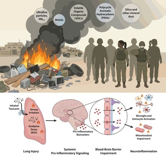
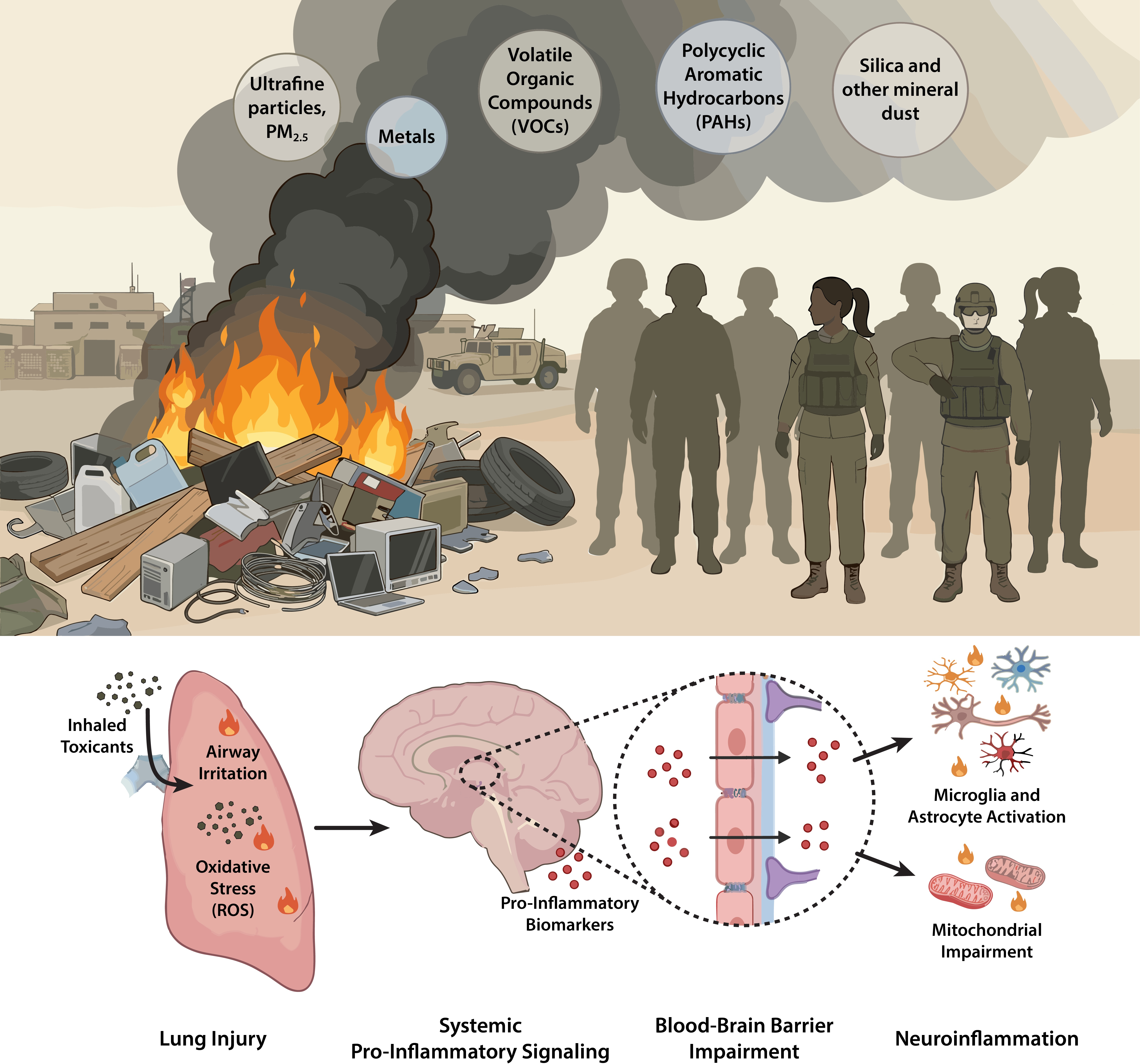
This narrative review synthesizes epidemiologic, experimental, and mechanistic evidence linking burn pit emissions to disruption of the lung–brain axis and downstream neurological outcomes, Figure 1. We focus on how combustion-derived toxicants drive neuroinflammation, blood–brain barrier (BBB) dysfunction, mitochondrial impairment, monoamine disruption, and hypothalamic–pituitary–adrenal (HPA) axis dysregulation, and how these processes contribute to TE, cognitive impairment, and mood disorders. By integrating fire science with environmental health, neurobiology, and Veteran-focused health research, this review aims to advance a more comprehensive understanding of the systemic and neurological consequences of combustion-derived inhalation exposures.

2. Materials and Methods

This review draws from peer-reviewed human and animal studies, cohort studies, meta-analyses, and clinical trials to examine the associations and mechanistic overlap between combustion emissions and the neurologic and psychological characteristics of toxic encephalopathy.

Search Strategy and Selection Criteria

This is a narrative review informed by systematic search principles, but not a formal systematic review. A comprehensive literature search strategy was used to compile evidence connecting fire emissions, cognitive decline, toxic encephalopathy, PTSD, and depression. Peer-reviewed articles, animal studies, cohort studies, meta-analyses, and clinical trials published in English from 1996 to March 2026 were considered for use. The 1996–2026 time frame was chosen to capture both early work on combustion emissions and more recent post-9/11 military burn pit literature. Primary searches were conducted in PubMed (including MEDLINE) and Google Scholar using the search terms: “neurotoxic effects of military burn pit emissions”, “neurotoxic effects of military burn pit emissions neuro* PTSD depression”, “HPA axis PTSD”, “firefighter exposure to neurotoxic chemicals”, “toxic encephalopathy neuropsychological symptoms”, “PACT Act”, “Lung-Brain axis”, “Blood-Brain Barrier”, “post-9/11 Veterans”, and “burn pit registry” (Table 1).
We focused on published peer-reviewed literature that assessed relationships between combustion emissions, neuroinflammation, and mental health in preclinical and clinical models. References of eligible studies were manually checked (snowballing) to identify additional sources not captured in the primary database searches. Literature that was clearly irrelevant, redundant, or outside the scope of combustion-related inhalation exposures was excluded based on title, abstract, and discussion review. Full texts of potentially relevant articles were then reviewed, and articles that remained irrelevant or could not be fully accessed were excluded. Systematic reviews were used to identify relevant primary research articles and were not included as primary evidence sources in the synthesis.
AI tools were used solely to assist with literature search term generation. All sources were independently verified and interpreted by the authors (ChatGPT5 OpenAI, Perplexity). All sources were independently retrieved, verified, and interpreted by the authors. AI tools were not used to summarize or extract data from individual articles.

3. Results

3.1. Military Burn Pits as Combustion Systems

The general composition of the waste burned in US military burn pits was measured to be approximately over 80% combustible materials such as plastics, wood, and other miscellaneous combustibles and under 20% noncombustibles such as metals and other miscellaneous noncombustibles, with jet and diesel fuel used as an accelerant at some bases [3]. Field investigations at Balad Air Base, Iraq, and other locations have identified that contents could include municipal and military waste, including munitions, petroleum, oils and lubricants, medical and human waste, and industrial chemicals, creating a heterogeneous combustion matrix [1]. The burn pit at Balad covered more than 25 acres, and the Department of Defense (DoD) estimated that larger bases burned up to 85,000 pounds of waste daily [9]. When burned, this combination of plastics, synthetic polymers, and chemically treated materials generates complex organic pollutants, including PAHs, dioxins, chlorinated hydrocarbons, and metal-rich particulates [3,4]. Jet propellant-8 (JP-8) jet fuel, routinely used as an accelerant for burn pits, contains both naphthalene and benzene [13,14]. Air sampling at multiple military installations identified elevated concentrations of PM, PAHs, and VOCs associated with burn pit operations [15,16]. Although measured benzene concentrations generally remained below occupational exposure limits and military exposure guidelines, environmental monitoring demonstrated concentrations exceeding U.S. Environmental Protection Agency (EPA) acceptable long-term exposure benchmarks in some deployment settings [9,15,16]. These data support the view that burn pit emissions represent a mixture of combustion-derived particulates and gases rather than a single-chemical exposure [15]. In contrast, other toxicants, including PM, select PAHs, and acrolein, exceeded the 1-year MEG during occupational sampling [9,15,16]. Importantly, exposure measurements varied substantially across deployment locations, reflecting differences in burn pit composition, waste streams, combustion conditions, meteorological factors, and sampling methodologies. This variability highlights one of the major challenges in burn pit research: characterizing exposure to a complex and heterogeneous mixture of combustion-derived toxicants across diverse deployment environments. Collectively, these findings suggest that deployed personnel may have experienced exposure through both occupational and environmental pathways and support the view that burn pit emissions represent a heterogeneous exposure mixture rather than a single-chemical exposure [15].
U.S. Bases were frequently in arid regions that experienced dust and sandstorms, resulting in sustained elevation of airborne mineral particles and crystalline silica [1]. Military operations, vehicle exhaust, and underregulated industrial sources further contributed to ambient PM levels, while regional power generation and transportation emissions elevated levels of PAHs and VOCs that were detected both up and downwind of large burn pits such as at Balad [1]. As a result, personnel in proximity to burn pits were exposed to a complex mixture of locally generated pit emissions, high background levels of desert dust, and combustion-related particles [1]. Table 2 summarizes major pollutants measured in multiple deployment locations.
Combustion conditions within the burn pits themselves were highly variable and strongly influenced by fuel type, moisture content, air flow, and various other factors [1,17]. Observational reports and experimental work suggest that burning often occurred at relatively low temperatures, resulting in smoldering, oxygen-poor combustion with incomplete oxidation of waste materials [18,19]. Smoldering results in different emissions from high temperature burning [18,19]. The repeated and documented use of JP-8 as an accelerant resulted in episodic periods of high-temperature burning [2,17]. Together, these fluctuating combustion levels generated highly variable toxicants that impacted nearby personnel.
Section 3.1.1, Section 3.1.2 and Section 3.1.3 provide a focused overview of key emission classes relevant to lung and brain health and neurotoxicity. This review is not exhaustive but instead emphasizes toxicant classes ubiquitous across burn pit environments and strongly linked to pulmonary injury, systemic inflammation, and downstream neurological effects. Together, PM2.5/UFP, VOCs, and PAHs represent the dominant emission classes with consistent evidence for biologic and neurologic relevance. Their mechanistic pathways are discussed and downstream impacts on human health are reviewed in Section 3.2.
Table 2. Air Monitoring Studies Reporting Combustion-Related Pollutants Near Military Burn Pits During U.S. Post-9/11 Deployments.
Deployment LocationMajor Pollutants
Measured
Reported Concentrations/Measurement TypeKey ObservationsReferences
Joint Base Balad (Iraq)PM2.5, PM10, PAHs, VOCs, metalsPM2.5 ~ 40–120 µg/m3 (ambient air monitoring, gravimetric particle sampling); PM10 ~ 100–300 µg/m3Elevated particulate levels reflecting mixed combustion emissions and regional desert dust[3]
Camp Victory/Baghdad (Iraq)PM2.5, VOCs, CO, NOxPM2.5 ~ 50–150 µg/m3 (ambient monitoring stations); combustion gases measured using portable gas analyzersCombustion gases and particulates detected near waste burning operations[3]
Bagram Air Base (Afghanistan)PM2.5, PAHs, metalsPM2.5 ~ 60–180 µg/m3 (air filter sampling); metals measured using ICP-MS; PAHs analyzed by GC-MSAirborne particles consisted of mixed mineral dust and combustion-derived pollutants[3,15]
Kuwait/CENTCOM staging basesPM2.5, VOCs, PAHsPM2.5 ~ 40–100 µg/m3 (ambient particulate monitoring); VOCs measured using sorbent tubes and GC-MSPM levels frequently exceeded U.S. ambient air quality standards[20]
Southwest Asia regional monitoring studiesPM2.5, PAHs, metals, VOCsPM2.5 commonly > 40–100 µg/m3 (regional air monitoring and exposure modeling)Regional air pollution reflected combined influences of desert dust and combustion sources including burn pits[3]

3.1.1. Fine and Ultrafine Particulate Matter

Fine particulate matter (PM2.5; aerodynamic diameter ≤ 2.5 µm) and ultrafine particulate matter (UFP; ≤0.1 µm) are dominant components of combustion-derived emissions, including wildfires, structural fires, aircraft exhaust, diesel combustion, and military burn pits [21]. These particles are composed of complex mixtures of elemental carbon (e.g., carbon black), adsorbed organic hydrocarbons (e.g., VOCs and PAHs), transition metals (iron, nickel, vanadium, zinc), silica, and other inorganic constituents [22,23]. The physicochemical properties of PM, particularly particle size, surface area, and chemical composition, determine deposition patterns in the respiratory tract and subsequent systemic effects [22].
Field measurements and epidemiologic studies in military and civilian populations consistently demonstrate elevated levels of particulate matter near burn pits and similar combustion sources, in some cases exceeding regulatory air quality standards [9,15]. Exposure intensity varies by proximity, wind patterns, and duration of deployment, complicating dose estimation but supporting repeated and sustained inhalation exposures in affected personnel. Particulate matter in burn pit environments rarely acts in isolation. Instead, it serves as a carrier for co-pollutants such as PAHs, VOCs, and metals, enhancing pulmonary retention and prolonging biological exposure [22,24]. This multipollutant context increases the likelihood of sustained pulmonary injury and systemic signaling with downstream effects on the CNS.
Fine and ultrafine particles deposit efficiently in the distal airways and alveolar regions, where their small size allows deep penetration into pulmonary tissue [25,26]. Ultrafine particles possess high surface reactivity and large surface-area-to-mass ratios, enhancing adsorption of redox-active organic compounds and metals [21]. This property amplifies their oxidative potential relative to larger particles. In combustion systems characterized by incomplete oxidation such as burn pits, particles often consist of carbonaceous cores with adsorbed organic and metallic constituents, forming highly reactive multipollutant complexes [22,27].
PM2.5 exposure is associated with respiratory disease, including airway inflammation, reduced lung function, and small airway remodeling, as well as systemic effects characterized by increased circulating inflammatory markers [28]. Importantly, growing evidence links chronic particulate exposure to neurologic outcomes, including cognitive impairment, mood disorders, and structural brain changes. Epidemiologic studies report associations between PM2.5 exposure and reduced gray matter volume, impaired memory, and increased risk of depression and anxiety, findings that are supported by experimental models demonstrating neuronal injury and behavioral deficits [29,30,31,32,33,34]. Overall, PM2.5 and UFP represent a central component of burn pit emissions, linking combustion exposure to pulmonary dysfunction, systemic inflammation, and adverse neurologic outcomes. Their role within mixed-exposure environments underscores the importance of considering PM as both a direct toxicant and a vector for other combustion-derived chemicals.

3.1.2. Volatile Organic Compounds

VOCs are carbon-containing chemicals found associated with PM and characterized by high vapor pressure and rapid volatilization under ambient conditions, making inhalation the dominant route of exposure in combustion environments [35,36]. In open-air burn pits and other uncontrolled combustion systems, VOCs are generated through incomplete oxidation of plastics, petroleum products, synthetic polymers, treated wood, medical waste, and munitions [37]. Smoldering combustion (low temperature, oxygen-limited) produces higher yields of VOCs and incomplete combustion products, whereas high-temperature flaming combustion produces different emission profiles characterized by increased oxidation and distinct particulate composition [37]. As a result, burn pit plumes likely contained variable concentrations of gas-phase toxicants including benzene, toluene, ethylbenzene, xylene (BTEX), aldehydes, ketones, and chlorinated hydrocarbons [37,38].
Benzene is of particular concern in military contexts because it is a known component of the JP-8 jet fuel routinely used as an accelerant in burn pits and is widely present in aircraft exhaust and fueling operations [3,14]. Another concerning VOC is acrolein, which is released from vehicle exhaust and the burning of organic matter from trash or fuel combustion alongside benzene [15]. Personnel monitoring studies in active-duty personnel have demonstrated measurable benzene and acrolein vapor exposure during fuel handling [14,15]. Acrolein levels were found to be considerably higher at Bagram Airfield, Afghanistan, exceeding the 1-year MEG (1.4 × 10⁻² mg/m3) established to protect the health of deployed military personnel against the development of chronic disease [15,39]. Acrolein is known to cause severe respiratory problems after exposure, making high levels of the toxicant in personnel breathing zones a concern [40].
Following inhalation, benzene is rapidly absorbed across the alveolar-capillary barrier due to their lipophilicity and distributed systemically [41]. Acute exposure is associated with neurologic symptoms such as fatigue, dizziness, impaired attention, and slowed reaction time, while chronic exposure to solvent mixtures has been linked to persistent cognitive deficits, mood disturbances, and syndromes consistent with chronic solvent-induced encephalopathy [13,42]. These clinical patterns provide an important framework for interpreting neurologic symptoms reported in exposed Veterans.
VOCs also contribute to pulmonary injury through airway irritation and epithelial dysfunction, which may increase permeability and facilitate systemic dissemination of inflammatory mediators. In burn pit environments, VOCs co-exist with particulate matter, PAHs, and metals, forming complex multipollutant mixtures [43,44]. Interactions between VOCs and particles can enhance pulmonary retention and prolong biological exposure, increasing the likelihood of sustained tissue injury. Importantly, multiple VOCs can distribute beyond the lung and have been associated with CNS effects, including cognitive impairment and mood-related symptoms observed in exposed populations [45,46]. Their ability to rapidly enter systemic circulation, combined with established links to neurobehavioral outcomes in occupational and environmental studies, supports their relevance as contributors to combustion-related neurotoxicity.

3.1.3. Polycyclic Aromatic Hydrocarbons

PAHs are a structurally diverse class of compounds whose physicochemical properties influence deposition, metabolism, and neurotoxic potential [47,48,49]. PAHs are a specific, high-molecular-weight subset of semi-volatile organic compounds composed of fused benzene rings. Unlike general VOCs, which evaporate easily at room temperature, many PAHs are less volatile, persist in soil/sediment, and often bind to particles. Unlike many VOCs, which readily evaporate at ambient temperatures, PAHs exhibit a range of volatilities depending on their molecular structure. Lower-molecular-weight PAHs may exist in the vapor phase, whereas higher-molecular-weight PAHs are more commonly adsorbed onto airborne particulate matter. As a result, inhalation exposure to PAHs can occur through both gaseous and particle-bound forms, facilitating deposition throughout the respiratory tract [21]. PAHs are synthesized from the incomplete combustion of organic matter, including domestic fuels such as residential wood and coal, area sources like forest fires, and even during the cooking of food [50]. Many PAHs are toxic and highly lipid-soluble and are suspected to be neurotoxic agents [50,51]. PAHs can remain in the body for weeks, allowing for accumulation and diffusion across the BBB as critical concentrations of PAHs outside of the BBB cause the PAH particles to shift to the lower concentration area in the CNS [52,53,54].
Naphthalene, a PAH found in JP-8 jet fuel and generated during the combustion of plastics and other organic materials, is included in Table 3 under both PAHs and jet fuel combustion products [13,50,55]. Elevated concentrations of naphthalene and dioxins/furans have been detected in serum samples from military personnel exposed to burn pits, suggesting that circulating PAH concentrations may serve as biomarkers of combustion-related exposure [56]. Epidemiological studies have associated firefighter exposures with increased risks of cerebrovascular disease, cognitive decline, and other adverse neurological outcomes, although multiple occupational and environmental factors likely contribute to these associations [57]. Firefighters therefore represent a useful comparator population for understanding the potential long-term health effects of repeated combustion-derived exposures. This comparison is particularly relevant because Veterans comprise a notable proportion of the U.S. wildland firefighter workforce, creating the potential for cumulative exposures across both military and civilian occupations [58,59].
PAHs with a higher molecular weight, such as benzo[a]pyrene, predominantly bind to fine and ultrafine particles, facilitating deep lung deposition and subsequent systemic distribution [50,51]. In comparison, lighter PAHs such as naphthalene remain predominantly in the gas phase and are more easily able to penetrate mucosal barriers directly [50,51]. Epidemiologic studies link PAH exposure to airway disease, including conditions associated with combustion exposures such as chronic obstructive pulmonary disease (COPD), as well as to systemic inflammation [60]. Importantly, increasing evidence supports a role for PAHs in adverse brain health outcomes. Human imaging, preclinical studies, and population-based studies have demonstrated associations between PAH exposure and cortical thinning, impaired learning and memory, and accelerated cognitive aging [54,61,62]. In younger populations, PAH exposure has been associated with alterations in basal ganglia structure and adverse neurodevelopmental outcomes, including attention-related deficits, with experimental studies suggesting possible involvement of dopamine and glutamate signaling pathways [63,64]. Overall, PAHs represent a key class of combustion-derived toxicants with established relevance to both pulmonary and neurologic health. Their persistence, particle association, and consistent links to cognitive and structural brain changes support their role as contributors to lung and brain injury in populations exposed to burn pit emissions.

3.2. Biological Pathways Linking Inhalation Exposure to Brain Injury

Combustion-derived toxicants from military burn pits can affect the brain through a combination of pulmonary, systemic, and direct neuroanatomical pathways. The lung–brain axis refers to the integrated biologic communication network through which inhaled toxicants and lung-derived inflammatory signals influence the CNS. In this framework, the lung serves not only as the primary site of toxicant deposition and injury, but also as a source of systemic inflammatory mediators, oxidative stress signals, and circulating toxicants that can propagate to the brain. In addition, some combustion-derived constituents may bypass systemic circulation and enter the CNS more directly through the olfactory and upper respiratory pathway, providing a second route of exposure. Together, these mechanisms help explain how burn pit emissions can translate from inhalation injury to neuroinflammation, neuroendocrine dysfunction, and downstream cognitive and neuropsychiatric effects.

3.2.1. The Lung–Brain Axis and Systemic Inflammatory Signaling

Mechanistic evidence linking combustion-derived exposures to neurological outcomes is summarized here, with detailed cellular and molecular pathways described in subsequent sections. Section 3.2.2 focuses on neuroinflammation and glial activation, including the roles of microglia and astrocytes, while Section 3.2.3 examines HPA axis dysregulation and neuroendocrine signaling.
Human and animal studies collectively support the lung–brain axis as a central pathway through which inhaled combustion-derived toxicants exert systemic and neurological effects. In preclinical models, including burn pit surrogates such as carbon black and naphthalene (CBN), inhalation exposure produces coordinated inflammatory responses in both the lung and brain, including activation of nuclear factor kappa B (NF-κB), a key transcription factor regulating inflammatory gene expression, and increased production of proinflammatory cytokines [65]. Complementary studies using ambient PM, diesel exhaust, and PAHs similarly demonstrate that inhalation exposure induces pulmonary inflammation, systemic cytokine release, oxidative stress, and downstream activation of CNS immune pathways [23,66,67,68].
Within this framework, the lung serves as the primary site of injury. Inhaled toxicants stimulate airway epithelial cells and alveolar macrophages to release circulating inflammatory mediators such as interleukin-6 (IL-6), a cytokine involved in immune signaling and acute-phase responses; tumor necrosis factor-α (TNF-α), a central regulator of systemic inflammation; and C-reactive protein (CRP), a liver-derived marker of systemic inflammatory burden [69,70]. These signals propagate beyond the lung and contribute to systemic inflammation, which can disrupt the integrity of BBB, a specialized endothelial interface that tightly regulates the passage of molecules and cells into the brain. Increased BBB permeability allows inflammatory mediators, oxidative stress signals, and potentially particle-associated toxicants to access the CNS and initiate downstream injury pathways [23,68].
Clinical and epidemiologic studies further support these mechanisms. Exposure to PM2.5 is consistently associated with elevated circulating IL-6, CRP, and TNF-α, reflecting lung-derived systemic immune activation [71,72]. These peripheral inflammatory signals are linked to neurobiological changes, including neuroinflammation detected through imaging markers such as increased translocator protein (TSPO) binding, which serves as a proxy for activation of brain immune cells, as well as structural and functional changes associated with cognitive decline and neuropsychiatric disease [68,73]. Additional biomarkers of oxidative stress, including 8-hydroxy-2′-deoxyguanosine (8-OHdG), an indicator of oxidative DNA damage, and F2-isoprostanes, which reflect lipid peroxidation, provide evidence of systemic redox imbalance following inhalation exposures [23,68]. In parallel, alterations in cortisol dynamics, including changes in the cortisol awakening response, indicate disruption of HPA axis regulation and link inflammatory signaling to neuroendocrine dysfunction [74,75].
Resident glial cells including microglia, the primary immune cells of the CNS, and astrocytes, which support neuronal metabolism, regulate neurotransmitter balance, and maintain BBB integrity, play a central role in responding to these systemic signals. In the context of combustion-related exposures, these cells can become activated and contribute to neuroinflammatory signaling and neuronal dysfunction [66,67]. Collectively, these data support a model in which inhaled combustion-derived toxicants initiate lung-centered inflammation that propagates through systemic immune signaling, oxidative stress, and BBB disruption, ultimately contributing to CNS injury [23,66,68]. The specific roles of glial activation, oxidative injury, and neuroendocrine disruption are discussed in detail in Section 3.2.2 and Section 3.2.3.

3.2.2. Neuroinflammation, Glial Activation, and Oxidative Stress

Microglia are primary mediators of neuroinflammation. Peripheral inflammatory signals, including circulating cytokines and oxidative stress mediators generated in the lung, promote a shift toward a pro-inflammatory microglial phenotype characterized by increased production of interleukin-1β (IL-1β), tumor necrosis factor-α (TNF-α), and reactive oxygen species [23,76,77]. In humans, microglial activation is inferred through TSPO signal using positron emission tomography (PET) imaging [68] and elevated soluble triggering receptor expressed on myeloid cells 2 (sTREM2) in cerebrospinal fluid (CSF) [78].
In experimental models, this activation is supported by increased expression of ionized calcium-binding adapter molecule 1 (IBA1), CD68, and major histocompatibility complex class II (MHC-II), reflecting enhanced phagocytic activity and antigen presentation [67,76]. Sustained microglial activation contributes to synaptic remodeling, impaired neuronal signaling, and increased susceptibility to cognitive and mood-related dysfunction [23,77].
Astrocytes play a complementary and essential role in maintaining neuronal homeostasis and BBB stability. Under conditions of oxidative and inflammatory stress, astrocytes transition to a reactive phenotype that can amplify neuroinflammation through dysregulated glutamate handling, impaired metabolic support, and additional reactive oxygen species production [79,80]. Astrocyte activation is commonly reflected by increased glial fibrillary acidic protein (GFAP) and S100 calcium-binding protein B (S100B), both of which are detectable in CSF and peripheral blood and may serve as indicators of glial injury and BBB disruption [81,82]. Dysfunction of astrocytes further contributes to excitotoxicity and neuronal vulnerability, particularly in regions involved in cognition and mood regulation.
Oxidative stress and mitochondrial dysfunction act as key upstream drivers of glial activation. Combustion-derived toxicants generate reactive oxygen species that damage cellular lipids, proteins, and DNA, impair mitochondrial ATP production, and promote apoptotic signaling [23,30,31]. Markers such as 8-OHdG and lipid peroxidation products reflect cumulative oxidative injury, although these are more extensively characterized in preclinical systems than in CNS-specific clinical studies [23,76]. Endoplasmic reticulum (ER) stress further contributes to this process by disrupting protein folding and amplifying inflammatory signaling pathways [23].
BBB disruption represents a critical amplifying step in combustion-related neurotoxicity. Increased permeability allows circulating cytokines, immune cells, and toxic mediators to access the CNS, sustaining glial activation and inflammatory signaling [66,68]. Biomarkers associated with BBB dysfunction include S100B, matrix metalloproteinase-9 (MMP-9), and altered CSF-to-serum albumin ratios, indicating compromised barrier integrity [81,83].
While direct CNS-specific biomarker data in exposed populations such as Veterans remain limited, emerging studies demonstrate increased systemic inflammation and associations with cognitive and neuropsychiatric symptoms, highlighting a critical gap in linking inhalation exposures to brain-specific mechanisms [71].
Emerging evidence suggests that combustion-related exposures may influence neurotoxicity through alterations in gene regulatory pathways, including changes in microRNA (miRNA) expression [64,84]. In firefighters, exposure to PFAS has been associated with altered circulating miRNA profiles, suggesting that fire-related exposures can produce measurable changes in non-coding RNAs involved in cellular regulation [64]. MicroRNAs regulate gene expression through post-transcriptional mechanisms and influence pathways involved in immune signaling, oxidative stress responses, cell survival, and neuronal plasticity, making these findings relevant to the broader question of how inhaled toxicants may contribute to adverse neurological outcomes [64,84].
Additional studies in firefighters identified altered miRNA expression following a major fire-related environmental disaster, supporting the possibility that both acute and chronic combustion-related exposures can affect molecular pathways involved in disease susceptibility and cellular stress responses [84]. Although these studies were not conducted in military populations, they support the plausibility that burn pit exposures could produce similar alterations in gene regulatory networks because both settings involve heterogeneous combustion mixtures and repeated inhalation of complex toxicants [84,85]. Pathways associated with altered miRNA expression included those related to neurological disease, neurodegeneration, lung cancer, Alzheimer’s disease, and pituitary neoplasms, suggesting potential effects on neurobiological and neuroendocrine signaling [84]. This is particularly important for burn pit research because direct biomarker evidence in deployed populations remains limited, making mechanistic data from firefighters a useful comparator [85,86].
Additional support for the relevance of miRNAs comes from studies demonstrating associations between circulating miRNA profiles and exposure biomarkers in Veterans, including cotinine, PAHs, and inflammatory markers [87]. In the military context, post-deployment serum studies have also identified potential biomarkers associated with burn pits and other environmental hazards [56], suggesting that RNA-based markers may eventually help characterize exposure burden, biological responses, or susceptibility in Veterans [86]. Together, these findings suggest that altered miRNA expression may contribute to combustion-related neuroinflammation, neuroendocrine dysfunction, and cognitive or mood-related outcomes while highlighting the need for additional studies to define the underlying molecular mechanisms [64,84].
Overall, pro-inflammatory mediators, oxidative stress, and mitochondrial dysfunction contribute to activation of microglia and astrocytes, promoting BBB disruption and CNS dysfunction. Emerging miRNA studies suggest that firefighters may serve as a useful comparator population for burn pit research and that alterations in gene regulatory pathways may contribute to combustion-related neuroinflammation and neuroendocrine dysregulation, including effects on the HPA axis.

3.2.3. HPA Axis Dysregulation and Stress Response Signaling

The HPA axis serves as a central regulator of stress and immune signaling and is highly sensitive to persistent inflammatory stimuli induced by combustion-derived toxicants. In the context of burn pit emissions, this pathway links inhalation exposure to sustained neuroendocrine dysfunction. Key brain regions, molecular pathways, and biomarkers involved in this process are summarized in Table 4.
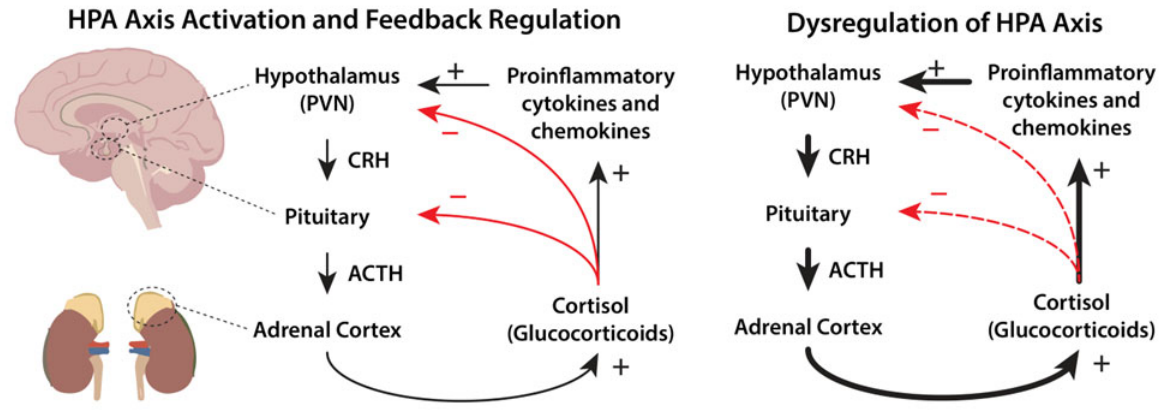
Activation of the HPA axis originates in the hypothalamic PVN (paraventricular nucleus, a neuroendocrine region of the hypothalamus that integrates neural and inflammatory signals to initiate the stress response), where corticotropin-releasing hormone (CRH) release is regulated by inflammatory signaling pathways, including NF-κB and circulating cytokines and chemokines [75,88,89].
These signals promote downstream activation of the anterior pituitary, which releases adrenocorticotropic hormone (ACTH), a peptide hormone that stimulates adrenal glucocorticoid production. ACTH acts on the adrenal cortex to regulate cortisol synthesis [90]. Dysregulation at this level is reflected clinically by altered ACTH levels and disrupted cortisol secretion patterns.
Negative feedback regulation is mediated primarily through glucocorticoid receptor (GR) signaling within the hippocampus, with additional modulation by the prefrontal cortex and amygdala, which together coordinate stress responsivity, cognition, and emotional processing (Figure 2) [75,91]. These regions are sensitive to inflammatory and endocrine disruption. Within these circuits, FK506-binding protein 5 (FKBP5) regulates GR sensitivity, while brain-derived neurotrophic factor (BDNF), a protein that supports neuronal survival and synaptic plasticity, contributes to adaptive stress responses [75,92]. Disruption of these pathways is associated with impaired feedback control and deficits in memory, executive function, and emotional regulation [83,93].
Combustion-driven inflammation disrupts GR signaling and promotes glucocorticoid resistance. Proinflammatory-responsive pathways impair GR function by reducing receptor sensitivity and limiting nuclear translocation [80,95,96]. In parallel, increased FKBP5 expression decreases receptor affinity for cortisol and attenuates downstream signaling [75,92,97,98]. These combined mechanisms weaken HPA axis negative feedback and sustain neuroendocrine activation and inflammatory signaling. Peripheral immune signaling further reinforces this dysregulation. Circulating proinflammatory biomarkers act on central regulatory regions, including the hypothalamus, to amplify HPA axis activation and integrate systemic inflammation with neuroendocrine responses [25,81,88,99].
Preclinical models of combustion-related exposures demonstrate altered glucocorticoid dynamics, impaired feedback regulation, and changes in GR-associated signaling pathways, supporting a direct impact of inhaled toxicants on HPA axis function [66,67,68]. Human studies further show that exposure to PM is associated with disrupted cortisol rhythms and increased systemic inflammation [68,72,94]. In Veterans with deployment-related exposures, these neuroendocrine alterations co-occur with respiratory disease, inflammatory biomarkers, and cognitive and mood-related symptoms, indicating coordinated dysregulation across physiological systems [71,81,82,83,100].
HPA axis dysfunction is closely linked to neuropsychiatric outcomes, including depression and PTSD. Altered glucocorticoid feedback, GR resistance, and FKBP5-mediated changes in receptor sensitivity are established mechanisms underlying stress-related psychiatric disorders [75,92,97,98,101,102,103]. In exposed populations, disrupted cortisol dynamics and persistent inflammation are associated with increased risk of mood and anxiety disorders [71,81,82,83,100,104]. A more detailed discussion of these relationships is provided in Section 3.3.
Table 3. Combustion-derived toxicants associated with neuroinflammation and HPA axis dysregulation. Evidence from clinical and experimental studies indicates that PM, PAH/VOCs, metals, and fuel combustion products can activate inflammatory and neuroendocrine pathways associated with cognitive impairment and neuropsychiatric outcomes following exposure to combustion emissions.
Toxicant ClassRepresentative
Compounds
Biological
Pathways
Affected
Effect on Brain HealthReferences
PM2.5/UFPCombustion particles, soot, secondary organic aerosols, carbon blackNeuroinflammation, oxidative stress, cytokine signaling (IL-1β, IL-6)HPA axis activation, microglial activation, cognitive impairment, and increased depression risk[8,33,48,50,51,66,105,106,107]
PAHsNaphthalene, benzo[a]pyreneAryl hydrocarbon receptor signaling, oxidative stress, inflammatory cytokine pathwaysAlters neurotransmitter systems and may impair cognition and emotional regulation[17,50,51,107,108]
VOCsBenzene, acrolein, toluene, xyleneNeurotransmitter disruption and solvent neurotoxicity pathwaysChronic toxic encephalopathy, mood disorders, and memory deficits[14,42,109,110]
Combustion
mixtures
Diesel exhaust particles, air pollutionSystemic inflammation, microglial activation, cytokine signalingIncreased inflammatory cytokines and altered stress hormone signaling[25,48,66,69,105,106,107]
Aerosolized
metals
Lead, manganese, nickel, vanadiumOxidative stress, mitochondrial dysfunctionCognitive impairment and neurodegeneration[17,29,69,106,107]
Jet fuel combustion productsJP-8 components including benzene and naphthaleneNeurotoxic solvent pathways and inflammatory signalingNeurobehavioral symptoms, fatigue, and impaired cognitive performance[13,14,46]
Environmental dust Crystalline silica, desert dust particlesPulmonary inflammation and systemic immune activationSystemic inflammation and neuroimmune signaling affecting brain health[1,3,15]
Table 4. Brain structures, molecular pathways, and biomarkers associated with HPA axis dysregulation and combustion-related toxicants.
Brain Region/
System
Role in HPA Axis RegulationKey BiomarkersClinical or Physiologic
Biomarkers
Associated
Combustion Toxicants
Supporting References
Hypothalamus (PVN)Initiates stress response via CRH secretionCRH, IL1B, IL6, TNF, NF-κBElevated CRH and altered cortisol rhythmsPM2.5, PAHs, VOCs[88,89,106,111]
PituitaryReleases ACTH in response to CRH stimulationACTH signaling pathwaysElevated ACTH levelsCombustion particles, diesel exhaust PM[75,89,112]
Adrenal CortexProduces glucocorticoids (cortisol) in response to ACTHSteroidogenic
enzymes
Dysregulated cortisol secretionAir pollution mixtures, PAHs[83,88,90,111]
HippocampusNegative feedback regulation of HPA axis via glucocorticoid
receptors
GR, FKBP5, BDNFReduced hippocampal volume, impaired memoryPM2.5, PAHs[83,88]
Prefrontal
Cortex
Modulates stress response and executive control over limbic signalingFKBP5, GR signaling, inflammatory signalingCognitive impairment, executive dysfunctionPM2.5, VOCs[51,71,89]
AmygdalaEmotional stress processing and stimulation of HPA activationCRH, IL-1β, inflammatory signalingAnxiety, PTSD-related symptomsPAHs, combustion pollutants[68,73,82,88]
Peripheral Immune SystemProinflammatory signaling activates central HPA pathwaysIL1B, IL6, TNFElevated inflammatory cytokinesAir pollution mixtures, burn pit emissions[25,81,88,99]
Glucocorticoid Receptor RegulationControls HPA negative feedback signalingFKBP5Glucocorticoid resistance, chronic stress signalingCombustion toxicants, inflammatory cytokines[75,92,111]

3.3. Neurological and Neuropsychiatric Outcomes

3.3.1. Toxic Encephalopathy in Burn Pit-Exposed Veterans

Toxic encephalopathy (TE) refers to brain dysfunction resulting from toxic exposures and encompasses a spectrum of neurocognitive and neurobehavioral impairments that complicate diagnosis [42,109]. In deployment settings, burn pit exposures represent a clinically relevant source of mixed combustion toxicants, including PM2.5, PAHs, VOCs, and metals, as documented in VA airborne hazard exposure guidance and Veteran registry data [113]. Diagnosis requires documentation of exposure, a compatible neurological syndrome, temporal progression, and exclusion of alternative causes, supported by environmental assessment and neuropsychological testing [42,109]. TE includes several subtypes, chronic toxic encephalopathy (CTE), chronic solvent-induced encephalopathy (CSE), acute diffuse toxic encephalopathy, and toxic-induced cerebellar syndromes (Table 5) [100,109] with symptom severity driven by exposure intensity and duration [42,109].
Multiple early clinical features of TE are also common manifestations of deployment-related stress or other conditions, including sleep disorders, traumatic brain injury (TBI), and psychiatric comorbidities, complicating early diagnosis and potentially delaying treatment [114,115]. Importantly, many symptoms associated with TE have been reported in burn pit-exposed Veterans, including fatigue, impaired concentration, memory deficits, and mood disturbances, often occurring in the absence of overt structural abnormalities. These symptoms, along with anxiety, sleep disruption, and reduced cognitive endurance, overlap with features of a CSE-like phenotype and have been documented in Veteran exposure registries and clinical guidance summaries [116]. Collectively, these observations highlight the challenges of distinguishing exposure-related neurotoxic effects from other deployment-associated conditions and underscore the need for improved diagnostic approaches, biomarkers, and longitudinal studies in exposed populations.
TE is closely linked to disruption of the HPA axis, providing a mechanistic bridge between environmental exposure and neuropsychiatric outcomes. Combustion-derived toxicants induce pulmonary inflammation and systemic cytokine release, which signal to the brain and alter hypothalamic regulation of CRH and downstream ACTH–cortisol dynamics, impairing GR-mediated negative feedback. This results in dysregulated cortisol exposure, altered FKBP5 expression, and reduced feedback sensitivity, reinforcing chronic stress signaling. These pathways are strongly associated with depression, PTSD, and suicidality, with HPA axis dysfunction representing one of the most predictive biomarker-associated risk factors for suicide [94].
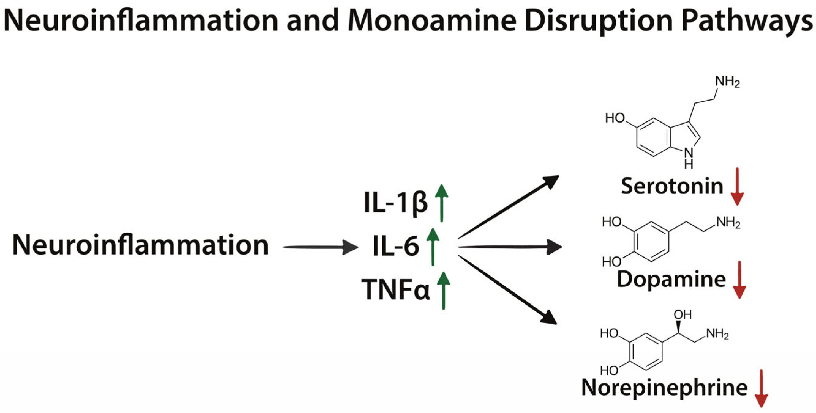
At the molecular level, TE reflects convergence across inflammatory, neuroendocrine, metabolic, and epigenetic mechanisms (Table 4). Cytokine signaling activates indoleamine 2,3-dioxygenase (IDO)-mediated tryptophan metabolism, reducing serotonin availability while generating neuroactive metabolites that contribute to neurotoxicity (Figure 3) [117]. Disruption of dopaminergic pathways further impairs executive function and reward processing. Synaptic plasticity is compromised through reduced cAMP response element-binding protein (CREB) activity and BDNF expression, limiting neuronal resilience [118]. In parallel, mitochondrial dysfunction and oxidative stress impair cellular energetics, while microglial activation sustains neuroinflammation and promotes structural injury [119]. Epigenetic remodeling, particularly DNA demethylation at GRE sites within the FKBP5 locus, further disrupts GR signaling and stress-response regulation [92,120].
Clinically, early-stage TE, including mild CSE, may be partially reversible with cessation of exposure; however, repeated or prolonged exposure is associated with persistent structural changes, including white matter demyelination and gliosis, which are often irreversible [42,101,121]. This progression reflects cumulative neuroinflammatory and metabolic injury and highlights a major challenge in early detection, as initial symptoms are nonspecific and frequently underrecognized.
TE should therefore be considered within a broader spectrum of exposure-related brain injury in Veterans, where cumulative toxicant burden interacts with stress-system pathology to increase risk for cognitive impairment, mood disorders, and suicidality. The integration of mechanistic pathways and associated biomarkers summarized in Table 4 provides a framework for identifying translational targets for early detection and intervention in exposed populations [1,7,42,104,109].
Table 5. Subtypes of Toxic Encephalopathy.
SubsetExposureSymptomsClinical ManifestationsReferences
Chronic Toxic Encephalopathy (CTE)Solvents or heavy metalsMemory, concentration, and moodPsychomotor function, learning deficits, and neurological deficits[109]
Chronic Solvent-Induced Encephalopathy (CSE)Organic solventsForgetfulness, concentration, fatigue, irritability, mood changesNeuropsychological deficits in speed of information processing and immediate memory[42]
Acute Diffuse Toxic EncephalopathyOrganic solvents and some gasesRange from mild euphoria to stuporSeizure and death[109]
Toxic-Induced Cerebellar SyndromesMetal intoxication, carbon monoxide, and VOCs Cerebellar dysfunction[100,109]

3.3.2. Cognitive Impairment and Executive Dysfunction

Deployment to military bases utilizing burn pits has been associated with an increased prevalence of disorders characterized by cognitive symptoms, including impairments in memory, attention, and executive function [122]. Many post-9/11 Veterans report “medically unexplained symptoms,” a clinical construct describing persistent symptoms without clear structural pathology, frequently including deficits in concentration, processing speed, and working memory [113]. Clinical evidence further supports a role for systemic inflammation in these outcomes, as elevated circulating biomarkers such IL-6 and CRP are associated with poorer memory performance and reduced cognitive efficiency [123]. In parallel, Veterans with deployment-related exposures exhibit increased levels of neuroinflammatory markers [71,72], supporting the concept that chronic, low-grade inflammation—both systemic and central—contributes to cognitive dysfunction in this population (Table 3).
Mechanistic and preclinical studies provide biological context for these clinical observations. Exposure to PM has been shown to directly disrupt neurocognitive pathways. Components of PM2.5, including phenyl-containing organic compounds, can interact with neurotransmitter systems by binding to dopamine receptors, thereby altering dopaminergic signaling [32]. The dopamine receptor D1 (Drd1), which plays a central role in prefrontal cortex-mediated functions such as working memory, attention, and planning, is reduced following PM exposure in rodent models and is associated with anxiety- and depression-like behaviors [32]. Disruption of dopaminergic tone is particularly relevant to executive dysfunction, as tightly regulated dopamine signaling is required for cognitive flexibility and goal-directed behavior [124]. Inflammatory signaling further amplifies these effects; pro-inflammatory cytokines such as IL-1 and IL-6 can alter activity in reward- and cognition-related brain regions leading to reduced motivation and impaired reward processing (Figure 3) [125]. Proinflammatory signaling also interacts with the HPA axis, altering glucocorticoid dynamics and further impairing memory and executive function [75].
In addition to neurotransmitter and inflammatory mechanisms, burn pit-related toxicants disrupt neuronal energy metabolism, a critical determinant of cognitive performance. In rodent exposure models there was impaired adenosine triphosphate (ATP) production and deficits in cellular respiration [65]. More broadly, PM2.5-induced oxidative stress alters mitochondrial regulators of energy metabolism, limiting the energy supply required for synaptic signaling and plasticity [23]. These mitochondrial impairments align with clinical symptoms commonly reported by exposed Veterans, including fatigue, reduced processing speed, and diminished cognitive endurance. Collectively, these findings support a model in which burn pit-related combustion exposures contribute to cognitive impairment through convergent mechanisms involving systemic and neuroinflammation, disruption of dopaminergic signaling, and mitochondrial dysfunction, ultimately affecting circuits that govern executive function, attention, and motivation (Table 4).
In individuals with PTSD, including Veterans, altered glucocorticoid (GC) signaling has direct functional consequences on cognition. Experimental cortisol administration has been shown to modify episodic and working memory performance, and fMRI studies demonstrate that corticosteroids modulate working memory tasks, linking HPA axis activity to executive function and cognitive control [126,127]. These neuroendocrine effects provide a mechanistic foundation for the cognitive and behavioral disturbances observed in PTSD and depression and, as discussed in Section 3.3.3, extend to broader disruptions in cognitive function and neurocircuitry associated with chronic toxicant exposure.

3.3.3. Depression and PTSD in Burn Pit-Exposed Veterans

PTSD and major depressive disorder are highly prevalent among post-9/11 Veterans and represent significant contributors to morbidity and suicide risk in this population. PTSD affects approximately 11–20% of Veterans annually, with lifetime prevalence approaching 20–30%, while major depressive disorder is reported in approximately 14–28%, frequently with substantial comorbidity [128,129]. Veterans with PTSD, depression, or substance use disorders exhibit markedly elevated suicide risk, more than twofold higher than those without these conditions [102]. In 2022, 6407 U.S. Veterans died by suicide, with rates rising faster than in non-Veteran populations since 2005 and highest among individuals aged 18–34, those most likely to have deployed to Iraq and Afghanistan [102].
Clinical evidence indicates that both PTSD and depression are characterized by convergent biological alterations, including systemic inflammation, impaired GR signaling, and chronic HPA axis dysregulation [81,89,99,103]. Disrupted cortisol dynamics, manifesting as altered diurnal rhythms, impaired feedback sensitivity, and changes in receptor responsiveness, are associated with hippocampal atrophy, impaired memory processing, and neuroinflammatory-driven alterations in the amygdala [80,83,104]. Neuroimaging studies demonstrate heightened amygdala reactivity and reduced hippocampal volume, consistent with impaired emotional regulation and stress adaptation [8,63,130]. In parallel, PET imaging studies show increased microglial activation, which correlates with depressive symptom severity and supports a role for neuroinflammation in mood disorders [68,96]. Clinical studies implicate FKBP5 in altered GR sensitivity, where exposure- and stress-associated demethylation at GRE sites disrupts feedback regulation and sustains HPA axis activation [92,120].
Importantly, burn pit-exposed Veterans report a high burden of mood and cognitive symptoms, including fatigue, impaired concentration, sleep disturbance, and depression/anxiety, often in the absence of overt structural abnormalities [7,131]. While these observations are primarily symptom-based and cannot be attributed solely to exposure, they are consistent with the biological pathways described above and support a potential role for combustion-related toxicants in amplifying neuropsychiatric risk.
Preclinical data provide mechanistic support for this relationship. Combustion-derived PM2.5 exposure induces depressive-like behavior in rodent models through disruption of monoaminergic signaling, including suppression of dopaminergic pathways (e.g., Drd1) and downstream reductions in serotonin, norepinephrine, and BDNF [32,33]. Inhibition of CREB signaling further reduces BDNF expression, impairing synaptic plasticity and reinforcing depressive phenotypes [33,96]. Additional model data demonstrate that combustion exposure alters circadian regulatory pathways [65,132]. Given the critical role of circadian rhythms in HPA axis function, these changes may contribute to the flattened cortisol awakening response and altered diurnal regulation observed in PTSD [75,133,134].
Clinical and experimental findings support a model in which burn pit-related toxicant exposure interacts with established neurobiological pathways HPA axis dysfunction, neuroinflammation, monoaminergic disruption, impaired neuroplasticity, and epigenetic remodeling to increase vulnerability to depression and PTSD in Veterans. While causality remains difficult to establish due to overlapping deployment-related stressors, the convergence of symptom data and mechanistic evidence provides a strong biological rationale for the role of combustion exposures in shaping neuropsychiatric outcomes.

4. Discussion

Burn pit toxic waste exposures are harmful to humans. Burn pits are not just another combustion source, they represent a distinct exposure system that is unusually complex from a combustion science and medical perspective. U.S. military personnel deployed to regions where burn pits were utilized were typically exposed for durations spanning months to years resulting in cumulative exposures [10]. Growing evidence across wildfire, industrial, and military combustion environments supports a role for toxic emissions in disrupting the lung–brain axis and impairing brain health. Within this framework, repeated acute or chronic exposures such as that experienced by personnel stationed near burn pits likely establishes a sustained neuroinflammatory continuous state that contributes to TE and related neuropsychiatric outcomes.
The potential consequences of this chronic neuroinflammatory state extend beyond mechanistic pathways and may manifest as a spectrum of cognitive, behavioral, and psychological symptoms that can complicate clinical recognition and diagnosis. Early manifestations of TE (e.g., fatigue, impaired concentration, mood changes) are frequently managed as isolated conditions rather than as components of an exposure-related syndrome, which may delay recognition of underlying neurotoxic injury [57,58]. The clinical relevance of these symptoms is underscored by the disproportionate burden of mental health outcomes among Veterans. Veterans are at higher risk of PTSD, major depressive disorders, and suicide risk [7,102,128,129]. Although these observations suggest a plausible link between burn pit exposure and adverse neuropsychiatric outcomes, establishing causality remains challenging because Veterans often experience multiple overlapping environmental, occupational, and psychosocial risk factors throughout their lifetime.
Emerging evidence has identified growing associations between military burn pit exposures and adverse neuropsychiatric outcomes. However, important limitations remain. Key challenges include the inability to isolate the specific contributions of burn pit toxicants from co-occurring deployment-related hazards and the difficulty distinguishing neuropsychiatric outcomes potentially associated with burn pit exposures from those related to other factors, such as deployment-related stress, TBI, sleep disorders, low socioeconomic status, and psychiatric comorbidities. As a result, some service members may have entered military service with pre-existing environmental exposure burdens that were subsequently compounded by deployment-related burn pit emissions. Additional post-service occupational exposures may further contribute to cumulative toxicant burden, particularly among Veterans working in firefighting and other combustion-related occupations where exposure to naphthalene, particulate matter, and other combustion byproducts is common.
Other factors such as socioeconomic and geography may influence healthy brain aging. In North America, lower socioeconomic status communities are disproportionately burdened by air pollution exposure [135]. As a result, some service members may have entered military service with pre-existing environmental exposure burdens that were subsequently compounded by deployment-related burn pit emissions. Additional post-service occupational exposures may further contribute to cumulative toxicant burden, particularly among Veterans working in firefighting and other combustion-related occupations where exposure to naphthalene, particulate matter, and other combustion byproducts is common. The multifactorial nature of these environmental and occupational exposures highlights the need for studies that integrate lifetime exposure burden with neurological outcomes and mechanistic biomarkers.
The predominance of respiratory-focused study designs has resulted in insufficient integration of neurocognitive outcomes, neuroimaging, and biomarker data, limiting mechanistic resolution [25,48,65]. Experimental models similarly rely on simplified exposure surrogates that do not fully capture the complexity of real-world combustion mixtures [136,137]. There are a limited number of mechanistic studies specifically addressing burn pit mixtures and a distinct lack of longitudinal neurological surveillance following exposure. This leaves an incomplete understanding of exposure characterization and long-term effects of key toxicants of complex combustion mixtures, many of which are not yet fully understood. Taken together, these findings suggest that combustion-related exposures, including burn pits and related fire environments, are likely underrecognized contributors to neurological and mental health outcomes in Veterans.
Most studies rely on retrospective or modeled exposure estimates, with limited direct exposure measurements and incomplete characterization of the complex mixtures involved. Furthermore, burn pit emissions contain a broad range of compounds, including organophosphate ester flame retardants, polybrominated diphenyl ethers, dioxins, and PFAS generated from the combustion of synthetic materials, creating significant challenges for exposure assessment, biomarker development, and experimental modeling of burn pit-related health effects [64]. These limitations underscore the need for improved exposure characterization and translational research approaches that more accurately reflect the complex exposure scenarios experienced by deployed service members and Veterans.
Future experimental models should employ complex exposure mixtures that resemble burn pit emissions. Such studies are needed to provide evidence for the effects of burn pit emissions on brain health, inform VA clinical screening protocols, and help guide policy decisions regarding long-term surveillance and compensation for neuro-toxic exposure-related injuries. Incorporating structured assessments about burn pit proximity, duration, and intensity of smoke exposure, solvent and fuel handling could help identify Veterans at higher risk for exposure-related brain injury. This could include standardized exposure questionnaires in primary care and mental health intake visits, routine documentation of deployment locations relative to known burn pit sites, and screening prompts about occupational roles (e.g., fuel handling, firefighting, aircraft maintenance) that signal higher neurotoxicant exposure risk. When feasible, targeted neuropsychological testing and neuroimaging could be added onto existing PTSD and depression assessments, aligning TE diagnostic criteria with the exposure mixtures and symptoms documented [42,109,138]. Embedding environmental exposure screening into primary care and post deployment programs in both VA and non-VA clinical settings could mitigate adverse neuropsychiatric outcomes [11].

5. Conclusions

Burn pits represent a highly heterogeneous and poorly controlled combustion, toxic system that generates complex, evolving mixtures of PM2.5, ultrafine particles, PAHs, VOCs, and metal-laden particulates. These emissions reflect dynamic fire chemistry rather than a single exposure source, with composition and toxicity shaped by waste streams, combustion conditions, and environmental factors. As such, understanding associated health effects requires integration of combustion chemistry, exposure science, and mechanistic toxicology.
This review highlights the lung–brain axis as a central framework linking combustion-derived exposures to neuroinflammation, blood–brain barrier disruption, mitochondrial dysfunction, monoamine dysregulation, and HPA axis impairment. Together, these interconnected mechanisms provide a biologically plausible basis for observed associations between burn pit exposures and adverse neurological and neuropsychiatric outcomes, including toxic encephalopathy, cognitive impairment, depression, and PTSD. However, the strongest evidence to date comes from mechanistic, toxicological, and observational studies that support biological plausibility and underscore the need for continued investigation. Longitudinal clinical studies with improved exposure characterization, objective exposure biomarkers, and brain-specific outcome measures are needed to strengthen causal inference and clarify how neurological health outcomes vary across exposure levels and populations.
Addressing these knowledge gaps will require coordinated multidisciplinary approaches spanning fire science, exposure modeling, environmental chemistry, toxicology, biomarker discovery, and clinical research, which aligns with the research priorities outlined in the U.S. PACT Act. Improved characterization of burn pit emissions, identification of exposure and disease biomarkers, and integration of longitudinal exposure and health data will be essential for advancing risk assessment, early detection, and intervention strategies aimed at protecting brain health in exposed military populations.

Author Contributions

Conceptualization, T.A.B., P.B., K.M.E., J.H.T. and J.M.T.; methodology, K.M.E. and Z.A.K.; formal analysis, J.M.T.; investigation, K.M.E., Z.A.K., and T.A.B.; resources, T.A.B.; data curation, T.A.B., P.B., and K.M.E.; writing—original draft preparation, K.M.E. and Z.A.K.; writing—review and editing, T.A.B., J.H.T., P.B., K.M.E., Z.A.K., and J.M.T.; visualization, T.A.B., J.P.N., K.M.E., Z.A.K., J.H.T., and J.M.T.; supervision, T.A.B. and P.B.; project administration, T.A.B.; funding acquisition, T.A.B. All authors have read and agreed to the published version of the manuscript.

Funding

This work was supported by the U.S. Department of Veterans Affairs I01 BX004146 (T.A.B.), I01 CX002701 (J.H.T.), CVRE Investigator Award (T.A.B.), and the U.S. National Institutes of Health (R21 HD109804 (P.B.), U3DMC327550100 (P.B.)).

Institutional Review Board Statement

Not applicable for studies not involving humans or animals.

Data Availability Statement

No new data were generated in this study. All information discussed in this review is derived from previously published sources cited within the article.

Acknowledgments

During the preparation of this manuscript, the authors used AI search tools (ChatGPT5.3 OpenAI, Perplexity) to assist in identifying additional search terms and potentially relevant literature. In particular, reference Henckens et al., 2011 [126] was initially identified through AI-assisted searches, but all sources were independently retrieved, verified, and interpreted by the authors. AI tools were not used to summarize or extract data from individual articles.

Conflicts of Interest

The authors declare no conflicts of interest.

Abbreviations

The following abbreviations are used in this manuscript:
PM2.5Fine particulate matter
PAHsPolycyclic aromatic hydrocarbons
VOCsVolatile organic compounds
PTSDPost-traumatic stress disorder
HPAHypothalamic–pituitary–adrenal axis
CENTCOMU.S. Central Command
OEFOperation Enduring Freedom
OIFOperation Iraqi Freedom
ONDOperation New Dawn
PMParticulate matter
UFPUltrafine particulate matter
CNSCentral nervous system
PACT ActSergeant First Class Heath Robinson Honoring our Promise to Address Comprehensive Toxics Act of 2022
TEToxic encephalopathy
BBBBlood–brain barrier
DoDDepartment of Defense
JP-8Jet Propellant-8
MEGMilitary exposure guideline
EPAEnvironmental Protection Agency
BTEXBenzene, toluene, ethylbenzene, and xylenes
COPDChronic obstructive pulmonary disease
CBNCarbon black naphthalene
NF-κBNuclear factor kappa B
IL-6Interleukin-6
TNFαTumor necrosis factor α
CRPC-reactive protein
TSPOTranslocator Protein
8-OHdG8-hydroxy-2′-deoxyguanosine
IL-1βinterleukin-1β
PETPositron emission tomography
sTREM2Soluble triggering receptor expressed on myeloid cells 2
CSFCerebral spinal fluid
IBA1Ionized calcium-binding adapter molecule 1
MHC-IIMHC Class II
GFAPGlial fibrillary acidic protein
S100BS100 calcium-binding protein B
EREndoplasmic reticulum
MMP-9Matrix metalloproteinase-9
miRNAMicroRNA
PFASPer- and polyfluoroalkyl substances
PVNParaventricular nucleus
CRHCorticotropin-releasing hormone
ACTHAdrenocorticotropic hormone
GRGlucocorticoid receptor
FKBP5FK506-binding protein 5
BDNFBrain-derived neurotrophic factor
CTEChronic toxic encephalopathy
CSEChronic solvent-induced encephalopathy
TBITraumatic brain injury
IDOIndoleamine 2,3-dioxygenase
CREBcAMP-response element-binding protein
Drd1Dopamine receptor D1
GREGlucocorticoid response element
GCGlucocorticoid

References

  1. Garshick, E.; Abraham, J.H.; Baird, C.P.; Ciminera, P.; Downey, G.P.; Falvo, M.J.; Hart, J.E.; Jackson, D.A.; Jerrett, M.; Kuschner, W.; et al. Respiratory Health after Military Service in Southwest Asia and Afghanistan. An Official American Thoracic Society Workshop Report. Ann. Am. Thorac. Soc. 2019, 16, e1–e16. [Google Scholar] [CrossRef]
  2. Woskie, S.R.; Bello, A.; Rennix, C.; Jiang, L.; Trivedi, A.N.; Savitz, D.A. Burn Pit Exposure Assessment to Support a Cohort Study of US Veterans of the Wars in Iraq and Afghanistan. J. Occup. Environ. Med. 2023, 65, 449–457. [Google Scholar] [CrossRef]
  3. Institute of Medicine; Board on the Health of Select Populations; Committee on the Long-Term Health Consequences of Exposure to Burn Pits in Iraq and Afghanistan. Long-Term Health Consequences of Exposure to Burn Pits in Iraq and Afghanistan; National Academies Press: Washington, DC, USA, 2011. [Google Scholar]
  4. Penuelas, V.L.; Lo, D.D. Burn pit exposure in military personnel and the potential resulting lung and neurological pathologies. Front. Environ. Health 2024, 3, 1364812. [Google Scholar] [CrossRef]
  5. Perveen, M.M.; Mayo-Malasky, H.E.; Lee-Wong, M.F.; Tomaska, J.M.; Forsyth, E.; Gravely, A.; Klein, M.A.; Trembley, J.H.; Butterick, T.A.; Promisloff, R.A.; et al. Gross Hematuria and Lower Urinary Tract Symptoms Associated with Military Burn Pits Exposures in US Veterans Deployed to Iraq and Afghanistan. J. Occup. Environ. Med. 2023, 65, 740–744. [Google Scholar] [CrossRef] [PubMed]
  6. Pun, V.C.; Manjourides, J.; Suh, H. Association of Ambient Air Pollution with Depressive and Anxiety Symptoms in Older Adults: Results from the NSHAP Study. Environ. Health Perspect. 2017, 125, 342–348. [Google Scholar] [CrossRef]
  7. Hoisington, A.J.; Stearns-Yoder, K.A.; Kovacs, E.J.; Postolache, T.T.; Brenner, L.A. Airborne Exposure to Pollutants and Mental Health: A Review with Implications for United States Veterans. Curr. Environ. Health Rep. 2024, 11, 168–183. [Google Scholar] [CrossRef]
  8. Zhang, Y.; Rosen, R.; Reibman, J.; Shao, Y. Posttraumatic Stress Disorder Mediates the Association between Traumatic World Trade Center Dust Cloud Exposure and Ongoing Systemic Inflammation in Community Members. Int. J. Environ. Res. Public Health 2022, 19, 8622. [Google Scholar] [CrossRef]
  9. Trembley, J.; Barach, P.; Tomáška, J.; Poole, J.; Ginex, P.; Miller, R.; Lindheimer, J.; Szema, A.; Gandy, K.; Siddharthan, T.; et al. Current understanding of the impact of United States military airborne hazards and burn pit exposures on respiratory health. Part. Fibre Toxicol. 2024, 21, 43. [Google Scholar] [CrossRef]
  10. Hoisington, L.C.A.J.; Lowry, C.A.; McDonald, L.T.; Krefft, S.D.; Rose, C.S.; Kovacs, E.J.; Brenner, L.A. First Annual PACT Act Research Symposium on Veterans Health: A Colorado PACT Act Collaboration (CoPAC) Initiative. Mil. Med. 2024, 189, 80–84. [Google Scholar] [CrossRef] [PubMed]
  11. Conti, M.A.; Bardes, J.M.; Oury, J.; Goodboy, A.K.; Shin, M.; Wilson, A. Prevalence of Burn Pit Associated Symptoms Among US Veterans Who Utilize Non-VA Private Healthcare. J. Occup. Environ. Med. 2024, 66, 439–444. [Google Scholar] [CrossRef] [PubMed]
  12. Braithwaite, I.; Zhang, S.; Kirkbride, J.B.; Osborn, D.P.J.; Hayes, J.F. Air Pollution (Particulate Matter) Exposure and Associations with Depression, Anxiety, Bipolar, Psychosis and Suicide Risk: A Systematic Review and Meta-Analysis. Environ. Health Perspect. 2019, 127, 126002. [Google Scholar] [CrossRef] [PubMed]
  13. National Research Council; Division on Earth and Life Studies, Board on Environmental Studies and Toxicology; Committee on Toxicology; Subcommittee on Jet-Propulsion Fuel 8. Toxicologic Assessment of Jet-Propulsion Fuel 8; National Academies Press: Washington, DC, USA, 2003. [Google Scholar]
  14. Pleil, J.D.; Smith, L.B.; Zelnick, S.D. Personal exposure to JP-8 jet fuel vapors and exhaust at air force bases. Environ. Health Perspect. 2000, 108, 183–192. [Google Scholar] [CrossRef]
  15. Blasch, K.W.; Kolivosky, J.E.; Heller, J.M. Environmental Air Sampling Near Burn Pit and Incinerator Operations at Bagram Airfield, Afghanistan. J. Occup. Environ. Med. 2016, 58, S38. [Google Scholar] [CrossRef]
  16. Blasch, K.; Kolivosky, J.; Hill, B. Occupational exposures among personnel working near combined burn pit and incinerator operations at Bagram Airfield, Afghanistan. Inhal. Toxicol. 2016, 28, 216–225. [Google Scholar] [CrossRef]
  17. Urbanski, S.P.; Hao, W.M.; Baker, S. Chapter 4 Chemical Composition of Wildland Fire Emissions. In Developments in Environmental Science; Elsevier: Amsterdam, The Netherlands, 2008; pp. 79–107. [Google Scholar] [CrossRef]
  18. Lemieux, P.M. Evaluation of Emissions from the Open Burning of Household Waste in Barrels; U.S. Environmental Protection Agency, National Risk Management Research Laboratory: Cincinnati, OH, USA, 1998. [Google Scholar]
  19. Kim, Y.H.; Warren, S.H.; Kooter, I.; Williams, W.C.; George, I.J.; Vance, S.A.; Hays, M.D.; Higuchi, M.A.; Gavett, S.H.; DeMarini, D.M.; et al. Chemistry, lung toxicity and mutagenicity of burn pit smoke-related particulate matter. Part. Fibre Toxicol. 2021, 18, 45. [Google Scholar] [CrossRef]
  20. Alrashidi, H.; Sibai, F.N.; Abonamah, A.; Alrashidi, M.; Alsaber, A. PM2.5: Air Quality Index Prediction Using Machine Learning: Evidence from Kuwait’s Air Quality Monitoring Stations. Sustainability 2025, 17, 9136. [Google Scholar] [CrossRef]
  21. Penn, A.; Murphy, G.; Barker, S.; Henk, W.; Penn, L. Combustion-Derived Ultrafine Particles Transport Organic Toxicants to Target Respiratory Cells. Environ. Health Perspect. 2005, 113, 956–963. [Google Scholar] [CrossRef]
  22. Gillespie, P.; Tajuba, J.; Lippmann, M.; Chen, L.-C.; Veronesi, B. Particulate Matter Neurotoxicity in Culture is Size-Dependent. Neurotoxicology 2013, 36, 112–117. [Google Scholar] [CrossRef] [PubMed]
  23. Xu, F.; Shi, X.; Qiu, X.; Jiang, X.; Fang, Y.; Wang, J.; Hu, D.; Zhu, T. Investigation of the chemical components of ambient fine particulate matter (PM2.5) associated with in vitro cellular responses to oxidative stress and inflammation. Environ. Int. 2020, 136, 105475. [Google Scholar] [CrossRef]
  24. Niu, X.; Ho, S.S.H.; Ho, K.F.; Huang, Y.; Sun, J.; Wang, Q.; Zhou, Y.; Zhao, Z.; Cao, J. Atmospheric levels and cytotoxicity of polycyclic aromatic hydrocarbons and oxygenated-PAHs in PM2.5 in the Beijing-Tianjin-Hebei region. Environ. Pollut. 2017, 231, 1075–1084. [Google Scholar] [CrossRef] [PubMed]
  25. Glencross, D.A.; Ho, T.-R.; Camiña, N.; Hawrylowicz, C.M.; Pfeffer, P.E. Air pollution and its effects on the immune system. Free Radic. Biol. Med. 2020, 151, 56–68. [Google Scholar] [CrossRef]
  26. Geiser, M.; Rothen-Rutishauser, B.; Kapp, N.; Schürch, S.; Kreyling, W.; Schulz, H.; Semmler, M.; Hof, V.I.; Heyder, J.; Gehr, P. Ultrafine Particles Cross Cellular Membranes by Nonphagocytic Mechanisms in Lungs and in Cultured Cells. Environ. Health Perspect. 2005, 113, 1555–1560. [Google Scholar] [CrossRef]
  27. Kreyling, W.G.; Semmler, M.; Möller, W. Dosimetry and Toxicology of Ultrafine Particles. J. Aerosol Med. 2004, 17, 140–152. [Google Scholar] [CrossRef] [PubMed]
  28. Elam, T.; Raiculescu, S.; Biswal, S.; Zhang, Z.; Orestes, M.; Ramanathan, M. Air Pollution Exposure and the Development of Chronic Rhinosinusitis in the Active Duty Population. Mil. Med. 2023, 188, e1965–e1969. [Google Scholar] [CrossRef]
  29. Chen, X.; Guo, J.; Huang, Y.; Liu, S.; Huang, Y.; Zhang, Z.; Zhang, F.; Lu, Z.; Li, F.; Zheng, J.C.; et al. Urban airborne PM2.5-activated microglia mediate neurotoxicity through glutaminase-containing extracellular vesicles in olfactory bulb. Environ. Pollut. 2020, 264, 114716. [Google Scholar] [CrossRef]
  30. Angelova, P.R.; Abramov, A.Y. Role of mitochondrial ROS in the brain: From physiology to neurodegeneration. FEBS Lett. 2018, 592, 692–702. [Google Scholar] [CrossRef]
  31. Gandhi, S.; Abramov, A.Y. Mechanism of Oxidative Stress in Neurodegeneration. Oxidative Med. Cell. Longev. 2012, 2012, 428010. [Google Scholar] [CrossRef]
  32. Ji, S.; Guo, Y.; Yan, W.; Wei, F.; Ding, J.; Hong, W.; Wu, X.; Ku, T.; Yue, H.; Sang, N. PM2.5 exposure contributes to anxiety and depression-like behaviors via phenyl-containing compounds interfering with dopamine receptor. Proc. Natl. Acad. Sci. USA 2024, 121, e2319595121. [Google Scholar] [CrossRef]
  33. Li, H.; Guo, X.; Li, P.; Gao, X.; Song, X.; Chen, X.; Liang, R.; Yang, J.; Li, Y.; Chen, H.; et al. Particulate matter induces depression-like behavior through systemic inflammation and brain-derived neurotrophic factors. Environ. Int. 2024, 194, 108883. [Google Scholar] [CrossRef] [PubMed]
  34. Keller, Z.A.; Eggers, K.M.; Nixon, J.P.; Butterick, T.A. The Impact of Air Pollution and Obesity on Cognitive Decline and Risk of Alzheimer’s Disease. Int. J. Mol. Sci. 2025, 27, 92. [Google Scholar] [CrossRef] [PubMed]
  35. Michanowicz, D.R.; Dayalu, A.; Nordgaard, C.L.; Buonocore, J.J.; Fairchild, M.W.; Ackley, R.; Schiff, J.E.; Liu, A.; Phillips, N.G.; Schulman, A.; et al. Home is Where the Pipeline Ends: Characterization of Volatile Organic Compounds Present in Natural Gas at the Point of the Residential End User. Environ. Sci. Technol. 2022, 56, 10258–10268. [Google Scholar] [CrossRef]
  36. Koppmann, R. Volatile Organic Compounds in the Atmosphere; John Wiley & Sons: Hoboken, NJ, USA, 2008. [Google Scholar]
  37. Lemieux, P.M.; Lutes, C.C.; Santoianni, D.A. Emissions of organic air toxics from open burning: A comprehensive review. Prog. Energy Combust. Sci. 2004, 30, 1–32. [Google Scholar] [CrossRef]
  38. Zhou, X.; Zhou, X.; Wang, C.; Zhou, H. Environmental and human health impacts of volatile organic compounds: A perspective review. Chemosphere 2023, 313, 137489. [Google Scholar] [CrossRef]
  39. U.S. Army Public Health Command. TG230 Environmental HRA and Chemical Military Exposure Guidelines (MEGs)—Defense Centers for Public Health—Aberdeen; Defense Centers for Public Health: Aberdeen, MD, USA, 2018; Available online: https://ph.health.mil/topics/envirohealth/hrasm/Pages/TG230.aspx (accessed on 28 May 2026).
  40. Roney, N. Acrolein ToxGuideTM; Agency for Toxic Substances and Disease Registry: Atlanta, GA, USA, 2025. Available online: https://www.atsdr.cdc.gov/toxguides/toxguide-124.pdf (accessed on 1 June 2026).
  41. Cui, Y.; Mo, Z.; Ji, P.; Zhong, J.; Li, Z.; Li, D.; Qin, L.; Liao, Q.; He, Z.; Guo, W.; et al. Benzene Exposure Leads to Lipodystrophy and Alters Endocrine Activity In Vivo and In Vitro. Front. Endocrinol. 2022, 13, 937281. [Google Scholar] [CrossRef]
  42. van Valen, E.; van Thriel, C.; Akila, R.; Nilson, L.N.; Bast-Pettersen, R.; Sainio, M.; van Dijk, F.; van der Laan, G.; Verberk, M.; Wekking, E. Chronic solvent-induced encephalopathy: European consensus of neuropsychological characteristics, assessment, and guidelines for diagnostics. NeuroToxicology 2012, 33, 710–726. [Google Scholar] [CrossRef]
  43. Engelbrecht, J.P.; McDonald, E.V.; Gillies, J.A.; Gertler, A.W. Department of Defense Enhanced Particulate Matter Surveillance Program (EPMSP); Desert Research Institute (DRI): Reno, NV, USA, 2008. [Google Scholar]
  44. Engelbrecht, J.; Mcdonald, E.; Gillies, J.; Jayanty, R.; Casuccio, G.; Gertler, A. Characterizing Mineral Dusts and Other Aerosols from the Middle East—Part 1: Ambient Sampling. Inhal. Toxicol. 2009, 21, 297–326. [Google Scholar] [CrossRef] [PubMed]
  45. National Research Council (US) Committe. Systemic Exposures to Volatile Organic Compounds and Factors Influencing Susceptibility to Their Effects. In Contaminated Water Supplies at Camp Lejeune: Assessing Potential Health Effects; National Academies Press (US): Washington, DC, USA, 2009. [Google Scholar]
  46. Drake, M.G.; Witzmann, F.A.; Hyde, J.; Witten, M.L. JP-8 jet fuel exposure alters protein expression in the lung. Toxicology 2003, 191, 199–210. [Google Scholar] [CrossRef]
  47. Zhong, Y.; Zhu, L. Distribution, input pathway and soil–air exchange of polycyclic aromatic hydrocarbons in Banshan Industry Park, China. Sci. Total Environ. 2013, 444, 177–182. [Google Scholar] [CrossRef]
  48. Costa, L.G.; Cole, T.B.; Coburn, J.; Chang, Y.-C.; Dao, K.; Roque, P. Neurotoxicants Are in the Air: Convergence of Human, Animal, and in vitro Studies on the Effects of Air Pollution on the Brain. BioMed Res. Int. 2014, 2014, 736385. [Google Scholar] [CrossRef] [PubMed]
  49. Delgado-Saborit, J.M.; Aquilina, N.; Baker, S.; Harrad, S.; Meddings, C.; Harrison, R.M. Determination of atmospheric particulate-phase polycyclic aromatic hydrocarbons from low volume air samples. Anal. Methods 2010, 2, 231–242. [Google Scholar] [CrossRef]
  50. Samanta, S.K.; Singh, O.V.; Jain, R.K. Polycyclic aromatic hydrocarbons: Environmental pollution and bioremediation. Trends Biotechnol. 2002, 20, 243–248. [Google Scholar] [CrossRef]
  51. Cho, J.; Sohn, J.; Noh, J.; Jang, H.; Kim, W.; Cho, S.-K.; Seo, H.; Seo, G.; Lee, S.-K.; Noh, Y.; et al. Association between exposure to polycyclic aromatic hydrocarbons and brain cortical thinning: The Environmental Pollution-Induced Neurological EFfects (EPINEF) study. Sci. Total Environ. 2020, 737, 140097. [Google Scholar] [CrossRef]
  52. Stahlhut, R.W.; Welshons, W.V.; Swan, S.H. Bisphenol A Data in NHANES Suggest Longer than Expected Half-Life, Substantial Nonfood Exposure, or Both. Environ. Health Perspect. 2009, 117, 784–789. [Google Scholar] [CrossRef] [PubMed]
  53. Hartz, A.M.S.; Bauer, B.; Block, M.L.; Hong, J.-S.; Miller, D.S. Diesel exhaust particles induce oxidative stress, proinflammatory signaling, and P-glycoprotein up-regulation at the blood-brain barrier. FASEB J. 2008, 22, 2723–2733. [Google Scholar] [CrossRef]
  54. Das, L.; Patel, B.; Patri, M. Adolescence benzo[a]pyrene treatment induces learning and memory impairment and anxiolytic like behavioral response altering neuronal morphology of hippocampus in adult male Wistar rats. Toxicol. Rep. 2019, 6, 1104–1113. [Google Scholar] [CrossRef] [PubMed]
  55. UL Research Institutes. Naphthalene—A Common Air Pollutant; Chemical Insights: Marietta, GA, USA, 2022; Available online: https://chemicalinsights.ul.org/resources/understanding-vaping-or-electronic-nicotin-delivery-systems-copy/ (accessed on 28 May 2026).
  56. Xia, X.; Carroll-Haddad, A.; Brown, N.; Utell, M.J.; Mallon, T.; Hopke, P.K. Polycyclic Aromatic Hydrocarbons and Polychlorinated Dibenzo-p-Dioxins/Dibenzofurans in Microliter Samples of Human Serum as Exposure Indicators. J. Occup. Environ. Med. Am. Coll. Occup. Environ. Med. 2016, 58, S72–S79. [Google Scholar] [CrossRef]
  57. Kim, Y.T.; Kim, W.; Bae, M.; Choi, J.E.; Kim, M.-J.; Oh, S.S.; Park, K.S.; Park, S.; Lee, S.-K.; Koh, S.-B.; et al. The effect of polycyclic aromatic hydrocarbons on changes in the brain structure of firefighters: An analysis using data from the Firefighters Research on Enhancement of Safety & Health study. Sci. Total Environ. 2022, 816, 151655. [Google Scholar] [CrossRef] [PubMed]
  58. White, A.R. The impact of military occupational combustion smoke inhalation on neuroinflammation and brain health. Neurotoxicology 2026, 113, 103394. [Google Scholar] [CrossRef]
  59. Scott, K.A.; Wingate, K.C.; DuBose, K.N.; Butler, C.R.; Ramirez-Cardenas, A.; Hale, C.R. The wildland firefighter exposure and health effect (WFFEHE) study: Cohort characteristics and health behavior changes in context. Ann. Work. Expo. Health 2024, 68, 122–135. [Google Scholar] [CrossRef]
  60. Pardo, M.; Li, C.; Fang, Z.; Levin-Zaidman, S.; Dezorella, N.; Czech, H.; Martens, P.; Käfer, U.; Gröger, T.; Rüger, C.P.; et al. Toxicity of Water- and Organic-Soluble Wood Tar Fractions from Biomass Burning in Lung Epithelial Cells. Chem. Res. Toxicol. 2021, 34, 1588–1603. [Google Scholar] [CrossRef]
  61. Peterson, B.S.; Rauh, V.A.; Bansal, R.; Hao, X.; Toth, Z.; Nati, G.; Walsh, K.; Miller, R.L.; Arias, F.; Semanek, D.; et al. Effects of Prenatal Exposure to Air Pollutants (Polycyclic Aromatic Hydrocarbons) on the Development of Brain White Matter, Cognition, and Behavior in Later Childhood. JAMA Psychiatry 2015, 72, 531–540. [Google Scholar] [CrossRef]
  62. Peiffer, J.; Grova, N.; Hidalgo, S.; Salquèbre, G.; Rychen, G.; Bisson, J.-F.; Appenzeller, B.M.R.; Schroeder, H. Behavioral toxicity and physiological changes from repeated exposure to fluorene administered orally or intraperitoneally to adult male Wistar rats: A dose–response study. NeuroToxicology 2016, 53, 321–333. [Google Scholar] [CrossRef] [PubMed]
  63. Mortamais, M.; Pujol, J.; van Drooge, B.L.; Macià, D.; Martínez-Vilavella, G.; Reynes, C.; Sabatier, R.; Rivas, I.; Grimalt, J.; Forns, J.; et al. Effect of exposure to polycyclic aromatic hydrocarbons on basal ganglia and attention-deficit hyperactivity disorder symptoms in primary school children. Environ. Int. 2017, 105, 12–19. [Google Scholar] [CrossRef]
  64. Furlong, M.A.; Liu, T.; Jung, A.; Beitel, S.; Hughes, J.; Krause, R.; Graber, J.M.; Calkins, M.M.; Calafat, A.M.; Botelho, J.C.; et al. Per- and polyfluoroalkyl substances (PFAS) and microRNA: An epigenome-wide association study in firefighters. Environ. Res. 2025, 279, 121766. [Google Scholar] [CrossRef]
  65. Haaning, A.M.; Sandri, B.J.; Wyneken, H.L.; Goldsmith, W.T.; Nixon, J.P.; Nurkiewicz, T.R.; Wendt, C.H.; Barach, P.; Trembley, J.H.; Butterick, T.A. RNA Sequencing Reveals Inflammatory and Metabolic Changes in the Lung and Brain After Carbon Black and Naphthalene Whole Body Inhalation Exposure in a Rodent Model of Military Burn Pit Exposures. Int. J. Mol. Sci. 2025, 26, 7238. [Google Scholar] [CrossRef]
  66. Mumaw, C.L.; Levesque, S.; McGraw, C.; Robertson, S.; Lucas, S.; Stafflinger, J.E.; Campen, M.J.; Hall, P.; Norenberg, J.P.; Anderson, T.; et al. Microglial priming through the lung–brain axis: The role of air pollution–induced circulating factors. FASEB J. 2016, 30, 1880–1891. [Google Scholar] [CrossRef]
  67. Milani, C.; Farina, F.; Botto, L.; Massimino, L.; Lonati, E.; Donzelli, E.; Ballarini, E.; Crippa, L.; Marmiroli, P.; Bulbarelli, A.; et al. Systemic Exposure to Air Pollution Induces Oxidative Stress and Inflammation in Mouse Brain, Contributing to Neurodegeneration Onset. Int. J. Mol. Sci. 2020, 21, 3699. [Google Scholar] [CrossRef] [PubMed]
  68. Setiawan, E.; Wilson, A.A.; Mizrahi, R.; Rusjan, P.M.; Miler, L.; Rajkowska, G.; Suridjan, I.; Kennedy, J.L.; Rekkas, P.V.; Houle, S.; et al. Increased Translocator Protein Distribution Volume, A Marker of Neuroinflammation, in the Brain During Major Depressive Episodes. JAMA Psychiatry 2015, 72, 268–275. [Google Scholar] [CrossRef] [PubMed]
  69. Lodovici, M.; Bigagli, E. Oxidative Stress and Air Pollution Exposure. J. Toxicol. 2011, 2011, 487074. [Google Scholar] [CrossRef]
  70. Wang, S.; Young, R.S.; Sun, N.N.; Witten, M.L. In vitro cytokine release from rat type II pneumocytes and alveolar macrophages following exposure to JP-8 jet fuel in co-culture. Toxicology 2002, 173, 211–219. [Google Scholar] [CrossRef]
  71. Casanova, R.; Wang, X.; Reyes, J.; Akita, Y.; Serre, M.L.; Vizuete, W.; Chui, H.C.; Driscoll, I.; Resnick, S.M.; Espeland, M.A.; et al. A Voxel-Based Morphometry Study Reveals Local Brain Structural Alterations Associated with Ambient Fine Particles in Older Women. Front. Hum. Neurosci. 2016, 10, 495. [Google Scholar] [CrossRef] [PubMed]
  72. Aurell, J.; Gullett, B.K.; Yamamoto, D. Emissions from Open Burning of Simulated Military Waste from Forward Operating Bases. Environ. Sci. Technol. 2012, 46, 11004–11012. [Google Scholar] [CrossRef]
  73. Invernizzi, A.; Rechtman, E.; Curtin, P.; Papazaharias, D.M.; Jalees, M.; Pellecchia, A.C.; Santiago-Michels, S.; Bromet, E.J.; Lucchini, R.G.; Luft, B.J.; et al. Functional changes in neural mechanisms underlying post-traumatic stress disorder in World Trade Center responders. Transl. Psychiatry 2023, 13, 239. [Google Scholar] [CrossRef]
  74. Capuron, L.; Raison, C.L.; Musselman, D.L.; Lawson, D.H.; Nemeroff, C.B.; Miller, A.H. Association of Exaggerated HPA Axis Response to the Initial Injection of Interferon-Alpha with Development of Depression During Interferon-Alpha Therapy. Am. J. Psychiatry 2003, 160, 1342–1345. [Google Scholar] [CrossRef]
  75. Leistner, C.; Menke, A. How to measure glucocorticoid receptor’s sensitivity in patients with stress-related psychiatric disorders. Psychoneuroendocrinology 2018, 91, 235–260. [Google Scholar] [CrossRef]
  76. Reisetter, A.C.; Stebounova, L.V.; Baltrusaitis, J.; Powers, L.; Gupta, A.; Grassian, V.H.; Monick, M.M. Induction of Inflammasome-dependent Pyroptosis by Carbon Black Nanoparticles. J. Biol. Chem. 2011, 286, 21844–21852. [Google Scholar] [CrossRef] [PubMed]
  77. Li, B.; Yang, W.; Ge, T.; Wang, Y.; Cui, R. Stress induced microglial activation contributes to depression. Pharmacol. Res. 2022, 179, 106145. [Google Scholar] [CrossRef]
  78. Ewers, M.; Biechele, G.; Suárez-Calvet, M.; Sacher, C.; Blume, T.; Morenas-Rodriguez, E.; Deming, Y.; Piccio, L.; Cruchaga, C.; Kleinberger, G.; et al. Higher CSF sTREM2 and microglia activation are associated with slower rates of beta-amyloid accumulation. EMBO Mol. Med. 2020, 12, e12308. [Google Scholar] [CrossRef] [PubMed]
  79. Halleskog, C.; Mulder, J.; Dahlström, J.; Mackie, K.; Hortobágyi, T.; Tanila, H.; Puli, L.K.; Färber, K.; Harkany, T.; Schulte, G. WNT signaling in activated microglia is pro-inflammatory. Glia 2011, 59, 119–131. [Google Scholar] [CrossRef]
  80. Ferrari, A.J.; Santomauro, D.F.; Mantilla Herrera, A.M.; Shadid, J.; Ashbaugh, C.; Erskine, H.E.; Charlson, F.J.; Degenhardt, L.; Scott, J.G.; McGrath, J.J.; et al. Global, regional, and national burden of 12 mental disorders in 204 countries and territories, 1990–2019: A systematic analysis for the Global Burden of Disease Study 2019. Lancet Psychiatry 2022, 9, 137–150. [Google Scholar] [CrossRef]
  81. Speer, K.; Upton, D.; Semple, S.; McKune, A. Systemic low-grade inflammation in post-traumatic stress disorder: A systematic review. J. Inflamm. Res. 2018, 11, 111–121. [Google Scholar] [CrossRef]
  82. Zhang, W.; Rutlin, J.; Eisenstein, S.A.; Wang, Y.; Barch, D.M.; Hershey, T.; Bogdan, R.; Bijsterbosch, J.D. Neuroinflammation in the Amygdala Is Associated with Recent Depressive Symptoms. Biol. Psychiatry Cogn. Neurosci. Neuroimaging 2023, 8, 967–975. [Google Scholar] [CrossRef] [PubMed]
  83. Keller, J.; Gomez, R.; Williams, G.; Lembke, A.; Lazzeroni, L.; Murphy, G.M.; Schatzberg, A.F. HPA Axis in Major Depression: Cortisol, Clinical Symptomatology, and Genetic Variation Predict Cognition. Mol. Psychiatry 2017, 22, 527–536. [Google Scholar] [CrossRef] [PubMed]
  84. Goodrich, J.M.; Xin, Y.; Beitel, S.C.; Gulotta, J.; Wang, L.; Thotakura, B.; Graber, J.M.; Urwin, D.; Mayer, A.C.; Jahnke, S.; et al. Differences in MicroRNA Expression in Firefighters Responding to a Train Derailment and Fire in East Palestine, Ohio. Epigenomes 2026, 10, 8. [Google Scholar] [CrossRef]
  85. Jeong, K.S.; Zhou, J.; Griffin, S.C.; Jacobs, E.T.; Dearmon-Moore, D.; Zhai, J.; Littau, S.R.; Gulotta, J.; Moore, P.; Peate, W.F.; et al. MicroRNA Changes in Firefighters. J. Occup. Environ. Med. 2018, 60, 469–474. [Google Scholar] [CrossRef]
  86. Thatcher, T.H.; Woeller, C.F.; Thakar, J.; Khan, A.; Hopke, P.K.; Smith, M.R.; Uppal, K.; Walker, D.I.; Go, Y.-M.; Jones, D.P.; et al. Analysis of Postdeployment Serum Samples Identifies Potential Biomarkers of Exposure to Burn Pits and Other Environmental Hazards. J. Occup. Environ. Med. 2019, 61, S45–S54. [Google Scholar] [CrossRef]
  87. Woeller, C.F.; Thatcher, T.H.; Van Twisk, D.; Pollock, S.J.; Croasdell, A.; Kim, N.; Hopke, P.K.; Xia, X.; Thakar, J.; Mallon, C.T.M.; et al. Detection of Serum microRNAs from Department of Defense Serum Repository: Correlation with Cotinine, Cytokine, and Polycyclic Aromatic Hydrocarbon Levels. J. Occup. Environ. Med. 2016, 58, S62–S71. [Google Scholar] [CrossRef]
  88. Miller, A.H.; Raison, C.L. Immune System Contributions to the Pathophysiology of Depression. Focus 2008, 6, 36–45. [Google Scholar] [CrossRef]
  89. Menke, A. The HPA Axis as Target for Depression. Curr. Neuropharmacol. 2024, 22, 904–915. [Google Scholar] [CrossRef]
  90. Agorastos, A.; Heinig, A.; Stiedl, O.; Hager, T.; Sommer, A.; Müller, J.C.; Schruers, K.R.; Wiedemann, K.; Demiralay, C. Vagal effects of endocrine HPA axis challenges on resting autonomic activity assessed by heart rate variability measures in healthy humans. Psychoneuroendocrinology 2019, 102, 196–203. [Google Scholar] [CrossRef] [PubMed]
  91. Modell, S.; Yassouridis, A.; Huber, J.; Holsboer, F. Corticosteroid Receptor Function Is Decreased in Depressed Patients. Neuroendocrinology 2008, 65, 216–222. [Google Scholar] [CrossRef] [PubMed]
  92. Menke, A.; Klengel, T.; Rubel, J.; Brückl, T.; Pfister, H.; Lucae, S.; Uhr, M.; Holsboer, F.; Binder, E.B. Genetic variation in FKBP5 associated with the extent of stress hormone dysregulation in major depression. Genes Brain Behav. 2013, 12, 289–296. [Google Scholar] [CrossRef]
  93. Barden, N. Modulation of Glucocorticoid Receptor Gene Expression by Antidepressant Drugs. Pharmacopsychiatry 1996, 29, 12–22. [Google Scholar] [CrossRef]
  94. Hennings, J.M.; Ising, M.; Uhr, M.; Holsboer, F.; Lucae, S. Recurrent suicide attempts affect normalization of HPA axis dysregulation after recovery from major depression. Front. Psychiatry 2022, 13, 937582. [Google Scholar] [CrossRef] [PubMed]
  95. Webster, J.C.; Oakley, R.H.; Jewell, C.M.; Cidlowski, J.A. Proinflammatory cytokines regulate human glucocorticoid receptor gene expression and lead to the accumulation of the dominant negative β isoform: A mechanism for the generation of glucocorticoid resistance. Proc. Natl. Acad. Sci. USA 2001, 98, 6865–6870. [Google Scholar] [CrossRef] [PubMed]
  96. Yin, Y.; Ju, T.; Zeng, D.; Duan, F.; Zhu, Y.; Liu, J.; Li, Y.; Lu, W. “Inflamed” depression: A review of the interactions between depression and inflammation and current anti-inflammatory strategies for depression. Pharmacol. Res. 2024, 207, 107322. [Google Scholar] [CrossRef]
  97. Zhang, X.; Clark, A.F.; Yorio, T. FK506-Binding Protein 51 Regulates Nuclear Transport of the Glucocorticoid Receptor β and Glucocorticoid Responsiveness. Investig. Ophthalmol. Vis. Sci. 2008, 49, 1037–1047. [Google Scholar] [CrossRef]
  98. Koh, D.S.Q.; Gan, W.H. Textbook of Occupational Medicine Practice, 5th ed.; World Scientific: Singapore, 2022. [Google Scholar]
  99. Yang, J.-J.; Jiang, W. Immune biomarkers alterations in post-traumatic stress disorder: A systematic review and meta-analysis. J. Affect. Disord. 2020, 268, 39–46. [Google Scholar] [CrossRef]
  100. Manto, M.; Perrotta, G. Toxic-induced cerebellar syndrome: From the fetal period to the elderly. In Handbook of Clinical Neurology; Elsevier: Amsterdam, The Netherlands, 2018; pp. 333–352. [Google Scholar] [CrossRef]
  101. Hurley, R.A.; Taber, K.H. Occupational Exposure to Solvents: Neuropsychiatric and Imaging Features. J. Neuropsychiatry Clin. Neurosci. 2015, 27, 1–6. [Google Scholar] [CrossRef]
  102. Ramchand, R. Suicide Among Veterans: Veterans’ Issues in Focus. RAND Health Q. 2022, 9, 21. [Google Scholar]
  103. Osimo, E.F.; Baxter, L.J.; Lewis, G.; Jones, P.B.; Khandaker, G.M. Prevalence of low-grade inflammation in depression: A systematic review and meta-analysis of CRP levels. Psychol. Med. 2019, 49, 1958–1970. [Google Scholar] [CrossRef]
  104. McEwen, B.S.; Bowles, N.P.; Gray, J.D.; Hill, M.N.; Hunter, R.G.; Karatsoreos, I.N.; Nasca, C. Mechanisms of stress in the brain. Nat. Neurosci. 2015, 18, 1353–1363. [Google Scholar] [CrossRef]
  105. Block, M.L.; Calderón-Garcidueñas, L. Air pollution: Mechanisms of neuroinflammation and CNS disease. Trends Neurosci. 2009, 32, 506–516. [Google Scholar] [CrossRef]
  106. Genc, S.; Zadeoglulari, Z.; Fuss, S.H.; Genc, K. The Adverse Effects of Air Pollution on the Nervous System. J. Toxicol. 2012, 2012, 782462. [Google Scholar] [CrossRef] [PubMed]
  107. Kim, K.-H.; Jahan, S.A.; Kabir, E.; Brown, R.J.C. A review of airborne polycyclic aromatic hydrocarbons (PAHs) and their human health effects. Environ. Int. 2013, 60, 71–80. [Google Scholar] [CrossRef] [PubMed]
  108. Kritikos, M.; Clouston, S.A.P.; Huang, C.; Pellecchia, A.C.; Mejia-Santiago, S.; Carr, M.A.; Kotov, R.; Lucchini, R.G.; Gandy, S.E.; Bromet, E.J.; et al. Cortical complexity in world trade center responders with chronic posttraumatic stress disorder. Transl. Psychiatry 2021, 11, 597. [Google Scholar] [CrossRef]
  109. Kim, Y.; Kim, J.W. Toxic Encephalopathy. Saf. Health Work 2012, 3, 243–256. [Google Scholar] [CrossRef]
  110. U.S. Deapartment of Health and Human Services. Toxicological Profile for JP-5, JP-8, And Jet A Fuels; Agency for Toxic Substances and Disease Registry: Atlanta, GA, USA, 2017. [Google Scholar]
  111. Pariante, C.M. Glucocorticoid Receptor Function in vitro in Patients with Major Depression. Stress 2004, 7, 209–219. [Google Scholar] [CrossRef] [PubMed]
  112. Ahlers, N.E.; Weiss, S.J. Exposure to particulate matter, prenatal depressive symptoms and HPA axis dysregulation. Heliyon 2021, 7, e07166. [Google Scholar] [CrossRef]
  113. Spelman, J.F.; Hunt, S.C.; Seal, K.H.; Burgo-Black, A.L. Post Deployment Care for Returning Combat Veterans. J. Gen. Intern. Med. 2012, 27, 1200–1209. [Google Scholar] [CrossRef]
  114. Mathias, J.L.; Beall, J.A.; Bigler, E.D. Neuropsychological and information processing deficits following mild traumatic brain injury. J. Int. Neuropsychol. Soc. 2004, 10, 286–297. [Google Scholar] [CrossRef]
  115. Bast-Pettersen, R. The neuropsychological diagnosis of chronic solvent induced encephalopathy (CSE)—A reanalysis of neuropsychological test results in a group of CSE patients diagnosed 20 years ago, based on comparisons with matched controls. NeuroToxicology 2009, 30, 1195–1201. [Google Scholar] [CrossRef]
  116. Agrawal, R.; Razjouyan, J.; Glick, D.R.; Jones, M.B.; Ramezani, A.; Maghsoudi, A.; Helmer, D.A.; Sharafkhaneh, A. Burn Pit Smoke Exposure and Sleep Apnea in US Veterans. Med. Care 2026, 64, S161–S167. [Google Scholar] [CrossRef]
  117. Wichers, M.C.; Maes, M. The role of indoleamine 2,3-dioxygenase (IDO) in the pathophysiology of interferon-α-induced depression. J. Psychiatry Neurosci. 2004, 29, 11–17. [Google Scholar] [CrossRef] [PubMed]
  118. Liu, J.; Liu, B.; Yuan, P.; Cheng, L.; Sun, H.; Gui, J.; Pan, Y.; Huang, D.; Chen, H.; Jiang, L. Role of PKA/CREB/BDNF signaling in PM2.5-induced neurodevelopmental damage to the hippocampal neurons of rats. Ecotoxicol. Environ. Saf. 2021, 214, 112005. [Google Scholar] [CrossRef]
  119. Hathaway, Q.A.; Majumder, N.; Goldsmith, W.T.; Kunovac, A.; Pinti, M.V.; Harkema, J.R.; Castranova, V.; Hollander, J.M.; Hussain, S. Transcriptomics of single dose and repeated carbon black and ozone inhalation co-exposure highlight progressive pulmonary mitochondrial dysfunction. Part. Fibre Toxicol. 2021, 18, 44. [Google Scholar] [CrossRef]
  120. Hawn, S.E.; Sheerin, C.M.; Lind, M.J.; Hicks, T.A.; Marraccini, M.E.; Bountress, K.; Bacanu, S.-A.; Nugent, N.R.; Amstadter, A.B. GxE effects of FKBP5 and traumatic life events on PTSD: A meta-analysis. J. Affect. Disord. 2019, 243, 455–462. [Google Scholar] [CrossRef]
  121. Snyder, R.; Hedli, C.C. An overview of benzene metabolism. Environ. Health Perspect. 1996, 104, 1165–1171. [Google Scholar] [CrossRef] [PubMed][Green Version]
  122. Hoover, C.; Trivedi, A.N.; Bello, A.; Culpepper, W.J.; Hoover, G.G.; Gasper, J.; Gaither, R.; Jiang, L.; Rennix, C.; Schneiderman, A.; et al. Deployment to Military Bases With Open Burn Pits and Mental Health Conditions and Injury Mortality Among US Veterans. J. Occup. Environ. Med. 2025, 67, e783. [Google Scholar] [CrossRef] [PubMed]
  123. Mac Giollabhui, N.; Slaney, C.; Hemani, G.; Foley, É.M.; van der Most, P.J.; Nolte, I.M.; Snieder, H.; Davey Smith, G.; Khandaker, G.M.; Hartman, C.A. Role of inflammation in depressive and anxiety disorders, affect, and cognition: Genetic and non-genetic findings in the lifelines cohort study. Transl. Psychiatry 2025, 15, 164. [Google Scholar] [CrossRef]
  124. Tsang, J.; Fullard, J.F.; Giakoumaki, S.G.; Katsel, P.; Katsel, P.; Karagiorga, V.E.; Greenwood, T.A.; Braff, D.L.; Siever, L.J.; Bitsios, P.; et al. The relationship between dopamine receptor D1 and cognitive performance. npj Schizophr. 2015, 1, 14002. [Google Scholar] [CrossRef] [PubMed]
  125. Felger, J.C.; Li, Z.; Haroon, E.; Woolwine, B.J.; Jung, M.Y.; Hu, X.; Miller, A.H. Inflammation is associated with decreased functional connectivity within corticostriatal reward circuitry in depression. Mol. Psychiatry 2016, 21, 1358–1365. [Google Scholar] [CrossRef]
  126. Henckens, M.J.A.G.; Van Wingen, G.A.; Joëls, M.; Fernández, G. Time-dependent corticosteroid modulation of prefrontal working memory processing. Proc. Natl. Acad. Sci. USA 2011, 108, 5801–5806. [Google Scholar] [CrossRef]
  127. Yehuda, R.; Harvey, P.D.; Buchsbaum, M.; Tischler, L.; Schmeidler, J. Enhanced Effects of Cortisol Administration on Episodic and Working Memory in Aging Veterans with PTSD. Neuropsychopharmacology 2007, 32, 2581–2591. [Google Scholar] [CrossRef]
  128. Yaffe, K.; Vittinghoff, E.; Lindquist, K.; Barnes, D.; Covinsky, K.E.; Neylan, T.; Kluse, M.; Marmar, C. Post-Traumatic Stress Disorder and Risk of Dementia among US Veterans. Arch. Gen. Psychiatry 2010, 67, 608–613. [Google Scholar] [CrossRef]
  129. Gould, C.E.; Rideaux, T.; Spira, A.P.; Beaudreau, S.A. Depression and Anxiety Symptoms in Male Veterans and Non-Veterans: The Health and Retirement Study. Int. J. Geriatr. Psychiatry 2015, 30, 623–630. [Google Scholar] [CrossRef]
  130. Clouston, S.A.P.; Hall, C.B.; Kritikos, M.; Bennett, D.; DeKosky, S.; Edwards, J.; Finch, C.; Kreisl, W.; Mielke, M.; Peskind, E.R.; et al. Cognitive impairment and World Trade Centre-related exposures. Nat. Rev. Neurol. 2022, 18, 103–116. [Google Scholar] [CrossRef]
  131. Tomaska, J.; Torres, L.R.; Provost, A.; Provost, A.; Torres, R. Clinician Exposure Guide. 2026. Available online: https://mail.google.com/mail/u/1/?ogbl#inbox/FMfcgzQgLFgmkPNrcTrWmSXpQdwPdcpk?projector=1&messagePartId=0.1 (accessed on 30 March 2026).
  132. Lane, J.M.; Qian, J.; Mignot, E.; Redline, S.; Scheer, F.A.J.L.; Saxena, R. Genetics of circadian rhythms and sleep in human health and disease. Nat. Rev. Genet. 2023, 24, 4–20. [Google Scholar] [CrossRef] [PubMed]
  133. Yehuda, R. Current status of cortisol findings in post-traumatic stress disorder. Psychiatr. Clin. 2002, 25, 341–368. [Google Scholar] [CrossRef] [PubMed]
  134. Yehuda, R.; Golier, J.A.; Kaufman, S. Circadian Rhythm of Salivary Cortisol in Holocaust Survivors with and Without PTSD. Am. J. Psychiatry 2005, 162, 998–1000. [Google Scholar] [CrossRef]
  135. Hajat, A.; Hsia, C.; O’Neill, M.S. Socioeconomic Disparities and Air Pollution Exposure: A Global Review. Curr. Environ. Health Rep. 2015, 2, 440–450. [Google Scholar] [CrossRef] [PubMed]
  136. Trembley, J.H.; So, S.W.; Nixon, J.P.; Bowdridge, E.C.; Garner, K.L.; Griffith, J.; Engles, K.J.; Batchelor, T.P.; Goldsmith, W.T.; Tomáška, J.M.; et al. Whole-body inhalation of nano-sized carbon black: A surrogate model of military burn pit exposure. BMC Res. Notes 2022, 15, 275. [Google Scholar] [CrossRef] [PubMed]
  137. Ranpara, A.; Goldsmith, W.T.; Batchelor, T.P.; Wang, J.; Burns, R.; Casuccio, G.; Bunker, K.; Rickabaugh, K.; Wilson, M.; Nurkiewicz, T.R. Establishing a novel combustion generator system to simulate and study emissions from diverse applications. Inhal. Toxicol. 2025, 37, 267–283. [Google Scholar] [CrossRef]
  138. Bergen-Cico, D.; Possemato, K.; Pigeon, W. Reductions in Cortisol Associated with Primary Care Brief Mindfulness Program for Veterans with PTSD. Med. Care 2014, 52, S25. [Google Scholar] [CrossRef] [PubMed]
Figure 1. Overview of the review framework and major themes discussed in the manuscript. This schematic summarizes the organization of the review and the progression of evidence linking military burn pit emissions to adverse neurological and neuropsychiatric outcomes. Section 1 introduces military burn pit exposures and their potential implications for brain health. Section 2 outlines the literature search, screening, and evidence synthesis approach. Section 3 reviews major combustion-derived toxicants and examines biological pathways linking inhalation exposure to brain injury through the lung–brain axis, including pulmonary inflammation, systemic immune signaling, neuroinflammation, oxidative stress, and HPA axis dysregulation, as well as current evidence for toxic encephalopathy, cognitive impairment, depression, and PTSD in exposed populations. Section 4 integrates mechanistic, toxicological, experimental, and epidemiological findings, highlighting knowledge gaps and limitations in exposure characterization and causal inference. Section 5 summarizes the biomedical, clinical, and environmental implications of burn pit exposures and identifies priorities for future research, biomarker development, improved exposure assessment, and Veteran health surveillance. Collectively, the evidence supports the biological plausibility of combustion-related neurotoxicity while emphasizing the need for additional longitudinal human studies to strengthen causal inference.
Fire 09 00249 g001
Figure 2. HPA axis activation and feedback regulation. Activation of the hypothalamic–pituitary–adrenal (HPA) axis in the hypothalamus paraventricular nucleus (PVN), stimulating the secretion of corticotropin-releasing hormone (CRH) in the hypothalamus. Adrenocorticotropic hormone (ACTH) is released by the pituitary, stimulating the adrenal cortex to release cortisol. Cortisol inhibits the secretion of CRH and ACTH while also activating proinflammatory biomarkers that inhibit the PVN [75,91]. The introduction of toxicants interferes with the normal negative feedback loop, preventing cortisol from decreasing HPA activation through increasing circulating proinflammatory cytokines and chemokines and desensitizing the glucocorticoid receptors in the PVN or pituitary resulting in a net increase in glucocorticoids that no longer dampen the HPA axis [68,72,94].
Fire 09 00249 g002
Figure 3. Neuroinflammation and monoamine disruption pathways. Neuroinflammation increases the expression of proinflammatory biomarkers IL-1β, IL-6, and TNFα. The increase in proinflammatory biomarkers decreases the synthesis of serotonin and disrupting the dopamine and norepinephrine pathways [117].
Fire 09 00249 g003
Table 1. Inclusion and exclusion criteria table.
CategoryInclusion CriteriaExclusion Criteria
Study types
  • Animal studies
  • Human studies
  • Systematic reviews
  • Abstracts only
  • Case series only
  • Editorials
  • Letters
Year publishedLiterature published between 1996 and March 2026Literature published before 1996 and after March 2026
LanguageLiterature published in EnglishLiterature published in non-English languages
Search ResultsLiterature results from PubMed and Google Scholar databases addressing combustion-derived emissions or lung–brain health using search query terms
n = 344
Literature that did not address content related to combustion-derived emissions or lung–brain health or content that was not accessible
ContentContent relating to combustion-derived emissions (including military burn pits, deployment-related airborne hazards, wildfires, structural fires, or diesel exhaust), neuroinflammation, toxic encephalopathy, HPA axis, cognitive impairment, or mental health outcomes
n = 138
Repetitive content, content not relating to combustion-derived inhalation exposures to neurologic or mental health outcomes
Disclaimer/Publisher’s Note: The statements, opinions and data contained in all publications are solely those of the individual author(s) and contributor(s) and not of MDPI and/or the editor(s). MDPI and/or the editor(s) disclaim responsibility for any injury to people or property resulting from any ideas, methods, instructions or products referred to in the content.