Article

The Influence of Si(C,N) Layer Composition on the Corrosion of NiCr Prosthetic Alloy

by
Zofia Kula
1,
Barbara Burnat
2,
Katarzyna Dąbrowska
3 and
Leszek Klimek
4,*
1
Department of Dental Technology, Medical University of Lodz, 251 Pomorska Street, 92-213 Lodz, Poland
2
Department of Inorganic and Analytical Chemistry, Faculty of Chemistry, University of Lodz, Tamka 12, 91-403 Lodz, Poland
3
Department of Endodontics, Chair of Conservative Dentistry and Endodontics, Medical University of Lodz, 251 Pomorska Street, 92-213 Lodz, Poland
4
Institute of Materials Science and Engineering, Faculty of Mechanical Engineering, Lodz University of Technology, Street B. Stefanowskiego 1/15, 90-924 Lodz, Poland
*
Author to whom correspondence should be addressed.
Ceramics 2025, 8(2), 50; https://doi.org/10.3390/ceramics8020050
Submission received: 6 April 2025 / Revised: 1 May 2025 / Accepted: 3 May 2025 / Published: 5 May 2025
(This article belongs to the Special Issue Advances in Ceramics, 3rd Edition)

Abstract

For decades, metal alloys have played a crucial role in medicine and dentistry as restorative materials. To enhance corrosion resistance and mitigate undesirable biological reactions, surface modifications of these alloys are widely employed. This study investigates the corrosion resistance and adhesion properties of a NiCr dental alloy coated with a Si(C,N) layer. The findings suggest that these coatings hold potential as protective layers for prosthetic components in future applications. Si(C,N) coatings were deposited using the reactive magnetron sputtering (RMS) method on the surface of a NiCr dental alloy. Four different carbon-to-nitrogen (C/N) ratio variations were examined. The results indicate that Si(C,N) coatings deposited via magnetron sputtering exhibit relatively low porosity (approximately 3%), enabling them to function effectively as barrier coatings. Among the tested coatings, the Si(39.6C/25.2N) layer demonstrated the highest polarization resistance (Rp) value and the lowest corrosion current density (icor), corrosion rate (CR), and mass loss rate (MR), suggesting that this composition achieves an optimal balance between carbon and nitrogen content. These findings are promising for the potential application of Si(C,N) coatings in dental techniques.

1. Introduction

The oral cavity presents a highly aggressive environment for prosthetic materials, particularly metals and their alloys [1,2]. Saliva plays a crucial role in maintaining the proper function of the oral cavity. Composed primarily of water (99%), it also contains a variety of inorganic constituents such as chloride, fluoride, phosphate, and bicarbonate as well as ions of sodium, potassium, magnesium, and calcium, alongside organic components including proteins, enzymes, urea, uric acid, and carbohydrates [3,4]. Saliva also contains dissolved gases, including nitrogen, oxygen, and carbon dioxide [3,5]. Within the oral cavity, metallic structures are subjected to a variety of mechanical and chemical stressors [6]. Chemical corrosion is one of the effects of their action, driven by the acids present in acidic foods and beverages, as well as acids generated by the oral biofilm [6,7]. Additionally, galvanic corrosion of dental alloys may occur. This phenomenon arises when two metallic structures with differing electrochemical potentials interact within the electrolytic environment of saliva [6,7,8,9]. The corrosion resistance of metal alloys used in prosthetic restoration elements is influenced by various factors, including the alloy’s composition, crystal structure, and surface morphology, as well as the composition and pH of the corrosive medium [10,11].
Modern dentistry employs a diverse range of materials for the reconstruction of lost tooth tissues, including composites, ceramics, and metal alloys. Among these, metal alloys represent the largest group of materials utilized in dental prosthetics, dental surgery, and orthodontics [12,13,14]. Precious metals commonly used in dental prosthetics include platinum, gold, palladium, iridium, and ruthenium, while non-precious metals encompass titanium, cobalt, chromium, nickel, molybdenum, and iron [15]. Since pure metals are not employed, alloys of these metals are used. The primary distinction between precious and non-precious metals lies in their biocompatibility and corrosion resistance [15]. Economic factors have contributed to the widespread use of non-precious metal alloys in dental techniques. However, these alloys exhibit significantly lower corrosion resistance and provoke a more pronounced biological response from the body [14,15,16,17]. Depending on the composition of the alloy, corrosion can release various products into the oral cavity, including metal ions, which may lead to local and systemic toxic and allergic reactions [18,19,20]. The severity of these harmful effects is directly related to the alloy’s corrosion resistance [18,19,20,21,22]. In dental prosthetics, the Ni(60)Cr(30) alloy is commonly utilized, although it is characterized by relatively low corrosion resistance [23,24].
The corrosion resistance of the investigated Heraenium NA, a NiCr dental alloy, is primarily attributed to its ability to form a stable passive oxide layer, which protects the material from degradation in the oral environment [25]. This passivation process is largely governed by the presence of chromium, the key alloying element, which promotes the formation of a dense and protective chromium oxide (Cr2O3) layer. This passive film effectively limits the alloy’s exposure to corrosive agents, enhancing its resistance to general corrosion. Additionally, Heraenium NA contains molybdenum, which plays a crucial role in improving its resistance to localized corrosion phenomena such as pitting and crevice corrosion. Molybdenum contributes to the stability of the passive layer, particularly in chloride-rich environments like artificial saliva, where it helps prevent the breakdown of the protective oxide film.
The combination of chromium and molybdenum in Heraenium NA enhances the alloy’s ability to maintain its structural integrity and corrosion resistance over time, making it a reliable material for dental prosthetic applications. However, despite its inherent corrosion resistance, exposure to aggressive conditions or disruptions in the passive layer could still lead to localized corrosion, particularly in the presence of mechanical stress or unfavorable electrochemical conditions.
To this date, no metal alloy has been developed that satisfies all the necessary criteria, such as ease of processing and casting, biocompatibility, corrosion resistance, hardness, and sufficient strength [13,15,25]. The corrosion of dental alloys can have detrimental effects on the human body, potentially inducing allergic reactions. These reactions arise from the release of metal ions during corrosion, which can penetrate the surrounding tissues of the patient. As a result, the corrosion resistance of metal materials is a critical factor in determining their suitability for use in the oral cavity [25,26,27,28,29,30].
Enhancing the corrosion properties of alloys can be achieved by applying surface layers of carbides, nitrides, or carbonitrides [25,26,27,28,29]. These coatings can significantly improve corrosion resistance and biocompatibility and reduce bacterial adhesion [18,30,31]. Additionally, they help mitigate the release of nickel ions from the alloy [18,32].
Surface modification of biomaterials through the application of protective layers, particularly those with mixed compositions, is accomplished using a variety of techniques, including vacuum and low-pressure CVD (Chemical Vapor Deposition), PVD (Physical Vapor Deposition), laser techniques, and ion implantation and active evaporation [33,34,35,36,37]. Electrochemical corrosion tests serve as the primary method for evaluating the changes induced by such modifications.
The studies presented highlight the potential of using SiC,N coatings in dental prosthetics, deposited through the reactive magnetron sputtering (RMS) method. To this date, Si(C,N) coatings have not been employed to modify the properties of Ni-Cr alloys in dental prosthetics. The objective of the research described in this paper was to evaluate the corrosion properties of Ni(60)Cr(30) alloy with Si(C,N) layers, featuring varying carbon-to-nitrogen ratios, in an artificial saliva solution.

2. Materials and Methods

The test specimens consisted of cylindrical discs composed of the Ni-Cr alloy Heraenium NA (Heraeus Kulzer, Hanau, Germany), with dimensions of 8 mm in diameter and 10 mm in height. The coatings were deposited using the magnetron sputtering technique. The deposition system was equipped with four independent circular magnetrons, each featuring a 100 mm diameter target and connected to medium-frequency power supplies. The specimen holder permitted rotation along the vertical axis at adjustable speeds ranging from 0 to 12 rpm. In all coating deposition processes, four high-purity (5 N) silicon (Si) targets with dimensions of 107 × 10 mm were utilized. Acetylene (2.5 N) and nitrogen (5 N) served as carbon and nitrogen precursors, respectively, while pure argon (5 N) was employed as the working gas. The deposition parameters were systematically varied across five technological processes to achieve coatings with compositions ranging from pure silicon carbide to pure silicon nitride, with the carbon-to-nitrogen ratio being progressively adjusted. Additional reactive gases were introduced into the vacuum chamber after the first five minutes of the deposition process. As a result, Si(C,N) layers with distinct chemical compositions were obtained, as detailed in Table 1. The table also presents symbolic specimen designations that will be referenced throughout this paper. In addition, the uncoated NiCr substrate was included in the analysis and designated as specimen A.

2.1. Corrosion Tests

Corrosion tests were conducted in an artificial saliva solution formulated according to the Fusayama–Meyer composition. The solution was prepared by dissolving the following analytical-grade reagents in 1 dm3 of demineralized water: KCl (0.4 g), NaCl (0.4 g), CaCl2 (anhydrous, 0.684 g), NaH2PO4·H2O (0.611 g), Na2S·9H2O (0.005 g), and urea (1.000 g). The resulting solution had a pH of 5.06. Between measurements, the solution was stored under refrigeration. Corrosion tests were performed in the preheated solution at 37 °C, without applying deoxygenation.
Electrochemical corrosion measurements were carried out using a PGSTAT 30 potentiostat/galvanostat (EcoChimie Autolab, Utrecht, The Netherlands). A three-electrode setup was employed, comprising the tested specimen as the working electrode, a platinum mesh as the counter electrode, and a saturated calomel electrode (SCE) as the reference electrode. The exposed active surface area of the working electrode in contact with the corrosive solution was 0.13 cm2.
Linear polarization resistance (LPR) and potentiodynamic polarization (PDP) methods were employed to evaluate corrosion resistance, specifically against general and pitting corrosion, respectively. LPR curves were obtained within a potential range of ±20 mV relative to the open circuit potential (EOCP), which was stabilized for 1800 s before measurement. The scan rate was set to 0.166 mV/s. For PDP analysis, the specimens were polarized from a potential 0.2 V below EOCP to a maximum of 2 V at a scan rate of 1 mV/s. Subsequently, the polarization direction was reversed, and the backward scan was also recorded. To ensure the reproducibility and reliability of the results, each measurement series included three specimens per specimen type.
Corrosion-induced damage was examined using a Hitachi S3000 N scanning electron microscope (SEM) (Hitachi, Tokyo, Japan) at magnifications ranging from 100× to 1000×.

2.2. Adhesion

Layer adhesion measurements were conducted in accordance with the VDI 3198 standard [38]. This method involves performing Rockwell C hardness testing and comparing the resulting indentation with reference models to assess the degree of coating delamination. The selection of this method was based on the relatively low hardness of the Ni-Cr alloy substrate (approximately 300 HV). The commonly used fracture test was found to be unsuitable, as it caused substrate deflection and premature layer fracture before delamination could be accurately assessed. The obtained indentations were examined using a Hitachi S3000N scanning electron microscope (SEM) at magnifications of 90× and 1000×.

3. Results and Discussion

The open circuit potential (EOCP) values of the tested specimens in artificial saliva solution, as shown in Figure 1, illustrate the influence of Si(C,N) coatings on the electrochemical behavior of the investigated Ni-Cr alloy. The specimens coated with SiC (specimen B) and Si(C,N) with a predominant carbon content (specimen C) exhibited EOCP values equal to or slightly higher than those of the uncoated reference specimen (specimen A). In contrast, a progressive decrease in EOCP values was observed with increasing nitrogen content in the Si(C,N) coatings (specimens D–F), reaching approximately −0.2 V for the SiN-coated specimen (specimen F).
The variation in EOCP across the specimens can be attributed to differences in coating composition, porosity, and continuity, as these factors significantly affect the electrochemical behavior of the substrate. However, EOCP alone does not directly indicate the corrosion resistance or susceptibility of the specimens. A more comprehensive assessment of corrosion resistance is provided by parameters such as polarization resistance (Rp), corrosion current density (icor), corrosion rate (CR), and mass loss rate (MR). In accordance with the ASTM G102 standard [39], Rp and icor values were determined for each specimen based on LPR characteristics. The obtained values, along with their standard deviations, are summarized in Table 2.
Subsequently, following the aforementioned ASTM G102 standard, the icor values were used to calculate the corrosion rate, either in terms of penetration rate (CR in mmPY) or mass loss rate (MR in g m−2 d−1) using Equations (1) and (2), respectively.
C R = K 1 i c o r ρ E W
M R = K 2 i c o r E W
where
K1 = 3.27·10−3 mm g µA−1 cm−1 yr−1,
icor—current density in µA cm−2, determined from LPR characteristics,
ρ—density in g cm−3 (for the investigated NiCr alloy, it is 8.3 g cm−3),
K2 = 8.954·10−3 g cm2 µA−1 m−2 d−1,
EW—equivalent weight for the metal/alloy (considered dimensionless).
The equivalent weight (EW) represents the mass of metal, in grams, that undergoes oxidation upon the passage of one Faraday (96,489 ± 2 C) of electric charge. For alloys, in accordance with the ASTM G102 standard, the calculation of EW assumes a uniform oxidation process, without preferential dissolution of individual alloy components. EW is determined using Equation (3):
E W = 1 n i f i W i
where
ni—the valence of the ith element of the alloy,
fi—the mass fraction of the ith element in the alloy,
Wi—the atomic weight of the ith element in the alloy.
In accordance with ASTM G102, only alloying elements present at concentrations exceeding 1 mass percent are considered in the EW calculation. The valence states of these elements under the test conditions, defined by the equilibrium potential and the pH of the artificial saliva solution in contact with the specimen, were estimated using Pourbaix diagrams referenced in [40]. The data used for EW calculations, along with the computed EW value for the Heraenium NiCr alloy in artificial saliva solution, are presented in Table 3.
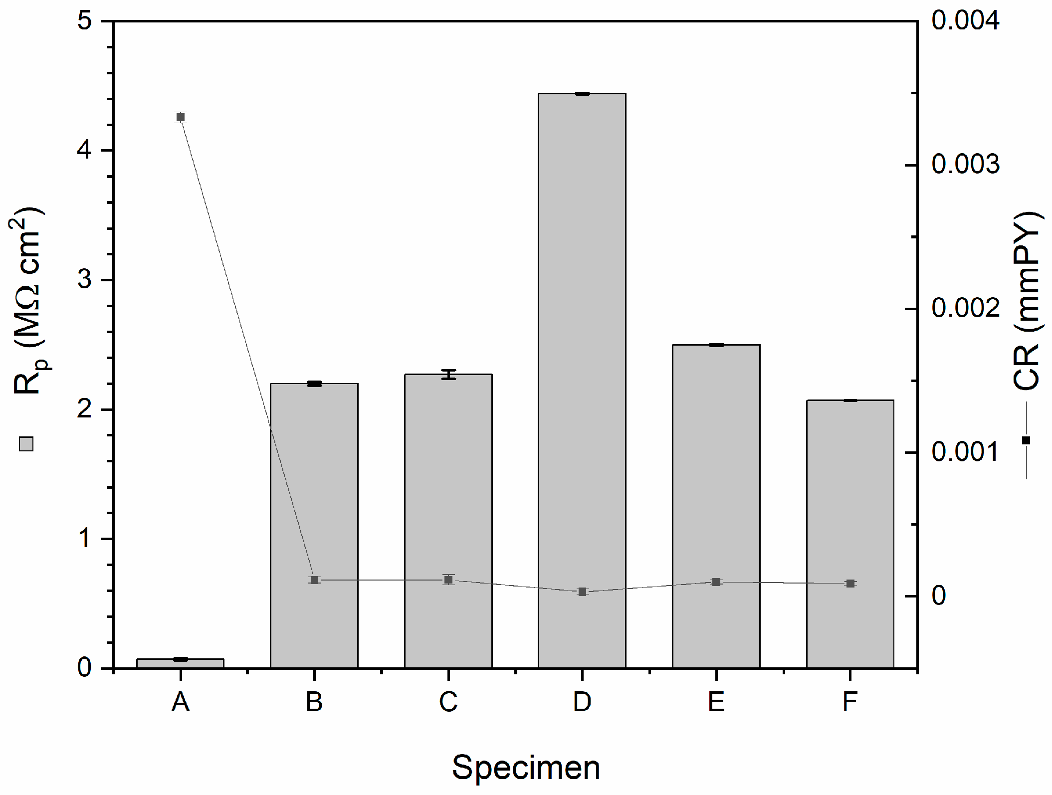
The calculated CR and MR values are presented in Table 2, alongside other corrosion parameters. Additionally, the Rp and CR values for individual specimens are illustrated in Figure 2.
As shown in Table 2 and Figure 2, all coated specimens exhibited a significant increase in Rp values compared to the uncoated specimen A, which displayed an Rp of 0.07 MΩ·cm2. Coated specimens B, C, E and F demonstrated Rp values ranging from 2.0 to 2.5 MΩ·cm2, representing an increase of approximately 29 to 35 times compared to specimen A. Notably, specimen D exhibited the highest Rp value of 4.44 MΩ·cm2, reflecting a 63-fold improvement over the uncoated specimen. Based on the obtained results, no clear correlation was observed between Rp and the carbon and nitrogen content in the Si(C,N) coatings.
The values of icor, CR, and MR further emphasize the enhanced corrosion resistance provided by the coatings (Table 2). Specimen A exhibited the highest icor, CR, and MR values, indicating its relatively high corrosion activity in the artificial saliva solution (Figure 2). In contrast, all coated specimens demonstrated significantly lower icor, CR, and MR values, with specimen D exhibiting the lowest values. These results confirm the superior protective capabilities of the Si(C,N) coatings, with specimen D providing the most effective barrier against general corrosion.
One of the parameters influencing the corrosion protection performance of the coatings is their thickness. Previous research [27] reported that the thicknesses of the Si(C,N) coatings deposited under the same conditions were as follows: B—2.6 µm, C—3.8 µm, D—4.2 µm, E—3.4 µm, and F—3.4 µm. These differences in thickness are attributed to variations in the carbon and nitrogen content in the precursor gases during the deposition process. A moderate correlation between coating thickness and corrosion resistance can be observed in the current study, with thicker coatings (e.g., specimen D) generally demonstrating superior protective performance. This is consistent with the expectation that thicker coatings offer more effective barriers against corrosive agents.
It is important to emphasize that Si(C,N) coatings, as highly effective barrier layers, play a crucial role in preventing the release of allergenic Ni2+ ions into surrounding tissues. By reducing ion release, these coatings mitigate the risk of allergic reactions and associated complications, thereby improving the biocompatibility and long-term safety of NiCr-based implants in clinical applications. This aspect will be explored further in future studies.
The anticorrosion properties of the coatings are closely linked to their porosity, which directly affects their permeability to the solution. Lower porosity/permeability corresponds to higher corrosion protection. In this study, the porosity of the coatings was evaluated based on electrochemical measurements using Equation (4).
p = R p , u R p , c · 100 %
where p is the total coating porosity, Rp,u is the polarization resistance of the uncoated alloy, and Rp,c is the polarization resistance of the coated substrate [41].
The calculated porosity values for the investigated coatings are as follows: 3.18%, 3.08%, 1.58%, 2.80%, and 3.37% for specimens B, C, D, E, and F, respectively. These results indicate that Si(C,N) coatings deposited via magnetron sputtering exhibit relatively low porosity (approximately 3%), enabling them to function effectively as barrier coatings in corrosive environments. The lowest porosity value of 1.58% was observed for specimen D, which correlates well with its superior anticorrosion performance as demonstrated by the highest Rp value and the lowest icor, CR, and MR values.
The observed differences in porosity (permeability to the corrosion solution) can be interpreted in relation to both the coating thickness and its chemical composition, particularly the C/N ratio. Specimen D, with a C/N ratio of 1.6 and a thickness of 4.2 µm, demonstrated not only the lowest porosity but also the best corrosion resistance. This suggests that, while the C/N ratio is an important factor, it does not solely determine porosity or protective performance. For instance, although specimen E has the lowest C/N ratio (0.6), its porosity (2.80%) is higher than that of specimen D, indicating that other factors, such as coating thickness and microstructural densification during deposition, also play critical roles.
Specimens B and F, representing SiC and SiN coatings, respectively, exhibit the highest porosity values (3.18% and 3.37%), consistent with their lower Rp values and lower coatings’ thickness (2.6 µm and 3.4 µm, respectively). Meanwhile, specimen C (C/N = 3.4) has a relatively high C/N ratio and intermediate porosity (3.08%). However, specimen D stands out due to the synergy of an optimized C/N ratio (1.6), greater thickness, and reduced structural defects, resulting in enhanced compactness and chemical stability.
These interpretations are supported by SEM cross-sectional observations presented in our previous work [27], which confirmed the uniform and dense morphology of the coating D. Therefore, while the C/N ratio influences coating porosity, it acts in concert with other deposition-related factors, such as thickness and growth mode, ultimately determining the coating’s protective capability.
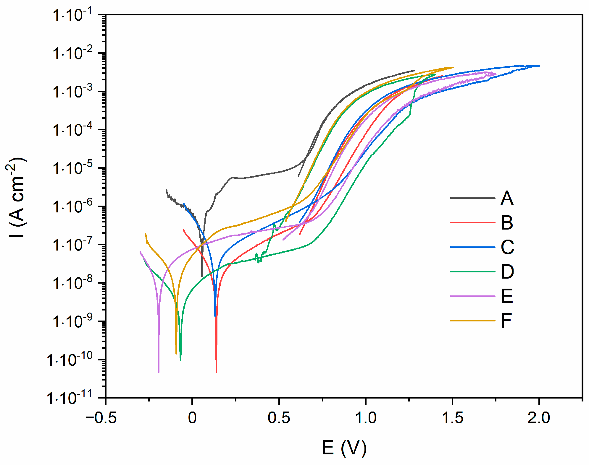
Potentiodynamic polarization curves (Figure 3) illustrate the electrochemical behavior of the investigated specimens under anodic polarization, providing insights into their passivity and resistance to pitting corrosion in an artificial saliva solution. As shown in Figure 3, all specimens exhibit passive behavior starting from the Tafel region, without an active-passive transition.
Among the investigated specimens, the uncoated specimen A demonstrated the highest electrochemical activity, with a narrow passive region (0.25–0.60 V) and corrosion current density of ca. 5–9 µA cm−2, followed by a subsequent increase in current density beyond 0.60 V. This behavior suggests that at potentials higher than 0.60 V, the transpassivation or pitting corrosion occurs, followed by immediate repassivation of pits, as indicated by the absence of a hysteresis loop in the reverse scan.
Si(C,N)−coated specimens exhibited broader passive regions and lower current densities within these regions, indicating reduced electrochemical activity. Notably, specimens D, E, and F demonstrated extended passive regions, initiating around 0 V or even at negative potentials. Among them, specimen D exhibited the lowest current density in the passive region, confirming its superior protective properties. At potentials above 0.75 V, an increase in current density was observed for all coated specimens. However, only in specimen D was a sharp increase at 1.15 V clearly identifiable, marking the onset of pitting corrosion. For the other specimens, pitting corrosion was also observed; however, the polarization curves did not provide sufficient resolution to accurately determine the potential at which it initiated. The presence of a hysteresis loop in the reverse scan confirms the development of pitting corrosion in Si(C,N)-coated specimens.
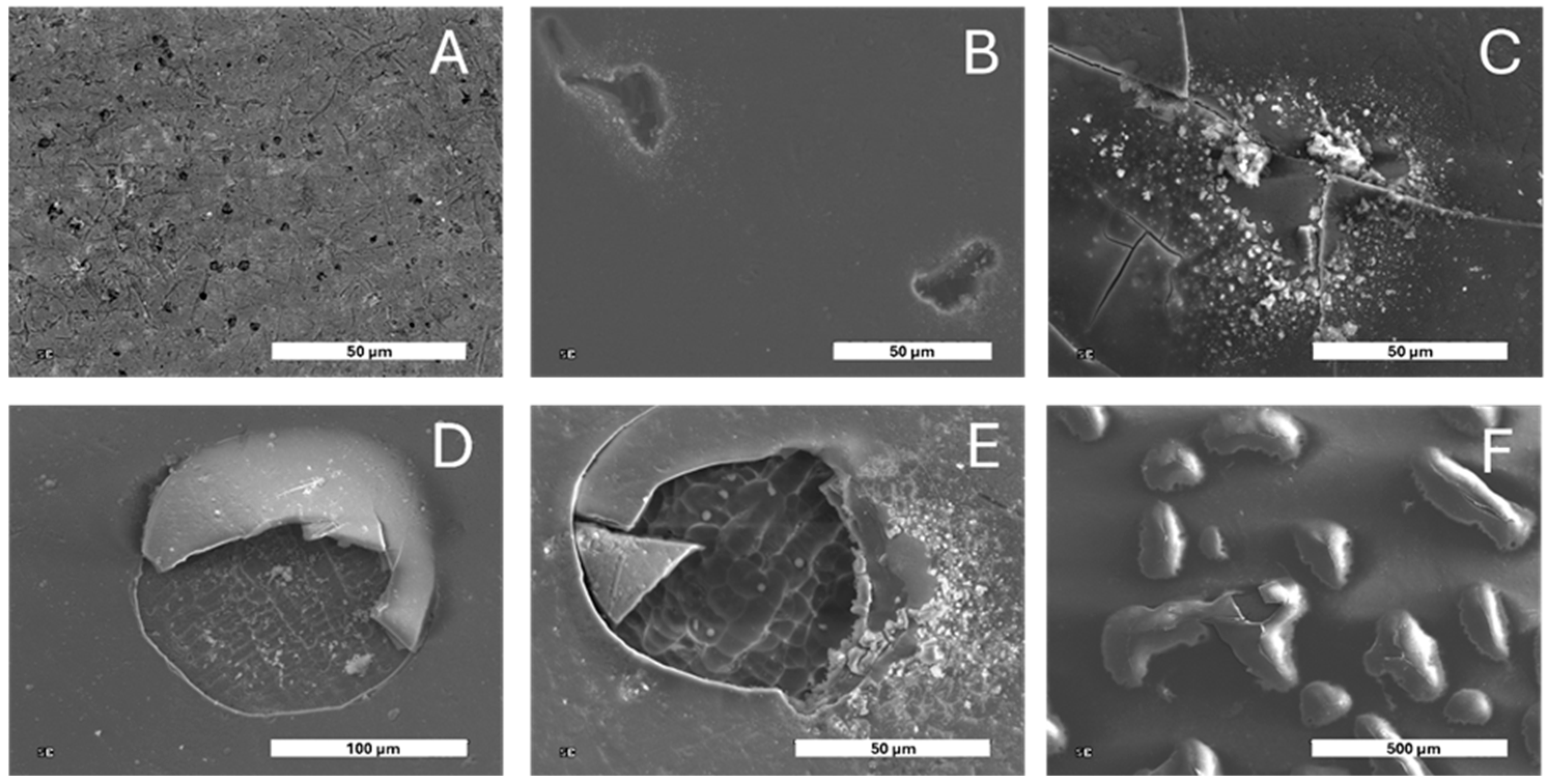
The formation of corrosion damage was confirmed through post-corrosion surface analysis using SEM (Figure 4).
As a result of potentiodynamic polarization, the uncoated specimen A exhibited minimal surface changes, while coated specimen B showed relatively small, randomly distributed irregular pits with limited growth during reverse polarization. In contrast, specimens C, D, E, and F displayed more extensive corrosion damage, characterized by the formation and subsequent rupture of blisters. Beneath these blisters, degradation of the NiCr alloy substrate was observed—a behavior typical of undercoating corrosion. The formation of blisters can be attributed to the mechanical properties of the Si(C,N) coatings, particularly their stiffness. Our previous studies have shown that the elastic modulus of Si(C,N) coatings increases with nitrogen content [42]. A higher modulus indicates greater stiffness, which can lead to increased internal stresses within the coating. Under electrochemical attack, these stresses contribute to localized delamination, allowing electrolyte penetration and facilitating undercoating corrosion. The trapped corrosion products and gases beneath the coating create internal pressure, leading to blister formation and eventual rupture. These findings indicate that under specific aggressive conditions, the presence of nitrogen in the Si(C,N) coating promotes localized undercoating corrosion of the NiCr alloy, ultimately leading to substrate deterioration and coating degradation. However, these processes are initiated at relatively high potential values, which do not typically occur in the human body. Therefore, the obtained corrosion test results for Si(C,N) coatings deposited on NiCr dental alloy can be considered fully satisfactory. These coatings are expected to provide sufficient corrosion protection under the operating conditions of prosthetic and orthodontic components.
Currently, the state of knowledge regarding the properties and effects of Si(C,N) layers on the substrates of dental alloys containing nickel and chromium is limited. Most researchers opt for alternative coatings, deposition methods, or substrates, such as titanium and chromium-cobalt alloys [43,44,45,46]. Mirzaev M.N. [47], in his research, employed carbonitride CNTi-(Zr, ZrNb, and ZrSi) coatings on Ti6Al4V substrates using a cathodic arc deposition system. The studies demonstrated that chloride ions (Cl) can penetrate through the oxide layers that form on materials in solutions containing chloride ions [47]. They suggested that carbonitride coatings can effectively block the penetration of Cl ions at the interface between the coating and the electrolyte, thus acting as an efficient anticorrosion barrier that prevents electrolyte infiltration [46]. The anticorrosion properties of coatings can be influenced by several factors, including the coating composition (carbon-to-nitrogen ratio, C/N), process parameters [46], and the thickness of the protective layer [32]. This conclusion was reached by the authors of [46]; however, these authors used a different method of sputtering the coating, a different substrate, and the coating also contained the Ti element in its composition. Therefore, there will be discrepancies with our results.
In recent years, silicon carbonitrides have been used as coating materials for dental implants. Until now, they have been used in medicine as joint implants. Interest in these materials has increased due to their potential properties that can be used in the field of dentistry. The properties of SiCN coatings can be modified by the ratio of silicon, carbon and nitrogen in the coating. In this way, coatings can be tailored to specific applications in dental prosthetics. These coatings can exhibit antibacterial properties, which can potentially reduce the risk of infections in the implant area. As mentioned earlier, few researchers have developed SiCN coatings for dental applications. The authors Xinyi Xia et al. [48] are among the few who have used SiCN coatings in the field of dental implantology.
Xinyi Xia et al. [48] deposited silicon carbon nitride (Si(C,N)) coatings onto the implant surface using the plasma-enhanced chemical vapor deposition (PECVD) method. Their study investigated the corrosion rate of Si(C,N) coatings under varying pH conditions. The results revealed a correlation between the corrosion rate and pH, indicating that acidic or alkaline substances in the oral environment could potentially influence the in vivo corrosion behavior of Si(C,N) layers. These studies indicate the possible anti-corrosion properties of these coatings and their potential use in dentistry, where we deal with an aggressive oral environment.
To complement the corrosion resistance assessment, adhesion tests were conducted to evaluate the mechanical integrity of the Si(C,N) coatings on the NiCr prosthetic alloy substrate. Understanding the adhesion behavior is essential, as insufficient coating–substrate bonding can critically impact long-term protective performance, especially in corrosive environments. Figure 5 presents the results of the adhesion measurements performed on the examined coatings.
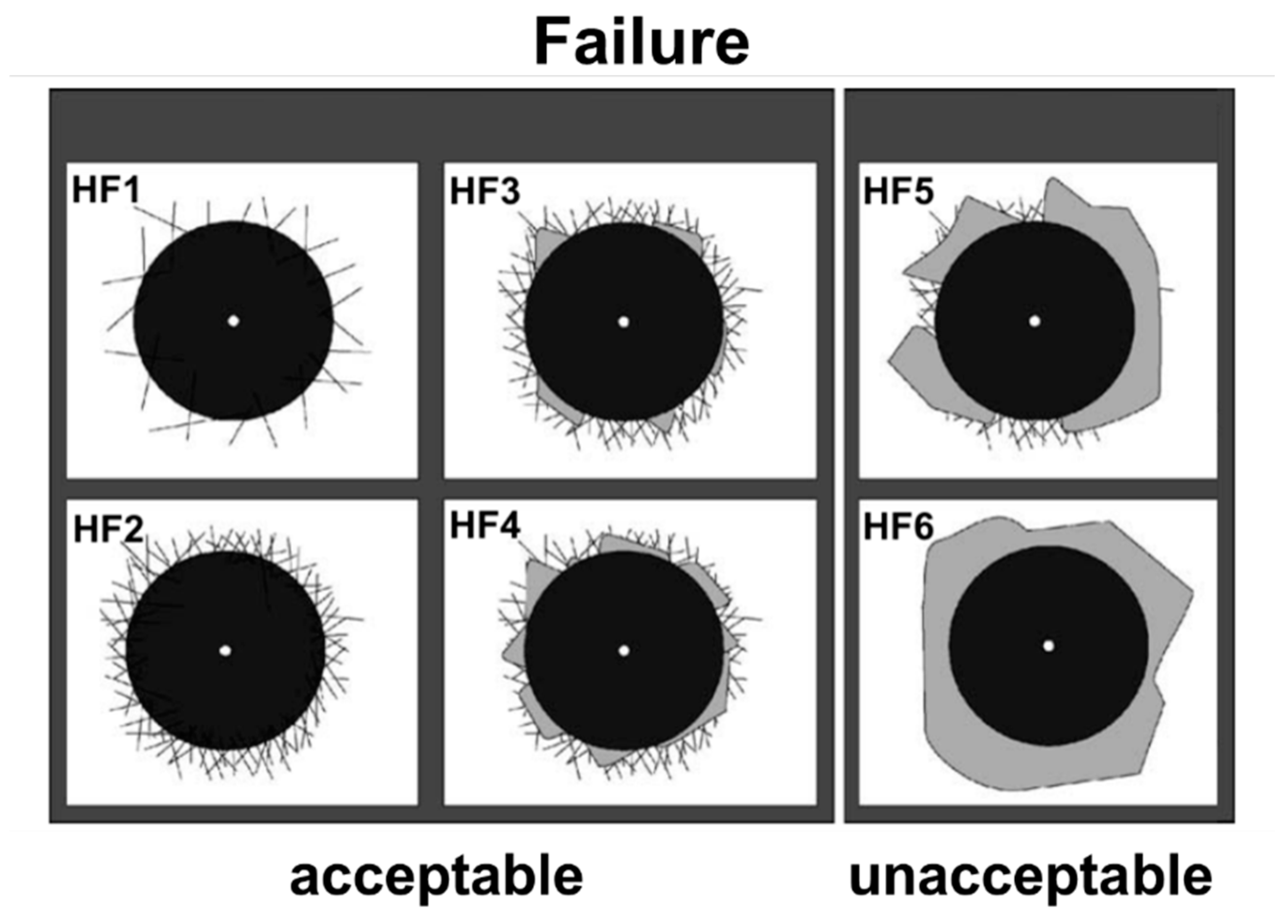
The microscopic images of the indentations presented in (Figure 5) show that, in all cases, the coating is detaching from the surface. By comparing these images with the reference patterns shown in (Figure 6), the individual samples can be classified into corresponding groups. Specifically, samples from groups B, C, D, and E align with the HF5 pattern, while sample F corresponds to the HF6 pattern. As observed, the adhesion of all samples is deemed inadequate according to the pattern classification criteria. This adhesion of the tested coatings may influence the nature of the corrosion damage presented in (Figure 4). If the adhesion is insufficient, the formation of corrosion pits can allow the corrosive medium to infiltrate between the substrate and the coating, which, as a consequence, can lead to the formation of blisters (as seen in specimen D–F in Figure 4) and subsequent coating delamination. Therefore further studies are needed to improve the adhesion of Si(C,N) coatings to the NiCr substrate.

4. Conclusions

The application of Si(C,N) coatings significantly improved the corrosion resistance of NiCr alloys in artificial saliva, as evidenced by increased polarization resistance (Rp) and reduced corrosion current density (icor), corrosion rate (CR), and mass loss rate (MR) when compared to uncoated alloy. A progressive increase in nitrogen content led to lower EOCP values and, under aggressive conditions, promoted localized undercoating corrosion. This behavior may be attributed to increased coating stiffness, which contributes to blisters formation and subsequent coating rupture. Among the tested coatings, specimen D (39.6%C, 25.2%N) exhibited the best corrosion protection, demonstrating the highest Rp value and the lowest icor, CR, and MR, suggesting an optimal balance between carbon and nitrogen content.
Summing up, Si(C,N) coatings act as effective barrier layers, potentially reducing the release of allergenic Ni2+ ions into surrounding tissues, thereby enhancing the biocompatibility and long-term safety of NiCr-based implants. Further studies are required to evaluate the long-term durability of these coatings in physiological conditions, as well as their biological interactions, to confirm their suitability for clinical applications.

Author Contributions

Conceptualization, Z.K. and L.K.; methodology, L.K. and B.B.; validation, K.D., B.B. and L.K.; formal analysis, L.K., K.D. and B.B.; investigation, Z.K., B.B. and K.D.; resources, Z.K.; data curation, Z.K.; writing—original draft preparation, Z.K., B.B., K.D. and L.K.; writing—review and editing, Z.K., B.B. and K.D.; visualization, L.K.; supervision, L.K.; project administration, Z.K. All authors have read and agreed to the published version of the manuscript.

Funding

This research received no external funding.

Data Availability Statement

Data are contained within the article.

Acknowledgments

The authors would like to thank Marcin Makówka for his cooperation in applying the coatings to the samples.

Conflicts of Interest

The authors declare that there are no conflicts of interest regarding the publication of this paper.

References

  1. Xu, W.; Yu, F.; Addison, O.; Zhang, B.; Guan, F.; Zhang, R.; Hou, B.; Sand, W. Microbial corrosion of metallic biomaterials in the oral environment. Acta Biomater. 2024, 184, 22–36. [Google Scholar] [CrossRef] [PubMed]
  2. Jia, R.; Unsal, T.; Xu, D.; Lekbach, Y.; Gu, T. Microbiologically influenced corrosion and current mitigation strategies: A state of the art review. Int. Biodeterior. Biodegrad. 2019, 137, 42–58. [Google Scholar] [CrossRef]
  3. Fischer, N.G.; Aparicio, C. The salivary pellicle on dental biomaterials. Colloids Surf. B Biointerfaces 2021, 200, 111570. [Google Scholar] [CrossRef] [PubMed]
  4. Sarkar, A.; Xu, F.; Lee, S. Human saliva and model saliva at bulk to adsorbed phases—Similarities and differences. Adv. Colloid Interface Sci. 2019, 273, 102034. [Google Scholar] [CrossRef]
  5. Proctor, G.B.; Shaalan, A.M. Disease-Induced Changes in Salivary Gland Function and the Composition of Saliva. J. Dent. Res. 2021, 100, 1201–1209. [Google Scholar] [CrossRef]
  6. Arakelyan, M.; Spagnuolo, G.; Iaculli, F.; Dikopova, N.; Antoshin, A.; Timashev, P.; Turkina, A. Minimization of Adverse Effects Associated with Dental Alloys. Materials 2022, 15, 7476. [Google Scholar] [CrossRef]
  7. Gopalakrishnan, U.; Felicita, A.S.; Mahendra, L.; Kanji, M.A.; Varadarajan, S.; Raj, A.T.; Feroz, S.M.A.; Mehta, D.; Baeshen, H.A.; Patil, S. Assessing the Potential Association Between Microbes and Corrosion of Intra-Oral Metallic Alloy-Based Dental Appliances Through a Systematic Review of the Literature. Front. Bioeng. Biotechnol. 2021, 9, 631103. [Google Scholar] [CrossRef]
  8. Dikopova, N.Z.; Volkov, A.G.; Arakelyan, M.G.; Makarenko, N.V.; Soxova, I.A.; Doroshina, V.J.; Arzukanyan, A.V.; Margaryan, E.G. The Study of the Electrochemical Potentials of Metal Structures in the Oral Cavity in Diseases of the Oral Mucosa. New Armen. Med. J. 2020, 14, 54–58. [Google Scholar]
  9. Mikulewicz, M.; Chojnacka, K.; Woźniak, B.; Downarowicz, P. Release of Metal Ions from Orthodontic Appliances: An in Vitro Study. Biol. Trace Elem. Res. 2012, 146, 272–280. [Google Scholar] [CrossRef]
  10. Moslehifard, E.; Ghasemzadeh, S.; Nasirpouri, F. Influence of pH Level of Artificial Saliva on Corrosion Behavior and Nickel Ion Release of a Ni-Cr-Mo Alloy: An In Vitro Study. Anti-Corros. Methods Mater. 2019, 66, 746–756. [Google Scholar] [CrossRef]
  11. Quezada-Castillo, E.; Aguilar-Castro, W.; Quezada-Alván, B. Ion release from non precious dental alloys in the oral cavity. Matéria 2022, 27, e202248593. [Google Scholar] [CrossRef]
  12. Motoyoshi, M. Current Techniques and Materials in Dentistry; MDPI AG: Basel, Switzerland, 2022; ISBN 3036544135. [Google Scholar]
  13. Łosiewicz, B.; Osak, P.; Kubisztal, J.; Górka-Kulikowska, K. Effect of Artificial Saliva Modification on the Corrosion Resistance and Electronic Properties of Bego Wirobond C Dental Alloys. Appl. Sci. 2023, 13, 12185. [Google Scholar] [CrossRef]
  14. Schmutz, P.; Quach-Vu, N.-C.; Gerber, I. Metallic Medical Implants: Electrochemical Characterization of Corrosion Processes. Electrochem. Soc. Interface 2020, 17, 35–40. [Google Scholar] [CrossRef]
  15. Smardz, J.; Skowron, M.; Florjański, W. The use of metals and their alloys in dental prosthetics. Prosthodontics 2016, 66, 461–467. [Google Scholar] [CrossRef]
  16. Wylie, C.M.; Sheltonb, R.M.; Fleming, G.J.P.; Davenporta, A.J. Corrosion of nickel-based dental casting alloys. Dent. Mater. 2007, 23, 714–723. [Google Scholar] [CrossRef]
  17. Upadhyaya, D.; Panchal, M.A.; Dubey, R.S.; Srivastava, V.K. Corrosion of alloys used in dentistry: A review. Mater. Sci. Eng. A 2006, 432, 1–11. [Google Scholar] [CrossRef]
  18. Kula, Z.; Semenov, M.; Klimek, L. Carbon Coatings Deposited on Prosthodontic Ni-Cr Alloy. Appl. Sci. 2021, 11, 4551. [Google Scholar] [CrossRef]
  19. Olms, C.; Schor, J.; Yahiaoui-Doktor, M. Potential Co-Factors of an Intraoral Contact Allergy—A Cross-Sectional Study. Dent. J. 2020, 8, 83. [Google Scholar] [CrossRef]
  20. Fletcher, R.; Harrison, W.; Crighton, A. Dental material allergies and oral soft tissue reactions. Br. Dent. J. 2022, 232, 620–625. [Google Scholar] [CrossRef]
  21. Mercieca, S.; Conti, C.M.; Buhagiar, J.; Camilleri, J. Assessment of corrosion resistance of cast cobalt- and nickel-chromium dental alloys in acidic environments. J. Appl. Biomater. Funct. Mater. 2018, 16, 47–54. [Google Scholar] [CrossRef]
  22. Asri, R.I.M.; Harun, W.S.W.; Samykano, M.; Lah, N.A.C.; Ghani, S.A.C.; Tarlochan, F.; Raza, M.R. Corrosion and surface modification on biocompatible metals: A review. Mater. Sci. Eng. 2017, 77, 1261–1274. [Google Scholar] [CrossRef]
  23. Rylska, D.; Sokolowski, G.; Sokolowski, K. Ni–Cr dental alloys—Porcelain firing impact on corrosion properties and surface characteristics. Mater. Res. Express 2024, 11, 076501. [Google Scholar] [CrossRef]
  24. Sampaio, N.A.; Silva, J.W.; Acciari, H.; Nakazato, R.Z.; Codaro, E.N.; Felipe, H. Influence of Ni and Cr Content on Corrosion Resistance of Ni-Cr-Mo Alloys for Fixed Dental Prostheses in 0.05% NaF Aqueous Solution. Mater. Sci. Appl. 2010, 1, 369–372. [Google Scholar] [CrossRef]
  25. Robles, D.; Brizuela, A.; Fernández-Domínguez, M.; Gil, J. Corrosion Resistance and Titanium Ion Release of Hybrid Dental Implants. Materials 2023, 16, 3650. [Google Scholar] [CrossRef] [PubMed]
  26. Petković Didović, M.; Jelovica Badovinac, I.; Fiket, Ž.; Žigon, J.; Rinčić Mlinarić, M.; Čanadi Jurešić, G. Cytotoxicity of Metal Ions Released from NiTi and Stainless Steel Orthodontic Appliances, Part 1: Surface Morphology and Ion Release Variations. Materials 2023, 16, 4156. [Google Scholar] [CrossRef]
  27. Klimek, L.; Makówka, M.; Sobczyk-Guzenda, A.; Kula, Z. Characteristics of Si (C, N) Silicon Carbonitride Layers on the Surface of Ni–Cr Alloys Used in Dental Prosthetics. Materials 2024, 17, 2450. [Google Scholar] [CrossRef] [PubMed]
  28. Rajib, P. Diamond-Like-Carbon Coatings for Advanced Biomedical Applications. Glob. J. Nanomed. 2017, 5, 1–5. [Google Scholar]
  29. Maher, N.; Mahmood, A.; Fareed, M.A.; Kumar, N.; Rokaya, D.; Zafar, M.S. An updated review and recent advancements in carbon-based bioactive coatings for dental implant applications. J. Adv. Res. 2024; in press. [Google Scholar] [CrossRef]
  30. Banaszek, K.; Wiktorowska-Owczarek, A.; Kowalczyk, E.; Klimek, L. Possibilities of applying Ti (C, N) coatings on prosthetic elements—Research with the use of human endothelial cells. Acta Bioeng. Biomater. 2016, 18, 119–126. [Google Scholar]
  31. Banaszek, K.; Klimek, L.; Zgorzynska, E.; Swarzynska, A.; Walczewska, A. Cytotoxicity of titanium carbonitride coatings for prostodontic alloys with different amounts of carbon and nitrogen. Biomed. Mater. 2018, 13, 045003. [Google Scholar] [CrossRef]
  32. Banaszek, K.; Klimek, L. Ti (C, N) as Barrier Coatings. Coatings 2019, 9, 432. [Google Scholar] [CrossRef]
  33. Bordbar-Khiabani, A.; Kovrlija, I.; Locs, J.; Loca, D.; Gasik, M. Octacalcium Phosphate-Laden Hydrogels on 3D-Printed Titanium Biomaterials Improve Corrosion Resistance in Simulated Biological Media. Int. J. Mol. Sci. 2023, 24, 13135. [Google Scholar] [CrossRef]
  34. Chouirfa, H.; Bouloussa, H.; Migonney, V.; Falentin-Daudré, C. Review of titanium surface modification techniques and coatings for antibacterial applications. Acta Biomater. 2019, 83, 37–54. [Google Scholar] [CrossRef] [PubMed]
  35. Dong, H.; Liu, H.; Zhou, N.; Li, Q.; Yang, G.; Chen, L.; Mou, Y. Surface modified techniques and emerging functional coating of dental implants. Coatings 2020, 10, 1012. [Google Scholar] [CrossRef]
  36. Ali, M.; Ali, F.; Yang, B.; Abbas, A. A comprehensive account of biomedical applications of CVD diamond coatings. J. Phys. D Appl. Phys. 2021, 54, 443001. [Google Scholar] [CrossRef]
  37. Saad, K.S.K.; Saba, T.; Rashid, A.B. Application of PVD Coatings in Medical Implantology for Enhanced Performance, Biocompatibility, and Quality of Life. Heliyon 2024, 10, e35541. [Google Scholar]
  38. VDI 3198:1992-08 Coating (CVD, PVD) of Cold Forging Tools. Available online: https://www.dinmedia.de/en/technical-rule/vdi-3198/930369 (accessed on 1 February 2025).
  39. ASTM G102-89; Standard Practice for Calculation of Corrosion Rates and Related Information from Electrochemical Measurements. ASTM International: West Conshohocken, PA, USA, 2015.
  40. Baraniak, M.; Ciszewski, A. Aktywność Chemiczna i Elektrochemiczna Pierwiastków w Środowisku Wody; Wydawnictwo Politechniki Poznańskiej: Poznań, Poland, 2006; ISBN 83-7143-222-4. [Google Scholar]
  41. Ahn, S.H.; Choi, Y.S.; Kim, J.G.; Han, J.G. A study on corrosion resistance characteristics of PVD Cr-N coated steels by electrochemical method. Surf. Coat. Technol. 2002, 150, 319–326. [Google Scholar] [CrossRef]
  42. Kula, Z.; Dąbrowska, K.; Klimek, L. Structure and Selected Properties of Si(C,N) Coatings on Ni-Cr Prosthetic Alloys. Processes 2025, 13, 624. [Google Scholar] [CrossRef]
  43. Moslehifard, E.; Moslehifard, M.; Ghasemzadeh, S.; Ghaiour, M.; Nasirpouri, F. Corrosion Behavior of a Nickel-Base Dental Casting Alloy in Artificial Saliva Studied by Weight Loss and Polarization Techniques. Front. Dent. 2019, 16, 13–20. [Google Scholar] [CrossRef]
  44. Metel, A.; Sotova, C.; Fyodorov, S.; Zhylinski, V.; Chayeuski, V.; Milovich, F.; Seleznev, A.; Bublikov, Y.; Makarevich, K.; Vereschaka, A. Improving the Wear and Corrosion Resistance of Titanium Alloy Parts via the Deposition of DLC Coatings. C 2024, 10, 106. [Google Scholar] [CrossRef]
  45. Singh, A.K.; Drunka, R.; Smits, K.; Vanags, M.; Iesalnieks, M.; Joksa, A.A.; Blumbergs, I.; Steins, I. Nanomechanical and Electrochemical Corrosion Testing of Nanocomposite Coating Obtained on AZ31 via Plasma Electrolytic Oxidation Containing TiN and SiC Nanoparticles. Crystals 2023, 13, 508. [Google Scholar] [CrossRef]
  46. Mirzayev, M.N.; Parau, A.C.; Slavov, L.; Dinu, M.; Neov, D.; Slavkova, Z.; Popov, E.P.; Belova, M.; Hasanov, K.; Aliyev, F.A.; et al. TiSiCN as Coatings Resistant to Corrosion and Neutron Activation. Materials 2023, 16, 1835. [Google Scholar] [CrossRef] [PubMed]
  47. Mirzayev, M.N.; Hasanov, K.M.; Parau, A.C.; Demir, E.; Abiyev, A.S.; Karaman, T.; Jabarov, S.H.; Dinu, M.; Popov, E.P.; Vladescu, A.D. Effect of the C/N ratio modification on the corrosion behavior and performance of carbonitride coatings prepared by cathodic arc deposition. J. Mater. Res. Technol. 2023, 27, 1724–1738. [Google Scholar] [CrossRef]
  48. Xia, X.; Chiang, C.-C.; Gopalakrishnan, S.K.; Kulkarni, A.V.; Ren, F.; Ziegler, K.J.; Esquivel-Upshaw, J.F. Properties of SiCN Films Relevant to Dental Implant Applications. Materials 2023, 16, 5318. [Google Scholar] [CrossRef] [PubMed]
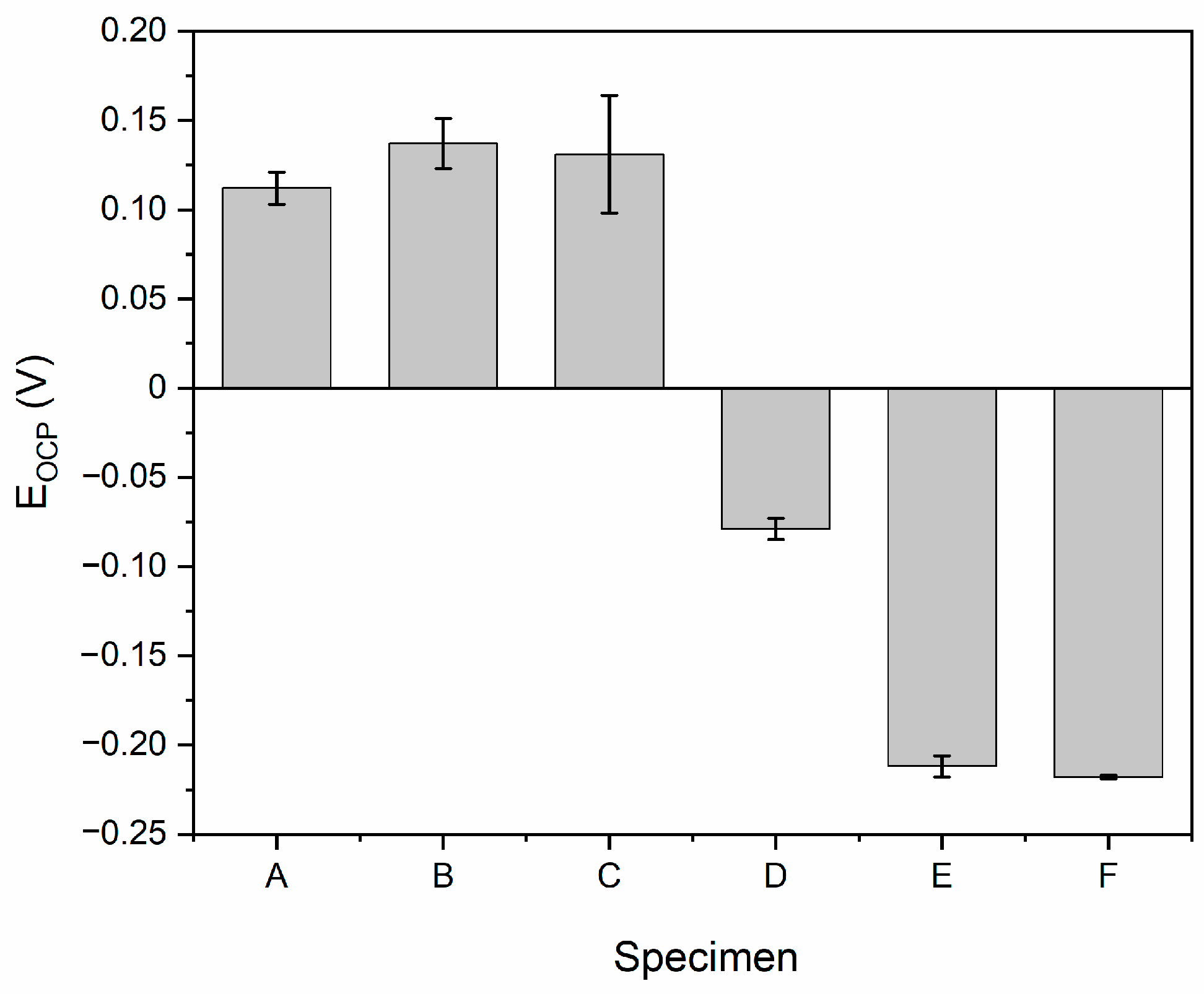
Figure 1. Open circuit potential (EOCP) values of NiCr specimens with Si(C,N) coatings in artificial saliva solution (pH 5.06). Specimen descriptions are provided in Table 1.
Ceramics 08 00050 g001
Figure 2. Polarization resistance (Rp) and corrosion resistance (CR) values of NiCr specimens with Si(C,N) coatings in artificial saliva solution (pH 5.06). Specimen descriptions are provided in Table 1.
Ceramics 08 00050 g002
Figure 3. Potentiodynamic characteristics of investigated NiCr alloy coated with Si(C,N) coatings in artificial saliva. Specimen descriptions are provided in Table 1.
Ceramics 08 00050 g003
Figure 4. Post-corrosion SEM morphologies of the investigated specimens (AF) (scale bars: 50 µm for specimens (AC,E); 100 µm for specimen (D); and 500 µm for specimen (F)).
Ceramics 08 00050 g004
Figure 5. Indentation of specimens with Si(C,N) coatings—backscattered electron images for specimens (BF).
Ceramics 08 00050 g005
Figure 6. Indentation of specimens models of the VDI 3198 standard.
Ceramics 08 00050 g006
Table 1. Chemical composition of Si(C,N) layers.
SpecimenElement
SiNCat. C/N
at. [%]wt. [%]at. [%]wt. [%]at. [%]wt. [%]
B24.838.5--75.261.5-
C29.646.715.912.554.540.83.4
D35.253.325.219.039.627.71.6
E42.961.035.325.021.814.00.6
F47.764.752.335.3---
Table 2. Values of corrosion parameters for individual specimens.
SpecimenRp (MOhm cm2)Icor (nA cm−2)CR (mmPY)MR (g m−2 d−1)
A0.07 ± 0.01355.6 ± 4.1(3.33 ± 0.04)·10−3(7.57 ± 0.09)·10−2
B2.20 ± 0.4512.2 ± 2.2(1.14 ± 0.21)·10−4(2.59 ± 0.48)·10−3
C2.27 ± 0.6912.3 ± 3.9(1.15 ± 0.36)·10−4(2.61 ± 0.83)·10−3
D4.44 ± 0.205.9 ± 2.1(3.24 ± 1.97)·10−5(7.36 ± 4.47)·10−4
E2.50 ± 0.3910.6 ± 1.6(9.92 ± 1.49)·10−5(2.25 ± 0.34)·10−3
F2.07 ± 0.219.6 ± 1.3(8.96 ± 1.23)·10−5(2.04 ± 0.28)·10−3
Table 3. Parameters considered in EW calculations.
ElementMass FractionValenceAtomic WeightEW
Ni0.6423258.6923.77
Cr0.2436352.00
Mo0.0891495.94
Si0.0143428.09
Fe0.0107255.84
Disclaimer/Publisher’s Note: The statements, opinions and data contained in all publications are solely those of the individual author(s) and contributor(s) and not of MDPI and/or the editor(s). MDPI and/or the editor(s) disclaim responsibility for any injury to people or property resulting from any ideas, methods, instructions or products referred to in the content.