Abstract
Substantial tooth bonding is the defining characteristic of effective minimally invasive all-ceramic restorations. Natural and synthetic cross-linkers that could strengthen the bonding quality are currently drawing enormous interest. Thus, this study aimed to assess the microtensile bond strength and nanoleakage of computer-aided design/computer-aided manufacturing (CAD/CAM)-fabricated ceramics to pretreated dentin with chlorhexidine or Salvadora persica extract, compared to no pretreatment, after thermomechanical cyclic loading. Consequently, forty-five extracted third-molar teeth (n = 45) were utilized to obtain mid-coronal dentin and assigned into three groups (n = 15) in accordance with dentin pretreatment; (group I: no dentin pretreatment (control), group II: 2% chlorhexidine, and group III: Salvadora persica extract pretreatments). Ceramic onlays were milled from lithium disilicate IPS e.max CAD/CAM blocks and cemented to prepared teeth with etch-and-rinse resin cement (Variolink Esthetic DC system kit). Microtensile bond strength and interfacial nanoleakage were accessed after thermomechanical cyclic loading. Statistical analysis was performed using one-way ANOVA, followed by Tukey’s post hoc test. Additionally, p-values < 0.05 were considered statistically significant. The chlorhexidine pretreated group showed the most favorable outcome compared to the control group. Conversely, using Salvadora persica pretreatment did not affect the bond strength and nanoleakage compared to the control group (p > 0.05). Consequently, unlike Salvadora persica extract, chlorhexidine–dentin pretreatment maintained superior bonding strength to ceramics after thermomechanical cyclic loading, facilitating minimally invasive, yet lasting, aesthetic restoration.
1. Introduction
The growing interest in esthetics and the swift advancement of digital dentistry technologies and materials have broadened the applications of all-ceramic restoratives throughout modern dentistry [1]. The fundamental quality of clinically durable ceramics relies not only on their flexural strength but also on effective adhesion to the tooth surface, particularly in minimally invasive procedures such as onlays and overlays [2].
Dentin bio-modifiers can be either synthetic, such as chlorhexidine [3], or natural, such as Salvadora persica extract [4]. Chlorhexidine is a matrix metalloproteinase inhibitor that also functions as a detergent, enhancing adhesive resin penetration into exposed collagen, and has recently demonstrated enhanced bond stability [3]. On the other hand, plants function as a natural supply of antimicrobial substances. Plant-based drugs have been historically included in our conventional healthcare system, with the antibacterial properties of plant-derived compounds being thoroughly documented [5]. Herbal medicines are highly effective and present minimum risk due to their low adverse reactions, low toxicity, and ease of management [5].
There is a growing interest in naturally generated cross-linkers that may enhance the bond stability between resin and dentin [6,7,8]. This is especially pertinent in the domain of phytotherapy, which has recently garnered attention [9]. Salvadora persica plant, sometimes called the Arak tree, is the source of miswak chewing sticks, which act as organic natural toothbrushes [10]. In 1986, the World Health Organization (WHO) approved the usage of miswak as an effective method for preserving oral hygiene [11] and part of special care dentistry [12].
The extract of miswak or Salvadora persica possesses several biological activities, including antibacterial, antifungal, anti-inflammatory, antioxidant, analgesic, and anticariogenic actions [10]. Furthermore, it has been found through phytochemical analysis in a former study [13] that Salvadora persica extract contains tannins and vitamin C [4,13], both of which have a cross-linking impact on collagen fibrils [14].
Most of the resin–dentin bonded interface disintegrates as dentinal matrix metalloproteinase (MMP) enzymes degrade exposed collagen [15,16]. MMPs are easily accessible and activated by acidic substances during adhesive bonding [3]. The collagen fibrils at the cement–dentin interface in partially resin-infiltrated hybrid layers may disintegrate if matrix-bound MMPs are not fully penetrated with adhesive resin, reducing the longevity of bonded restorations [3]. Self-etch or etch-and-rinse adhesives activate MMPs [17,18], which degrade type I collagen [19,20,21].
Previous studies have proposed multiple techniques to impede collagen breakdown by MMPs to enhance the longevity of resin–dentin interfaces [22,23,24]. The utilization of cross-linking compounds was employed to enhance the rigidity of collagen and augment its resistance to degradation by facilitating the creation of supplementary hydrogen bonding and intermolecular covalent bonds [25,26,27]. Previous research [6,7,8,25,26,27,28] has utilized several cross-linking agents on bonding resin composite; however, the optimal inhibitory effect on MMP for bonding ceramics has not been investigated thus far.
Nevertheless, a complete investigation of the impact of Salvadora persica extract on the preservation of dentinal collagen for enhancing the bond strength of ceramics has not been investigated yet. Thus, this study aimed to investigate the microtensile bond strength (μTBS) and nanoleakage (nL) of computer-aided design/computer-aided manufacturing (CAD/CAM)-fabricated ceramics to pretreated dentin with chlorhexidine or Salvadora persica extract, compared to no pretreatment, after thermomechanical cyclic loading. The null hypothesis examined was that the utilization of chlorhexidine or Salvadora persica extract had no impact on the μTBS and nL of CAD/CAM-fabricated ceramics to pretreated dentin compared to the control group after being subjected to thermomechanical cyclic loading.
2. Materials and Methods
2.1. Study Design, and Ethical Approval
The current parallel randomized controlled study was conducted at the College of Dentistry, Prince Sattam Bin Abdulaziz University and Tanta University. Ethical approval was granted by the Standing Committee of Bioethics Research (SCBR) of Prince Sattam Bin Abdulaziz University (approval No. SCBR-363/2024 and approval date 1 December 2024) and by the Research Ethics Committee of Tanta University (approval No. #R-RD-7-24-3128 and approval date July 2024). The study complied with CONSORT guidelines [29]. As an in vitro study, it did not require registration in the Clinical Trials Registry; and the study was conducted according to the guidelines of the Declaration of Helsinki.
2.2. Sample Size Calculation
A power analysis was conducted to ensure sufficient power for testing the null hypothesis that no differences exist across the various tested groups. Utilizing an alpha (α) level of 0.05 (5%), a beta (β) level of 0.2 (20%) (resulting in a power of 80%), and an effect size (ω) of 0.517, derived from the findings of Abu-Nawareg et al.’s study [30], the minimum necessary sample size (n) was determined to be 45, equating to 15 samples per group: control, chlorhexidine, and Salvadora persica extract-pretreated dentin groups. Sample size computation was conducted utilizing G*Power version 3.1.9.7 (Heinrich-Heine-Universität Düsseldorf, Düsseldorf, Germany) [31].
2.3. Randomization, Grouping, and Blinding
Forty-five intact third molars were obtained from the Department of Oral and Maxillofacial Surgery at Tanta University with the informed consent of the donors, following a protocol authorized by the Research Ethics Committee (approval No. #R-RD-7-24-3128 and approval date July 2024). Teeth were taken from unhealthy patients with periodontal disease and placed in acrylic blocks for full visibility of the crowns, and were consecutively numbered. Teeth were randomly allocated into three equal groups (n = 15) based on dentin pretreatment: group I (control group): no dentin pretreatment; group II: chlorhexidine (Consepsis™ 2% CHX gluconate, Ultradent, Inc., South Jordan, UT, USA); and group III: Salvadora persica extract in a 20% concentration solution.
The lab assistant developed a block randomization sequence using Excel 2010 (Microsoft, Redmond, WA, USA) with a block size of 6. Opaque-sealed envelopes were utilized for the concealing of randomization sequences. The operator was cognizant of the randomization sequence immediately prior to the experimental procedures, whereas the assessor was completely blinded.
2.4. Preparation of 20% Salvadora persica Extract
A botany expert meticulously selected a fresh half-kilogram specimen of Salvadora persica root from a retail outlet (Riyadh, Saudi Arabia). A solution of Salvadora persica extract was formulated utilizing a methodology described in prior research [6]. The roots were purified, rinsed with distilled water, permitted to dry at ambient temperature for several days, subsequently fragmented into small pieces, and processed into a fine powder using a ball mill. An aqueous solution was formulated by combining 20 g of Salvadora persica powder with 100 mL of deionized sterile water. The amalgamation was thereafter preserved at 4 °C for a duration of 48 h.
Following centrifugation of the mixture for 10 min at 2200× g, a 20% aqueous extract was acquired. The liquid phase was filtered using Millipore filters with a pore size of 0.45 mm (Sigma-Aldrich Chemie GmbH, Steinheim, Germany) to remove any potential bacterial contamination. A source of ultraviolet radiation (Camag UV-Cabinet II, Basel, Switzerland) emitting approximately 10 mW/cm2 at a wavelength of 366 nm was employed for 1 h to sterilize the extract at a distance of 10 cm, without affecting the extract’s components. The sterilized extract was stored in a sterile container at 4 °C and utilized within one week of production.
2.5. Specimen Preparation
The teeth were maintained in a 0.5% chloramine T solution at a temperature of 4 °C. They were employed within two months of extraction. The occlusal enamel and superficial dentin were sectioned with a low-speed diamond saw (IsoMetTM 4000, Buehler Ltd., Lake Bluff, IL, USA) with water cooling. The cut was executed in a manner along the tooth’s longitudinal axis. Mid-coronal dentinal surfaces were polished with 600-grit silicon carbide paper (Norton Saint, Gobain Abrasives, Worcester, MA, USA). This procedure was conducted to remove any residual enamel and provide consistent smear layers.
2.6. Digital Impression Fabrication and Cementation of Ceramic Onlays
CEREC Omnicam system was used to scan the specimens. Additionally, the restorations were fabricated using IPS e.max CAD/CAM blocks (Ivoclar Vivadent, Amherst, NY, USA) on the CEREC milling machine.
2.7. Dentin Pretreatment
Group I, as per the randomization sequence, underwent no dentin pretreatment (control), but group II got a 60 s pretreatment with 2% chlorhexidine using an applicator tip, followed by blot-drying to eliminate excess chlorhexidine without causing overdrying. Group III underwent pretreatment with a 20% Salvadora persica extract solution for 60 s via an applicator tip, followed by a 20 s water rinse followed by blot-drying.
2.8. Cementation of Ceramic Onlays
The restorations were cemented using an etch-and-rinse technique. The Variolink Esthetic DC system kit (Ivoclar Vivadent, Amherst, NY, USA) was utilized for cementation treatments in accordance with the manufacturer’s instructions, adhering to a step-by-step protocol outlined in a previous study [32]. Materials utilized in the current study are described in Table 1.

Table 1.
Materials utilized in the current study.
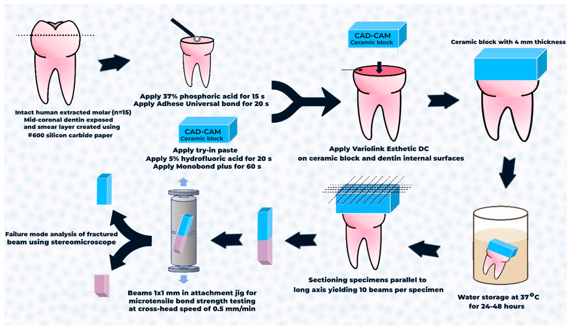
Restorations were applied to teeth with Variolink Esthetic try-in paste (Ivoclar Vivadent, Amherst, NY, USA), and the adaptability was assessed. The fitting surface was first etched with 5% hydrofluoric acid (IPS Ceramic Etching Gel; Ivoclar Vivadent, Amherst, NY, USA) for 20 s, followed by rinsing with water for 40 s. Monobond Etch & Prime (Ivoclar Vivadent, Amherst, NY, USA), a self-etching glass-ceramic primer, was applied to the internal surfaces of restorations using a microbrush for 60 s and, subsequently, dispersed with a gentle stream of air. Prepared dentin surfaces were etched with 37% orthophosphoric acid gel for 10 to 15 s. Then, the etching agent was meticulously washed with a water spray and the tooth was dried. Dentin teeth surfaces were coated with Adhese Universal for 20 s and dispersed using oil- and moisture-free compressed air. Light curing was conducted for 20 s utilizing the light curing unit (Light Emitting Diode curing unit, 3M Oral Care Elipar, Seefeld, Germany). Variolink Esthetic DC was administered using an application tip directly onto the internal surface of the restoration. The ceramic prosthesis was thereafter positioned and secured while surplus material was removed. Excess material was polymerized using a curing light for 2 s at a distance of 10–15 mm, with the light probe traversing the whole cement line, and it was promptly removed with a scaler. Restoration margins were coated with liquid strips immediately following excess removal to avert oxygen inhibition and light-cured for 10 s. The margins and cement lines were refined with Kenda polishers (Figure 1).

Figure 1.
A flowchart of the microtensile bond strength test showing the step-by-step procedure, including specimens’ selection, cementation steps, storage time, sectioning procedures, testing, and failure mode analysis.
2.9. Thermomechanical Cyclic Loading
A masticatory simulator (CS-4.4 SD Mechatronik GmbH, Feldkirchen-Westerham, Germany) was employed to administer mechanical cycles of axial compressive loads with a specially designed cone-shaped stainless-steel bar with a rounded point. At a velocity of 60 mm/s, this bar exhibited a vertical displacement of 2.5 mm. The simulator exerted a force of fifty newtons (N) on the central fossa of each restoration. The chewing simulator was modified to facilitate temperature cycles with the thermo-cycling device (TC-3; SD Mechatronik GmbH, FeldkirchenWesterham, Germany). Thermocycling occurred in distilled water for 30 s at temperatures ranging from 5° to 55 °C. Each restoration underwent 5000 thermal cycles and 120,000 mechanical cycles. The simulations approximately depicted an oral service duration of six months. Subsequently, each specimen was examined with a 40× magnification stereomicroscope (Nikon SMZ-10; Nikon Corporation, Tokyo, Japan) to verify the integrity of the restoration.
2.10. Microtensile Bond Strength Test (μTBS)
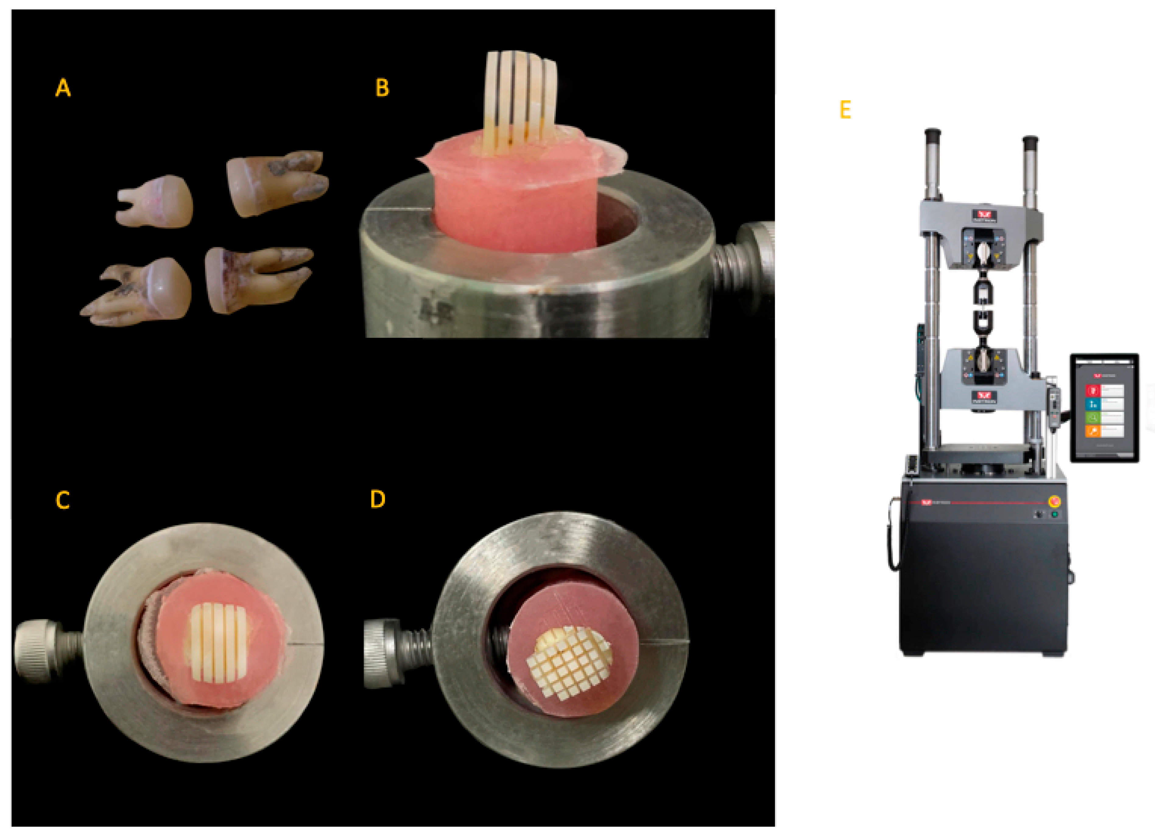
Subsequently, each specimen was sectioned parallel to its long axis at the ceramic–dentin interface, yielding 10 beams (1 × 1 mm ± 0.12 mm) per specimen. This was accomplished via a low-speed diamond saw (IsoMetTM 4000, Buehler Ltd., Lake Bluff, IL, USA) with water-cooling equipment. Dimensions of each beam were precisely measured to 0.01 mm with a digital caliper (HSL 246–15, Karl Hammacher GmbH, Solingen, Germany). A total of 150 beams were analyzed for each group, with (n = 150) being the number of specimens/beams. In the nanoleakage analysis, one stick was obtained from each tooth in every group, resulting in a total of ten sticks (n = 15). The beams were bonded with cyanoacrylate adhesive to a testing jig and subsequently subjected to tensile testing until failure using a universal testing machine (Instron Model 6800, Canton, MA, USA) at a cross-head speed of 0.5 mm/min. The μTBS was measured in megapascals (MPa) (Figure 2).

Figure 2.
Representative images of specimens and devices used for μTBS testing: (A) teeth with lithium disilicate ceramics cemented; (B) longitudinal sectioning of the specimen in one direction from an axial view; (C) longitudinal sectioning of the specimen in one direction from an occlusal view; (D) longitudinal sectioning of the specimen in two directions from an occlusal view; (E) Instron device model 6800.
2.11. Nanoleakage Analysis (nL)
Sections were cut along the axial surface of the specimens, parallel to their long axis, at the ceramic–dentin interface, in order to obtain beams. The axial wall served as the point of analysis for the beams at the ceramic–dentin interface. Nanoleakage analysis was performed on one beam from each tooth within every group, with a sample size of 10. A dual layer of nail polish (Shenzhen Meixin Industry Co., Ltd., Shenzhen, China) was meticulously applied to all surfaces, maintaining a 1 mm gap from the bonding interface. The samples were immersed in a 50% by-weight ammoniacal silver nitrate solution (pH = 9.5) for 24 h at 37 °C after drying the nail polish. Following five minutes of cleaning with distilled water, the samples were immersed in a photo-developing solution (Kodak GBX fixer and replenisher, Kodak, Rochester, NY, USA) for eight hours under fluorescent illumination. Subsequently, a periodontal scaler was employed to eliminate the nail polish following a five-minute wash of the specimens in distilled water.
The specimens were refined utilizing waterproof paper of silicon carbide (Norton Saint-Gobain Abrasives, Worcester, MA, USA) with grit sizes of 800, 1200, 2000, and 4000. Final polish was attained using progressively finer diamond pastes with particle sizes of 20 μm, 6 μm, 4 μm, and 1 μm, applied using a polishing cloth. All debris was eliminated by submerging the slabs in an ultrasonic bath (Ultrasonic Cleaning System, LR Manufacturing, Kearny, NJ, USA) for 10 min. Subsequently, the samples were air-dried for 24 h and affixed to aluminum stubs utilizing graphite paint and carbon adhesive tapes (Ted Pella, Inc., Moores-town, NJ, USA). The samples were subsequently coated with a gold layer by a sputtering procedure and analyzed using a scanning electron microscope (SEM; Quanta 200 ESEM, FEI France, Mérignac, France). The SEM functioned at an accelerating voltage of 30 kV and a working distance of 17–19 mm, with images acquired at a magnification of 1000×. Image analysis software (Version 1.32 NIH picture, Scion Corp., Fredrick, MD, USA) was used to quantify the amount of silver nitrate that precipitated at the contact.
2.12. Mode of Failure Analysis
The failure modes were documented by examining both surfaces of each fragmented specimen at 40× magnification with a stereomicroscope (model SZH, Olympus, Tokyo, Japan). Fracture modes were classified into four categories: (1) cohesive failures in dentin; (2) adhesive junction failures; (3) cohesive failures in ceramics; and (4) mixed failures [33].
2.13. Statistical Analysis
The statistical software package for the social sciences (SPSS) version 28 (IBM Corp., Armonk, NY, USA) was used to collect and process the data. The results were subjected to the Shapiro–Wilk test at a significance level of 0.05 to verify their conformity to a normal distribution. Levene’s test was employed to assess the homogeneity of variances. Data exhibited parametric distribution and homogeneity of variance and then were examined using one-way ANOVA, followed by Tukey’s post hoc test. The significance level was established at p < 0.05 for all analyses.
3. Results
3.1. Microtensile Bond Strength Values
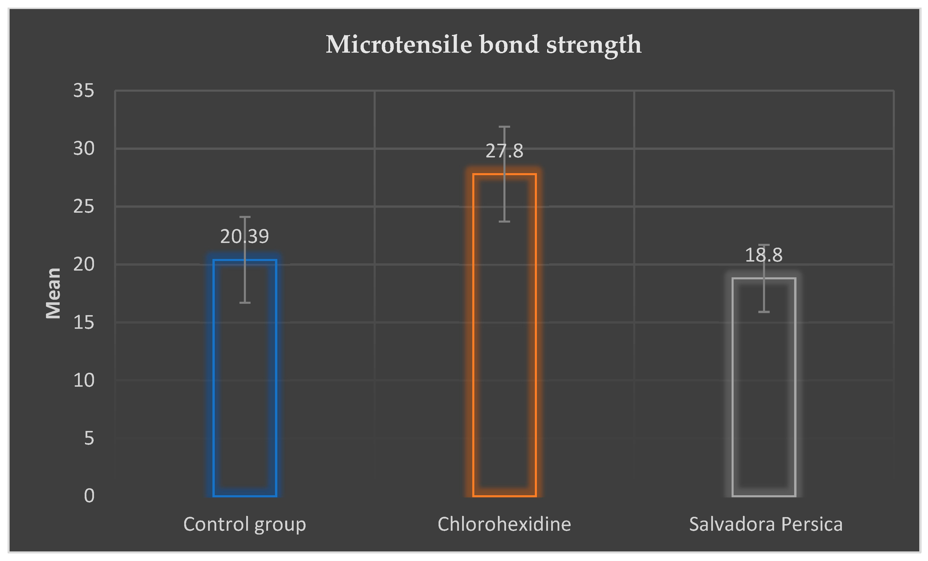
Shapiro–Wilk confidence test proved that data of all groups were normally distributed (p > 0.05). Moreover, Levene’s test proved the homogeneity of variances. Descriptive statistics for μTBS values are presented in Table 2 and Figure 3. The chlorhexidine pretreatment group exhibited the greatest mean value (27.8 ± 4.1 MPa), followed by the control group (20.39 ± 3.7 MPa), with the lowest value recorded in the Salvadora persica pretreatment group (18.8 ± 2.9 MPa).
Table 2.
A comparison of microtensile bond strength (μTBS) expressed in mean values ± standard deviations between all groups in MPa.

Figure 3.
Microtensile bond strength mean values ± standard deviations (SDs) in MPa for each group.
The one-way ANOVA revealed no statistically significant difference in μTBS between the control and Salvadora persica pretreatment group (p = 0.591). Conversely, significant differences in μTBS were detected between the chlorhexidine pretreatment group and the control group (p = 0.0003).
3.2. Nanoleakage Values
Shapiro–Wilk confidence test proved that data of all groups were normally distributed (p > 0.05). Moreover, Levene’s test proved the homogeneity of variances. Descriptive statistics for nL values are presented in Table 3. Salvadora persica pretreatment group exhibited the greatest mean value (49.27 ± 9.1), followed by the control group (47.39 ± 8.6), with the lowest value recorded in the chlorhexidine pretreatment group (39.62 ± 7.9).
Table 3.
A comparison of nanoleakage (%) expressed in mean values ± standard deviations between all groups.
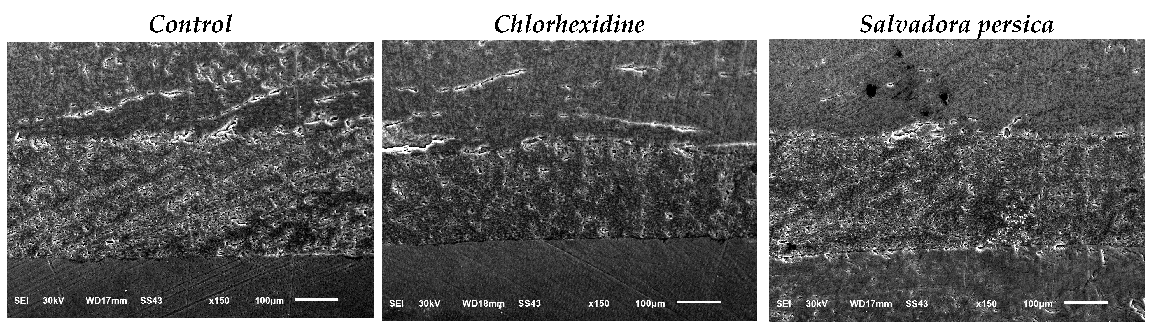
The one-way ANOVA revealed no statistically significant difference in nL between the control and Salvadora persica pretreatment group (p = 0.876). Conversely, a significant difference in nL was detected between the chlorhexidine pretreatment group and the control group (p = 0.03). Representative SEM images of nL (%) of all groups at 1000× magnification are shown in Figure 4.
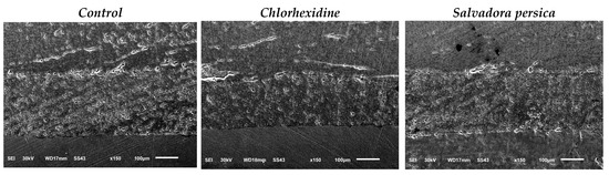
Figure 4.
Representative SEM images at 1000× magnification of nanoleakage (%) of all groups.
3.3. Distribution of the Failure Mode
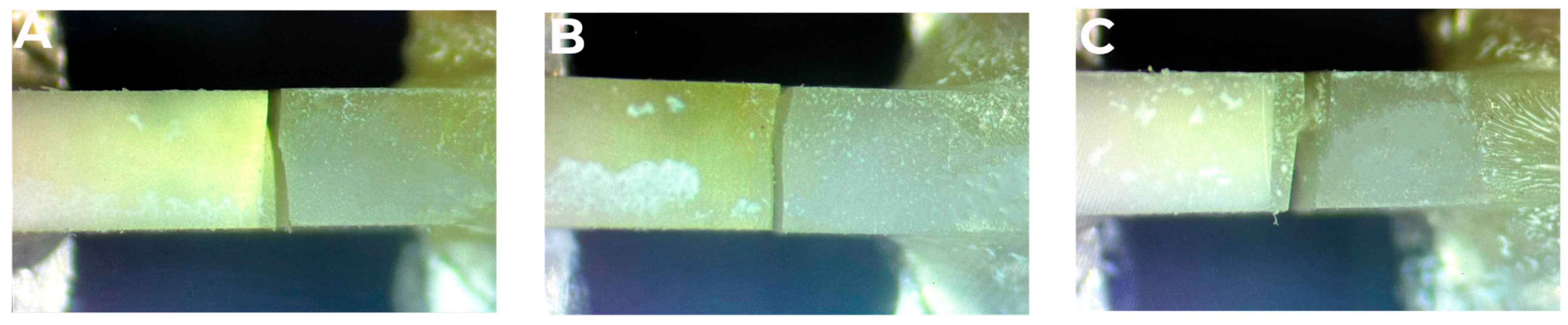
The distribution of the failure modes is summarized in Table 4 and Figure 5. In the control, chlorhexidine, and Salvadora persica pretreatment groups, mixed failures were the most frequent fracture types, followed by adhesive junction failures. Few cohesive failures occurred in the ceramic and dentin. All mixed failures were the result of a combination of cohesive failures and adhesive junction failures. Representative stereomicroscope images of different failure modes are shown in Figure 6.
Table 4.
Distribution of failure modes.
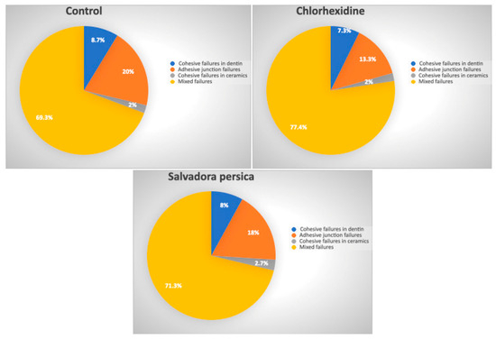
Figure 5.
Pie charts showing the percentage of different failure modes for control, chlorohexidine, and Salvadora persica groups.

Figure 6.
Representative stereomicroscope images of different failure modes: (A) mixed failure; (B) adhesive junction failure; (C) cohesive failure in ceramic.
4. Discussion
Despite significant advancements in adhesive dental restoratives, bond deterioration remains a concern threatening their clinical longevity [34]. The primary reason, recently detailed in the literature, is nonbacterial deterioration attributed to intrinsic dentinal enzymes known as MMPs [35]. MMPs are enzymes that have the ability to break down collagen in the hybrid layer, which, in turn, decreases the lifespan of adhesive restorations. With the progress of scientific knowledge about MMPs, effective approaches focused on inhibiting the activity of these enzymes with MMP inhibitors have rapidly emerged in various medical disciplines [35].
Chlorhexidine has been shown to effectively inhibit MMPs by methods that involve the binding of metal ions, including zinc and calcium [36]. This binding inhibits the catalytic activation of MMPs, hence preserving the integrity of resin–dentin-bonded surfaces [22,25]. Unfortunately, the prolonged effects of these compounds remain ambiguous due to the potential for leaking, which would render the bonded interface vulnerable to degradation [25]. Moreover, numerous cross-linking agents have been utilized to augment the cross-linking of dentinal collagen fibrils, hence enhancing their resistance to degradation by MMPs [8,25,27]. Glutaraldehyde, grape seed extract, riboflavin, and chitosan have been utilized as cross-linking agents [25,27,37]. Salvadora persica, a natural chemical, has shown promise as a natural cross-linker for resin–dentin bonding [30]. However, more research is needed to fully understand its effect on dentinal collagen [25].
Salvadora persica extract was used in this study as a conditioner to cross-link dentinal collagen at the CAD/CAM lithium disilicate–ceramics bonded interface. Moreover, the application of Salvadora persica extract on an etched dentinal surface did not enhance µTBS or nL in comparison to the control group. Conversely, the results demonstrated the effectiveness of chlorhexidine in maintaining μTBS and reducing nL, consistent with the findings of Milani et al. [38], Zaghloul et al. [39], and Yiu et al. [22]. Thus, we should partially accept the null hypothesis. This may be attributed to its anti-collagenolytic properties resulting from the suppression of MMPs through the chelation of calcium and zinc, which are essential for MMP activity [40]. Chlorhexidine possesses a positive charge, enabling it to connect to the negatively charged dentin collagen, thus maintaining its protective effect for up to six months [40].
Numerous factors can significantly affect the µTBS and the proportion of nL, including the preparation method of Salvadora persica extract [10], the type of curing light employed [41], the period of conditioning [42], and the selection between etch-and-rinse or self-etch adhesive strategies [43]. Therefore, further research is necessary, carefully examining these factors with the application of Salvadora persica extract [4]. The timing of extract application, along with the rinse duration, may significantly influence the outcome [4,10].
Factors such as dentin surface area, tubule architecture, smear layer, moisture content, and abrasion and erosion can impact the µTBS of adhesive systems on dentin [44]. A 20% Salvadora persica concentration did not influence resin cement adhesion in etch-and-rinse mode. Interactions between Salvadora persica and adhesive components may reduce wettability and dentin conditioning [44]. Furthermore, Salvadora persica may hinder resin penetration into cross-linked dentin collagen, leading to inferior µTBS results. In addition, Salvadora persica has probably interfered with the adhesive interaction with the dentin substrate [39]. Likewise, it is challenging to compare the findings of the current study with other research as there are no available data in the literature on the impact of Salvadora persica extract on resin–dentin bond stability. According to the study by Abu-Nawareg et al. [30], a 20% concentration of Salvadora persica applied to etched dentin for 60 s enhanced and maintained resin–dentin interfaces for a duration of 6 months [30]. However, these results are contrary to our findings.
In addition, the fragmented samples from the chlorhexidine and Salvadora persica groups exhibited predominantly mixed failures. This conclusion may be ascribed to the use of etch-and-rinse cement; thereby, the inhibitory effect of chlorhexidine on MMPs is contingent upon the type of adhesive utilized, as found in the study by Collares et al. [45]. The optimal effect of chlorhexidine on bond persistence is achieved using etch-and-rinse methods (applied subsequent to phosphoric acid etching) rather than with self-etch adhesives [45].
However, other studies [46,47] are in disagreement with our results. Meiers et al. [46] performed a study to evaluate the effects of various disinfectants employed before the application of different adhesive systems. It was discovered that a disinfectant containing chlorhexidine resulted in elevated amounts of microleakage. This indicates that the selection of cavity disinfectant for ceramic restorations must be contingent upon the individual materials employed and their interactions with various dentin bonding systems [46]. Furthermore, the data by Tulunoglu et al. [47] regarding heightened microleakage and reduced bond strength values associated with chlorhexidine support this theory.
However, on the basis of the literature that is currently available, it is possible to draw the conclusion that there is evidence that supports the utilization of chlorhexidine in order to improve the prognosis of ceramic restorations that are adhesively bonded. Furthermore, it is worth considering if the bonded interface may be preserved by pretreating the dentine surface with chlorhexidine before luting lithium disilicate ceramic restorations utilizing resin cements. Thus, it would enable general practitioners to routinely use it in dental practice.
5. Limitations
Due to the challenges of accurately simulating the oral environment, the current in vitro study has several limitations. This includes the lack of masticatory forces, saliva, dentinal fluid, pH levels, temperature fluctuations, and pulpal pressure. Furthermore, the specimens’ design differs from that of dental restorations frequently seen in the oral cavity. Hence, in vitro studies may not fully replicate the clinical environment, which may affect the external validity of the outcomes. Additionally, more research is needed in the future to assess the effects of extract from Salvadora persica at different concentrations and etching techniques.
6. Conclusions
Given the constraints of the present investigation, it may be inferred that, in contrast to Salvadora persica extract, chlorhexidine–dentin pretreatment maintained superior bonding strength to ceramics after thermomechanical cyclic loading, facilitating minimally invasive, yet lasting, aesthetic restoration. Failure modes were exclusively admixed at the dentin–cement interface, suggesting the need for further research employing chlorhexidine and Salvadora persica dentin bio-modifiers with various cement types for ceramic bonding, ideally with a larger sample size and extended aging.
Author Contributions
Conceptualization, A.A. (Abdulellah Almudahi); data curation, A.R.A., B.A. and R.S.A.; formal analysis, M.A.A.; funding acquisition, B.A., A.A.E., R.S.A. and M.A.A.; investigation, A.A. (Abdulellah Almudahi) and A.A.E.; methodology, A.A. (Abdulellah Almudahi), A.A. (Abdullah AlShehri) and A.A.E.; project administration, A.A. (Abdullah AlShehri), B.A., A.A.E. and M.A.A.; resources, A.A. (Abdulellah Almudahi) and R.S.A.; supervision, A.A. (Abdullah AlShehri) and A.R.A.; validation, A.A. (Abdullah AlShehri); visualization, A.R.A.; writing—review and editing, A.R.A., B.A., A.A.E. and M.A.A. All authors have read and agreed to the published version of the manuscript.
Funding
The study was supported via funding from Prince Sattam bin Abdulaziz University project number (PSAU/2025/R/1446).
Institutional Review Board Statement
The Standing Committee of Bioethics Research (SCBR) of Prince Sattam Bin Abdulaziz University approved the study No. SCBR-363/2024 on 1 December 2024 and the Research Ethics Committee of Tanta University approved the study No. #R-RD-7-24-3128 in July 2024.
Informed Consent Statement
Informed consent was obtained from the participants involved in the study before the teeth extraction.
Data Availability Statement
The original contributions presented in this study are included in the article. Further inquiries can be directed to the corresponding author.
Conflicts of Interest
The authors declare no conflicts of interest.
References
- Juntavee, N.; Juntavee, A.; Wongnara, K.; Klomklorm, P.; Khechonnan, R. Shear bond strength of ceramic bracket bonded to different surface-treated ceramic materials. J. Clin. Exp. Dent. 2018, 10, e1167–e1176. [Google Scholar] [PubMed]
- Oztürk, E.; Hickel, R.; Bolay, S.; Ilie, N. Micromechanical properties of veneer luting resins after curing through ceramics. Clin. Oral. Investig. 2012, 16, 139–146. [Google Scholar] [PubMed]
- Carrilho, M.; Geraldeli, S.; Tay, F.; De Goes, M.; Carvalho, R.M.; Tjäderhane, L.; Reis, A.; Hebling, J.; Mazzoni, A.; Breschi, L. In vivo preservation of the hybrid layer by chlorhexidine. J. Dent. Res. 2007, 86, 529–533. [Google Scholar] [CrossRef] [PubMed]
- Abdeltawab, S.S.; Abu Haimed, T.S.; Bahammam, H.A.; Arab, W.T.; Abou Neel, E.A.; Bahammam, L.A. Biocompatibility and antibacterial action of Salvadora persica extract as intracanal medication (in vitro and ex vivo experiment). Materials 2022, 15, 1373. [Google Scholar] [CrossRef]
- Deshpande, R.R.; Kale, A.A.; Ruikar, A.D.; Panvalkar, P.S.; Kulkarni, A.A.; Deshpande, N.R.; Salvekar, J.P. Antimicrobial activity of different extracts of Juglans regia L. against oral microflora. Int. J. Pharm. Pharm. Sci. 2011, 3, 200–201. [Google Scholar]
- Castellan, C.S.; Bedran-Russo, A.K.; Antunes, A.; Pereira, P.N. Effect of dentin biomodification using naturally derived collagen cross-linkers: One-year bond strength study. Int. J. Dent. 2013, 2013, 918010. [Google Scholar]
- Castellan, C.S.; Bedran-Russo, A.K.; Karol, S.; Pereira, P.N.R. Long-term stability of dentin matrix following treatment with various natural collagen cross-linkers. J. Mech. Behav. Biomed. Mater. 2011, 4, 1343–1350. [Google Scholar]
- El Gindy, A.H.; Sherief, D.I.; El-Korashy, D.I. Effect of dentin biomodification using natural collagen cross-linkers on the durability of the resin-dentin bond and demineralized dentin stiffness. J. Mech. Behav. Biomed. Mater. 2023, 138, 105551. [Google Scholar]
- Aljarbou, F.; Almobarak, A.; Binrayes, A.; Alamri, H.M. Salvadora persica’s biological properties and applications in different dental specialties: A narrative review. J. Evid. Based. Complementary. Altern. Med. 2022, 2022, 8667687. [Google Scholar]
- Khunkar, S.; Hariri, I.; Alsayed, E.; Linjawi, A.; Khunkar, S.; Islam, S.; Bakhsh, T.A.; Nakashima, S. Inhibitory effect of Salvadora persica extract (Miswak) on collagen degradation in demineralized dentin: In vitro study. J. Dent. Sci. 2021, 16, 208–213. [Google Scholar]
- World Health Organization. Consensus statement on oral hygiene. Int. Dent. J. 2000, 50, 139. [Google Scholar]
- Hassona, Y. An Interview with ChatGPT About Special Care Dentistry. In Special Care in Dentistry; American Association of Hospital Dentists, The Academy of Dentistry for the Handicapped, The American Society for Geriatric Dentistry: Chicago, IL, USA, 2024. [Google Scholar]
- Al Bratty, M.; Makeen, H.A.; Alhazmi, H.A.; Syame, S.M.; Abdalla, A.N.; Homeida, H.E.; Sultana, S.; Ahsan, W.; Khalid, A. Phytochemical, cytotoxic, and antimicrobial evaluation of the fruits of miswak plant, Salvadora persica L. J. Chemi. 2020, 2020, 4521951. [Google Scholar] [CrossRef]
- Ghahri, S.; Chen, X.; Pizzi, A.; Hajihassani, R.; Papadopoulos, A.N. Natural tannins as new cross-linking materials for soy-based adhesives. Polymers 2021, 13, 595. [Google Scholar] [CrossRef] [PubMed]
- Mazzoni, A.; Nascimento, F.; Carrilho, M.; Tersariol, I.; Papa, V.; Tjäderhane, L.; Di Lenarda, R.; Tay, F.; Pashley, D.H.; Breschi, L. MMP activity in the hybrid layer detected with in situ zymography. J. Dent. Res. 2012, 91, 467–472. [Google Scholar] [CrossRef]
- Tjäderhane, L.; Nascimento, F.D.; Breschi, L.; Mazzoni, A.; Tersariol, I.L.; Geraldeli, S.; Tezvergil-Mutluay, A.; Carrilho, M.R.; Carvalho, R.M.; Tay, F.R. Optimizing dentin bond durability: Control of collagen degradation by matrix metalloproteinases and cysteine cathepsins. Dent. Mater. 2013, 29, 116–135. [Google Scholar] [CrossRef]
- Nishitani, Y.; Yoshiyama, M.; Wadgaonkar, B.; Breschi, L.; Mannello, F.; Mazzoni, A.; Carvalho, R.M.; Tjäderhane, L.; Tay, F.R.; Pashley, D.H. Activation of gelatinolytic/collagenolytic activity in dentin by self-etching adhesives. Eur. J. Oral. Sci. 2006, 114, 160–166. [Google Scholar] [CrossRef]
- Mazzoni, A.; Pashley, D.H.; Nishitani, Y.; Breschi, L.; Mannello, F.; Tjäderhane, L.; Toledano, M.; Pashley, E.L.; Tay, F.R. Reactivation of inactivated endogenous proteolytic activities in phosphoric acid-etched dentine by etch-and-rinse adhesives. Biomaterials 2006, 27, 4470–4476. [Google Scholar] [CrossRef]
- Carrilho, M.R.; Tay, F.R.; Donnelly, A.M.; Agee, K.A.; Tjäderhane, L.; Mazzoni, A.; Breschi, L.; Foulger, S.; Pashley, D.H. Host-derived loss of dentin matrix stiffness associated with solubilization of collagen. J. Biomed. Mater. Res. A Appl. Biomater. 2009, 90, 373–380. [Google Scholar] [CrossRef]
- De Munck, J.; Van den Steen, P.; Mine, A.; Van Landuyt, K.; Poitevin, A.; Opdenakker, G.; Van Meerbeek, B. Inhibition of enzymatic degradation of adhesive-dentin interfaces. J. Esthet. Restor. Dent. 2011, 23, 350–352. [Google Scholar] [CrossRef]
- Breschi, L.; Martin, P.; Mazzoni, A.; Nato, F.; Carrilho, M.; Tjäderhane, L.; Visintini, E.; Cadenaro, M.; Tay, F.R.; Dorigo, E.D.S. Use of a specific MMP-inhibitor (galardin) for preservation of hybrid layer. Dent. Mater. 2010, 26, 571–578. [Google Scholar] [CrossRef]
- Yiu, C.K.; Hiraishi, N.; Tay, F.R.; King, N.M. Effect of chlorhexidine incorporation into dental adhesive resin on durability of resin-dentin bond. J. Adhes. Dent. 2012, 14, 355. [Google Scholar] [PubMed]
- Mazzoni, A.; Angeloni, V.; Apolonio, F.M.; Scotti, N.; Tjäderhane, L.; Tezvergil-Mutluay, A.; Di Lenarda, R.; Tay, F.R.; Pashley, D.H.; Breschi, L. Effect of carbodiimide (EDC) on the bond stability of etch-and-rinse adhesive systems. Dent. Mater. 2013, 29, 1040–1047. [Google Scholar] [PubMed]
- Montagner, A.; Sarkis-Onofre, R.; Pereira-Cenci, T.; Cenci, M. MMP inhibitors on dentin stability: A systematic review and meta-analysis. J. Dent. Res. 2014, 93, 733–743. [Google Scholar] [PubMed]
- Abunawareg, M.; Abuelenain, D.; Elkassas, D.; Haimed, T.A.; Al-Dharrab, A.; Zidan, A.; Hassan, A.; Pashley, D. Role of dentin cross-linking agents in optimizing dentin bond durability. Int. J. Adhes. Adhes. 2017, 78, 83–88. [Google Scholar] [CrossRef]
- Mazzoni, A.; Angeloni, V.; Comba, A.; Maravic, T.; Cadenaro, M.; Tezvergil-Mutluay, A.; Pashley, D.H.; Tay, F.R.; Breschi, L. Cross-linking effect on dentin bond strength and MMPs activity. Dent. Mater. 2018, 34, 288–295. [Google Scholar]
- Zhou, J.; Chiba, A.; Scheffel, D.L.; Hebling, J.; Agee, K.; Tagami, J.; Tan, J.; Abuelenain, D.; Nawareg, M.A.; Hassan, A.H. Cross-linked dry bonding: A new etch-and-rinse technique. Dent. Mater. 2016, 32, 1124–1132. [Google Scholar] [CrossRef]
- Husain, S.; Al-Samadani, K.H.; Najeeb, S.; Zafar, M.S.; Khurshid, Z.; Zohaib, S.; Qasim, S.B. Chitosan biomaterials for current and potential dental applications. Materials. 2017, 10, 602. [Google Scholar]
- Moher, D.; Hopewell, S.; Schulz, K.F.; Montori, V.; Gøtzsche, P.C.; Devereaux, P.J.; Elbourne, D.; Egger, M.; Altman, D.G. CONSORT 2010 explanation and elaboration: Updated guidelines for reporting parallel group randomised trials. Int. J. Surg. 2012, 10, 28–55. [Google Scholar]
- Abu-Nawareg, M.M.; Abouelseoud, H.K.; Zidan, A.Z. Effect of Salvadora persica on resin-dentin bond stability. BMC Oral Health 2024, 24, 505. [Google Scholar]
- Faul, F.; Erdfelder, E.; Lang, A.-G.; Buchner, A. G* Power 3: A flexible statistical power analysis program for the social, behavioral, and biomedical sciences. Behav. Res. Methods 2007, 39, 175–191. [Google Scholar]
- Tisler, C.E.; Chifor, R.; Badea, M.E.; Moldovan, M.; Prodan, D.; Carpa, R.; Cuc, S.; Chifor, I.; Badea, A.F. Photodynamic Therapy (PDT) in Prosthodontics: Disinfection of Human Teeth Exposed to Streptococcus mutans and the Effect on the Adhesion of Full Ceramic Veneers, Crowns, and Inlays: An In Vitro Study. Biomedicines 2022, 10, 144. [Google Scholar] [CrossRef] [PubMed]
- Lin, C.-P.; Douglas, W.H. Failure mechanisms at the human dentin-resin interface: A fracture mechanics approach. J. Biomech. 1994, 27, 1037–1047. [Google Scholar] [PubMed]
- Al-karadsheh, O.; Albalsheh, M.; Zabadi, S.; Alhaddad, A.; Hassona, Y.; Sawair, F. The Social Impact of Dental Implant Research: An Altmetric Analysis. J. Int. Dent. Med. Res. 2024, 17, 1371–1379. [Google Scholar]
- De Moraes, I.Q.S.; do Nascimento, T.G.; da Silva, A.T.; de Lira, L.M.S.S.; Parolia, A.; de Moraes Porto, I.C.C. Inhibition of matrix metalloproteinases: A troubleshooting for dentin adhesion. Restor. Dent. Endod. 2020, 45, e31. [Google Scholar]
- Hannas, A.R.; Pereira, J.C.; Granjeiro, J.M.; Tjäderhane, L. The role of matrix metalloproteinases in the oral environment. Acta Odontol. Scand. 2007, 65, 1–13. [Google Scholar]
- Vasei, F. Effect of chitosan treatment on shear bond strength of composite to deep dentin using self-etch and total-etch adhesive systems. Braz. Dent. Sci. 2021, 24, 2440. [Google Scholar]
- Milani, S.; Seraj, B.; Khoshlafz, Z.; Abazarian, N. Effect of dentin pretreatment with chlorhexidine on push-out bond strength of composite restorations in severely damaged primary anterior teeth. Front. Dent. 2020, 17, 1–6. [Google Scholar]
- Zaghloul, S.A.; Gabal, Z.A.; Abdul-Rahman, F.A.-Z.M. Shear Bond Strength of Ceramics to Chlorhexidine versus Pomegranate Peel Extract–Pretreated Dentin: A Randomized Control Study. J. Nat. Sci. Med. 2023, 6, 109–113. [Google Scholar]
- Borompiyasawat, P.; Putraphan, B.; Luangworakhun, S.; Sukarawan, W.; Techatharatip, O. Chlorhexidine gluconate enhances the remineralization effect of high viscosity glass ionomer cement on dentin carious lesions in vitro. BMC Oral Health 2022, 22, 60. [Google Scholar]
- Uctasli, M.; Mutluay, M.M.; Tezvergil-Mutluay, A. Effect of High Intensity Light Curing, Cavity Depth and Aging on Bond Strength. Dent. Mater. 2023, 39, e77. [Google Scholar]
- Burrer, P.; Dang, H.; Par, M.; Attin, T.; Tauböck, T.T. Effect of over-etching and prolonged application time of a universal adhesive on dentin bond strength. Polymers 2020, 12, 2902. [Google Scholar] [CrossRef] [PubMed]
- Elkaffas, A.A.; Hamama, H.H.; Mahmoud, S.H.; Fawzy, A.S. Effect of acid etching on dentin bond strength of ultra-mild self-etch adhesives. Int. J. Adhes. Adhes. 2020, 99, 102567. [Google Scholar]
- Campos, E.A.d.; Correr, G.M.; Leonardi, D.P.; Pizzatto, E.; Morais, E.C. Influence of chlorhexidine concentration on microtensile bond strength of contemporary adhesive systems. Braz. Oral. Res. 2009, 23, 340–345. [Google Scholar] [PubMed]
- Collares, F.M.; Rodrigues, S.B.; Branco Leitune, V.C.; Celeste, R.K.; De Araújo, F.B.; Werner Samuel, S.M. Chlorhexidine application in adhesive procedures: A meta-regression analysis. J. Adhes. Dent. 2013, 15, 11–18. [Google Scholar]
- Meiers, J.; Kresin, J. Cavity disinfectants and dentin bonding. Oper. Dent. 1996, 21, 153–159. [Google Scholar]
- Tulunoglu, O.; Ayhan, H.; Olmez, A.; Bodur, H. The effect of cavity disinfectants on microleakage in dentin bonding systems. J. Clin. Pediatr. Dent. 1998, 22, 299–305. [Google Scholar]

Disclaimer/Publisher’s Note: The statements, opinions and data contained in all publications are solely those of the individual author(s) and contributor(s) and not of MDPI and/or the editor(s). MDPI and/or the editor(s) disclaim responsibility for any injury to people or property resulting from any ideas, methods, instructions or products referred to in the content. |
© 2025 by the authors. Licensee MDPI, Basel, Switzerland. This article is an open access article distributed under the terms and conditions of the Creative Commons Attribution (CC BY) license (https://creativecommons.org/licenses/by/4.0/).