1. Introduction
Osteochondroplastic tracheopathy is a rare disease of the trachea and bronchi that appears in the fourth-fifth decade of life, characterized by nodules or cartilaginous or bone spicules that project from the submucosa into the tracheobronchial lumen. It was first described by Wilks in 1837 and has been extensively studied from a clinical and pathogenic point of view by Balgaard in 1947. The etiology is highly uncertain. It seems possible that the connective cells of the tracheal wall can pose themselves to cartilaginous elements forming islands that will undergo partial calcification. These islands can also merge with tracheal cartilages. Some authors relate this disease to tracheobronchial amyloidosis due to the tendency of the latter to cause calcification or ossification. In this perspective, it could represent the terminal state of amyloidosis. According to other authors, this pathology is not the end result of amyloidosis and cannot be associated in any way with the latter. The endoscopic picture is typical for the presence of numerous cartilaginous and bony nodular growths (masserelle and spicules) that produce sessile or pedunculate endoluminary protrusions that give the trachea and bronchi a rosary crown appearance. The mucosa is intact and ulcerations that can cause hemoptysis are rarely found, in any case there is a marked rigidity of the wall. Tracheal obstruction can be mild, moderate and sometimes even severe. Over the years the cartilaginous and nodular protrusions tend to extend, thus affecting more and more vast trachea tracts and large bronchi; in more advanced cases they flow together forming plaques or masses which determine a marked rigidity of the wall and a considerable narrowing of the tracheobronchial lumen. Performing a biopsy is very difficult due to the hard consistency of the tissue, however bronchoscopy is diagnostic.
2. Clinical Cases
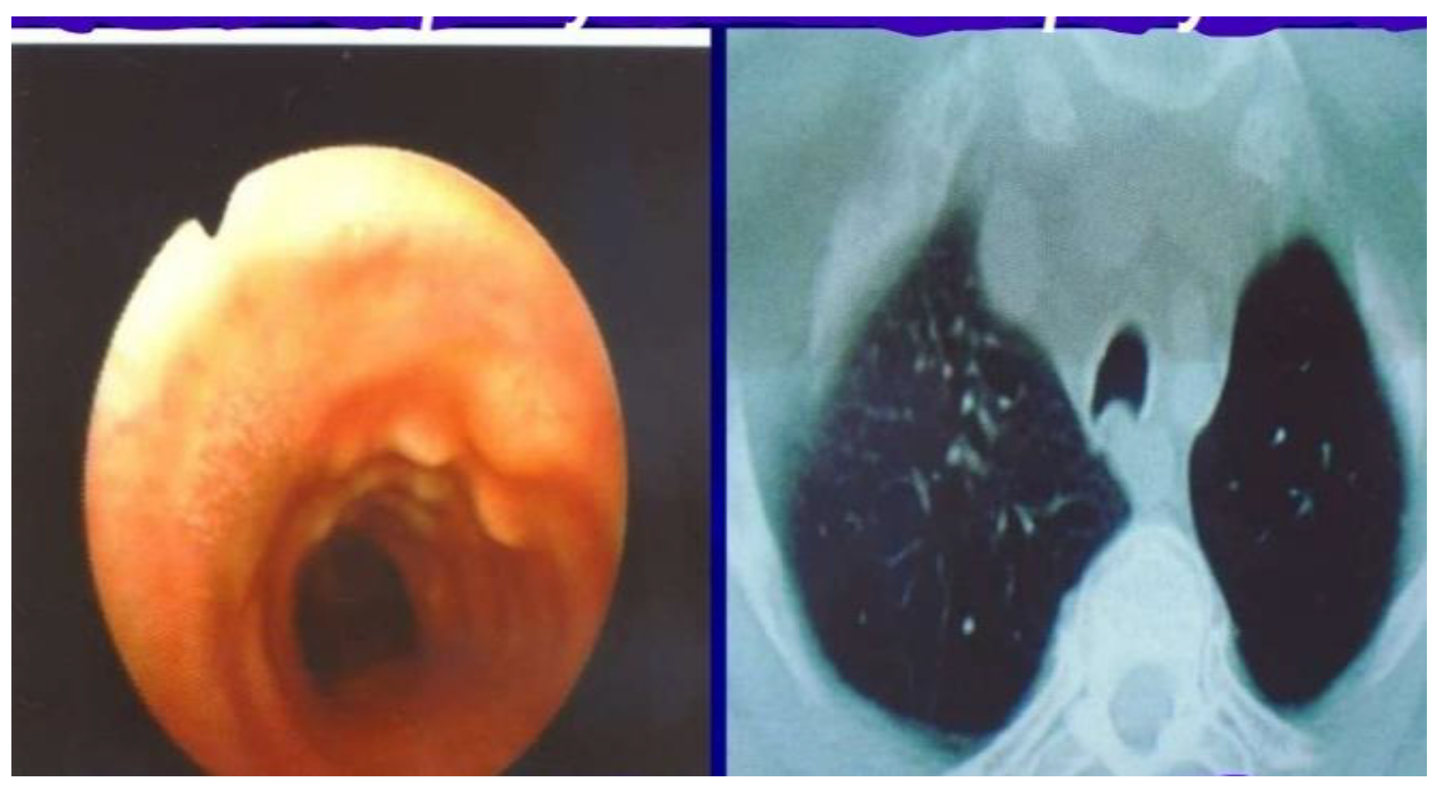
The first clinical case concerns a 51-year-old man, chronic bronchitis, smoker since the age of 15. He reported recurrent episodes of productive cough with sputum sometimes streaked with blood. Laboratory and respiratory function tests were normal. The same subsequently performed bronchoscopy and chest CT scan. Bronchoscopy highlighted a picture of early stage osteochondroplastic tracheopathy, characterized by cartilaginous and bone nodules affecting the wall of the trachea, sparing the pars membranacea. The CT scan, on the other hand, showed a slight reduction in the size of the trachea which showed a modest deformation.
Figure 1.
Presence of cartilage and bone tissue proliferation islands affecting the tracheal wall excluding the posterior wall.
Figure 1.
Presence of cartilage and bone tissue proliferation islands affecting the tracheal wall excluding the posterior wall.
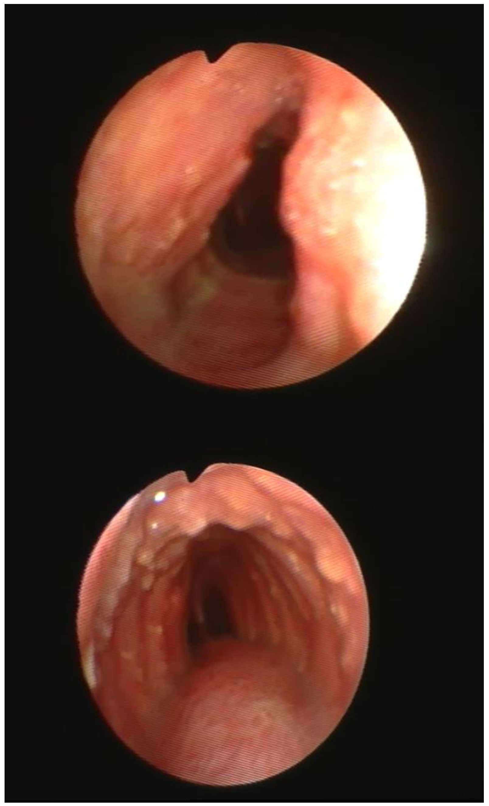
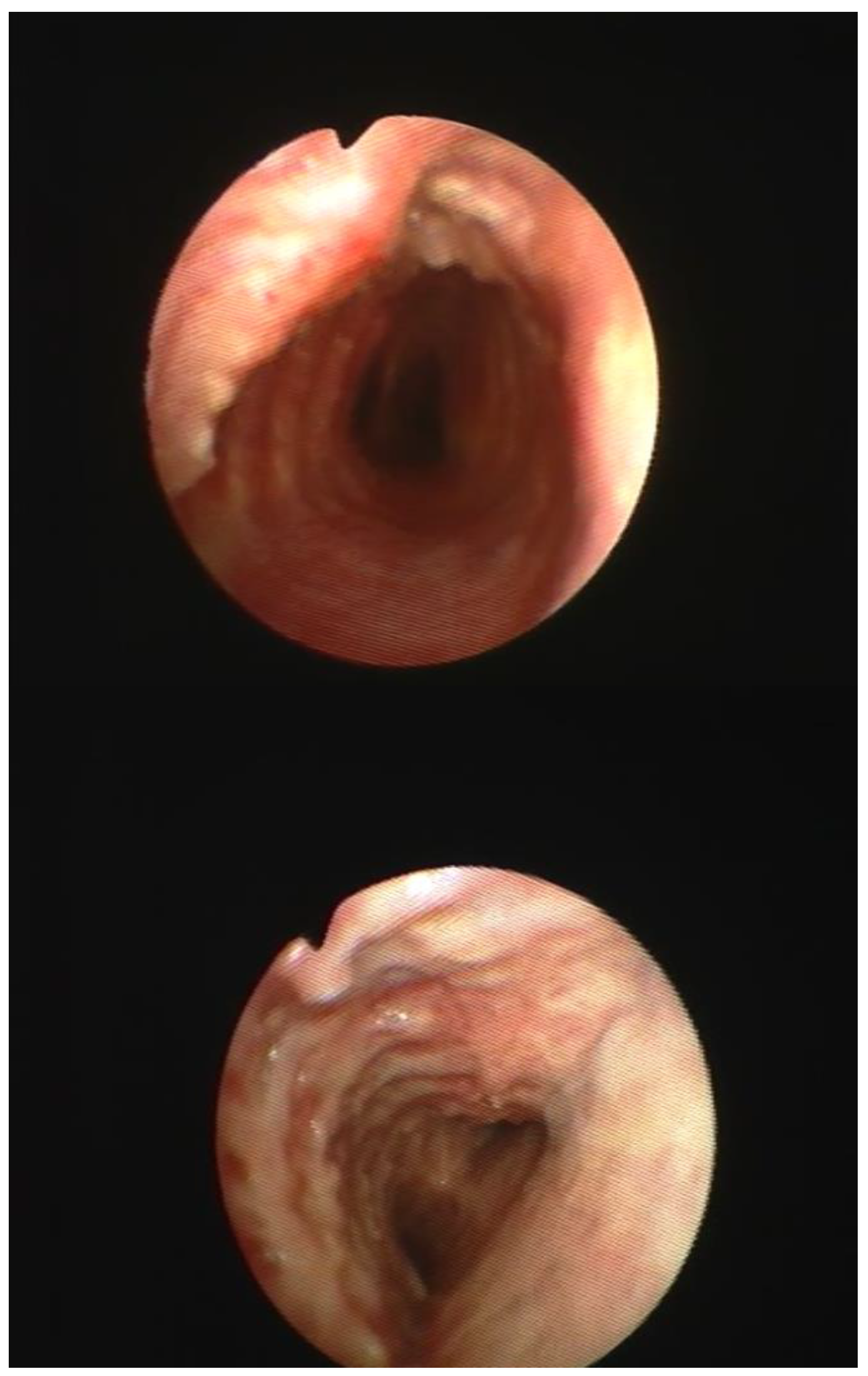
The second clinical case that presented itself to me was that of a 62-year-old woman who smoked, suffering from chronic bronchopathy for several years. For about a week, she has been reporting an accentuation of the cough with the emission of mucopurulent sputum, exertional dyspnea and a minor episode of hemoptysis. Laboratory tests revealed only a slight increase in white blood cells, while lung function tests showed a modest reduction in respiratory function parameters (FV1 and FVC). The fibrobronchoscopic examination was characterized by the presence on the walls of the trachea (excluding the posterior wall, pars membranacea) of numerous nodular bony and cartilaginous growths which adorned the trachea as beads with a rosary crown appearance which in part flowed together forming plaques. In the light of this endoscopic finding, the diagnosis of osteochondroplastic tracheobroncopathy was made.
Figure 2.
Nodular growths of the tracheal wall, with a rosary crown appearance.
Figure 2.
Nodular growths of the tracheal wall, with a rosary crown appearance.
Figure 3.
Nodular growths partly confluent at the level of the tracheal wall that do not affect the pars membranacea(rear wall).
Figure 3.
Nodular growths partly confluent at the level of the tracheal wall that do not affect the pars membranacea(rear wall).
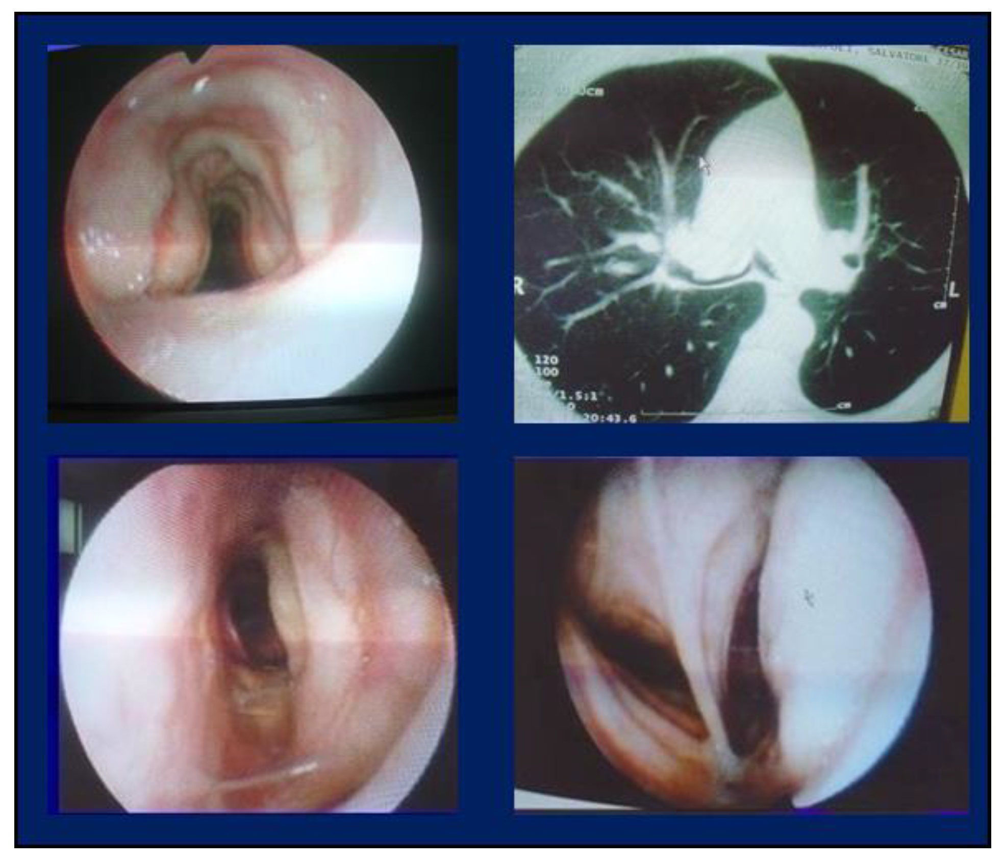
The third case report concerns a 65-year-old non-smoker man treated for chronic obstructive bronchopathy who reported that he had poorly productive cough and dyspnoea for many years. This blood gas analysis showed a reduction in P02 (60 mmHg). Bronchoscopy showed a significant reduction in size of the trachea and main bronchi due to the presence of plaques due to the confluence of nodular growths. Thoracic CT scan confirmed this reduction in size.
Figure 4.
Stenosis of the trachea and large bronchi due to the confluence of nodular excrements that form plaques or masses.
Figure 4.
Stenosis of the trachea and large bronchi due to the confluence of nodular excrements that form plaques or masses.
3. Conclusions and considerations
This is a very rare pathology characterized, as we have seen, by nodular cartilaginous and bony growths affecting the wall of the trachea (excluding pars membranacea) and bronchi. This pathology in the most severe form causes a significant obstruction of the trachea and large bronchi causing a symptomatology characterized by dyspnea with minimal effort. In these cases, however, the Yag laser treatment of the plaques or tracheal masses reduces stenosis and consequently improves the symptomatology.